Université de Montréal
Genetic risk factors of chronic insomnia disorder
par Maryam El Gewely
Département de psychiatrie Faculté de médecine
Mémoire présenté
en vue de l’obtention du grade de maîtrise ès sciences (M.Sc.) en sciences biomédicales
option sciences psychiatriques
Août, 2018
Résumé
Problématique: Le trouble d'insomnie chronique (TIC) affecte 3,5 millions de Canadiens. Malgré sa prévalence élevée, les mécanismes sous-jacents du TIC demeurent méconnus. Une meilleure compréhension de sa base biologique est possible grâce aux études génétiques. Deux études d'associations pangénomiques (GWAS) ont suggéré que le gène MEIS1, antérieurement associé au syndrome des jambes sans repos (SJSR), est indépendamment associé à l'insomnie. Cependant, la méthode de phénotypage des GWAS s'avère limitée et les résultats n'ont pas été répliqué.
Objectif: Évaluer l'association entre MEIS1 et le TIC. Si le gène MEIS1 est pléiotrope au TIC et au SJSR, la fréquence des variantes génétiques du gène sera équivalente chez les groupes TIC et TIC+SJSR.
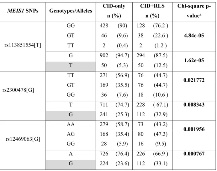
Méthodologie: Au total, 646 patients insomniaques ont participé à l'étude. Trois variantes du gène MEIS1 ont été génotypé. Nous avons comparé les distributions d'allèles et des génotypes de la cohorte TIC aux groupes contrôle et SJSR de la cohorte canadienne française.
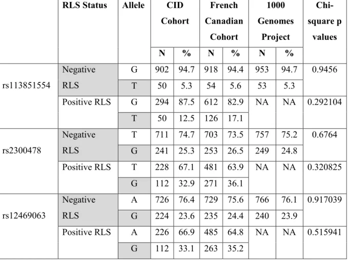
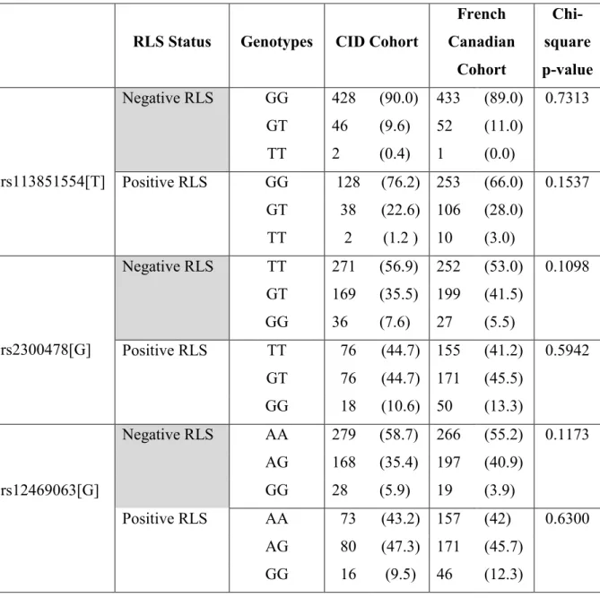
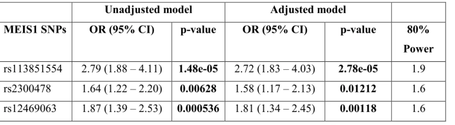
Résultats: Des spécialistes en sommeil ont émis les diagnostics TIC+SJSR à 26% de la cohorte. Nos résultats suggèrent des différences significatives dans les distributions alléliques et génotypiques entre les groupes TIC et TIC+SJSR. De plus, les distributions d'allèles et de génotypes des trois variantes génétiques étaient similaires entre les groupes TIC et contrôle, et les groupes TIC+SJSR et SJSR (p > 0.05).
Conclusion: Nos données confirment l'association entre MEIS1 et le SJSR mais elles ne sont pas en faveur de l'effet pléiotropique avec le TIC. Nous soulignons l’importance du phénotypage et le fait de distinguer le TIC du SJSR.
Mots-clés : Troubles du sommeil, génétique de l'insomnie, insomnie chronique, SJSR, GWAS,
ii
Abstract
Background: Chronic insomnia disorder (CID) affects 3.5 million Canadians. Despite its high prevalence, we do not fully understand the underlying mechanisms of CID. Genetic studies of insomnia contribute to better understanding its biological basis. The latest two genome-wide association studies (GWAS) suggest that MEIS1 gene, previously associated with restless legs syndrome (RLS), is independently associated to insomnia. However, the GWAS phenotyping method was limited and the finding was not yet replicated.
Objective: Evaluate the association between MEIS1 and CID. If MEIS1 is pleiotropic to CID and RLS, the minor allele frequency of MEIS1 variants will be equivalent in CID patients with and without RLS.
Methods: Overall, 646 CID patients participated in the study. We genotyped three MEIS1 variants. To confirm our results, we compared the allelic and genotypic distributions of the CID cohort to ethnically matched controls and RLS cases from the French Canadian cohort.
Results: Patients were diagnosed by sleep specialists and 26% of the sample were diagnosed with CID+RLS. We find significant differences in allele and genotype distributions between CID-only and CID+RLS groups. Allele and genotype distributions of the three MEIS1 SNPs were similar in between CID-only and control groups and in between CID+RLS and RLS-only groups (all p>0.05).
Conclusion: Our data confirms the association between MEIS1 and RLS but it does not support the pleiotropic effect of MEIS1 in CID. Further, our study highlights the critical importance of phenotyping and the need to carefully isolate CID from other disorders that can cause sleep difficulties, particularly RLS.
Keywords : Sleep disorders, sleep genetics, insomnia genetics, chronic insomnia disorder,
Table des matières
Résumé ... i
Abstract ... ii
Table des matières... iii
Liste des tableaux ... vi
Liste des figures ... vii
Liste des sigles ... viii
Liste des abréviations ... x
Remerciements ... xii
Introduction ... 1
1.Insomnia disorder: diagnostic criteria, economic burden and treatment ... 3
1.1 Diagnostic classification of insomnia disorder ... 3
1.1.1 Current clinical diagnostic criteria of insomnia disorder ... 3
1.1.2 Current research evidence for the subcategorization of insomnia ... 5
1.2 Economic burden of insomnia symptoms and chronic insomnia disorder ... 5
1.3 Insomnia Treatment ... 6
1.3.1 Primary treatment for insomnia: Cognitive behavioral therapy for insomnia (CBT-I) ... 6
1.3.2 Pharmacological treatment of insomnia... 8
1.3.3 Limits of the current insomnia treatments ... 9
2. Theoretical models of insomnia etiology ... 11
2.1 The "3P" model ... 11
2.2 The hyperarousal model of insomnia ... 12
2.2.1 What is the hyperarousal model of insomnia? ... 12
2.2.2 The hyperarousal model explains the link between insomnia and other sleep and psychiatric disorders ... 12
iv
3. Evidence of insomnia heritability ... 15
3.1 Family studies of insomnia ... 15
3.2 Twin studies of insomnia ... 17
4. Genetic Studies of insomnia ... 20
4.1 Candidate gene studies of insomnia ... 22
4.1.1 Chronic insomnia disorder and circadian genes ... 22
4.1.2 Chronic insomnia disorder and serotonin (5-HT) pathway ... 23
4.1.3 Chronic insomnia disorder and γ -aminobutyric acid (GABA) pathway ... 24
4.2 Genome wide association studies of insomnia ... 26
5. Objective and hypothesis ... 31
6. Article ... 32 Abstract ... 33 Introduction ... 35 Methods... 38 Participants ... 38 Polysomnography ... 38 Genotyping ... 39 Statistical Analysis ... 40 Results ... 41 Discussion ... 43 References ... 49 7. Discussion ... 58
7.1 Insomnia phenotying: a major concern in the methodology of previous genetic studies of insomnia ... 59
7.1.1 Pittsburgh Sleep Quality Index (PSQI) ... 60
7.1.2 Insomnia Severity Index (ISI ... 60
7.2 Insomnia severity index versus Athens Insomnia Scale and Sleep Condition Indicator 66 7.2.1 Athens Insomnia Scale (AIS)... 66
7.3 Subcategorization of insomnia in future genetic studies ... 68
7.3.1 The use of polysomnography for the subcategorization of insomnia in future genetic studies ... 69
7.4 Clinical Implications ... 70
7.4.1 Phenotyping of insomnia disorder in clinical practices ... 70
7.4.2 From insomnia genetic studies towards precision medicine... 71
Conclusion ... 72
vi
Liste des tableaux
Tableau I. Comparison between candidate gene studies and genome wide association studies (GWAS). Abbreviations: SNP=single nucleotide polymorphism. ... 21 Tableau II. Brief description of the five insomnia Genome wide association studies ... 30
Liste des figures
Figure 1. Direct and indirect economic burden of insomnia in the province of Quebec (Canada). Note that 76% of the economic burden of insomnia are attributed to reduced productivity (Daley et al., 2009). ... 6 Figure 2. Representation of the "3P" Model of insomnia (Spielman et al., 1987) ... 11
viii
Liste des sigles
AIS: Athens Insomnia Scale
BHLHE41: basic helix-loop-helix family member e41 gene (also known as DEC2) CACNA1A : calcium voltage-gated channel subunit alpha1 A gene
CACNA1C: Calcium Voltage-Gated Channel Subunit Alpha1 C gene
CBT-I: cognitive behavioral therapy for insomnia CID: chronic insomnia disorder
DZ: dizygotic
EEG: electroencephalogram
FDA: Food & Drug Administration GABA: γ -aminobutyric acid
GABRB3: GABA A receptor beta 3 subunit gene GABRA3: A receptor gene beta 3 subunit gene GABRA6: GABA A receptor gene 6 subunit gene
GWAS: genome wide association study Hcrt: hypocretin gene
ICSD: International classification of sleep disorders MAF: minor allele frequency
MDD: major depressive disorder MSLT: multiple sleep latency test
MEIS1: Myeloid Ecotropic Viral Insertion Site 1 gene
Meis1: Myeloid Ecotropic Viral Insertion Site 1 gene in mice
MZ: monozygotic
NREM: non-rapid eye movement OXA: orexin-A
OXB: orexin-B
PER: PERIOD gene
PLCB1: Phospholipase C Beta 1
PSQI: Pittsburgh Sleep Quality Index
RLS: restless legs syndrome
ROR1: Receptor Tyrosine Kinase Like Orphan Receptor 1 gene
SDB: sleep disordered breathing SNP: single nucleotide polymorphism SSRI: serotonin selective reuptake inhibitors SCI: Sleep Condition Indicator
5-HT: serotonin
5-HTT: serotonin transporter
x
Liste des abréviations
xii
Remerciements
It is on Febuary 28th 2016 that I received the offer of admission to the master's program. I was surrounded by my friends, celebrating my 24th birthday. I couldn't have wished for a better way to start the new year, feeling prosperous, enthusiastic and loved. Since that date a new journey started and made who I am today. During that journey I grew up on the personal and career levels. I met new people, whom I am blessed and grateful to have in my life. Others have left, they were just meant to share a limited period of this journey with me. Finally, my beloved ones who have always been there can't be thanked enough for who they are.
I would like to start by thanking Dr. Simon Warby for welcoming me in his lab since March 2015. Thank you for being a role model and the best mentor. Thank you for who you and for being genuine. You believed in me since we started to work together and ever since you have been fostering me ahead. Working with you has been a great pleasure that will always be engraved in my memory. You have, and you are still teaching me a lot. You offered me many opportunities, among them learning about sleep, genetics, technology, communication skills, mentoring and many more. Being part of the Warby lab also made me meet great people who helped me achieve the project.
Mélanie Welman, thank you for teaching me how to pipette and genotype. I really enjoyed our time together. Marie-Josée Quinn, thank you for taking care of the blood draws and for always helping me coordinate with the patients. Julien Beaudry, thank you for cooperating with me and help me organize the data. Karine Lacrousse and Houda Kaddioui thank you for your technical and moral supports. Working at Warby lab also allowed me to be part of the "CÉAMS family".
Being at CÉAMS made me meet the pioneers in sleep research and witness the history of the domain. By doing the retrospective analyses of more than a thousand clinical cases, I got the opportunity to work closely with the Canadian pioneer in sleep research and founder of CÉAMS, Dr. Jacques Montplaisir. I feel grateful to be in the student office that is right beside yours. You taught me about many sleep disorders and I am honored to have met you.
Dr. Alex Desautels, thank you for reviewing many clinical files with me and ensuring a careful knowledge transfer. Dr. Daniel Filippini, I would also like to thank you for your patience and for welcoming me in your office whenever I had enquiries.
Dr. Marie Dumont, thank you for the time you gave me to prepare and practice my first oral presentation for the research day of the department of psychiatry in September 2016. You were also my "godmother" during my master's and you have accepted to be a member of my thesis jury. Thank you for always providing me instructive advice.
Dr. Julie Carrier, you have also given me many advice and recommendations for my career. It was always a pleasure to see and talk to you. You have also helped in deciding the title of my first scientific article, which will also be an unforgettable memory.
Mrs. Régine Desnesle, thank you for giving me the opportunity to shadow you during your CBT-I sessions. Your therapeutic approach and skills taught me a lot. You are an inspirational model. Dr. Mélanie Vendette, I would also like to thank you for welcoming me as an intern in your CBT-I sessions and for discussing with me many clinical cases.
I will forever be grateful to the CÉAMS for making me meet you Cloé! You are a supportive, genuine, kind, dedicated and competent friend that I will always be blessed to have. The second CÉAMS Christmas party will always be a memorable day because it marks the beginning of our friendship. We attended classes together, we provided each other support and we traveled to Calgary together. To many more memories.
Thaina, attending my first sleep meeting at the Canadian Sleep Society meeting in 2015 is when we first met. Since that trip, we became closer and our friendship grew. You have always been supportive and understanding, your door is always open and you have always provided meaningful and thoughtful advice. Thank you for being who you are.
Neus, you are a hard-worker and you are a model of dedication. Since we first met during your internship at CÉAMS in fall 2016, we clicked and our friendship began. We have shared a lot together, deep conversations, sleepovers, movies, studying and traveling. We have been to Montreal, Philadelphia and Houston together. We had many adventures and many more are yet to come.
xiv
Thank you to many more CÉAMS colleagues. Namely, Jean Paquet for meeting me on Monday mornings to help me do my stats and make it look so easy. Mireille Charron for helping me find old files and for always being there when needed. Dominique Petit for the valuable advice and for always being a reference. Benoit and the other lab technicians, thank you for always sharing your knowledge. Sylvie Décarie for having a beautiful smile and showing support. Tyna Paquette for the beautiful morning conversations and endless support. Pauline Brayet, my previous office mate, thank you for spreading positivity and for the great moments we spent together at the lab and at the gym.
My presence in Canada would not have been possible without my parents planning and sacrifices. Words are not enough to thank you for all you have been providing. Being in Canada has greatly impacted my career path. It made me discover the domain of research and enhanced my independence. If I can quickly summarize my gratitude then I would like to thank you papi for your strength, for being our backbone and for making us who we are. You believed in me and encouraged me to follow my instinct by going a bachelor's in Psychology. Also, thank you for helping me practice my presentations and for the valuable comments. Mami, my soul mate and source of warm-heartedness, thank you for standing my ups and downs and for always being there. The pride I see in both of your eyes is what pushes me forward.
Esslam, thank you for always being there and making me practice my presentations with you. Joudy, thank you for always being pure and genuine and for being proud of what I have achieved. Dana, thank you for studying hard to get a full mark in your Science sleep-related exam.
My family includes many other members that I left back home in Egypt. Thank you for always cheering me and for your presence in my life. I would also like to thank my friends outside of the lab who have always being providing an unconditional social support. Thank you for the valuable time and for your positive energy.
Also, I would like to thank the funding sources: Chaire Pfizer, Bristol-Myers Squibb, SmithKline Beecham, Eli Lilly en psychopharmacologie de l'Université de Montréal, Faculté des études supérieures et postdoctorales de l'Université de Montréal, and the Centre de Recherche de l'Hôpital du Sacré-Coeur de Montréal.
Thanks for the studentships, your funding made it possible for me to devote the past two years and a half to conduct my master's research project. Finally, thanks in advance to all members of the master's jury for reading and correcting my thesis.
Introduction
The field of sleep research was born almost fifty years ago but is still in its infancy to investigate how and why we sleep (Montplaisir, 2015). Sleep is "one of the least understood phenomena in biology" (Sehgal & Mignot, 2011) and advances in sleep genetics contribute to a better understanding of the underlying mechanisms of sleep (Gehrman, Keenan, Byrne, & Pack, 2015; Sehgal & Mignot, 2011). There is considerable evidence suggesting that genetic factors influence the amount and the organization of sleep (Dauvilliers, Maret, & Tafti, 2005; Sehgal & Mignot, 2011). A decade ago, it was shown that electroencephalogram (EEG) patterns are highly heritable (Ambrosius et al., 2008). For instance, twin studies demonstrate strong concordance between monozygotic (MZ) twins in slow wave sleep, sleep onset and sleep disruption, suggesting a heritability rate of 50% (Dauvilliers et al., 2005; Harvey, Gehrman, & Espie, 2014; Tafti, Maret, & Dauvilliers, 2005). Also, power spectral analysis of EEG frequencies during NREM and wakefulness of MZ twins were significantly higher than dizygotic (DZ) twins; which highlights genetic contributions (Ambrosius et al., 2008). These findings support the hypothesis that there are common inherited neuronal mechanisms that generate EEG oscillations in humans (Ambrosius et al., 2008).
There is also considerable evidence supporting the heritability of sleep disorders (Barclay & Gregory, 2013; Dauvilliers & Tafti, 2008; Gehrman, Keenan, Byrne, & Pack, 2015; Tafti, 2009; Tafti, Maret, & Dauvilliers, 2005). On average, the heritability rate of sleep disorders such as narcolepsy, restless legs syndrome (RLS) and sleep disordered breathing (SDB) ranges from 40 to 50% (Gehrman et al., 2015; Schormair et al., 2017; Tanizawa & Chin, 2018). Current knowledge on the sleep genetics of RLS, narcolepsy and SDB is more advanced than in other disorders such as insomnia disorder (Gehrman et al., 2015; Lind & Gehrman, 2016; Parish, 2013). This is partly due to clearer phenotyping strategies and large sample sizes used in RLS, narcolepsy and SDB genetic studies.
In the case of insomnia disorder, heterogeneous phenotyping of the disorder limits the advancement of the field and the replication of findings (Lind & Gehrman, 2016). Poor insomnia phenotyping results from the use of a wide diversity of questionnaires that are not validated to assess insomnia. The majority of previous genetic studies identified insomnia cases using broad
sleep related questionnaires (Barclay et al., 2011; Brower, Wojnar, Sliwerska, Armitage, & Burmeister, 2012; Polito et al., 2015), sleep queries in psychiatric questionnaires (Feusner et al., 2001; Serretti et al., 2003) or in population based surveys (Gass et al., 2010; Hammerschlag et al., 2017; Lane et al., 2017; Rétey et al., 2005). A few studies used validated insomnia questionnaires in community cohorts (Huang et al., 2014; Li, Huang, Lan, & Wang, 2015) or assessed insomnia in psychiatric samples (Perlis et al., 2003; Serretti et al., 2010; Utge et al., 2010) and a very few studies used clinically diagnosed insomnia samples (Buhr et al., 2002; Deuschle et al., 2010). However, the sample sizes of these latter studies were very small, ranging from a minimum of one clinical case (Buhr et al., 2002) to a maximum of 167 insomnia patients (Deuschle et al., 2010). To address this limitation, genome wide association studies are using very large samples (4 × 10⁵ cases and 9 × 10⁵ controls) but cases are very poorly phenotyped (Oexle, 2018). This project will illustrate how the use of large sample sizes do not compensate poor phenotyping. To do so, we will provide the clinical and research background on insomnia disorder, going from the current diagnostic criteria, economic impact, treatments, etiological models of insomnia to the current knowledge of the genetics of insomnia.
1.Insomnia disorder: diagnostic criteria, economic burden
and treatment
1.1 Diagnostic classification of insomnia disorder
1.1.1 Current clinical diagnostic criteria of insomnia disorder
Insomnia disorder is the most common sleep disorder affecting 3.5 million Canadians (Morin et al., 2011). While 30% of the general population report insomnia symptoms, 10% meet the full diagnostic criteria of insomnia disorder (American Academy of Sleep Medicine, 2014; Morin et al., 2011). Females report higher prevalence of insomnia compared to males (Zhang & Wing, 2006) and insomnia prevalence increases with age (Riemann et al., 2017).
Insomnia disorder is characterized by a subjective complaint of poor sleep quality or quantity that is associated to difficulty initiating, maintaining or undesired early morning awakenings from sleep (American Academy of Sleep Medicine, 2014). The main criterion that distinguishes insomnia disorder from non-clinical insomnia symptoms is the impact of the sleep disturbance on the daytime functioning. For insomnia to be considered as a disorder, sleep difficulties or their consequences should cause significant clinical distress or occupational or social impairment (American Psychiatric Association, 2013; Bastien et al., 2014). When sleep disturbance and associated daytime symptoms have been present for at least three times a week for at least three months, it is called chronic insomnia disorder (CID) (American Academy of Sleep Medicine, 2014). When sleep disturbance and associated daytime symptoms have been present for less than three months, it is called short-term insomnia disorder (American Academy of Sleep Medicine, 2014).
The diagnostic criteria of insomnia disorder have greatly evolved with time (Vgontzas & Fernandez-Mendoza, 2013). While the previous diagnostic guidelines of insomnia disorder distinguished different subtypes of insomnia, current diagnostic criteria aggregate insomnia disorder into a single category due to the lack of empirical evidence to support the sub-categorizations (American Academy of Sleep Medicine, 2014; American Psychiatric
Association, 2013; Riemann et al., 2015). In 1997, the revised first edition of the International classification of sleep disorders (ICSD-R) included twenty-one insomnia subtypes (Vgontzas & Fernandez-Mendoza, 2013). The second edition of the ICSD listed twelve insomnia subtypes (Vgontzas & Fernandez-Mendoza, 2013). Finally, the third edition of the ICSD only distinguishes the periodicity of insomnia disorder: less (short-term insomnia disorder) or more than three months (CID) (American Academy of Sleep Medicine, 2014; Vgontzas & Fernandez-Mendoza, 2013). On a parallel note, our study included patients who had insomnia for more than three months so we focused on CID.
With the current diagnostic criteria, insomnia disorder is considered independent of other psychiatric disorders. In case of comorbidity, it is recommended to treat insomnia disorder and other psychiatric disorders simultaneously (Riemann et al., 2017). It is also important to note that insomnia is often comorbid to other sleep disorders and psychiatric disorders (Riemann et al., 2015). Insomnia can precede a comorbid condition, persist even after successfully treating the comorbid condition or aggravate symptoms of the comorbid condition (Riemann et al., 2015). In fact, more than 75% of RLS patients complain of insomnia symptoms resulting from their leg discomfort (Allen et al., 2014; Montplaisir et al., 1997; Ulfberg et al., 2007).
In psychiatric disorders, an illustrative example is the bidirectional relationship between insomnia disorder and major depressive disorder (MDD). Insomnia disorder and MDD are two of the most prevalent psychiatric disorders and they are frequently comorbid (American Psychiatric Association, 2013). Insomnia disorder is believed to be a predictor to MDD and vice versa (Staner, 2010). In a European study, insomnia preceded depression in 41% of mood disorder cases, 29% of the time both disorders were comorbid and in another 29% insomnia appeared after the onset of MDD (Sutton, 2014). Additionally, insomnia is one of MDD diagnostic criteria (American Psychiatric Association, 2013). When sleep disturbances are severe in MDD patients, response to antidepressant treatment and remission rates are lower than in those without sleep problems (Krystal, 2012).
Despite these clinical guidelines, it is still strongly believed in the research domain that there are at least two types of insomnia that merit attention: psychophysiological insomnia and paradoxical insomnia (Bastien et al., 2014; Edinger et al., 2004).
5
1.1.2 Current research evidence for the subcategorization of insomnia
In psychophysiological insomnia, the subjective complaints of the patient can be objectively observed using polysomnography (Bastien et al., 2014). Patients with psychophysiological insomnia with short sleep duration (less than six hours) are believed to have the most severe phenotype of the disorder (Vgontzas, Fernandez-Mendoza, Liao, & Bixler, 2013). Indeed, insomnia with objective short sleep duration is associated with physiological hyperarousal and higher risk of developing diseases such as hypertension, diabetes, neurocognitive impairment and mortality (Vgontzas & Fernandez-Mendoza, 2013). This subgroup of insomnia is also more likely to have a persistent course compared to insomnia with normal sleep duration (equal or more than six hours).
Paradoxical insomnia is characterized by normal sleep duration, but there is a discrepancy between subjective complaints and objective measures of sleep time. Despite the lack of objective sleep loss, there are subtle sleep microstructural differences that exist between good sleepers and patients with paradoxical insomnia (Bastien et al., 2014). Paradoxical insomnia is also associated with cognitive, emotional and cortical arousal (Bastien et al., 2014).
It is also important to note that these two types are not mutually exclusive. One can display objective sleep onset or sleep maintenance difficulties on polysomnographic records and express extreme subjective complaints of insomnia that are not objectively measured (Bastien et al., 2014). The distinction between these types of insomnia is crucial when considering treatment options. It is proposed that insomnia with short sleep duration may better respond to pharmacological treatments whereas insomnia with normal sleep duration may respond primarily to psychological therapy (Bathgate, Edinger, & Krystal, 2017; Vgontzas & Fernandez-Mendoza, 2013).
1.2 Economic burden of insomnia symptoms and chronic insomnia
disorder
The economic burden of insomnia is excessively high on society. In 2009, the total direct and indirect annual cost of insomnia in the province of Quebec was estimated at 6.6 billion Canadian dollars (Daley, Morin, LeBlanc, Grégoire, & Savard, 2009). Direct costs include
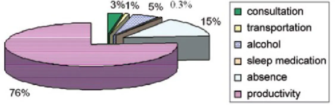
medical consultations, transport to consultations, pharmacological prescriptions, over the counter products and alcohol used as a sleep aid (Figure 1) (Daley et al., 2009). Indirect costs include work absenteeism and productivity losses, which comprises 76% of economic burden (Figure 1) (Daley et al., 2009). The average annual direct and indirect cost per patient with insomnia disorder was five times higher than the annual cost of an individual with insomnia symptoms, $5,010 versus $1,431 respectively (Daley et al., 2009). Compared to good sleepers, the cost of those with insomnia disorder was eleven times higher, $5,010 versus $421 respectively (Daley et al., 2009). Moreover, untreated insomnia expenses are much higher than those of treated insomnia (Daley et al., 2009). The same research group has shown that in Canada, 74% of patients with insomnia disorder show persistent symptoms over the course of a year and 46% report insomnia persisting over three years (Morin et al., 2009). This finding indicates that insomnia disorder is often a persistent condition. Hence, there is a need to understand its biological basis. Such knowledge will lead to precision medicine and to methods of preventing insomnia.
Figure 1. Direct and indirect economic burden of insomnia in the province of Quebec (Canada). Note that 76% of the economic burden of insomnia are attributed to reduced productivity (Daley et al., 2009).
1.3 Insomnia Treatment
1.3.1 Primary treatment for insomnia: Cognitive behavioral therapy for
insomnia (CBT-I)
Cognitive behavioral therapy for insomnia (CBT-I) is the primary recommended treatment for insomnia (Morin et al., 2006). CBT-I also has positive therapeutic effects on
7
comorbid anxiety and depressive symptoms to insomnia (Riemann et al., 2017). CBT-I is composed of four main elements: psychoeducation about sleep–wake behaviour (sleep hygiene), behavioral strategies (sleep restriction and stimulus control), relaxation and cognitive techniques. The psychoeducation component of CBT-I instructs health practices (ie: clockwatching, exercise and substance use) and environmental factors (ie: light, noise and temperature) that promote or disrupt sleep (Riemann et al., 2017). Behavioral strategies are composed of sleep restriction method and stimulus control therapy. Sleep restriction method aims at reducing time in bed to the actual amount of sleep. Hence, a patient is recommended an individualized sleep window by the therapist, which is adjusted weekly over the course of treatment by the therapist. When sleep efficiency, ratio of the total time spent asleep to the total time spent in bed, reaches 85% or more the recommended time in bed increases by 15-30 minutes. However, if the sleep efficiency did not improve or is decreased (<80%), the sleep window is kept stable or decreased by 15-30 minutes until the optimal sleep duration is reached (Riemann et al., 2017; Spielman, Caruso, & Glovinsky, 1987).
Stimulus control therapy is a series of behavioural instructions that target the re-association of the sleep environment (bed/bedroom) with sleep and the reestablishment of a consistent sleep-wake schedule (Bootzin, 1972; Riemann et al., 2017). The set of behavioral instructions are as follows: going to bed only when sleepy, getting out of bed if unable to sleep after twenty minutes, using the bed/bedroom only for sleep and sex, waking up at the same time every morning and no napping during the day (Riemann et al., 2017).
Relaxation and cognitive techniques aim at reducing somatic tension (muscle relaxation and autogenic training) and intrusive thoughts at bedtime (ie: imagery training and meditation) (Riemann et al., 2017). Cognitive techniques identify and change misconceptions and beliefs about sleep and daytime consequences of insomnia (Riemann et al., 2017). Meta-analyses have shown that for long-term treatment, CBT-I is more prominent that pharmacological treatment (Smith et al., 2002). Nevertheless, for acute treatment pharmacological treatments and CBT-I have equal efficiency (Smith et al., 2002).
1.3.2 Pharmacological treatment of insomnia
Pharmacological treatment of insomnia includes benzodiazepines, benzodiazepines agonists, antidepressants, antipsychotics, antihistamines, phytotherapeutic substances and melatonin (Riemann et al., 2017). In 2014, Suvorexant, a reversible dual orexin receptor agonist, was approved by the U.S. Food & Drug Administration (FDA) and added as a possible pharmacological treatment for insomnia (Traynor, 2014).
Benzodiazepines and benzodiazepines agonists are the most prescribed classes of medication for insomnia (Riemann et al., 2015). These drugs bind to benzodiazepine receptor binding sites of the γ -aminobutyric acid (GABA) A receptor, which increases GABA inhibition in brain regions that promote arousal (ie: brain stem and hypothalamus) (Riemann et al., 2015). These classes of drugs are safe and effective for the short term treatment of acute insomnia (≤ 4 weeks) but their long term use is associated with great risk of tolerance and dependence (Riemann et al., 2017, 2015). In fact, millions of people worldwide are dependent on these drugs and suffer from long term side effects and increased morbidity and mortality (Ashton, 2005; Riemann et al., 2015). Moreover, a recent meta-analysis reported that more than 60% of the effectiveness of benzodiazepines and benzodiazepine agonists are due to placebo effect (Winkler & Rief, 2015). This placebo effect was proven using both subjective and polysomnography measures of sleep (Riemann et al., 2017; Winkler & Rief, 2015). Consequently, the use of these drugs can only be used in short term if CBT-I is ineffective or unavailable but its long term use is not recommended (Riemann et al., 2017, 2015).
Alternatively, antidepressants can be prescribed to for short term treatment of insomnia (Riemann et al., 2017, 2015). Dosages for antidepressants to treat insomnia are less than the recommended doses for depression (Riemann et al., 2017). However, the magnitude of antidepressants efficiency remains controversial. While two meta-analyses reported that antidepressants efficiency is less than benzodiazepines (Buscemi et al., 2007; Riemann et al., 2017; Winkler, Auer, Doering, & Rief, 2014), others report positive effects (McCleery, Cohen, & Sharpley, 2014; Yeung, Chung, Yung, & Ng, 2015). Even though antihistamines and antipsychotics are being prescribed to treat insomnia, there is very low quality evidence about their efficacy. Also, they have major side effects such as remission of insomnia after withdrawal, liver dysfunction, and heart rhythm disturbances (Riemann et al., 2015); therefore, they are not
9
recommended treatments of insomnia. Similarly, the low quality evidence of the use of melatonin and phytotherapy to treat insomnia limits their recommendation (Riemann et al., 2017).
To overcome these limitations and negative side effects, Suvorexant, a reversible dual orexin receptor agonist, was introduced to the U.S. market since 2014 as a potential insomnia treatment. Orexin is a neuropeptide secreted from the lateral hypothalamus neurons, known for the role it plays in regulating the sleep-wake cycle, particularly in maintaining the wake state (Krystal, Benca, & Kilduff, 2013). There are two types of orexin neuropeptides, orexin-A (OXA) and orexin-B (OXB). Both types act with different affinities through binding to two G-protein coupled receptors, OX1R and OX2R. Suvorexant binds reversibly to these two receptors and inhibits the activation of the arousal system, which facilitates sleep onset and sleep maintenance (Kishi, Matsunaga, & Iwata, 2015). A meta-analysis conducted on four clinical trials concluded that Suvorexant is effective in treating insomnia and it is better tolerated compared to the other pharmacological treatments in the market (Kishi et al., 2015). However, Suvorexant has major side effects such as next-morning somnolence and safety as seen in driving tests, with possible signs of muscle weakness, weird dreams, sleep walking, other night time behaviors and suicidal ideation (Jacobson, Callander, & Hoyer, 2014).
1.3.3 Limits of the current insomnia treatments
Meta-analyses have shown that for acute treatment of insomnia, CBT-I has equal efficiency to pharmacological treatment and for long-term treatment CBT-I is more effective (Smith et al., 2002). Despite this well documented efficacy of CBT-I, some questions remain unanswered. In fact, there is not enough evidence on the effect of CBT-I on long-term health outcomes (Morin, 2015). In other words, it remains unclear if treating insomnia disorder with CBT-I also decreases the risk for hypertension, depression and occupational disability (Morin, 2015). Moreover, 40% of CID patients treated with both CBT-I and medication do not sustain remission past six months (Morin et al., 2009).
In addition, CBT-I does not seem to be equally efficient for all types of insomnia (Vgontzas et al., 2013). Insomnia patients with the most severe phenotype of the disorder, with short sleep duration, are less responsive to CBT-I than those with normal sleep duration
(Bathgate et al., 2017). It is proposed that insomnia patients with short sleep duration might respond better to biologically-based treatments (Vgontzas & Fernandez-Mendoza, 2013). However, current pharmacological treatments are not convenient to all patients and have major adverse effects such as medication dependency (Ashton, 2005). Insomnia patients with comorbid depression and who experienced the onset of any of these disorders in childhood are less responsive to antidepressant medication compared to those with adulthood onset (Edinger et al., 2016). Hence, genetic studies can lead to better understanding the biological differences in this heterogeneous group of insomnia patients, which will optimize future treatment choices with less severe side effects. Genetic studies will also advance the field towards precision medicine, which will allow the identification of optimal treatments for patients based on their genetic screening.
2. Theoretical models of insomnia etiology
2.1 The "3P" model
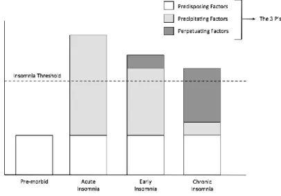
The 3P model, proposed by Arthur Spielman (1987), is one of the most influential models of the etiology of insomnia (Spielman et al., 1987). The 3P model explains the factors that contribute to the development and the maintenance of insomnia. The 3P model suggests that insomnia results from the interaction between predisposing, precipitating and perpetuating factors (Figure 2). Predisposing factors are biological vulnerabilities that confer risk for insomnia (ie: genetic susceptibility); therefore, they are present before the manifestation of insomnia. When these predisposing factors interact with precipitating factors (ie: stressful life events), it is hypothesized that the risk of insomnia increases in vulnerable individuals. Finally, perpetuating factors are maladaptive behaviors ie daytime napping and prolonged stay in bed (Riemann et al., 2010) and beliefs that contribute to the persistence of insomnia over time. Perpetuating factors were further studied by the hyperarousal model hypothesizing that cognitive aspects such as hyperarousal (Riemann et al., 2010) and attention biases (Woods, Marchetti, Biello, & Espie, 2009) lead to the maintenance of insomnia over time.
2.2 The hyperarousal model of insomnia
2.2.1 What is the hyperarousal model of insomnia?
The hyperarousal model is inspired by Spielman behavioral paradigm explained above. The hyperarousal model expands the behavioral perspective by suggesting that conditioned arousal (somatic, cognitive and cortical activation that interfere with one's ability to disengage from the environment) may also act as a perpetuating factor (Levenson, Kay, & Buysse, 2015; Riemann et al., 2010). Conditioned arousal refers to classical conditioning by which the sleep environment (ie: bed and bedroom) and sleep circumstances become stimuli of arousal instead of de-arousal (Riemann et al., 2010). In fact, enhanced sensory processing at sleep onset and during sleep inhibit insomnia patients from disengaging from the environment, which leads to difficulties initiating or maintaining sleep. This constant alertness can also explain the discrepancy between objective polysomnography sleep measures and subjective reports of wake observed in paradoxical insomnia (Perlis, Giles, Mendelson, Bootzin, & Wyatt, 1997; Riemann et al., 2010). Physiologic hyperarousal can be in both central (cortical) (Perlis et al., 1997; Riemann et al., 2010) and peripheral (autonomic) nervous systems (Bonnet & Arand, 2010; Levenson et al., 2015). Arousal can be measured via cortisol level, heart rate variability, EEG and/or self-reports (Levenson et al., 2015).
Espie and colleagues (2006) also added a new perspective to the hyperarousal model called the AIE (attention-intention-effort) (Espie, Broomfield, MacMahon, Macphee, & Taylor, 2006). The AIE perspective suggests that patients' focused attention on sleep and their explicit intention to fall asleep lead to the development and maintenance of insomnia over time. In fact, selective direct attention to sleep counters the natural automaticity and involuntary process of sleep, which leads to maladaptive sleep preventing behavior.
2.2.2 The hyperarousal model explains the link between insomnia and other
sleep and psychiatric disorders
The hyperarousal model also explains the link between insomnia disorder and other sleep and psychiatric disorders. As shown in neuroimaging studies of insomnia hyperarousal, patients with RLS also display arousal sleep disturbance related to the balance between glutamate and
13
GABA (Allen, Barker, Horská, & Earley, 2013; Spiegelhalder et al., 2016). The hyperarousal model also suggests that through classical conditioning, insomnia patients develop depression via learned helplessness (Riemann et al., 2010). Finally, the model demonstrates that insomnia patients are also at risk to suffer from anxiety as a consequence of their sleep related anxiety (Riemann et al., 2010). Consequently, the mechanism of stress is used by research studies to identify subjects who are likely to suffer from insomnia (Drake, Pillai, & Roth, 2014) and insomnia patients with low resilience (Palagini et al., 2018).
2.3 Stress diathesis model
The stress-diathesis model is the most robust etiological model of many psychiatric disorders such as in depression (Hammen, 2005). This model explains how a disorder results from the interaction between genetic predisposition and environmental stressors. Even though this model is incorporated in the 3P model (Figure 1), few studies examined how stressful life events trigger the onset of insomnia.
Studies on the stress-diathesis model of insomnia used sleep reactivity (sleep disturbance in response to a sleep challenge) to assess insomnia pre-morbidity (state that precedes the onset of a disorder) (Drake et al., 2014). The choice of using sleep reactivity was based on previous sleep research conducted on healthy participants. These studies indicated that sleep reactivity is subject to individual differences. Indeed, healthy participants exhibited different stress levels in response to four types of events that caused sleep disruption: first night effect, caffeine administration and advanced sleep phase (by 3 and 6 hours) (Bonnet & Arand, 2003; Drake, Jefferson, Roehrs, & Roth, 2006). In these studies, stress level was assessed using Ford Insomnia in Response to Stress Test (FIRST; questionnaire composed of nine items which assess the likelihood of experiencing sleep disturbance in response to common environmental stressors), nocturnal polysomnographic recordings, multiple sleep latency test (MSLT), performance testing, metabolic and heart-rate observations (Bonnet & Arand, 2003; Drake, Jefferson, Roehrs, & Roth, 2006).
Results of these studies show that the subgroup of healthy participants who displayed the highest level of stress in response to several stressful conditions were likely to experience sleep disruption (Bonnet & Arand, 2003; Drake et al., 2006). A subsequent study confirmed that individuals displaying the highest sleep reactivity have two times higher risk of
developing insomnia over a period of twelve months compared to good sleepers (Drake et al., 2014). Also, sleep reactivity seems to be a precipitant of depression, as mediated by insomnia (Drake et al., 2014).
Additionally, sleep reactivity plays a role in the maintenance of insomnia. In a group of patients diagnosed with insomnia disorder, sleep reactivity correlated to low resilience (psychobiological determinant of one's capacity to adapt successfully to stressful events) (Palagini et al., 2018). Overall, the theoretical models of insomnia suggest that there is a strong role for stress-response and predisposing factors, such as genetics, in the etiology of insomnia. In the following section we will review additional evidence of insomnia heritability that stems from family and twin studies. Particular focus will be presented regarding the link between genetic predisposition and age of onset, specific family members and stress induced insomnia.
3. Evidence of insomnia heritability
3.1 Family studies of insomnia
Familial aggregation is the clustering of disease within families (Matthews, Finkelstein, & Betensky, 2008). Familial aggregation studies determine one's risk to have a disease, given its presence in one or more family members, relative to others without a family history. If a disease is influenced by genetic factors, family members of an affected individual will be more likely to be affected compared to relatives of non-affected individuals (Lind & Gehrman, 2016). As the degree of relatedness is closer, it is expected that the genetic relationship increases, and family members will be more alike (Lind & Gehrman, 2016).
A positive family history of insomnia is common among insomnia patients. In the province of Québec, 35% of patients consulting for insomnia in a sleep clinic report positive family history of sleep disturbances (Bastien & Morin, 2000; Beaulieu-Bonneau, LeBlanc, Mérette, Dauvilliers, & Morin, 2007). The highest sleep disturbance among relatives was insomnia (76%) and 7% of the other reported sleep disturbances included apnea, RLS and daytime sleepiness (Bastien & Morin, 2000). Another study listed the prevalence of the other sleep disturbances among first degree relatives of insomnia patients as follows: sleep apnea (5%), RLS (3%) and excessive daytime sleepiness (2.4%) (Beaulieu-Bonneau et al., 2007). The same research group evaluated the risk factors of insomnia in a population-based sample. They observed that participants with positive family history of insomnia are three times more likely to experience new onset of insomnia over a period of twelve months compared to those without family history of insomnia (LeBlanc et al., 2009). This robust familial aggregation was stable even after adjustment of shared environment and socioeconomic factors (Zhang et al., 2009), which underscores the important role of genetic factors. Incidence of familial cases of insomnia is reported to be similar between males and females (Jarrin et al., 2017). Furthermore, positive family history of insomnia seems to be more frequent in individuals with childhood onset insomnia compared to adulthood onset.
3.1.1 Higher reports of positive family history before mid-adulthood
Three studies reported that positive family history of insomnia is more frequent in patients with childhood onset insomnia compared to adulthood onset insomnia (Bastien & Morin, 2000; Hauri & Olmstead, 1980). In fact, one study reported that patients with insomnia onset before the age of 18 have higher family history of insomnia compared to those with adulthood onset, 55% versus 39% respectively (Hauri & Olmstead, 1980). Similarly, another study reported higher familial incidence in patients with insomnia onset in childhood, adolescence and early adulthood compared to middle age or late life onset, 33 to 30% versus 13 to 25% respectively (Bastien & Morin, 2000). Finally, Dauvillers and colleagues reported that cases of familial insomnia are more likely to exist in those with early onset compared to sporadic insomnia (Dauvilliers et al., 2005).
3.1.2 The mother: most affected family member
Studies using different ethnic groups and different medical cases (InsomniaDisorder-only vs InsomniaDisorder+PsychiatricDisorder) report that the most frequently affected family member by insomnia is the mother (Bastien & Morin, 2000). Similar to a French Canadian cohort (Bastien & Morin, 2000), a French cohort (France) showed that 42% of patients with InsomniaDisorder-only and 45.5% of those with InsomniaDisorder+PsychiatricDisorder report that the family relative the most affected is the mother (Dauvilliers et al., 2005). In the latter study, the ratio of maternally versus paternally transmitted cases is at 1.85 in both InsomniaDisorder-only and InsomniaDisorder+PsychiatricDisorder (Dauvilliers et al., 2005). Two Chinese cohorts also suggested that there is stronger maternal than paternal association of familial insomnia (Wing et al., 2012; Zhang et al., 2009). Despite this evidence, the mode of inheritance of insomnia does not seem to be linked to the X (female sex) chromosome.
3.1.3 Increased risk of stress-induced insomnia and pre-sleep somatic arousal in offspring of parents suffering from stress-induced insomnia
The vulnerability to develop insomnia is present in both family members of insomnia patients and in the offspring of individuals with stress induced insomnia. A nuclear family study (estimating heritability rate based on the two parents and one offspring) has shown that 29% of stress-induced insomnia is heritable. Further, offspring of one or two parents with stress-induced
17
insomnia had threefold to sevenfold risk of having similar sleep reactivity as their parent(s) following stressful events (Fernandez-Mendoza et al., 2014). Mothers with such stress-induced insomnia were likely to have an offspring with increased anxiety levels. Fathers with stress-induced insomnia were likely to have offspring with high pre-sleep cognitive arousability (Fernandez-Mendoza et al., 2014). There is also evidence supporting that sleep-related stress is heritable.
The heritability rate of sleep related stress in non-insomnia participants is 37% (Drake, Scofield, & Roth, 2008). Hence, vulnerability to sleep disturbances caused by stressful events is common in the general population. Since participants in this study were not complaining of insomnia, this data presumes that there is an interaction between environmental and genetic factors that predisposes to insomnia disorder.
Overall, familial aggregation studies provide significant evidence of the heritability of insomnia disorder. Nevertheless, a major limitation of the family study approach is that familial aggregation studies do not discriminate between genetic or shared environmental factors (Lind & Gehrman, 2016). Consequently, twin studies are needed to disentangle these two factors (Lind & Gehrman, 2016).
3.2 Twin studies of insomnia
Twin studies examine the degree of genetic and environmental influences on the disorder of interest by comparing identical MZ, theoretically sharing 100% of the genome, to non-identical dizygotic twins DZ, sharing on average 50% of their genes (Lind & Gehrman, 2016). Increased concordance among MZ compared to DZ twins is suggestive of genetic contribution (Zondervan & Cardon, 2007). Typically, the MZ correlation is twice the correlation between DZ twins for traits that have strong genetic basis (Lind & Gehrman, 2016).
Twin studies report that the heritability rate of insomnia is moderate; reported estimates of the additive genetic variance of insomnia ranged from 0.22 (Lind, Aggen, Kirkpatrick, Kendler, & Amstadter, 2015) to 0.57 (Watson, Goldberg, Arguelles, & Buchwald, 2006). The difference in the heritability rate between studies is explained by methodological differences. While Lind et al. (2015), which reported the lowest heritability rate, used the Symptom
Checklist-90 questionnaire evaluating insomnia symptoms in adults in the past month, Watson et al. (2006), reporting the highest heritability rate, estimated the heritability of childhood onset insomnia and assessed insomnia using the following question: "How often do you have trouble falling asleep or staying asleep?".
Despite the methodological differences between studies, they have consistently shown that the MZ correlation is higher than DZ (Barclay, Gehrman, Gregory, Eaves, & Silberg, 2015; Drake, Friedman, Wright, & Roth, 2011; Heath, Kendler, Eaves, & Martin, 1990; Hublin, Partinen, Koskenvuo, & Kaprio, 2011; Lind et al., 2015; McCarren, Goldberg, Ramakrishnan, & Fabsitz, 1994; Watson et al., 2006). Some studies have even shown that the MZ correlation is twice or more the DZ twins correlation (Drake et al., 2011; Heath et al., 1990; Hublin et al., 2011; McCarren et al., 1994; Watson et al., 2006), which highlights the important role of genetic variance in the predisposition of insomnia.
3.2.1 Stability of insomnia heritability rate over the life span and between sexes
Evidence of the stability of insomnia heritability across time comes from children and adult twin studies. A twin study examined the stability of insomnia in children longitudinally at four ages: 8, 10, 14 and 15 years (Barclay et al., 2015). These results suggest that the genetic influence at the age of 8 impacts the subsequent timeframes. Interestingly, new genetic variance came into play at the age of 10, contributing to the presence of insomnia in adolescence. Hence, there are stable genetic influences that exist since the age of 8 and new ones interfere at the age of 10 (Barclay et al., 2015). Similarly, adulthood longitudinal twin studies have shown that the heritability of insomnia is consistent across time (Hublin et al., 2011; Lind et al., 2015). Further, twin studies conducted in youth (Gehrman et al., 2011; Gregory, Rijsdijk, Dahl, McGuffin, & Eley, 2006), young and senior adults (Gregory et al., 2016; Lind et al., 2017) also suggest that the shared genetic variants between insomnia disorder and MDD (genetic correlation ~0.6) are stable across the life-span.
Genetic stability over time is equivalent for both, females and males (Hublin et al., 2011; Lind et al., 2015). Even though there are no sex-based genetic differences, some studies reported that the heritability rate of insomnia in women is higher than in men (Drake et al., 2011; Lind et al., 2015). Drake et al. (2011) argued that this sex difference is mediated by gender differences
19
in sleep reactivity, showing that women experience more sleep disruption due to stressful events than men (Drake et al., 2011).
Taken together, familial aggregation studies and twin studies provide evidence of the heritability of insomnia disorder and its stability across time (Barclay et al., 2015; Hublin et al., 2011; Lind et al., 2015). Positive family history of insomnia is more common in first degree relatives of insomnia patients, especially in individuals with childhood onset insomnia (Riemann et al., 2015). Twin studies disentangled the genetic and environmental factors arguing that the heritability rate of insomnia is around 50%, which is equivalent to the heritability rate other sleep disorders (refer to introduction) (Riemann et al., 2015). Even though there is no statistically significant difference in the heritability rate of insomnia between sexes, reported heritability rate of insomnia is sometimes higher in women. Also, the mother is the most affected family member across ethnic groups and across medical conditions (Bastien & Morin, 2000; Dauvilliers et al., 2005; Wing et al., 2012; Zhang et al., 2009). Hence, investigation of sex differences need to be further studied in the future studies. Finally, we have seen that insomnia shares some genetic factors with other sleep and psychiatric disorders (ie: MDD). Positive family history of other sleep disorders (ie: sleep apnea and RLS) exists in insomnia patients (Bastien & Morin, 2000; Beaulieu-Bonneau et al., 2007).
4. Genetic Studies of insomnia
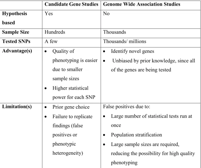
Candidate gene studies and genome wide association studies (GWAS) have contributed greatly to our understanding of biology and mechanisms of disease in humans (Lind & Gehrman, 2016). A comparison between the two basic types of genetic studies is presented in Table 1. Briefly, in candidate gene studies, genes of interest are identified a priori based on existing knowledge of biology and potential mechanisms (Lind & Gehrman, 2016). For instance, genes of interest can be identified for the role they play in a neurotransmitter system or based on animal studies or GWAS findings. Usually, the sample size of these studies includes hundreds of participants. Candidate gene studies compare the frequency of a genetic variation between affected and unaffected individuals or between different levels of symptoms (Lind & Gehrman, 2016). Genetic variations are base pair changes at a specific position in the gene, called single nucleotide polymorphism (SNP). SNP frequency differs between ethnic groups and between disease status (affected vs unaffected) (Lind & Gehrman, 2016). In a case-control design, we compare the minor allele frequency (MAF) to assess if the frequency of either of the SNP alleles (or genotypes) are altered in cases versus controls (Lind & Gehrman, 2016). Despite the popularity of candidate gene studies, the prior choice of gene does not lead to the discovery of new pathways, which limits what is believed to be “biologically plausible”. In addition, the majority of candidate genes have failed to replicate, which can be due to phenotypic heterogeneity and false positives (Lind & Gehrman, 2016).
In contrast, GWAS are conducted with no prior hypotheses which allows to identify novel genes that one would not have hypothesized to be related to the phenotype of interest (Zondervan & Cardon, 2007). This methodology is deemed to be unbiased and results in the assessment of thousands to millions of SNPs (Lind & Gehrman, 2016). Usually, GWAS sample sizes include thousands of individuals. GWAS association analyses are obtained either from a chip of a specific set of SNPs or from whole genome-sequencing (Lind & Gehrman, 2016). However, this method is not without limitations. GWAS results can be false positives as a consequence to the large number of statistical tests that are run at once or due to population stratification (Lind & Gehrman, 2016). Consequently, independent replication of GWAS findings in smaller, but more precisely phenotyped cohorts is mandatory (Gehrman et al., 2013).
21
Candidate Gene Studies Genome Wide Association Studies Hypothesis
based
Yes No
Sample Size Hundreds Thousands
Tested SNPs A few Thousands/ millions
Advantage(s) • Quality of
phenotyping is easier due to smaller
sample sizes • Higher statistical
power for each SNP
• Identify novel genes
• Unbiased by prior knowledge, since all of the genes are being tested
Limitation(s) • Prior gene choice • Failure to replicate
findings (false positives or phenotypic heterogeneity)
False positives due to:
• Large number of statistical tests run at once
• Population stratification
• Large sample sizes are required,
reducing the possibility for high quality phenotyping
Tableau I.Comparison between candidate gene studies and genome wide association studies (GWAS). Abbreviations: SNP=single nucleotide polymorphism.
4.1 Candidate gene studies of insomnia
Studies on the predisposing factors of insomnia are scarce and have smaller sample sizes compared to genetic studies of psychiatric disorders (Gehrman et al., 2013; Lind & Gehrman, 2016). Candidate gene studies of insomnia investigated genes involved in the regulation of circadian rhythms and others related to the neurotransmitter systems involved in sleep-wake regulation such as serotonin and GABA pathways (Gehrman et al., 2013; Lind & Gehrman, 2016). The overview of these studies will provide an example of how candidate gene studies of insomnia can be hard to replicate due to the heterogeneity in phenotyping between studies.
4.1.1 Chronic insomnia disorder and circadian genes
Circadian genes (CLOCK, TIMELESS and PERIOD) were used as a starting point to assess the genetics of insomnia due to the interplay between circadian and sleep mechanisms (Gehrman et al., 2013). Studies that tested the role of circadian genes in insomnia did not assess insomnia directly. The majority of these studies evaluated sleep of patients with psychiatric disorders and results were mixed (Serretti et al., 2003, 2010; Utge et al., 2010). In fact, Serretti et al. (2003) found higher recurrence of insomnia in MDD patients with the homozygotes C variant of 3111T/C CLOCK (Serretti et al., 2003). However, the same research group was not able to replicate this finding in another sample of MDD patients with sleep disturbances (Serretti et al., 2010). Genetics of MDD patients with sleep problems was also studied in a large cohort from Finland (Utge et al., 2010). This study examined 113 SNPs across 18 clock genes and found an association between TIMELESS gene and early morning awakenings in male MDD patients only (Utge et al., 2010).Nonetheless, this finding was not replicated by other studies.
PERIOD (PER) genes -circadian rhythm related genes that contribute to individual
differences in sleep timing- were also associated to insomnia symptomatology (Brower et al., 2012; Gehrman et al., 2013; Li et al., 2015; Lind & Gehrman, 2016; Viola et al., 2007). Viola and colleagues (2007) showed that healthy participants with the short allele of PER3 gene (PER3 4/4) had longer sleep latency and less slow wave sleep compared to those with the long allele (PER3 5/5) (Viola et al., 2007). This finding was concordant to what was found in patients with alcohol dependence. Patients with PER3 4/4 genotype had the most severe insomnia symptoms compared to PER3 4/5 and PER3 5/5 carriers (Brower et al., 2012). Eventhough this finding
23
was never tested in a cohort of patients with insomnia disorder, another PER gene was associated to insomnia in a Chinese cohort.
PER2 gene is suggested to be mediating the interaction between work stress and the risk
of insomnia. A Chinese study showed that Chinese workers with the C allele of PER2 gene have five times higher risk of having insomnia than controls (Li et al., 2015). This allelic effect increased when combined to high work stress. In fact, those with high work stress and AC genotype were fifteen times more likely to have insomnia compared to those with AA genotype and low work stress (Li et al., 2015). These findings were never replicated by other studies and insomnia was assessed using Athens insomnia scale. Moreover, generalizability of these results are limited because the association between insomnia and PER2 gene was not assessed in other ethnic groups than Chinese. Consequently, future genetic studies need to replicate the finding in patients with insomnia disorder and in other ethnic groups. Based on these studies, Barclay and colleagues (2011) tested the link between PER3 and CLOCK genes and sleep quality (using the Pittsburgh Sleep Quality Index) but failed to replicate previous findings (Barclay et al., 2011). They argued that the effects of these genes on sleep quality are small and mixed findings between studies may be related to population composition (Barclay et al., 2011). In fact, we have seen that previous studies were focused on patients with MDD, alcohol dependence or they were questionnaire based. In addition to the circadian genes, other candidate genes studies of insomnia investigated serotonin and GABA pathways. The choice of these pathways was based on the role they play in sleep-wake regulation and because they are targeted by the most common insomnia pharmaceutical drugs.
4.1.2 Chronic insomnia disorder and serotonin (5-HT) pathway
The link between CID and the serotonin pathway is interesting for three main reasons. First, serotonin (5-HT) plays a role in sleep. In fact, 5-HT is a monoamine, group of neurotransmitters that are wake-promoting that receive excitatory input from the hypothalamic hypocretin/orexin system (Schwartz & Kilduff, 2015). Second, the third insomnia GWAS by Amin et al. (2016) suggested the implication of monoamines in insomnia disorder (Amin et al., 2016). Third, serotonin transporter (5-HTT) expression modulates serotonin selective reuptake inhibitors (SSRI), which is the primary pharmaceutical treatment for MDD and often prescribed
to treat insomnia disorder (Nautiyal & Hen, 2017; Riemann et al., 2017). Consequently, the serotonin transporter linked polymorphic region (5-HTTLPR) of the serotonin transporter gene -encoding 5-HTT protein and influencing synaptic serotonin levels- is an interesting genetic target for understanding the common biological basis between both disorders, CID and MDD (Harvey et al., 2014).
In a pharmacogenetic study of MDD, patients with the short (S) allele of 5-HTTLPR had greater risk of developing new or worsening insomnia and showed greater agitation with fluoxetine treatment compared to the long allele carriers (Perlis et al., 2003). This finding was replicated in a German cohort of CID patients (Deuschle et al., 2010). Nevertheless, the statistically significant difference between insomnia cases and controls at the genotype analysis (SS vs SL vs LL) was borderline, chi-square p-values of 0.052 (Deuschle et al., 2010). Furthermore, possible link between 5-HTTLPR, job-related stress, and the risk for insomnia was explored in a group of Chinese workers (Huang et al., 2014). This study reported that those with the 5-HTTLPR short allele had six times higher risk of developing insomnia when exposed to high work stress level compared to those with the long allele. Finally, a study conducted on community-dwelling individuals has shown that the 5-HTTLPR short allele is associated with sleep onset disturbance (Polito et al., 2015). Moreover,
5-HTTLPR genotype mediated the association between sleep onset latency and depressive symptoms (Polito et al., 2015). Taken together, only one study investigated the link between CID and 5HTTLPR. However, this study was underpowered (n=157) and genotyping results were borderline (Deuschle et al., 2010). Thus, assessment of the role of 5-HTTLPR in a larger CID cohort is needed.
4.1.3 Chronic insomnia disorder and γ -aminobutyric acid
(GABA) pathway
The GABA system is another relevant neurotransmitter for the role it plays in sleep. In fact, GABA is the main inhibitory neurotransmitter in the brain and sleep-promoting nuclei are GABAergic in nature (Schwartz & Kilduff, 2015). These nuclei are found in the preoptic area, brainstem and lateral hypothalamus. A decrease in GABA function could lead to excitation of wake-promoting neurons instead, which leads to sleep disorders such as CID (Schwartz & Kilduff, 2015). To treat these dysfunctions, sleep medications such as benzodiazepines target
25
GABA A receptors (Bateson, 2004), which makes GABA A receptor genes relevant genetic candidates to understand the etiology of insomnia. Further, Amin et al. (2016) GWAS finding also suggested the implication of GABA in insomnia disorder (Amin et al., 2016). Despite its relevance, few studies have investigated the link between GABA A receptor genes and CID.
The four genetic studies that suggest a link between GABA receptor genes and insomnia did not assess insomnia directly (Agosto et al., 2008; Buhr et al., 2002; Feusner et al., 2001; Uhart, McCaul, Oswald, Choi, & Wand, 2004). The first study used a population of patients with post-traumatic stress disorder and phenotyped insomnia using the General Health Questionnaire (Feusner et al., 2001). Results suggest that heterozygosity of the GABA A receptor beta 3 subunit gene (GABRB3) major allele (G1) is associated to anxiety and insomnia symptoms (Feusner et al., 2001). Another study identified a missense mutation in GABA A receptor gene beta 3 subunit gene (GABRA3) in a patient diagnosed with CID with positive family history of insomnia (Buhr et al., 2002). Functional analysis showed a slower rate of the fast phase of desensitization of the beta3 subunit compared to the other GABA A receptor subunits. Moreover, current deactivation of the receptor revealed a slower rate of the fast phase of desensitization; which suggests that decreased GABAergic inhibition is potentially contributing to insomnia (Buhr et al., 2002). Finally, the T allele of GABA A receptor gene 6 subunit (GABRA6) was associated to increased blood pressure and activation of the HPA axis in response to psychological stress in healthy white Caucasians (Uhart et al., 2004). The association between GABA A receptor and sleep difficulty was also found in the animal model. Indeed, Drosophila with the mutant GABAA receptor gene, RdlA302S, displayed decreased sleep latency (Agosto et al., 2008).
To sum up, poor phenotying has been a major limitation in all of the above-mentioned studies. Another challenge in conducting candidate gene studies is that one must know which genes to examine while little is known about the underlying mechanisms of insomnia and sleep/wake regulation. To overcome this limitation, a great effort has been made since 2010 to conduct insomnia related GWAS.