HAL Id: hal-02187739
https://hal.archives-ouvertes.fr/hal-02187739
Submitted on 17 Dec 2020HAL is a multi-disciplinary open access archive for the deposit and dissemination of sci-entific research documents, whether they are pub-lished or not. The documents may come from teaching and research institutions in France or abroad, or from public or private research centers.
L’archive ouverte pluridisciplinaire HAL, est destinée au dépôt et à la diffusion de documents scientifiques de niveau recherche, publiés ou non, émanant des établissements d’enseignement et de recherche français ou étrangers, des laboratoires publics ou privés.
Evidence of Wolframite-Type Structure in Ultrasmall
Nanocrystals with a Targeted Composition MnWO 4
Pascaline Patureau, Remi Dessapt, Pierre-Emmanuel Petit, Gautier Landrot,
Christophe Payen, Philippe Deniard
To cite this version:
Pascaline Patureau, Remi Dessapt, Pierre-Emmanuel Petit, Gautier Landrot, Christophe Payen, et al.. Evidence of Wolframite-Type Structure in Ultrasmall Nanocrystals with a Targeted Compo-sition MnWO 4. Inorganic Chemistry, American Chemical Society, 2019, 58 (12), pp.7822-7827. �10.1021/acs.inorgchem.9b00464�. �hal-02187739�
1
Evidence of wolframite-type structure in ultrasmall nanocrystals
with a targeted composition MnWO
4Pascaline Patureau
1, Rémi Dessapt
1, Pierre-Emmanuel Petit
1, Gautier Landrot
2,
Christophe Payen
1,*, Philippe Deniard
1,*1 Institut des Matériaux Jean Rouxel (IMN), Université de Nantes, CNRS, 2 rue de la
Houssinière, BP 32229, 44322 Nantes cedex 3, France
2 Synchrotron SOLEIL, F-91192 Gif sur Yvette, France
2
ABSTRACT: Here, we report a study of white-ochre powders with targeted composition
MnWO4 prepared via a coprecipitation method. Through X-ray total scattering combined with
pair distribution function analysis and Rietveld refinement of X-ray diffraction data we find that their crystal structure is similar to that of bulk-MnWO4, despite a mean crystallite size of
1.0-1.6 nm and a significant deviation of the average chemical composition from MnWO4.
The chemical formula derived from elemental and thermogravimetric analyses is Mn0.8WO3.6(OH)0.4.3H2O. X-ray absorption and magnetic susceptibility measurements show
that Mn and W have the same oxidation states as in MnWO4. No magnetic ordering or spin
glass or superparamagnetic behavior is observed above 2 K, unlike in the case of MnWO4
3 INTRODUCTION
Transition metal tungstates of the MWO4 type (M = Mn, Fe, Co, Ni, Cu, or Zn) have attracted
great interest due to potential applications in various fields, including humidity sensors1,
multiferroic materials2, photocatalysts3, heterogeneous catalysts4, biomedical imaging5,
energy storage materials6, and color pigments7. These compounds contain M(II) and
tungsten(VI) cations in octahedral environments. They crystallize with the wolframite (monoclinic) structure8, with the exception of CuWO4 which exhibits a lower (triclinic)
crystal symmetry associated with cooperative Jahn-Teller distortions of the Cu2+ (d9)
environment9. Crystal structures comprise infinite zigzag (MnO
4) and (WO4) chains,
running parallel to the same direction, of either edge-sharing MO6 distorted octahedra or
edge-sharing WO6 distorted octahedra.8-10 Their interesting properties have been investigated
at different length scales, from bulk in single crystals or fine particles to nanoscale in nanosized materials. For applications or studies at the nanoscale, a possible key challenge is keeping crystal structures with high crystallinity and their associated bulk properties when decreasing particle size. For MnWO4, which is a well-studied multipurpose material1,2,5,6,
phase-pure nanoparticles of controlled size and shape have been prepared by mild solution methods11,12, and single-crystal nanoparticles with mean size in the range of 10-50 nm have
been studied13-15. When looking at their structure-dependent bulk electronic properties, one
can note that magnetic transitions associated with type-II multiferroicity in bulk-MnWO4 are
still visible in temperature-dependent magnetic susceptibility curves of 50 nm single-crystal nanopellets and nanorods15 but are absent when mean crystallite size is 10 nm14.
Here, we address the problem of how the wolframite structural order of MnWO4
evolves when downscaling towards the ultrasmall nanocrystallite range ( 5-10 nm) where unique physicochemical properties are generally observed16. This unresolved issue also
4 formation.17 In this work, we have studied the crystal structure and metal oxidation states in
ultrasmall nanocrystals with targeted composition MnWO4 through X-ray total scattering
combined with pair distribution function analysis (PDF), X-ray diffraction (XRD), X-ray Absorption Near Edge Structure (XANES) spectroscopy, and magnetic susceptibility measurements. Samples were prepared by using a coprecipitation method that produces a white-ochre solid. Although the formation of a white-ochre precipitate with unknown chemical formula has already been observed in a few previous works18,19 which employed a
co-precipitation method, no characterization data has been reported so far. We therefore have also determined the actual macroscopic chemical formula and have examined the thermal stability of these precipitates in air by variable temperature XRD.
EXPERIMENTAL SECTION
Synthesis, Elemental and Thermogravimetric Analyses. High-purity reagent chemicals
were purchased from commercial vendors. Elemental analyses (Mn, W, Na, and H contents) were performed on as-prepared powders at the “Service Central d’Analyse du CNRS”, Vernaison, France. Thermogravimetric measurements were performed in flowing air. Powders were placed in alumina crucibles and heated up to 800°C at 3 K /min.
Characterization. Attempts at carrying out transmission electron microscopy experiments
failed because samples were unstable. X-ray diffraction (XRD) patterns were collected at room temperature on a Bruker D8 Advance instrument using monochromatic CuK-L3 (=
1.540598 Å) X-rays and a LynxEye detector. Temperature variable XRD experiments were carried out on a Bruker D8 Advance instrument using CuKL2-L3 (= 1.540598, 1.54433 Å)
5 performed using JANA 200620 and the Cheary-Coelho fundamental approach for XRD profile
parameters21.
X-ray Absorption Spectroscopy analyses were done at SAMBA beamline, Synchrotron SOLEIL. The storage ring was operated with multi-bunch and top-up modes, with 2.75 GeV electron energy and 450 mA current. Powder samples were pelletized prior acquisition and analyzed at room temperature. X-ray Absorption Near Edge Structure (XANES) spectra were collected at the W L3 edge (10207 eV) and Mn K edge (6539 eV) in fluorescence mode using
a Si(220) monochromator, a Ge multi-pixel fluorescence detector (Canberra), and step-by-step acquisition mode. At each edge, multiple spectra were collected until the signal-to-noise ratio of the corresponding merge spectrum was satisfactory. All spectra were averaged, normalized and corrected from self-absorption effects using Athena featured in Demeter Software Package.22
X-ray total scattering measurements were performed at room temperature on a Bruker D8 Advance instrument using MoKL2-L3 (= 0.70929, 0.71358 Å) radiation and a Våntec
detector. This set up produces, for a 2 range up to 150°, a Qmax of 17 Å-1. Observed PDFs
were calculated using PDFgetX2 software23 and real space refinements were performed using
PDFgui24.
A Quantum Design MPMS-XL7 magnetometer was used to collect temperature-dependent DC magnetization data. Zero field cooled (ZFC) and field cooled (FC) magnetization measurements were taken from 2 to 300 K in an applied field of µ0H = 0.01 or 0.1 T. Data
were corrected for the diamagnetism of the sample holder as well as for core diamagnetism using Pascal’s constants25.
6 RESULTS AND DISCUSSION
Synthesis and Compositional Analysis. Samples with a targeted Mn:W molar ratio of 1.0
were prepared in a reproducible manner by using a coprecipitation method.26 Aqueous
solution of manganese chloride MnCl2.4H2O (0.6 mmol in 10 mL) was added slowly to an
aqueous solution of Na2WO4.2H2O (0.6 mmol in 10 mL)at room temperature under vigorous
magnetic stirring, leading to immediate precipitation of a white-ochre solid. The pH was kept constant at about 7 by adding manually HCl 0.1 M and the slurry was stirred at room temperature for five minutes. After filtration, the powder was washed with water, ethanol, and dried in air at room temperature. The final yield in W was 22 mol%. Elemental analyses showed no evidence for incorporation of sodium cations and were in good agreement with the chemical composition H6.4Mn0.8WO7.0 that fulfills electroneutrality (see the Supporting
Information). Thermogravimetric data are shown in Figure 1. A total mass loss of about 19 % took place in three steps in the temperature range of 25-800 °C. The small mass loss of 1.5 % observed below 50 °C is likely due to the removal of free and/or physisorbed water. Thermogravimetric data also revealed a mass loss of 15.5 % in the temperature range of 50-150° C and 2% in the temperature range of 150-800 °C. By rewriting the chemical formula H6.4Mn0.8WO7.0 as Mn0.8WO3.6(OH)x.yH2O, mass loss calculations indicated that the weight
losses of 15.5 % and 2% correspond to the losses of 3 H2O and 0.4 (OH), respectively.
Therefore, the chemical formula was considered to be Mn0.8WO3.6(OH)0.4.3H2O. Quantitative
analysis of the XRD pattern at 700°C and magnetic susceptibility data were also fully consistent with this formula as described hereafter. The low Mn:W ratio in the precipitate (0.8) agrees with the results in ref. 26 and implies the presence of unreacted Mn(II) cations in solution. These Mn(II) cations actually react with Mn0.8WO3.6(OH)0.4.3H2O to yield MnWO4
7 Figure 1. Thermogravimetric curve for Mn0.8WO3.6(OH)0.4.3H2O in flowing air.
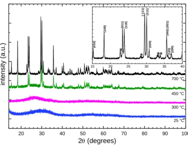
Variable Temperature X-ray Diffraction. Figure 2 (25° C) presents the room temperature
XRD pattern of as-prepared powder. No distinct Bragg reflections was observed and thus the compound could at first sight be considered to be amorphous. Three very broad features centered at 2 30, 55, and 65° were however visible in the XRD pattern and the absence of narrow Bragg reflections could instead be due to strong peak broadening and overlap. Syntheses of powder samples of MnWO4 via co-precipitation are usually followed by a brief
heating in air at a moderate temperature, e.g., 400-500 °C, that makes it possible to observe narrow Bragg reflections in XRD patterns26. Figure 2 also shows the evolution of the
diffraction pattern as a function of temperature. At 450 °C, the diffraction pattern was similar to that of MnWO4 (ICSD # 67907). Above 450 °C small additional peaks appeared. They
were observed up to 700 °C as evidenced by the inset of Figure 2 where they are marked with asterisks. They correspond to the krasnogorite variety of WO3 (pdf file 71-0131). A
two-phase refinement carried out by the Rietveld method for 700°C (see Figure S1 in the Supporting Information), leaded to 14.8 +/- 1.2 wt% of WO3 in perfect agreement with
complete thermal decomposition of Mn0.8WO3.6(OH)0.4.3H2O into MnWO4 plus WO3, in air.
100 200 300 400 500 600 700 800 80 85 90 95 100 1.5 wt% 2 wt% 15.5 wt% Weight (%) Temperature (°C)
8 20 30 40 50 60 70 80 90 100 700 °C 450 °C 300 °C inte ns ity (a.u .) 2 (degrees) 25 °C 15 20 25 30 35 40 (200) (120 ) (021 ) (002 ) (020) (111) (-11 1) (110 ) (01 1 ) (100) * * * * (0 10)
Figure 2. X-ray diffraction patterns of Mn0.8WO3.6(OH)0.4.3H2O taken with CuKL2-L3 radiation.
Patterns were collected in air at several temperatures from 25 to 700 °C (temperature is indicated below the corresponding pattern) using the same powder for the whole data set. The inset shows a portion of the pattern recorded at 700°C. Bragg reflections marked with asterisks are from WO3. hkl
indices are those of MnWO4 (ICSD 67907).
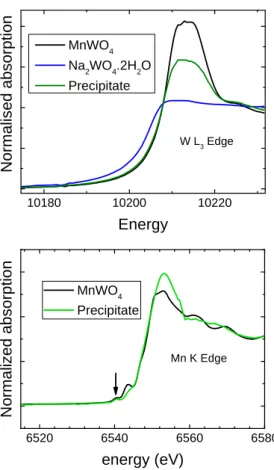
X-ray Absorption Near Edge Structure (XANES). XANES was used in order to determine
oxidation states and local environments of Mn and W in Mn0.8WO3.6(OH)0.4.3H2O. Figure 3
presents W L3 and Mn K edge spectra for both the Mn0.8WO3.6(OH)0.4.3H2O precipitate and a
well characterized micrometric powder of MnWO4. This figure also shows our W L3 XANES
data for the reactant Na2WO4.2H2O, which contains W(VI)ions in tetrahedral environment.27
For each edge, spectra were collected under the same experimental conditions. The ionic state and local environment of W can be identified by comparing the onset energies and shapes of the W L3 spectra.28 From data in Figure 3, it is evident that tungsten is exclusively found in
9 10180 10200 10220 W L3 Edge No rma lise d ab so rp ti o n Energy MnWO4 Na2WO4.2H2O Precipitate 6520 6540 6560 6580 MnWO4 Precipitate N o rma lized ab so rp tio n energy (eV) Mn K Edge
Figure 3. XANES spectra for Mn0.8WO3.6(OH)0.4.3H2O (termed Precipitate) and for micrometric
powder of MnWO4. Top panel: W L3-edge spectra. Data for Na2WO4.2H2O were also recorded for the
sake of comparison. Bottom panel: Mn K edge spectra. The arrow indicates pre-edge features at 6540 eV which are related to electronic transitions to 1s core levels to empty hybridized 3d/4p states.
determination of the Mn oxidation state from the main edge energy is not possible due to single and multiple scattering effects.29 However, the weak pre-edge features observed at
about 6540 eV for both Mn0.8WO3.6(OH)0.4.3H2O and MnWO4 clearly indicate the presence
of Mn(II) in octahedral symmetry.29
Pair Distribution Function (PDF) Analysis. A PDF analysis of X-ray total scattering data
was then performed to study the interatomic distances r. Total scattering structure factors
S(Q) of both Mn0.8WO3.6(OH)0.4.3H2O and micrometric MnWO4 are presented in Figure S2 in
the Supporting Information to visualize the quality of the data treatment. Figure 4 shows the PDF, G(r), of Mn0.8WO3.6(OH)0.4.3H2O. Peaks in the low r-region of the PDF were compared
10 to those observed in the PDF of a micrometric powder sample of MnWO4, which is also
shown in Figure 4. The calculated PDF of the published wolframite structure of bulk MnWO4
(ICSD # 67907)10 is presented in Figure 5 for the sake of comparison. For
Mn0.8WO3.6(OH)0.4.3H2O, the two peaks at about 1.8 and 2.3 Å correspond well to the mean
metal-oxygen distances in WO6 and MnO6 octahedra of the bulk-MnWO4 structure,
respectively. As in bulk-MnWO4, a weak peak at 2.9 Å can be associated with
oxygen-oxygen distances. The large and intense peak at 3.6 Å is likely due to the shortest metal-metal distances; Mn-Mn or W-W distances for edge-sharing MnO6 or WO6 octahedra that form the
(MnO4)n or (WO4)n chains, and Mn-W distances for two MnO6 and WO6 octahedra sharing a
corner. Furthermore, the two peaks found between 4 and 5 Å match with the W-W distances of 4.45 and 4.8 Å in bulk-MnWO4. All these observations prompted us to perform a
refinement of the PDF of Mn0.8WO3.6(OH)0.4.3H2O using the bulk-MnWO4 crystallographic
model with a size damping envelope. The obtained fit, shown in Figure 4, was of good quality up to about 6 Å. 2 4 6 8 10 12 14 16 18 20 -1 0 1 G(r) r (Å) Mn0.8WO3.6(OH)0.4.3H2O
11 2 4 6 8 10 12 14 16 18 20 -3 -2 -1 0 1 2 3 4 G(r) r (Å) Micrometric MnWO4
Figure 4. Experimental PDF G(r) for Mn0.8WO3.6(OH)0.4.3H2O (top panel) and for a micrometric
powder sample of MnWO4 (bottom panel) derived from X-ray total scattering data taken with Mo
K1 K2 radiations (blue dots). The red lines show fits obtained using (i) a MnWO4 particle model in
the case of Mn0.8WO3.6(OH)0.4.3H2O (ii) the published wolframite structure model of bulk-MnWO4 in
the case of micrometric MnWO4. The lower green curves show the differences between the PDF and
the refinement. 1 2 3 4 5 6 7 -4 -2 0 2 4 G(r ) r (Å) W‐O Mn‐O O∙∙∙O 1 Mn∙∙∙W 2 3 4 5 Mn∙∙∙W
12 Figure 5. Calculated PDF G(r) of the published wolframite bulk-MnWO4 model in the low r-region.
Some interatomic distances are shown above or below the PDF. The five shortest W-W distances are marked with numbers (1 to 5) and are illustrated on the perspective views of the crystal structure shown below the graph.
Difficulty in refining the PDF for longer distances correlates with the ultrasmall fitted particle size, 1.3 ± 0.3 nm. A spherical nanocrystal of MnWO4 with a diameter of 1.3 nm would
consist of about 8 unit cells and 100 atoms, a significant number of which would be located on the surface. Deviation from the bulk MnWO4 composition, which includes a lower
Mn-to-W ratio of 0.8 and the incorporation of hydroxides into particle structure, could also explain differences in local structure.
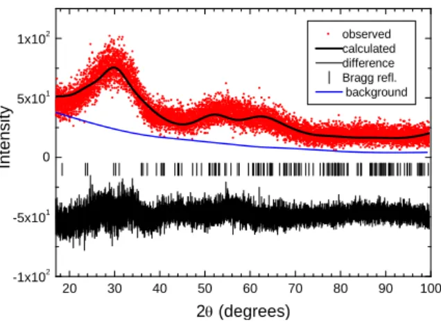
Rietveld refinement and simulation of room-temperature XRD data. According to our
PDF analysis, the room-temperature XRD pattern of Mn0.8WO3.6(OH)0.4.3H2O should be
consistent with ultrasmall 1 nm nanocrystallites having a MnWO4-type structure. Thus, we
performed a Rietveld refinement using the published bulk-MnWO4 structural model.10 Given
the low signal-to-noise ratio and the large width of Bragg reflections, only the following parameters were refined: scale factor, crystallite size and background (5-term Legendre polynomial). All MnWO4 structural parameters (taken from ICSD # 67907) were held fixed.
The refinement converged to obtain the results shown in Figure 6, which were in very good agreement with a nanoscale MnWO4-like compound. The refined crystallite size 0.82(2) nm
compares well with the value 1.3(3) nm derived from PDF analysis, given that microstrains could not be taken into account in the contribution to line broadening. An additional clue about the nanocrystalline nature of Mn0.8WO3.6(OH)0.4.3H2O is provided by Figure S3
(Supporting Information). In this latter figure, the X-ray diffraction pattern of MnWO4 (ICSD
# 67907) was simulated for two different crystallite sizes (50 and 1 nm) and for CuK-L3
13 Figure 6, results solely from overlap of many Bragg reflections and does not correspond to that of an amorphous material.
20 30 40 50 60 70 80 90 100 -1x10² -5x101 0 5x101 1x10² observed calculated difference Bragg refl. background Intensity 2 (degrees)
Figure 6. Final Rietveld refinement plot of the room-temperature XRD data (taken with CuK-L3
radiation) for Mn0.8WO3.6(OH)0.4.3H2O. This refinement was performed using the published
bulk-MnWO4 structural model. The blue line shows the fitted background. Each hkl reflection, as
represented by a vertical bar, overlaps with its neighbors, leading to a few broad features in the pattern.
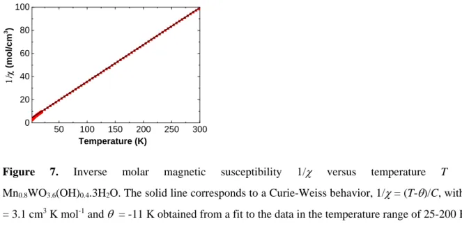
Magnetic susceptibility. Finally, we turn to the magnetic properties. Temperature dependent
magnetic susceptibility data, , of as-prepared powders were collected from 2 to 300 K. As can be seen in Figure 7, the magnetic susceptibility obeys a Curie-Weiss law above 25 K. The molar Curie constant calculated using the molar mass of Mn0.8WO3.6(OH)0.4.3H2O is
actually the one expected for 0.8 mole of Mn(II) ions having S = 5/2 spin and for nonmagnetic W(VI) ions. Data in Figure 8 also indicate the absence of magnetic orders or superparamagnetic or spin-glass behaviors as the susceptibility keeps rising down to the base temperature (with no difference between the data measured under zero-field-cooled and that measured under field-cooled conditions). In bulk-MnWO4, three magnetic structures have
14
T2 and the associated ferroelectric transition were still observed in nanocrystals with mean
diameter of 50 nm15, but only one magnetic transition was visible at 6 K in nanocrystals with
mean size of 10 nm.14 In the multiferroic state of bulk-MnWO
4 below T2, the incommensurate
magnetic structure has a large magnetic unit cell volume of 2.8 nm3, which reflects the
existence of long-ranged magnetic interactions.30,31 Therefore a loss of spin-driven
multiferroicity is expected in any MnWO4 nanocrystallite having a volume lower than or
comparable with the bulk magnetic cell volume. Deviation from MnWO4 composition should
also affect the magnetic properties of Mn0.8WO3.6(OH)0.4.3H2O.
50 100 150 200 250 300 0 20 40 60 80 100 (m o l/c m 3 ) Temperature (K)
Figure 7. Inverse molar magnetic susceptibility 1/ versus temperature T for Mn0.8WO3.6(OH)0.4.3H2O. The solid line corresponds to a Curie-Weiss behavior, 1/= (T-)/C, with C
= 3.1 cm3 K mol-1 and = -11 K obtained from a fit to the data in the temperature range of 25-200 K.
2 4 6 8 10 12 14 16 18 20 0.0 0.1 0.2 0.3 0.4 (c m 3 /m o l) Temperature (K)
15 CONCLUDING REMARKS
To summarize, white-ochre powder samples of Mn0.8WO3.6(OH)0.4.3H2O were reproducibly
obtained by a simple salt metathesis reaction involving stoichiometric amounts of manganese(II) chloride and sodium tungstate dihydrate. A common belief is that this co-precipitation method gives dispersions containing amorphous particles which would act as precursors for the hydrothermal synthesis of crystallized nanoparticles of transition metal tungstates.13 However our study indicates that coprecipitation can lead to the formation of
ultrasmall nanocrystals which retain important structural features of the bulk when synthesis conditions are chosen so as to prevent the formation of polyoxometalates in solution. In this case, there are similarities in the crystal structures between the precursor and the final nanosized MnWO4 material.
AUTHOR INFORMATION
Corresponding authors
*Email: philippe.deniard@cnrs-imn.fr *Email: christophe.payen@cnrs-imn.fr
Notes
The authors declare no competing financial interest.
ACKNOWLEDGMENTS
The authors are grateful for the beamtime obtained at beamline SAMBA at the SOLEIL synchrotron radiation source, France (project 20141046). We thank Eric Gautron and Nicolas Stephant for their attempts at performing electron microscopy.
16 REFERENCES
(1) Qu, W.; Wlodarski, W.; Meyer, J.U. Comparative study on micromorphology and humidity sensitive properties of thin‐film and thickfilm humidity sensors based on semiconducting MnWO4. Sens. Actuators B 2000, 64, 76–82.
(2) Arkenbout, A. H.; Palstra, T. T. M.; Siegrist, T.; Kimura, T. Ferroelectricity in the Cycloidal Spiral Magnetic Phase of MnWO4. Phys. Rev. B 2006, 74, 184431.
(3) Montini, T.; Gombac, V.; Hameed, A.; Felisari, L.; Adami, G.; Fornasiero, P. Synthesis, characterization and photocatalytic performance of transition metal tungstates. Chem. Phys. Lett 2010, 498, 113‐119.
(4) Jibril, B. Y. Catalytic Performances and Correlations with Metal Oxide Band Gaps of Metal‐ Tungsten Mixed Oxide Catalysts in Propane Oxydehydrogenation. React. Kinet. Catal. Lett. 2005, 86, 171‐177. (5) Zou, Q.; Tang, R.; Zhao, H.; Jiang, J.; Li, J.; Fu, Y. Hyaluronic‐Acid‐Assisted Facile Synthesis of MnWO4 Single‐Nanoparticle for Efficient Trimodal Imaging and Liver−Renal Structure Display ACS Appl. Nano Mater. 2018, 1, 101−110. (6) Zhang, E.; Xing, Z.; Wang, J.; Ju, Z.; Qian, Y. Enhanced energy storage and rate performance induced by dense nanocavities inside MnWO4 nanobars. RSC Advances 2012, 2, 6748–6751. (7) Dey, S.; Ricciardo, R.A.; Cuthbert, H.L.; Woodward, P.M. Metal‐to‐Metal Charge Transfer in AWO4 (A = Mg, Mn, Co, Ni, Cu, or Zn) Compounds with the Wolframite Structure. Inorg. Chem. 2014, 53, 4394–4399. (8) Sleight, A.W. Accurate Cell Dimensions for ABO4 Molybdates and Tungstates. Acta Cryst.B 1972, 28, 2899−2902. (9) Kihlborg, L.; Gebert, E. CuWO4, a Distorted Wolframite‐Type Structure. Acta Cryst.B 1970, 26, 1020‐1026. (10) Macavei, J.; Schulz, H. The crystal structure of wolframite type tungstates at high pressure. Z.Kristallogr. 1993, 207, 193‐208.
(11) Chen, S.‐J.; Chen, X.‐T.; Xue, Z.; Zhou, J.‐H.; Li, J.; Hong, J‐M.; You, X.‐H. Morphology control of MnWO4 nanocrystals by solvothermal route. J. Mater. Chem. 2003, 13, 1132‐1135. (12) Tong, W.; Li, L..; Hu, W.; Yan, T.; Guan, X.; Li, G. Kinetic control of MnWO4 Nanoparticles for Tailored Structural Properties. J. Phys. Chem. C 2010, 114, 15298‐15305. (13) Yu, S.‐H.; Liu, B.; Mo, M.‐S.; Huang, J.‐H.; Liu, X.‐M., and Qian, Y.‐T. General Synthesis of Single‐Crystal Tungstate Nanorods/Nanowires: A Facile, Low‐Temperature Solution Approach. Adv. Funct. Mater. 2003, 13, 639‐647.
(14) Ungelenk, J.; Roming, S.; Adler, P.; Schnelle, W.; Winterlik, J.; Felser, C.; Feldmann, C. Ultrafine MnWO4 nanoparticles and their magnetic properties. Solid State Sciences 2015, 46, 89‐94.
(15) Patureau, P.; Dessapt, R.; Deniard, P.; Chung, U.‐C.; Michau, D.; Josse, M.; Payen, C.; Maglione, M. Persistent Type‐II Multiferroicity in Nanostructured MnWO4 Ceramics. Chem. Mater. 2016, 28, 7582‐7585.
(16) Kim, B.H.; Hackett, M.J.; Park, J.; Hyeon, T. Synthesis, Characterization, and Application of Ultrasmall Nanoparticles. Chem. Mater. 2014, 26, 59‐71.
(17) Bojesen, E.D.; Iversen, B.B. The chemistry of nucleation. CrystEngComm 2016, 18, 8332‐ 8353.
(18) Thongtem, S.; Wannapop, S.; Phuruangrat, A.; Thongtem, T. Cyclic microwave‐assisted spray synthesis of nanostructured MnWO4. Materials Letters 2009, 63, 833‐836.
(19) Zhou, S.; Huang, J.; Zhang, T.; Ouyang H.; Li, A.; Zhang, Z. Effect of variation Mn/W molar ratios on phase composition, morphology and optical properties of MnWO4. Ceramics International 2013, 39, 5159‐5163.
17 (20) Petricek, V.; Dusek, M.; Palatinus, L. Crystallographic Computing System JANA2006: General Features. Z. Für Krist. 2014, 229, 345–352.
(21) Cheary, R. W.; Coelho, A.A. Axial Divergence in a Conventional X‐ray Powder Diffractometer. I. Theoretical Foundations. J. Appl. Crystallogr. 1998, 31, 851–861.
(22) Ravel, B.; Newville, M., ATHENA, ARTEMIS, HEPHAESTUS: data analysis for X‐ray absorption spectroscopy using IFEFFIT. Journal of Synchrotron Radiation 2005, 12, 537‐541. (23) Qiu, X.; Thompson, J. W.; Billinge, S. J. PDFgetX2: a GUI‐driven program to obtain the pair distribution function from X‐ray powder diffraction data. J. Appl. Crystallogr. 2004, 37, 678–678.
(24) Farrow, C. L.; Juhas, P.; Liu, J. W.; Bryndin, D.; Božin, E. S.; Bloch, J.; Proffen, T.; Billinge, S. J. L. PDFfit2 and PDFgui: computer programs for studying nanostructure in crystals. Journal of Physics: Condensed Matter 2007, 19, 335219.
(25) Bain, G. A.; Berry, J.F. Diamagnetic Corrections and Pascal’s Constants. J. Chem. Educ. 2008, 85, 532‐536.
(26) Krustev, S.; Ivanov, K.; Klissurski, D. Preparation of manganous(II) tungstate by a precipitation method. J. Alloys Compounds 1992, 182, 189‐193. (27) Farrugia, L.J. Sodium tungstate dihydrate: a redetermination. Acta Cryst. 2007, E63, i142. (28) Yamazoe, S.; Hitomi, Y.; Shishido T.; Tanaka T. XAFS Study of Tungsten L1‐ and L3‐Edges: Structural Analysis of WO3 Species Loaded on TiO2 as a Catalyst for Photo‐oxidation of NH3. J. Phys. Chem. C 2008, 112, 6869‐6879. (29) Farges F. Ab initio and experimental pre‐edge investigations of the Mn K‐edge XANES in oxide‐type materials. Phys. Rev. B 2005, 71, 155109.
(30) Lautenschläger, G.; Weitzel, H.; Vogt, T.; Hock, R.; Böhm, A.; Bonnet, M.; Fuess, H. Magnetic Phase Transitions of MnWO4 Studied by the Use of Neutron Diffraction. Phys. Rev. B 1993, 48, 6087–6098.
(31) Ye, F.; Fishman, R. S.; Fernandez‐Baca, J. A.; Podlesnyak, A. A.; Ehlers, G.; Mook, H. A.; Wang, Y.; Lorenz, B.; Chu, C. W. Long‐range magnetic interactions in the multiferroic antiferromagnet MnWO4. Phys. Rev. B 2011, 83, 140401.
18
Table of Contents Synopsis
The structure of ultrasmall nanocrystals of Mn0.8WO3.6(OH)4.3H2O, which were obtained by a
simple salt metathesis reaction involving stoichiometric amounts of manganese(II) chloride and sodium tungstate, was found to be similar to the wolframite structure of bulk-MnWO4.
Table of Contents Graphic
2 4 6 8 10 12 14 16 18 20 -1 0 1 G(r ) r (Å)


