A vaccine against porcine reproductive
and respiratory syndrome virus
COMMUNAUTÉ FRANÇAISE DE BELGIQUE
UNIVERSITÉ DE LIÈGE – GEMBLOUX AGRO-BIO TECH
A vaccine against porcine reproductive and
respiratory syndrome virus
Xiukun SUI
Dissertation originale présentée en vue de l’obtention du grade de docteur en sciences agronomiques et ingénierie biologique
Co-promoteur: Hongfei Zhu
Promoteur: Luc Willems
Année civile: 2019
i
Résumé
Xiukun SUI. (2019). Un vaccin contre le virus du syndrome reproducteur et respiratoire porcin (Thèse de doctorat en anglais). Gembloux, Belgique,
Gembloux Agro-Bio Tech, Université de Liège. 151 pages, 7 tableaux, 32 figures.
Résumé- Le syndrome reproducteur et respiratoire porcin (PRRS) est causé par le virus du syndrome reproducteur et respiratoire du porc (PRRSV). Dans les principaux pays producteurs de porc, c’est une maladie importante sur le plan économique, qui provoque une défaillance de la reproduction chez les truies et des maladies respiratoires chez les jeunes porcs. Pour la prévention et le contrôle du PRRS, la vaccination est le choix principal pour la majorité des producteurs de porcs. Il existe actuellement deux types de vaccins commerciaux, les vaccins atténués vivants atténués et les vaccins inactivés, actuellement utilisés dans les élevages porcins. Ces deux vaccins présentent chacun des avantages et des inconvénients, mais ils ne permettent pas de remplir l’objectif de prévention et de contrôle de ce virus. Ainsi, un vaccin doit être efficace (i.e. induire une réponse immunitaire suffisante) et sûr (e.g. pas de réversion de la virulence). Actuellement, la plupart des recherches sur les vaccins inactivés contre le PRRSV sont axées sur leur efficience de protection. Il manque des informations au niveau de la prévalence des souches virales, de l’état inactivation des vaccins et de leur qualité.
Dans notre étude, nous avons isolé une souche prévalente du virus du PRRS dans le but de développer un vaccin. Nous avons prélevé des échantillons de tissus et de sang dans la ferme suspecte, puis nous avons isolé et identifié le virus PRRS. Après cela, nous avons comparé le génome et la virulence de différentes souches. Ensuite, une souche semblable à HP-PRRSV, qui a une virulence élevée, a été choisie comme souche vaccinale candidate pour une étude ultérieure.
La meilleure procédure d'inactivation du virus du PRRS a été déterminée au niveau du type de composés (la β-propiolactone, la éthylène imine binaire, ou le formaldéhyde), de la durée et de la température. La β-propiolactone diluée à 1:2000 pendant 24 h à 4 °C permet d’inactiver rapidement et complètement le PRRS tout en conservant la réactivité du vaccin.
Deux méthodes de purification, basées respectivement sur l’ultracentrifugation à gradient de densité de saccharose et la chromatographie en phase liquide, ont été réalisées. Lorsque nous avons comparé la capacité de purification de ces méthodes, nous avons constaté qu’avec la méthode de chromatographie en phase liquide, il était possible d’obtenir des particules virales très pures ainsi que des antigènes de meilleure immunogénicité et de haute spécificité. Cette méthode décrite ici devrait être utile pour la production à grande échelle de virus PRRS hautement purifié.
Enfin, nous présentons une évaluation préliminaire du vaccin purifié et inactivé avec la β-propiolactone. Après immunisation et injection d'épreuve, il y avait des signes cliniques significatifs dans le groupe contrôle. Les titres moyens d'anticorps
ii
lésions macro-et microscopiques étaient moins graves chez les porcs vaccinés. Ces résultats ont donc indiqué que le vaccin anti-PRRSV inactivé et purifié possédait une capacité à réduire les signes cliniques induits par le virus.
En conclusion, cette thèse a contribué à la mise en œuvre d'un nouveau vaccin contre le PRRSV destiné à améliorer le degré de réponse des anticorps anti-VN et à protéger les porcs contaminés contre l'infection homologue par le PRRSV.
iii
Abstract
Xiukun SUI. (2019). A vaccine against porcine reproductive and respiratory syndrome virus. (PhD Dissertation in English). Gembloux, Belgium, Gembloux
Agro-Bio Tech, Université de Liège. 151 pages, 7 tables, 32 figures.
Abstract-Porcine reproductive and respiratory syndrome (PRRS) is caused by
porcine reproductive and respiratory syndrome virus (PRRSV). It is an economically important disease responsible for reproductive failure in sows and respiratory disease in young pigs. For prevention and control of PRRS, vaccination is the primary choice for the majority of pig producers. There are two kinds of commercial vaccines: modified live-attenuated vaccines (MLVs) and inactivated vaccines. These two types of vaccines cannot prevent and control of PRRSV. Thus, efficient (i.e. induce protective immunity) and safe (e.g. cannot revert to virulence) vaccines are required. At present, most researches on PRRSV inactivated vaccines are focused on the protection efficiency. Information on strain prevalence, inactivation and quality of the vaccine is lacking.
In our study, a prevalent PRRSV strain was chosen for vaccine development. We collected tissue and blood samples from a suspect farm, then isolated and identified the virus. After that, we compared the genome and the virulence of the different prevalent strains. Then, a strain similar to the virulent HP-PRRSV was selected for further vaccine development.
To determine the best inactivation procedure of PRRSV, different concentrations of β-propiolactone (BPL), binary ethylenimine (BEI) and formaldehyde (F) were tested at different times and temperatures. BPL diluted at 1:2000 for 24 h at 4 °C completely inactivates PRRSV while maintaining adequate reactivity. This study thus provides a detailed inactivation procedure for PRRSV.
Two purification methods, based on sucrose density gradients ultracentrifugation or liquid chromatography were conducted. We found that the liquid chromatography method yields highly pure and immunogenic viral particles. The purification method described here should thus be useful in large-scale production of highly pure PRRS virus.
Finally, we describe a preliminary evaluation of the purified PRRSV vaccine inactivated with BPL. After immunization and challenge, significant clinical signs were observed in the mock group. Mean anti-PRRSV neutralizing antibody titers were higher in the inactivated vaccine group while the mean copy number of virus was significantly lower. There were less severe macroscopic and microscopic lesions in vaccinated pigs. These results indicate that the purified inactivated vaccine has the ability to reduce clinical signs of PRRSV infected pigs.
In conclusion, this thesis has contributed to the implementation of a novel experimental inactivated PRRSV vaccine aiming at priming antibody response and protecting pigs from PRRSV infection.
v
Acknowledgements
How time flies! My PhD study research will finish this year. Many former images have emerged in front of me, and I really miss those times in the past and appreciate those who have helped me. First of all, I would like to express my sincere gratitude to my supervisors Prof. Hongfei Zhu and Prof. Luc Willems for continuously supporting my PhD study, for their trust in my ability and academic support for my research and for their continuously guidance, encouragement and useful suggestions on my thesis. They pointed out the direction for my research life.I am also deeply grateful to other members of my thesis committee: Micheline Vandenbol, Martine Schroyen, Claude Saegerman and Nicolas Gillet for insightful comments and encouragement, and also for the hard questions which prompted me to broaden my research to encompass various perspectives. I also wish to sincerely thank Dr. Ting Xin, Dr. Xiaoyu Guo, Dr. Weifeng Yuan, Dr. Zhanzhong Zhao, Prof. Shaohua Hou, Prof. Hong Jia, Yitong Jiang and Di Rao for theirs valuable professional guidance and advice on my experiments and for theirs help with my living life.
Lots of my work would not have been possible without efficient collaboration with Xintao Gao, Ming Li, Xixi Wang, Jing Wu, Weidong Lin, Lichun Fang, Zhian Mao, Hongyan Jin, Shan Zhang, Yingtong Wu, Xiao Ren, and Zhaoyang Wang during my stay at Institute of Animal Sciences of CAAS. I would also like to express my gratitude to Prof. Arsène Burny, post-doctoral Roghaiyeh Safari, technician Jean-Rock Jacques and Dr Clotilde Hoyos, Dr Lin Li for their warm-hearted help to me during my studies in the Molecular Biology in Gembloux Agro-Bio Tech.
Most importantly, I really appreciate my parents for supporting me to come to Gembloux for my PhD and raising me so many years. Their love, strength and encouragement are my motivation to move forward in my life.
Xiukun Sui 2019
vii
Table of Contents
Title 1 General Introduction... 1
1 The origin, transmission route and classification of PRRS ... 3
2 PRRSV genome structure, protein function and biological characteristics ... 5
2.1 PRRSV genome structure ... 5
2.2 PRRSV Gene-encoded proteins and their function ... 7
2.3 Biological characteristics of PRRSV ... 8
3 Vaccines ... 8
3.1 Inactivated vaccine ... 8
3.2 Attenuated vaccine ... 10
3.3 Genetic engineering vaccine ... 11
3.4 Factors affecting the efficacy of PRRS vaccine ... 12
3.5 Future perspective ... 13
4 References ... 14
Title 2 Objective and dissertation structure... 23
Title 3 Genomic characterization and pathogenic study of two porcine reproductive and respiratory syndrome viruses with different virulence in Fujian, China ... 27
1 Introduction ... 30
2 Materials and Methods ... 31
2.1 Ethical approval ... 31
2.2 Clinical samples ... 31
2.3 Virus isolation ... 31
2.4 RNA extraction and RT-PCR ... 31
2.5 Electron microscopy ... 33
2.6 Sequence alignments and phylogenetic analyses ... 34
2.7 Amino acid analysis ... 35
2.8 Animals and experimental design ... 36
viii
2.11 Measurement of PRRSV-specific antibody ... 36
2.12 Detection of viruses in tissue samples ... 36
2.13 Gross pathology and histological evaluations of lungs ... 37
2.14 Statistical analyses ... 37
3 Results ... 37
3.1 Genomic Characteristics of the FZ06A and FZ16A isolate strain ... 37
3.2 Analysis of full-length genomic sequence ... 37
3.3 Phylogenetic analysis ... 40
3.4 Observation of clinical signs post infection ... 42
3.5 Viral loads in the serum samples ... 43
3.6 Antibody detection post infection ... 43
3.7 Virus detection in tissue samples ... 44
3.8 Gross Pathology and histological evaluations of lungs ... 44
4 Discussion ... 46
5 References ... 49
6 Supplementary data ... 53
Title 4 Genomic sequence and virulence of a novel NADC30-like porcine reproductive and respiratory syndrome virus isolate from the Hebei province of China... 55
1 Introduction ... 58
2 Materials and methods ... 59
2.1 Clinical samples ... 59
2.2 Virus isolation ... 59
2.3 Primer design and synthesis ... 59
2.4 RNA extractions and RT-PCR ... 60
2.5 Genome and phylogenetic analysis ... 60
2.6 Amino acid analysis ... 60
2.7 Recombination analyses ... 60
ix
2.9 Clinical observations ... 61
2.10 Quantification of PRRSV ... 61
2.11 Humoral immune response ... 61
2.12 Cytokine detection in serum ... 61
2.13 Detection of virus in nasal secretions and tissues ... 62
2.14 Gross pathology and histological examination ... 62
2.15 Statistical analysis ... 62
3 Results ... 62
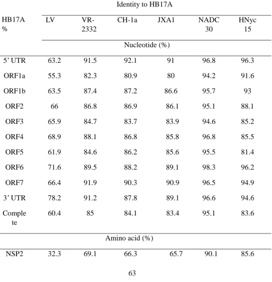
3.1 Characterizations of the full genome of PRRSV isolate HB17A ... 62
3.2 Comparative analysis of the Nsp2 region ... 64
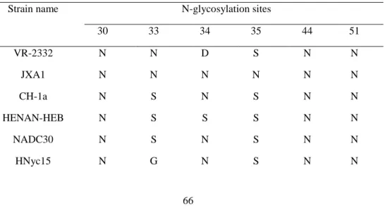
3.3 Sequence alignment and analysis of GP5 ... 65
3.4 Phylogenetic analysis ... 67
3.5 Recombination analysis ... 68
3.6 Observation of clinical signs during infection ... 69
3.7 Viral loads in serum samples ... 70
3.8 Antibody detection post infection ... 71
3.9 Virus detection in nasal secretions and tissue samples ... 71
3.10 Cytokine concentrations in serum ... 71
3.11 Gross pathology and histological evaluations of lungs ... 72
4 Discussion ... 74
5 References ... 79
Title 5 The preliminary evaluation of efficacy of PRRS inactivated vaccine based on the viral purification and inactivation methods ... 83
1 Introduction ... 86
2 Methods ... 87
2.1 Virus propagation ... 87
2.2 Concentration of the virus ... 87
2.3 Purification ... 88
x
2.4 Quantitative analysis of purified PRRS virus ... 88
2.5 Protein assays ... 88
2.6 Virus inactivation ... 89
2.6.1 Binary ethylenimine (BEI) ... 89
2.6.2 Formaldehyde (F) ... 89
2.6.3 β-propiolactone (BPL) ... 89
2.7 Analysis of virus inactivation ... 89
2.7.1 Virus inactivation verification test ... 89
2.7.2 RT-PCR ... 89
2.7.3 Detection of the integrity of inactivated virus antigens by SDS-PAGE and Westernblot ... 90
2.8 Experimental design of animal studies ... 90
2.9 Clinical signs ... 90
2.10 ELISA and virus neutralization assay ... 90
2.11 Quantification of PRRSV ... 91
2.12 Pathological examinations ... 91
3 Results ... 91
3.1 Comparison of the efficacy of the two purification methods ... 91
3.1.1 Virus recovery and concentration ... 91
3.1.2 SDS-PAGE and Western blot analysis ... 92
3.1.3 Analysis of the immunogenicity of the purified virus ... 94
3.2 Comparison of the efficacy of the inactivants ... 95
3.2.1 Inactivation of PRRSV-FZ06A using different concentrations of BEI at 30°C ... 95
3.2.2 Inactivation of PRRSV-FZ06A using different concentrations of formaldehyde at 37°C ... 96
3.2.3 Inactivation of PRRSV-FZ06A using different concentrations of BPL at 4°C ... 96
xi
3.2.5 Inactivated virus antigen integrity test ... 98
3.3 Clinical examination ... 99
3.4 Antibody detection ... 101
3.5 PRRSV VN titer assay ... 102
3.6 Viral loads in serum samples ... 102
3.7 Gross pathology and histological evaluations of lungs ... 102
4 Discussion ... 103
5 References ... 107
Title 6 General discussion and perspectives ... 113
1 Surveillance and genetic evolution analysis ... 115
2 Chromatography methods for virus purification ... 116
3 Persistent infection ... 117
4 Vaccine adjuvant ... 118
5 Regional elimination ... 118
6 Limitations and prospects for future research ... 119
7 Conclusion ... 121
8 References ... 122
xiii
List of Figures
Figure 1. Clinical presentation of pigs with “high fever” disease. ... 4
Figure 2. Severe damage to multiple organs in dead pigs. ... 4
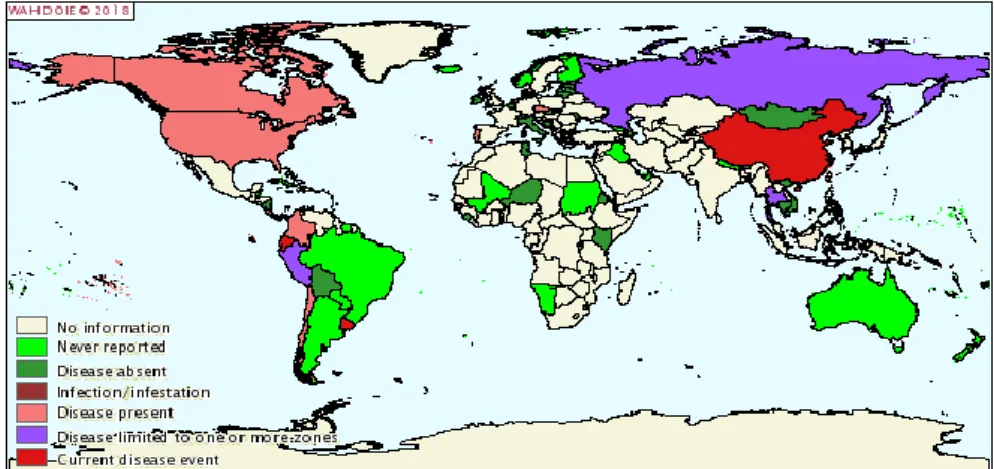
Figure 3. Worldwide distribution of PRRS by 2018 ... 5
Figure 4. The genome organisation and structure of PRRSV... 6
Figure 5. Cytopathic effects of isolated PRRS virus cultured in Marc-145 cells. .... 37
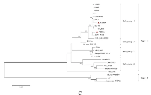
Figure 6. Phylogenetic tree of 24 PRRSV isolates based on analysis of nucleotide sequences of the complete genomic sequences, Nsp2, and ORF5 of PRRSV strains... 42
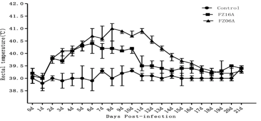
Figure 7. Mean rectal temperature in negative-control pigs and pigs infected experimentally with FZ06A and FZ16A PRRSV. ... 43
Figure 8. Serum antibody levels over time.. ... 44
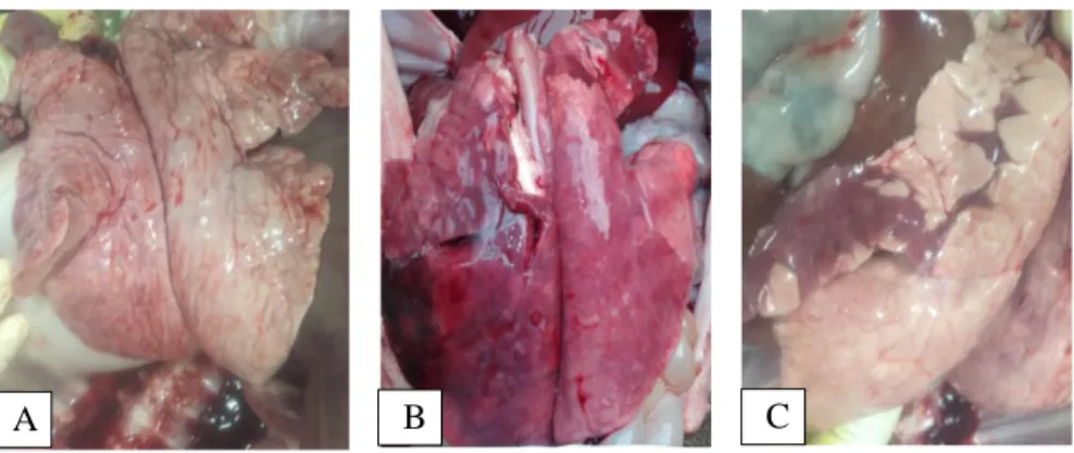
Figure 9. Macroscopic observation of the lungs of the control and HP-PRRSV infected pigs. ... 45
Figure 10. Histopathological lesions in the lung of control and HP-PRRSV infected pigs. ... 45
Figure 11. Alignment of the putative amino acid sequence of Nsp2. ... 65
Figure 12. Alignment of ORF5 amino acid sequences.. ... 66
Figure 13. Phylogenetic trees of the nucleotide sequences of complete genomic sequences, Nsp2, and ORF5 of 28 PRRSV strain. ... 68
Figure 14. Rectal temperatures, body weights, clinical scores, viremias and viral RNA loads in lungs, and virus-specific antibody responses in piglets ... 70
Figure 15. Mean pro-inflammatory cytokines levels. ... 72
Figure 16. Gross and microscopic observations of lungs and the detection of PRRSV in lungs of piglets by IHC.. ... 74
Figure 17. Purification of PRRSV on a C26/100 sepharose 4 fast flow column. ... 93
Figure 18. Linear gradient elution of PRRSV in Q Sepharose High Performance column. ... 93
xiv
Figure 20. Immunogenicity of purified PRRSV was tested by immunoperoxidase
monolayer assay. ... 95
Figure 21. The inactivated effect by BEI at 30°C with different concentrations... 96
Figure 22. The inactivated effect by formaldehyde at 37°C with different concentrations. ... 96
Figure 23. The inactivated effect by BPL at 4°C with different concentrations. ... 97
Figure 24. Inactivated virus passage on Marc-145 cell. ... 97
Figure 25. PCR identification for inactivated PRRSV. ... 98
Figure 26. Western blot analysis... 99
Figure 27. Clinical signs observation of vaccinated pigs and unvaccinated pigs infected experimentally with FZ06A strain. ... 100
Figure 28. Mean rectal temperatures in vaccinated pigs and unvaccinated pigs challenged experimentally with FZ06A strain. ... 100
Figure 29. Body weight in vaccinated pigs and unvaccinated pigs challenged experimentally with FZ06A strain. ... 101
Figure 30. PRRSV-specific antibody titer in pigs. ... 101
Figure 31. Macroscopic observation of the lungs of the vaccinated pigs and unvaccinated pigs after challenge with FZ06A. ... 102
xv
List of Tables
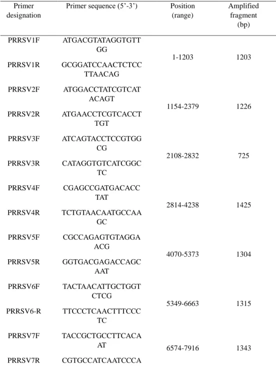
Table 1. Primers used for amplification and sequencing of gene fragments of
PRRSV strains FZ06A and FZ16A. ... 32
Table 2. Representative PRRSV strains used in this study. ... 34 Table 3. Nucleotide and deduced amino acid identities of the full-length genomes of
FZ06A and FZ16A with 8 reference strains of PRRSV. ... 39
Table 4. The potential N-glycosylation sites in various PRRSV strains. ... 40 Table 5. Detailed comparison of the full-length genomes of HB17A and six
reference strains of PRRSV. ... 63
Table 6. Potential N-glycosylation sites in PRRSV strains. ... 66 Table 7. The recovery and protein removal of virus purified by ultrafiltration and
xvii
List of Abbreviations
AA Amino acid
ADE Antibody-dependent enhancement APC Antigen-presenting cells
BEI Binary ethylenimine
bp Base pair
BPL β-propiolactone BSA Bovine serum albumin
conc Concentration
CPE Cytopathic effect
CpG ODN CpG Oligodeoxynucleotides CSFV Classical swine virus
CTL Cytotoxic T-cell
d Day
DC Dendritic cells
DIVA Differentiating Infected from Vaccinated Animals DMEM Dulbecco’s modified Eagle’s medium
dpi Days post-infection
E Envelope
EAV Equine arteritis virus
ELISA Enzyme-linked immunosorbent assay ELISpot Enzyme-linked immunospot assay
F Formaldehyde
FBS Fetal bovine serum
FMD Foot and mouth disease
g Gravity
h
HourHIV Human immunodeficiency virus
HP-PRRSV Highly pathogenic porcine reproductive and respiratory syndrome virus HVJ-E Hemagglutinating Virus of Japan Envelope
IEC Ion exchange chromatography IFA Indirect fluorescent antibody
IFN Interferon
IHC Immunohistochemistry
IL Interleukin
i.n. Intranasal
IPMA Immunoperoxidase monolayer assay
kb Kilo base
KD Kilo dalton
LDV Lactate dehydrogenase-elevating virus
LV Lelystad virus
xviii
MLV Modified live-attenuated vaccine
N Nucleocapsid NGS N-glycosylation site NK Natural killer nm Nano meter NSP Non-structural protein nt Nucleotide OD Optical density
ORFs Open reading frames
OTU Ovarian tumor
PAM Porcine alveolar macrophage PBL Peripheral blood lymphocytes PBS Phosphate-buffered saline PCV-2 Porcine circovirus type 2 PLGA Poly(d,l-lactide-co-glycolide)
PLP2 Papain-like protease domain 2 PNE Primary neutralizing epitope
PRRS Porcine reproductive and respiratory syndrome PRRSV Porcine reproductive and respiratory syndrome virus
PRV Pseudorabies virus
q-PCR
Quantitative polymerase chain reactionRT-PCR Reverse transcription polymerase chain reaction
sec Second
SHFV Simian hemorrhagic fever virus
S/P Sample-to-positive
SPF Specific-pathogen-free
TCID50 50% tissue culture infective doses TEM Transmission electron microscopy TGF Transforming growth factor
TM Transmembrane region
TNF Tumor necrosis factor
TRS Transcription-regulating sequence
UTR Untranslated region
UV Ultraviolet
V Voltage
VN Virus neutralizing
μ Micro
xix
Supplementary Data
Figure S1. Transmission electron microscopy image of isolated porcine
1
3
1 The origin, transmission route and classification of
PRRS
Porcine reproductive and respiratory syndrome (PRRS) is caused by porcine reproductive and respiratory syndrome virus (PRRSV). It is also known as high fever pig disease, swine plague and blue ear disease (Paton et al., 1991). In the late of 1980s, PRRS was first discovered in North Carolina in the United States and later in European countries and other areas of the world (Collins et al., 1992; Wensvoort et al., 1992). At the end of 1995, the first case report on PRRS occurred in northern China. In 1996, China’s first PRRSV epidemic strain CH-1a was isolated from miscarried fetuses (Guo et al., 1996). After that, other epidemic strains such as BJ-4, HB-1(sh)/2002 and HB-2(sh)/2002 were successively isolated (Zhou et al., 2010). In 2006, a new PRRSV variant strain, also known as highly pathogenic PRRSV (HP-PRRSV), was identified in Jiangxi, China. It led to high morbidity (50 %-100 %) and mortality (20 %-100 %) in pigs (Tian et al., 2007). The outbreak of HP-PRRSV resulted in a dramatic decline of pig stock and high prices of pork meat in China (Feng et al., 2008). Since 2013, PRRS became prevalent again in China caused by new PRRSV variants, NADC30-like strain, which was considered to be imported from North American and went through extensive variation in China (Zhao et al., 2015; Zhou et al., 2015). Now, HP-PRRSV and NADC30-like strain became the dominant circulating PRRSV strain in China.
Currently, the World Animal Health Organization has listed highly pathogenic PRRS as one of the animal diseases that must be promptly reported. Pigs are the only natural hosts of PRRSV. Piglets or adult pigs of different breeds and genders can be infected by PRRSV. After being infected, pigs often have the following common clinical symptoms (Figure 1): respiratory infections, lameness, shivering, diarrhea, and secondary infections caused by other viruses or bacteria (Tian et al., 2007). Serve damages in multiple organs include lung haemorrhage, interstitial pneumonia, and less often mild vasculitis (Pol et al., 1991; Michel Morin & Yves Robinson 1992; Tian et al., 2007). PRRSV can be transmitted by pig-to-pig infection and transplacental infection to fetuses during mild-to-late gestation (Nathues et al., 2016; Thanawongnuwech et al., 2010). Wild boars and domestic pigs have the same susceptibility to PRRSV (Albina et al., 2000). Wild boars can act as a reservoir for infectious diseases of domestic pigs (Al Dahouk et al., 2005). The potential role of wild boars as a reservoir for PRRSV has been reported in France, Germany, and the USA with serological evidence of infection (Reiner et al., 2009). The outlined ways of spread and infection between domestic pig herds might also potentially affect wild boars, and vice versa. Rates of PRRSV infected wild boars vary drastically with location, season, and PRRSV-strain. Exchange of the virus between domestic pigs and wild boars can be expected because of high prevalences of PRRSV in domestic pigs in China. PRRSV persistence in pigs plays an important role in viral transmission because the virus is present at low levels in the infected animals (Rochon et al., 2015; Horter et al., 2001).
4
Figure 1. Clinical presentation of pigs with “high fever” disease (Tian et al., 2007). (A) Sick
pigs with a fat and healthy appearance. (B) Sick pigs with thin and debilitated feature. (C) The shivering piglet. (D) The limping pig with erythematous blanching rash in its ears. (E) & (F) Pimples observed on the back of an infected pig. (G) & (H) Grown pigs killed during this
epidemic.
Figure 2. Severe damage to multiple organs in dead pigs (Tian et al., 2007). Lung
haemorrhage (indicated at the arrow). (B) Lung edema. (C) Spleen infarct and bladder dilatation filled with mahogany urine. (D) Kidney with many blood spots (at the arrows). (E)
Heart with disorders. (F) Liver with yellow-white necrosis or haemorrhage. (G) Encephala softened slightly. (H) Brain putamen with blood egression emission. (I) Lymph node with
5
Depending on the genetic diversity and geographic distribution, PRRSV can be divided into European type (I type) and North American type (II type) (Benfield et al., 1992; Collins et al., 1992). Representative strain of European type is LV (Lelystad virus), which was named after its first isolation by Wensvoort et al (Wensvoortet al., 1991) at the Lelystad Institute in the Netherlands. The representative strain of North America is VR-2332. These two types of strains are widespread in North America, Europe, and Asia, but a few countries (Sweden, Switzerland, New Zealand, and Australia) have claimed that they have not yet detected PRRSV (Carlsson et al., 2009; Cannon et al., 1998; Cobb et al., 2015) (Figure 3).
Figure 3. Worldwide distribution of PRRS by 2018.
These two genotypes are associated with a similar clinical symptoms and concurrent emergence (Halbur et al., 1995). They show about 40 % genetic divergence, with a high degree of antigenic variation (Forsberg et al., 2002; Frossard et al., 2012; Le et al., 1997). At present, a number of genetically heterogeneous PRRSV widely co-circulate throughout the world and cause huge economic losses (Van et al., 2011; Stadejek et al., 2013; Zhang et al., 2018). However, there is still a lack of effective vaccines that can prevent PRRSV infection. Therefore, in-depth study of PRRSV gene structure, protein function, virus biological activity, pathogenic mechanism and development of safe and effective vaccines is mandatory.
2 PRRSV genome structure, protein function and
biological characteristics
2.1 PRRSV genome structure
PRRSV is a single-stranded, positive-sense RNA virus. It belongs to the family
6
relevance such as lactate dehydrogenase-elevating virus (LDV) of mice, equine arteritis virus (EAV), and simian hemorrhagic fever virus (SHFV) (Cavanagh et al., 1997; Dunowska et al., 2012). The mature virus particle has a diameter of 50-72 nm and has an icosahedral viral capsid. The capsid contains the viral RNA genome (Kim et al., 1993). The PRRSV genome is about 15 kb in length. It has a polycistronic structure, and contains at least 10 open reading frames (ORFs) flanked by two untranslated regions 5’-untranslated region (5’UTR) and 3’-untranslated region (3’UTR) (5’UTR-ORF1a-ORF1b-ORF2a-ORF2b-ORF3-ORF4- ORF5/ORF5a-ORF 6-ORF7-3’UTR). The PRRSV genome encodes a 5’UTR of 217-222 nucleotides (nt; Type 1) and 188-191 nt (Type 2) in length (Yun & Lee, 2013). Type 1 and 2 strains share approximately 50 % genetic homology (Nelsen et al., 1999; Tan et al., 2001). The 3’UTR of 114 nt (Type 1) and 148 nt (Type 2) in length. They also share approximately 60 % nucleotide identity between Type 1 and Type 2 (Choi et al., 2006; Yin et al., 2013). ORF1a and ORF1b encode the non-structural proteins (NSPs), and account for about 80 % of the total length of the viral genome. These 2 ORFs produce 14 non-structural proteins upon enzymatic cleavage. ORF2 through ORF7 encode eight structural proteins including GP2, small envelope (E), GP3, GP4, GP5a, GP5, matrix (M) and nucleocapsid (N) protein (Figure 4) (Allende et al., 1999; Firth et al., 2011; Johnson et al., 2011). After PRRSV infections, viral genomic RNA and subgenomic RNAs are generated. As a template for the viral genome, the full-length RNA also encodes non-structural proteins (NSPs). The subgenomic RNAs encodes the viral structural protein (Yoo et al., 2004).
Figure 4. The genome organisation and structure of PRRSV.
One of the characteristics of the ORFs encoding PRRSV structural proteins is that an antisense transcription-regulating sequence (TRS) at or near the 5’ end of each structural protein coding region (ORF2-7) can form a kissing-loop interaction with a conserved TRS sequence (UUAACC). This sequence located at the 3’ terminus of
7
the 5’ UTR, and it acts similarly to a eukaryotic gene promoter (Kimman et al., 2009; Kappes & Faaberg, 2015). There is a marked difference in the ORFs and TRS sequence of European and North American PRRSV strains (Allende et al. 1999; Nelsen et al., 1999). In addition, the amino acid sequence of the Nsp2 protein of these two viruses only shares 32 % similarity (Allende et al., 1999). Because there are large differences in nucleotide and amino acid sequences between European and North American strains, it has been speculated that these two types of virus may have originated from the same ancestor and then spread and evolved in Europe and North America respectively. Although possible, it should be taken into account that the evolution of the virus in nature requires a long period and other factors. Some researchers also doubt this evolution. Nucleotide alignment of European and North American PRRSV assigned, the epidemic virus isolated in China to North American strains (Zhou et al., 2010; Guo et al., 2018).
2.2 PRRSV Gene-encoded proteins and their function
The PRRSV genome can encode 21 viral proteins. ORF2-ORF7 encodes eight structural proteins of the virus (Figure 4). One of which is the nucleocapsid N protein and the other is envelope proteins. These eight structural proteins are all essential for the production of infectious PRRSV (Terje 2010). The GP5 and M proteins are the major envelope proteins of PRRSV. They are disulfide-linked, exist as heterodimers and interact with PRRSV cell receptors (Hicks et al., 2018). GP5 contains the virus major neutralizing epitope, which has the highest degree of variation in all PRRSV structural proteins (Mu et al., 2015). The M protein is involved in the assembly and release of the virus, and its amino acid sequence is most conserved among PRRSV structural proteins (Wang et al., 2017). The M protein also contains T cell epitopes and neutralizing epitopes (Bautista et al., 1999).
The minor envelope proteins of PRRSV include GP2, GP3, GP4 and E proteins, which are encoded by ORF2a, ORF3, ORF4 and ORF2b, respectively (Matthew et al., 2015). GP2, GP3 and GP4 proteins contain weaker neutralizing epitopes. Gp3 is highly glycosylated among PRRSV structural proteins (Kim et al., 1993; Chen et al., 2014). The ORF7-encoded viral nucleocapsid N protein is the most abundant and most immunogenic protein in viral particles. It accounts for approximately 30 % of the total viral protein (Olasz et al., 2016; LeGall et al., 1998). The N protein is rich in basic amino acids and exists as a homodimer through cysteine interactions. This dimer is good for binding of the N protein to the viral RNA genome. The N protein mainly produces non-neutralization antibodies (Rahe et al., 2017; Li et al., 2015).
Non-structural proteins are encoded by ORF1a and ORF1b. ORF1a expresses 1a protein, while 1a-1b fusion protein is produced by translation frame shifting (Yoo et al., 2004). 1a protein and 1a-1b fusion protein are cleaved by cellular proteases, producing 14 non-structural proteins of PRRSV (Charerntantanakul 2012) (Figure 4). Many of these NSPs have enzymatic activities, such as Nsp2 with cysteine protease activity, Nsp4 with serine protease activity, Nsp11 with endonuclease activity, and Nsp9 with RNA-dependent RNA polymerase activity (Charerntantanakul 2012; Li et al., 2015). Nsp1α, Nsp1β, Nsp2, and Nsp11 can inhibit IFN-mediated signaling
8
pathways, and they also can block the action of IFN-induced genes with antiviral activity (Charerntantanakul 2012; Li et al., 2015).
2.3 Biological characteristics of PRRSV
The host cells of PRRSV are porcine alveolar macrophages (PAMs: porcine alveolar macrophages) and blood mononuclear cells. Macrophages and testicular germ cells derived from other tissues are also susceptible to PRRSV (Sang et al., 2012; Sur et al., 1997). PRRSV was first isolated from primary cultured PAMs (Wensvoort et al., 1991). PRRSV infects PAMs causing apoptosis (Wensvoort et al., 1992; Karniychuk et al., 2011). The virulence of PRRSV is closely related to structural protein GP5 and non-structural protein Nsp3-Nsp8 (Robinson et al., 2013; Rascón-Castelo et al., 2015). The 118 amino acids at the N-terminus of GP5 protein can induce apoptosis (Roques et al., 2013). The common cellular receptors for PRRSV are pCD163 and pCD169, which are associated with the virus husking and entry into the cytosol, respectively (Li et al., 2015; Van et al., 2013). After infection, PRRSV replicates in the cytoplasm (Jourdan et al., 2012).
European PRRSV strains can easily multiply in PAMs. North American strains can infect primary PAMs or replicate in cell lines such as CL2621, Marc-145, and CRL11171. CL2621 and Marc-145 are derived from Rhesus monkey kidney cell lines (Wang et al., 2014; Faaberg et al., 1998). Until now, most of the North American PRRSV strains are isolated using in vitro culture of monkey kidney cell lines. PRRSV can cause viremia after infection in pigs. But due to the delay in the body’s protective immune response, the virus is often difficult to be cleared and becomes a persistent infection. Persistent infection plays an important role in maintaining the replication and spread of PRRSV in pigs. Therefore, it is necessary to further study the mechanism of how PRRSV interacts with the host to cause persistent infection. Although the virulence of different PRRSV strains varies greatly, there is a similar organizational tropism of the strong and attenuated strains (Shang et al., 2013).
Control and eventually eradication of PRRSV is an important issue for swine production. OIE manual of diagnostic tests and vaccines for terrestrial animals provides guidelines for PRRS management: (http://www.oie.int/en/animal-health -in-the-world/animal-diseases/porcine-reproductive-and-respiratory-syndrome/). Upon PRRS outbreak, control measures should be applied at the individual farm level to prevent the spread of the disease. Available vaccines are effective in controlling outbreaks and preventing economic losses.
3 Vaccines
3.1 Inactivated vaccine
PRRSV inactivated vaccine is safe, does not interfere with maternal antibodies, and is easy to store and transport. Its disadvantage is that it requires a high immunization dose, needs multiple immunizations and insufficiently protects against
General introduction
9
heterologous strains. Now the existing commercial inactivated vaccines have been widely used in the European market, including the Ingelvac® PRRS (P120 strain) vaccine from Boehringer AG in sows and piglets, and Suipravac-PRRS (5710 strain) vaccine from Hipra company, Spain, and Progressis® vaccine from Merial company, France, and the SuivacPRRS-IN (VD-E1/E2 and VD-A1 strain) vaccines that can be applied simultaneously to boars, sows and piglets from Dyntec company, Czech; In USA, only briefly appeared in the PRRomiSeTM vaccine of Intervet, which produced from Netherlands. In Asia, South Korea mainly uses the SuiShot® PRRS vaccine, which is produced by its local company. In addition to several European brands, the common PRRSV inactivated vaccine in China mainly include the inactivated vaccine PRRSV-SD1 produced by Shandong Qilu animal health products co., LTD; inactivated NVDC-JXA1 strain produced by Chinese animal husbandry group and Guangdong Winsun Bio-pharmaceutical Co., Ltd..
Dai et al used Hemagglutinating Virus of Japan Envelope (HVJ-E) as an immune adjuvant to add JXA1-R as inactivated vaccine, and the inactivated vaccine had better immune protection for 28-day-old piglets than that without adjuvant (Dai et al., 2011). Inactivated vaccine with HVJ-E as an adjuvant induced greater lymphocyte proliferation, up-regulation of γ-interferon and interleukin-2 (IL-2), and down- regulation of interleukin-10 (IL-10) than the inactivated vaccine without adjuvant. What’s more, after challenge with this inactivated vaccine containing HVJ-E adjuvant can significantly reduce the clinical signs of piglets. Geldhof et al compared the efficacy of three European commercial vaccines and two self-made inactivated vaccines (07V063 and LV strains) (Geldhof et al., 2012). They found that three commercial vaccines could reduce viremia for at least one week. But neither of the two self-made inactivated vaccines affected the duration of viremia. Interestingly, compared to the three commercial inactivated vaccines and attenuated vaccines, two self-made inactivated vaccines induced the production of neutralizing antibodies against 07V063. Then, Geldhof et al performed a second experiment to compare the immunoprotective effect of inactivated vaccines by LV strain, 08V194 strain, and 07V064 strain with commercial attenuated vaccine (Porcilis® PRRS) against the wild type 08V194 strain. The inactivated vaccine against 07V064 strain and LV strain had no significant effect on the duration of viremia. Specific neutralizing antibodies against strain 08V194 were detected in all immunized animals. The attenuated vaccine could quickly induce the production of neutralizing antibodies. These two results suggest that when the field-mutant strains escape the immunity provided by the existing attenuated vaccines, the same type of inactivated vaccine may provide some protection. Karniychuk et al also used PRRSV-07V063 strain to prepare inactivated vaccines (Karniychuk et al., 2012). Three immunizations were performed on the 27th, 55th, and 83th days of pregnancy in the pregnant sow, then the 07V063 strain was used to challenge on the 90th day. This inactivated vaccine did not provide complete protection for sows and fetuses, but it could slightly reduce the viremia of the sows, and improve the survival rate of fetal pigs. Dwived et al use the biodegradable nanoparticle poly (D, L-Lactide-co-glycolide) (PLGA) encapsulated VR-2332 strain inactivated vaccine to intranasally immunize piglets
10
from 3 to 4 weeks of age, and challenge at 21 days (Dwived et al., 2013). They found that the duration of viremia in piglets immunized with nanoparticles- inactivated vaccine was reduced to two weeks. γ-interferon (IFN-γ) was up-regulated in lung homogenate while transforming growth factor-β (TGF-β) was down-regulated. The titer of the lung homogenate in the neutralization test was significantly higher than that in the control and the common inactivated vaccine group. The nanoparticle PLGA encapsulated inactivated vaccine can be used as an effective means to immunize piglets by the nasal route. The immunization route can rapidly clear viremia and defense the PRRSV infection.
3.2 Attenuated vaccine
At present, PRRSV attenuated vaccine is the most widely used in the field. Compare to the inactivated vaccine, attenuated vaccine induce a strong humoral immunity. It also has the ability to replicate in vivo and maintain long-lasting immunity. But a full protection is only achieved against homologous strains. Moreover, it has been shown that an attenuated vaccine virus can return to virulence and cause disease (Bøtner et al., 1997; Nielsen et al. 2001). In 1995, Boehringer AG developed the first commercially North American PRRSV attenuated vaccine-Ingelvac® PRRSV MLV (VR-2332 strain, entered the Chinese market in 2005), which mainly used for pigs aged 3 to 15 weeks. After that, the company developed another North American PRRSV attenuated vaccine-Ingelvac® PRRS ATP (JA-142 strain), which was used for piglets and finisher pigs. There are also attenuated vaccines only used for Europe: Merck’s Porcilis PRRS® (DV strain) attenuated vaccines for sows and gilts, and Amervac-PRRS® (VP046 strain) attenuated vaccine for piglets and gilts from Hipra of Spainsh and Spanish Syva company’s Pyrsvac-183®
(All-183 strains) attenuated vaccine for pigs at each stage. The CH-1R attenuated vaccine, which is produced by Harbin Veterinary Research Institute of Chinese Academy of Agricultural Sciences, was the first attenuated vaccine in China. This vaccine was officially put into production in 2007. After that, other attenuated vaccines were developed: attenuated vaccine JXA1-R strain, the HUN4-F112 strain and the TJM-F92 strain for the HP-PRRSV. When immunized with these three attenuated vaccines, piglets were able to resist well homologous and heterologous strains infection (Tian et al., 2009). In 2009, the attenuated strain R98 was approved as a new veterinary vaccine. The GDr180 attenuated strain produced by Guangdong Winsun bio, also had shown excellent immunity and safety characteristics in clinical trials.
Martelli et al immunized 4-week-old piglets with the attenuated Porcilis PRRS® (DV strain, European type) vaccine by intramuscular injection and intradermal needle-free injection, respectively (Martelli et al., 2009). After a boost at day 45, immunized pigs and the control pigs were maintained in a farm contaminated with the Italian European subtype sharing 84 % nucleotide identity with the attenuated vaccine DV strain. It was observed that the immunized piglets felt better than the control group. Clinical symptoms were reduced by 68 % to 72 %, and respiratory symptoms were reduced by 72 % to 80 %. The clinical protective effect was closely
11
related to significantly elevated cellular immune responses, indicating that injection of the vaccine in two different ways can effectively resist infection of virus having only 84 % nucleotide identity. In addition, experiments also suggest that among the factors affecting the protective effect of vaccines, the ability of the vaccine strain to induce cellular immunity is more important than its homologous relationship with field strains. Wang et al used A2MC2 strain, attenuated vaccine Ingelvac® PRRSV MLV, VR-2332 strain and VR-2385 strain (middle virulent strains) to inoculate animals (Wang et al., 2013). The results showed that A2MC2 strains could induce animals to produce neutralizing antibodies earlier and produce higher levels of neutralizing antibodies and IFN-γ levels than MLV strain. In addition, after immunization with this strain, animals could resist infection of the same strains or atypical strains. It also caused similar pathological damage to the VR-2385 strain, indicating that it could be a better candidate strain for the vaccine. Li et al divided 15 pigs into 3 groups and inoculated with HP-PRRSV BB0907 strain, the Invirvac® PRRSV MLV attenuated vaccine and phosphate-buffered saline (PBS), respectively (Li et al., 2013). Then they placed these three groups in the same pig rearing. When immunized with Ingelvac attenuated vaccine, the infected pigs showed low clinical morbidity, mild viremia, mild fever and lung injury, and a higher level of IFN-γ secretion. It was speculated that the emergency immunization with attenuated vaccine could reduce the risk of exposure to HP-PRRSV in animals.
3.3 Genetic engineering vaccine
Genetic engineering vaccine refers to the use of DNA recombinant biotechnology to insert natural or synthetic genetic material into bacterial, yeast or mammalian cells, so that it can be fully expressed and purified. These types of vaccines include Differentiating Infected from Vaccinated Animals (DIVA) vaccine, vector vaccine, nucleic acid vaccine and subunit vaccine.
DIVA vaccine is a new generation of recombinant live vaccines that use molecular engineering techniques to introduce molecular markers into viral genomes to distinguish them from wild strains. Marked vaccine can be used to effectively distinguish between immunized and wild-infected pigs. This plays a vital role in the prevention and control of PRRSV. Lin et al inserted a marker gene into the N protein of the PRRSV vAPRRS strain to obtain the v7APMa strain, which can be used stably to distinguish between immunized and wild-infected pigs (Lin et al., 2012). Wang et al passed the HP-PRRSV JX143 strain in vitro with 100 times to obtain the attenuated strain JXM100 strain (Wang et al., 2013). Then the original and attenuated strains were sequenced. They found there were a continuous 88 amino acids (AA) deletion of Nsp2 and a total of 75 scattered bases mutation in the whole genome. The 88 AA deletion of JXM100 strain can be used as a marker vaccine to diagnose the vaccine immunized pigs and wild-type naturally infected pigs.
In vector vaccine, the sequence encoding the immunogen is inserted into the genome of the vector. After inoculation, the antigen is expressed in large amounts as the vaccine strain proliferates in vivo. Baculovirus is widely used in recent years as a carrier system for the efficient expression of foreign proteins. Wang et al, Nam et al,
12
and Wu et al all use baculovirus as a vector to express GP5 and M genes of PRRSV (Wang et al., 2007; Nam et al., 2013; Wu et al., 2013). The results showed that the new vaccine had a greater improvement in immunogenicity than ordinary nucleic acid vaccines. The ability to stimulate the body to produce IFN-γ was positively correlated with the vaccine dose.
In nucleic acid vaccines, foreign DNA or RNA encoding certain antigenic proteins is injected directly into animal cells and synthesizes antigenic proteins through the host cell’s expression system, thereby stimulating an immune response to the antigenic protein. Zhang et al mixed the molecular complement protein adjuvant with GP5 protein of PRRSV, and prepared the pcDNA3.1-C3d-p28. n-GP5 nucleic acid vaccine, which containing multiple mC3d-p28 genes (Zhang et al., 2011). Anti-GP5 antibodies, GP5 neutralizing antibodies, IFN-γ and IL-4 on pcDNA3.1- C3d-p28.n-GP5 were significantly higher in inoculated mice compared to the pcDNA3.1-GP5 immunized group. It indicated that the adjuvant protein could effectively enhance the specific immune response of the antigen.
Subunit vaccines use surface structure component (antigen) of a microorganism. Prieto et al inoculated piglets with the purified GP5 subunit vaccine expressed in
E.coli (Prieto et al., 2011). The immunized group showed more severe clinical
symptoms such as dyspnea and progressive weight loss than the blank-injection group. This indicated that the subunit vaccine not only fails to provide immune protection, but promoted PRRSV infection in piglets. Its specific mechanism remains to be further studied. Yang et al fused ORF1b, ORF7, M and GP5 gene of PRRSV into a vector based on Pseudomonas exotoxin, and successfully made a subunit vaccine inducing a cytotoxic response (Yang et al., 2013). Clinical trials showed that this subunit vaccine could effectively protect piglets and sows from being infected by PRRSV. It also could reduce clinical symptoms and viremia in sows.
3.4 Factors affecting the efficacy of PRRS vaccine
Although several commercial attenuated and inactivated PRRS vaccines have been widely used to control PRRSV, there also remain concerns about the safety of attenuated vaccines and the efficacy of inactivated vaccines. First, it is still unclear which PRRSV antigen stimulates the protective immunity. So we cannot identify the best immunogen to produce the vaccine; secondly, because PRRSV is a RNA virus, the viral gene has a high frequency of mutation associated with antigenic variability. So, there is a risk to produce a mutant strain that escapes immune recognition. Thirdly, PRRSV can suppress the immune response and use other mechanisms to escape the body’s immune surveillance system. PRRSV can inhibit expression of IFN-α and tumor necrosis factor-α (TNF-α) in the infected cells (Albina et al., 1998; Murtaugh and Foss, 2002; Van Reeth et al., 1999); fourthly, it remains to be elucidated what factors determine virulence among different PRRSV strains (Guo et al., 2018).
13
(Yoon et al., 1996). The ADE of PRRSV infection is a phenomenon in which PRRSV-specific antibodies enhance the entry of virus, and in some cases the replication of virus, into monocytes or macrophages through interaction with Fc and/or complement receptors (Tirado & Yoon, 2003). In the early stage of infection by PRRSV, pigs mainly produce non-neutralizing antibodies against viral proteins. Neutralizing antibodies are generally produced three weeks later PRRSV infection, and the titer is very low (Lopez-Fuertes et al., 2000; Kimman et al., 2009). The M and N proteins of PRRSV stimulate the production of non-neutralizing antibodies (Choi et al., 2016). In addition, GP5 neutralizing epitopes are masked by surrounding non-neutralizing epitopes (Popescu et al., 2017). In addition, there are multiple glycosylation sites near the non-neutralizing GP5 epitopes. When these sites are glycosylated, they may mask adjacent neutralizing epitopes. This mechanism impairs the production of neutralizing antibodies (Lopez et al., 2004).
Non-specific immunity in pigs also affects the production of specific immunity. In the early stage of PRRSV infection, the changes in cytokine expression profiles may affect the type of immune response. High expression of IFN-γ may be beneficial for stimulation cellular immune response (Bautista & Molitor, 1999; Rowland et al., 2001; Binjawadagi et al., 2016). Therefore, it is important to study the role of different cytokines in protective immunity. In addition, the route, the dose and the boosting of the vaccine may affect the vaccination effect of the vaccine.
3.5 Future perspective
The ideal PRRS vaccine should enhance the innate and adaptive immunity against PRRSV, while blocking the immunosuppression of PRRSV (Renukaradhya et al., 2015; Renukaradhya et al., 2015). Follwing the legislation of the ministry of agriculture and rural affairs of the People’s Republic of China, a PRRS vaccine must be safe. This means that, after immunization, the pigs should not have any adverse reaction, allergy symptom or stress response. The pregnant pigs should give birth normally to healthy piglets. There should be no transmission of the virus through the placenta and between pigs. The vaccine should also contain enough viral antigens able to elicit an optimal immunity. After immunization, pigs must resist the viral infection, reduce the viremia and/or attenuate clinical symptoms. In addition, the new PRRS vaccine should provide protection to different PRRSV lineage. Wild-type PRRSV infection and vaccinated animals should be clearly identified. The important sign of an effective PRRS vaccine is that when a vaccinated pig is exposed to PRRSV again, the pig should be able to rapidly produce a strong humoral and cellular immune recall response, and quickly clear the virus from the body.
Because there are different kinds of PRRSV genotypes, it may be considered to develop a multivalent PRRS live vaccine, which includes several types or different antigenic PRRSVs in order to achieve broad-spectrum immune effects against different PRRSV genotypes. The closer the antigenicity of the PRRS vaccine strain to its epidemic strain, the better the protective effect of the vaccine. It is worthwhile to consider the virus strain closest to the epidemic strain when preparing the vaccine.
14
4 References
Al Dahouk, S., Nöckler, K., Tomaso, H., Splettstoesser, W.D., Jungersen, G., Riber, U., Petry, T., Hoffmann, D., Scholz, H.C., Hensel, A., Neubauer, H., 2005. Seroprevalence of brucellosis, tularemia, and yersiniosis in wild boars (Sus scrofa) from north-eastern Germany. J. Vet. Med. B. Infect. Dis. Vet. Public. Health. 52, 444-455.
Albina, E., Carrat, C., Charley, B., 1998. Interferon-alpha response to swine arterivirus (PoAV), the porcine reproductive and respiratory syndrome virus. J. Interferon. Cytokine. Res. 18, 485-490.
Albina, E., Mesplède, A., Chenut, G., Le Potier, M.F., Bourbao, G., Le Gal, S., Leforban, Y., 2000. A serological survey on classical swine fever (CSF), Aujeszky's disease (AD) and porcine reproductive and respiratory syndrome (PRRS) virus infections in French wild boars from 1991 to 1998. Vet. Microbiol. 77, 43-57.
Allende, R., Lewis, T.L., Lu, Z., Rock, D.L., Kutish, G.F., Ali, A., Doster, A.R., Osorio, F.A., 1999. North American and European porcine reproductive and respiratory syndrome viruses differ in non-structural protein coding regions. J. Gen. Virol. 80, 307-315.
An, T.Q., Tian, Z.J., Leng, C.L., Peng, J.M., Tong, G.Z., 2011. Highly pathogenic porcine reproductive and respiratory syndrome virus, Asia. Emerg. Infect. Dis. 17, 1782-1784.
Bautista, E.M., Suárez, P., Molitor, T.W., 1999. T cell responses to the structural polypeptides of porcine reproductive and respiratory syndrome virus. Arch. Virol. 144, 117-134.
Benfield, D.A., Nelson, E., Collins, J.E., Harris, L., Goyal, S.M., Robison, D., Christianson, W.T., Morrison, R.B., Gorcyca, D., Chladek, D., 1992. Characterization of swine infertility and respiratory syndrome (SIRS) virus (isolate ATCC VR-2332). J. Vet. Diagn. Investig. 4, 127-133.
Binjawadagi, B., Lakshmanappa, Y.S., Longchao, Z., Dhakal, S., Hiremath, J., Ouyang, K., Shyu, D.L., Arcos, J., Pengcheng, S., Gilbertie, A., Zuckermann, F., Torrelles, J.B., Jackwood, D., Fang, Y., Renukaradhya, G.J., 2016. Development of a porcine reproductive and respiratory syndrome virus-like-particle-based vaccine and evaluation of its immunogenicity in pigs. Arch. Virol. 161, 1579-1589.
Bøtner, A., Strandbygaard, B., Sørensen, K.J., Have, P., Madsen, K.G., Madsen, E.S., Alexandersen, S., 1997. Appearance of acute PRRS-like symptoms in sow herds after vaccination with a modified live PRRS vaccine. Vet. Rec. 141, 497-499.
Cannon, N., Audige, L., Denac, H., Hofmann, M., Grinot, C., 1998. Evidence of freedom from porcine reproductive and respiratory syndrome virus infection in Switzerland. Vet. Res. 142, 142-143.
Carlsson, U., Wallgren, P., Renström, L.H., Lindberg, A., Eriksson, H., Thorén, P., Eliasson-Selling, L., Lundeheim, N., Nörregard, E., Thörn, C., Elvander, M., 2009. Emergence of porcine reproductive and respiratory syndrome in Sweden: detection, response and eradication. Transbound. Emerg. Dis. 56, 121-131.
Cavanagh, D., 1997. Nidovirales: a new order comprising Coronaviridae and Arteriviridae. Arch. Virol. 142, 629-633.
15
Charerntantanakul, W., 2012. Porcine reproductive and respiratory syndrome virus vaccines: Immunogenicity, efficacy and safety aspects. World. J. Virol. 1, 23-30.
Chen, J.Z., Wang, Q., Bai, Y., Wang, B., Zhao, H.Y., Peng, J.M., An, T.Q., Tian, Z.J., Tong, G.Z., 2014. Identification of two dominant linear epitopes on the GP3 protein of highly pathogenic porcine reproductive and respiratory syndrome virus (HP-PRRSV). Res. Vet. Sci. 97, 238-243.
Choi, K., Park, C., Jeong, J., Kang, I., Park, S.J., Chae, C., 2016. Comparison of commercial type 1 and type 2 PRRSV vaccines against heterologous dual challenge. Vet. Rec. 178, 291.
Choi, Y.J., Yun, S.I., Kang, S.Y., Lee, Y.M., 2006. Identification of 5’ and 3’ cis-acting elements of the porcine reproductive and respiratory syndrome virus: acquisition of novel 5’ AU-rich sequences restored replication of a 5’-proximal 7-nucleotide deletion mutant. J. Virol. 80, 723-736.
Cobb, S.P., Pharo, H., Stone, M., Groenendaal, H., Zagmutt, F.J., 2015. Quantitative risk assessment of the likeihood of introducing porcine reproductive and respiratory syndrome virus into New Zealand through the importation of pig meat. Res. Sci. Tech. 34, 961-975.
Collins, J.E., Benfield, D.A., Christianson, W.T., Harris, L., Hennings, J.C., Shaw, D.P., Gpyal, S.M., McCullough, S., Morrison, R.B., Joo, H.S., 1992. Isolation of swine infertility and respiratory syndrome virus (isolate ATCC VR-2332) in North America and experimental reproduction of the disease in gnotobiotic pigs. J. Vet. Diagn. Invest. 4, 117-126.
Dai, Z., Zhang, Q., Wang, W., Zhang, Z., Guo, P., Zhao, D., 2011. Hemagglutinating virus of Japan envelope (HVJ-E) can enhance the immune responses of swine immunized with killed PRRSV vaccine. Biochem. Biophys. Res. Commun. 415, 1-5.
Dokland, T., 2010. The structural biology of PRRSV. Virus. Res. 154, 86-97. Dunowska, M., Biggs, P.J., Zheng, T., Perrott, M.R., 2012. Identification of a novel nidovirus associated with a neurological disease of the Australian brushtail possum (Trichosurus vulpecula). Vet. Microbiol. 156, 418-424.
Dwivedi, V., Manickam, C., Binjawadagi, B., Renukaradhya, G.J., 2013. PLGA nanoparticle entrapped killed porcine reproductive and respiratory syndrome virus vaccine helps in viral clearance in pigs. Vet. Microbiol. 166, 47-58.
Faaberg, K.S., Elam, M.R., Nelsen, C.J., Murtaugh, M.P., 1998. Subgenomic RNA7 is transcribed with different leader-body junction sites in PRRSV (strain VR-2332) infection of CL2621 cells. Adv. Exp. Med. Biol. 440, 275-279.
Feng, Y., Zhao, T., Nguyen, T., Inui, K., Ma, Y., Nguyen, T.H., Nguyen, V.C., Liu, D., Bui, Q.A., To, L.T., Wang, C., Tian, K., Gao, G.F., 2008. Porcine respiratory and reproductive syndrome virus variants, Vietnam and China, 2007. Emerg. Infect. Dis. 14, 1774-1776.
Firth, A.E., Zevenhoven-Dobbe, J.C., Wills, N.M., Go, Y.Y., Balasuriya, U.B., Atkins, J.F., Snijder, E.J., Posthuma, C.C., 2011. Discovery of a small arterivirus gene that overlaps the GP5 coding sequence and is important for virus production. J. Gen. Virol. 92, 1097-1106.
16
Forsberg, R., Storgaard, T., Nielsen, H.S., Oleksiewicz, M.B., Cordioli, P., Sala, G., Hein, J., Bøtner, A., 2002. The genetic diversity of European type PRRSV is similar to that of the North American type but is geographically skewed within Europe. Virology. 299, 38-47.
Frossard, J.P., Fearnley, C., Naidu, B., Errington, J., Westcott, D.G., Drew, T.W., 2012. Porcine reproductive and respiratory syndrome virus: antigenic and molecular diversity of British isolates and implications for diagnosis. Vet. Microbiol. 158, 308-315.
Geldhof, M.F., Vanhee, M., Van, Breedam, W., Van, Doorsselaere, J., Karniychuk, U.U., Nauwynck, H.J., 2012. Comparison of the efficacy of autogenous inactivated Porcine Reproductive and Respiratory Syndrome Virus (PRRSV) vaccines with that of commercial vaccines against homologous and heterologous challenges. BMC, Vet. Res. 8, 182.
Guo, B. Q., Chen, Z. S., Liu, W. X., 1996. Isolation and identification of porcine reproductive and respiratory syndrome (PRRS) virus from aborted fetuses suspected of PRRS. Chin. J. Prev. Vet. Med. 2, 1-5.
Guo, Z., Chen, X.X., Li, R., Qiao, S., Zhang, G., 2018. The prevalent status and genetic diversity of porcine reproductive and respiratory syndrome virus in China: a molecular epidemiological perspective. Virol. J. 15, 2.
Halbur, P.G., Paul, P.S., Frey, M.L., Landgraf, J., Eernisse, K., Meng, X.J., Lum, M.A., Andrews, J.J., Rathje, J.A., 1995. Comparison of the pathogenicity of two US porcine reproductive and respiratory syndrome virus isolates with that of the Lelystad virus. Vet. Pathol. 32, 648-660.
Hicks, J.A., Yoo, D., Liu, H.C., 2018. Interaction of porcine reproductive and respiratory syndrome virus major envelope proteins GP5 and M with the cellular protein Snapin. Virus. Res. 249, 85-92.
Horter, D., Chang, C.C., Pogranichnyy, R., Zimmerman, J., Yoon, K.J., 2001. Persistence of porcine reproductive and respiratory syndrome in pigs. Adv. Exp. Med. Biol. 494, 91-94.
Jourdan, S.S., Osorio, F., Hiscox, J.A., 2012. An interactome map of the nucleocapsid protein from a highly pathogenic North American porcine reproductive and respiratory syndrome virus strain generated using SILAC-based quantitative proteomics. Proteomics. 12, 1015-1023.
Johnson, C.R., Griggs, T.F., Gnanandarajah, J., Murtaugh, M.P., 2011. Novel structural protein in porcine reproductive and respiratory syndrome virus encoded by an alternative ORF5 present in all arteriviruses. J. Gen. Virol. 92, 1107-1116.
Kappes, M.A., Faaberg, K.S., 2015. PRRSV structure, replication and recombination: Origin of phenotype and genotype diversity. Virology. 479, 475-486.
Karniychuk, U.U., Saha, D., Geldhof, M., Vanhee, M., Cornillie, P., Van, den, Broeck, W., Nauwynck, H.J., 2011. Porcine reproductive and respiratory syndrome virus (PRRSV) causes apoptosis during its replication in fetal implantation sites. Microb. Pathog. 51, 194-202.
Karniychuk, U.U., Saha, D., Vanhee, M., Geldhof, M., Cornillie, P., Caij, A.B., De, Regge, N., Nauwynck, H.J., 2012. Impact of a novel inactivated PRRS virus vaccine
17
on virus replication and virus-induced pathology in fetal implantation sites and fetuses upon challenge. Theriogenology. 78, 1527-1537.
Kim, H.S., Kwang, J., Yoon, I.J., Joo, H.S., Frey, M.L., 1993. Enhanced replication of porcine reproductive and respiratory syndrome (PRRS) virus in a homogeneous subpopulation of MA-104 cell line. Arch. Virol. 133, 477-483.
Kimman, T.G., Cornelissen, L.A., Moormann. R.J., Rebel, J.M., Stockhofe- Zurwieden, N., 2009. Challenges for porcine reproductive and respiratory syndrome virus (PRRSV) vaccinology. Vaccine. 27, 3704-3718.
Le Gall, A., Albina, E., Magar, R., Gauthier, J.P., 1997. Antigenic variability of porcine reproductive and respiratory syndrome (PRRS) virus isolates. Influence of virus passage in pig. Vet. Res. 28, 247-257.
Le Gall, A., Legeay, O., Bourhy, H., Arnauld, C., Albina, E., Jestin, A., 1998. Molecular variation in the nucleoprotein gene (ORF7) of the porcine reproductive and respiratory syndrome virus (PRRSV). Virus. Res. 54, 9-21.
Li, H., Zhou, E.M., Liu, C.Q., Yi, J.Z., 2015. Function of CD163 fragments in porcine reproductive and respiratory syndrome virus infection. Int. J. Clin. Exp. Med. 8, 15373-15382.
Li, J., Tao, S., Orlando, R., Murtaugh, M.P., 2015. N-glycosylation profiling of porcine reproductive and respiratory syndrome virus envelope glycoprotein 5. Virology. 478, 86-98.
Lin, T., Li, X., Yao, H., Wei, Z., Tan, F., Liu, R., Sun, L., Zhang, R., Li, W., Lu, J., Tong, G., Yuan, S., 2012. Use of reverse genetics to develop a novel marker porcine reproductive and respiratory syndrome virus. Virus. Genes. 45, 548-555.
Li, X., Qiu, L., Yang, Z., Dang, R., Wang, X., 2013. Emergency vaccination alleviates highly pathogenic porcine reproductive and respiratory syndrome virus infection after contact exposure. BMC Vet. Res. 9, 26.
Li, Y., Tas, A., Sun, Z., Snijder, E.J., Fang, Y., 2015. Proteolytic processing of the porcine reproductive and respiratory syndrome virus replicase. Virus. Res. 202, 48-59.
Lopez, O.J., Osorio, F.A., 2004. Role of neutralizing antibodies in PRRSV protective immunity. Vet. Immuno. Immunopathol. 102, 155-163.
López-Fuertes, L., Campos, E., Doménech, N., Ezquerra, A., Castro, J.M., Domínguez, J., Alonso, F., 2000. Porcine reproductive and respiratory syndrome (PRRS) virus down-modulates TNF-alpha production in infected macrophages. Virus. Res. 69, 41-46.
Martelli, P., Gozio, S., Ferrari, L., Rosina, S., De Angelis, E., Quintavalla, C., Bottarelli, E., Borghetti, P., 2009. Efficacy of a modified live porcine reproductive and respiratory syndrome virus (PRRSV) vaccine in pigs naturally exposed to a heterologous European (Italian cluster) field strain: Clinical protection and cell-mediated immunity. Vaccine. 27, 3788-3799.
Michel Morin, Yves Robinson, 1992. Causes of mystery swine disease. Can. Vet. J. 33, 6.
Mu, Y., Li, L., Zhang, B., Huang, B., Gao, J., Wang, X., Wang, C., Xiao, S., Zhao, Q., Sun, Y., Zhang, G., Hiscox, J.A., Zhou, E.M., 2015. Glycoprotein 5 of porcine
18
reproductive and respiratory syndrome virus strain SD16 inhibits viral replication and causes G2/M cell cycle arrest, but does not induce cellular apoptosis in Marc-145 cells. Virology. 484, 136-145.
Murtaugh, M.P., Foss, D.L., 2002. Inflammatory cytokines and antigen presenting cell activation. Vet. Immunol. Immunopathol. 87, 109-121.
Nam, H.M., Chae, K.S., Song, Y.J., Lee, N.H., Lee, J.B., Park, S.Y., Song, C.S., Seo, K.H., Kang, S.M., Kim, M.C., Choi, I.S., 2013. Immune responses in mice vaccinated with virus-like particles composed of the GP5 and M proteins of porcine reproductive and respiratory syndrome virus. Arch. Virol. 158, 1275-1285.
Nathues, C., Perler, L., Bruhn, S., Suter, D., Eichhorn, L., Hofmann, M., Nathues, H., Baechlein, C., Ritzmann, M., Palzer, A., Grossmann, K., Schüpbach-Regula, G., Thür, B., 2016. An Outbreak of Porcine Reproductive and Respiratory Syndrome Virus in Switzerland Following Import of Boar Semen. Transbound. Emerg. Dis. 63, e251-261.
Nelsen, C.J., Murtaugh, M.P., Faaberg, K.S., 1999. Porcine reproductive and respiratory syndrome virus comparison: divergent evolution on two continents. J. Virol. 73, 270-280.
Niederwerder, M.C., Rowland, R.R., 2017. Is There a Risk for Introducing Porcine Reproductive and Respiratory Syndrome Virus (PRRSV) Through the Legal Importation of Pork? Food Environ. Virol. 9, 1-13.
Nielsen, H.S., Oleksiewicz, M.B., Forsberg, R., Stadejek, T., Bøtner, A., Storgaard, T., 2001. Reversion of a live porcine reproductive and respiratory syndrome virus vaccine investigated by parallel mutations. J. Gen. Virol. 82, 1263-1272.
Olasz, F., Dénes, B., Bálint, Á., Magyar, T., Belák, S., Zádori, Z., 2016. Immunological and biochemical characterisation of 7ap, a short protein translated from an alternative frame of ORF7 of PRRSV. Acta. Vet. Hung. 64, 273-287.
Paton, DJ., Brown, I.H., Edwards, S., Wensvoort, G., 1991. ‘Blue ear’ disease of pigs. Vet. Rec. 128, 617.
Pol, J.M., van, Dijk, J.E., Wensvoort, G., Terpstra, C., Loula, T., 1991. Pathological, ultrastructural, and immunohistochemical changes caused by Lelystad virus in experimentally induced infections of mystery swine disease (synonym: porcine epidemic abortion and respiratory syndrome (PEARS)). Vet. Q. 13, 137-143. Popescu, L.N., Trible, B.R., Chen, N., Rowland, R.R.R., 2017. GP5 of porcine reproductive and respiratory syndrome virus (PRRSV) as a target for homologous and broadly neutralizing antibodies. Vet. Microbiol. 209, 90-96.
Prieto, C., Martínez-Lobo, F.J., Díez-Fuertes, F., Aguilar-Calvo, P., Simarro, I., Castro, J.M., 2011. Immunisation of pigs with a major envelope protein sub-unit vaccine against porcine reproductive and respiratory syndrome virus (PRRSV) results in enhanced clinical disease following experimental challenge. Vet. J. 189, 323-329.
Rahe, M.C., Murtaugh, M.P., 2017. Mechanisms of Adaptive Immunity to Porcine Reproductive and Respiratory Syndrome Virus. Viruses. 9, 148.
Rascón-Castelo, E., Burgara-Estrella, A., Mateu, E., Hernández, J., 2015. Immunological features of the non-structural proteins of porcine reproductive and