0021-9193/82/121042-07$02.00/0
Copyright©1982, American Society for Microbiology
Penicillin-Binding Proteins and
Carboxypeptidase/Transpeptidase
Activities in Proteus
vulgaris P18 and
Its
Penicillin-Induced Stable L-Forms
ANDRtROUSSET,V
MARTINENGUYEN-DISTECHE,2 RAYMOND MINCK,1 ANDJEAN-MARIEGHUYSEN2*
Universite de Strasbourg, Faculte de Medecine,InstitutdeBacteriologie, F-67000Strasbourg,
France';
andService deMicrobiologie appliquee auxsciencespharmaceutiques, FacultedeMedecine,InstitutdeChimie,
UniversitedeLiege,B4000Sart Tilman(Liege), Belgium2 Received25June1982/Accepted 3 September 1982
Theoriginally penicillin-induced, wall-less stable L-forms ofProteus vulgaris P18, isolated by Tulasne in 1949 and since then cultured in the absence of penicillin, have kept the abilitytosynthesize thesevenpenicillin-binding proteins
andthe variousDD-andLD-peptidase activities foundin theparental bacteria and known to be involved in wall peptidoglycan metabolism. The stable L-forms, however, secreteduringgrowth both the highly penicillin-sensitive, DD-carboxy-peptidase-transpeptidase penicillin-binding protein PBP4 (which in normal bacte-ria is relatively loosely bound to the plasma membrane) and the penicillin-insensitive LD-carboxypeptidase (which in normal bacteria is located in the periplasmic region).
The
rod-shaped
enterobacteria
cangive rise
by
apenicillin-induced
process to two typesof
coccal
L-forms.
The unstable L-forms
(sphero-plasts) have defects in their wall peptidoglycan
and
areosmotically fragile. The induced lesion is
reversible, and
uponremoval
of penicillin from
the growth medium, both normal peptidoglycan
synthesis and reversion
tothe
original
bacteria
occur.
In
contrast,the stable
L-forms
(proto-plasts)
grow aspermanently wall-less, ovoid
organisms
and
cando
sounder
conditions of
normal
osmotic stabilization whether penicillin
is
present or not (1,8, 25). Like the other
enterobacteria
studied (4, 5, 20),
Proteusmirabi-lis
possesses sevenpenicillin-binding proteins
(PBPs), referred
to asPBP1A, 1B, 2, 3, 4, 5, and
6
(in
the order of
decreasing
molecular
weight).
On the basis of the PBP
pattern
of the normal
bacteria and thatof the corresponding stable
L-forms,
Martin
etal. (13) have
suggested that the
permanent
inability of these L-forms
tosynthe-size
anormal
peptidoglycan might
be related tothe hereditary and selective loss of
PBP4. PBP4is known
tobe
aD-alanyl-D-alanine-cleaving
peptidase which is able
toshow,
onwell-defined
substrates, carboxypeptidase, transpeptidase,
and
endopeptidase activities (22).
Inview of
theimportance
of
the wall-lessgrowth forms of
bacteria in clinical
cases,another
stableL-form
originating
from
Proteusvulgaris
P18has been
investigated
andcompared with
itsparental
strain.
This stable L-form
wasisolated
by
Tu-lasne
in
1949 as apenicillin-resistant organism
(25). Since
then, the cells have been subcultured
twice
weekly
in
suitable
growth
medium in the
absence
of
penicillin.
(This
paperis
from
adissertation submitted
by
A.R.
in
partial
fulfillment
of
the
requirements
for
a
degree
of
Docteur
6sSciences
atthe
Universi-ty
of
Strasbourg,
Strasbourg,
France.)
MATERIALSANDMETHODS
Strains and growth conditions. P. vulgaris P18 was grownat37° Cwithshaking in standard Merck I broth medium, and the cells were harvested at the midlog phase (optical density at 600 nm, 1). The stable L-formsweregrownat37° Ceither withoutshakingin the modifiedMedill-O'Kane medium (17) or with shaking inthemodified Medill-Brown medium (16). Both cul-turesgaveidentical results.
Preparation of the cell envelopes. Three procedures were used to prepare the cell envelopes from the normal bacteria.
(i) Standardprocedure (via spheroplasts formation). Cells (1 g,wetweight)weresuspended in30ml of 50 mMTris-hydrochloride buffer (pH 8.0) containing 0.75 Msucrose and40 mg oflysozyme, and the suspen-sionsweresubmitted to successivefreezingand thaw-ing. Afteranosmoticshock in distilled water, the cell envelopes were collected by centrifugation. Formore
details, seereferences 9 and11.
(ii) Ribi procedure. Cells (1g, wet weight) were suspended in 5 ml of10 mMTris-maleate buffer (pH 7.0)containing1 mMMgCl2,and thesuspensionwas
treated withaRibi press at 30,000
lb/in2
andatempera-turerangingbetween4and15° C (21). The cell
enve-lopeswerecollectedbycentrifugation.
(uii) Modified Nossal-Heppel procedure (19). Cells (collectedfrom1liter ofculture)weresuccessively (i) 1042
1043 plasmolyzed for 15 minat25C in 100 ml of 10 mM
Tris-hydrochloride buffer (pH 7.3) containing 30 mM NaCI,0.5 mM EDTA, and 0.58 M sucrose (centrifuga-tion gave rise to a supematant S1); (ii) rapidly homoge-nized in 100 ml of distilled water at 4° C(centrifugati'on gaverisetoasupernatant
S2); (iii)
treated for 30 minat4° Cwith 5 ml of 10 mMTris-hydrochloridebuffer (pH 7.3)containing 1 MNaCI(centrifugation gave rise to a supernatant
S3);
and (iv) submitted to sonication with aMeasuringScientificEquipment apparatus at 25,000 Hz and at0° C(centrifugation gave rise to a superna-tantS4 and a pellet which consisted of cell envelopes). Each supematant was dialyzed against 0.25 M Tris-hydrochloride buffer (pH 8.9) and concentrated by ultrafiltrationto 4 ml.The cell envelopes of the stable L-forms were prepared according to the standard procedure de-scribed above, butwithoutsupplementallysozyme.In all cases,thecellenvelopesweresuspended in0.25 M Tris-hydrochloride buffer(pH 8.9) at aconcentration ofabout 50 mgofproteinper ml.Theywerestoredin thefrozenstate at-20° C.
Proteindetermination.Theproteinswereestimated by the technique ofLowry et al. (10) with bovine serumalbuminas astandard and in the case of thecell envelope,inthepresenceof sodium dodecyl sulfateas described previously (23).
PBPanalysis. Samples (45
p1)
ofthecellenvelopes (150pg
ofprotein)orthesolubilizedenzyme prepara-tions (at a properconcentration)wereincubatedwith[14C]benzylpenicillin
(fromthe RadiochemicalCentre, Amersham;final specific radioactivity, 50mCi/mmol) attheindicated concentration for10 min at 37° C, and the reactionwas terminated by the addition of 8 mM (final concentration) nonradioactive benzylpenicillin and 1% (wt/vol, final concentration) Sarkosyl. After centrifugation (inthecase of the cell envelopes), thetween the PBPs and [14C]benzylpenicillin were esti-matedbythetechniqueofSpratt(24).The thermola-bility of the native PBPs (before reaction with radioactive benzylpenicillin) was determined as de-scribed by Ohya et al. (20). The apparent molecular weights of the PBPs were determined by comparing their mobilities on thegels withthose ofmyoglobin
(Mr,
17,800), chymotrypsinogen A(Mr,
25,000), oval-bumin(M1,
43,000), andbovine serum albumin(Mr, 68,000).Analytical polyacrylamide gel electrophoresis atpH 8.3 under nondenaturing conditions. Electrophoresis was carried out on cylindrical gels (0.9 by 12 cm; containing 7% acrylamide and 0.2%NN'-methylene bisacrylamide)in 25 mMTris-glycinebuffer(pH8.3). Afterpreelectrophoresisof thegelsfor 1 h at 1 mA/gel, the enzyme samples were submitted toelectrophoresis forabout 3 h at 4° C and 2 mA/gel. Bromophenol blue wasused as a marker. Afterelectrophoresis,thegels were sliced into 2-mm-thick disks, the disks were
eluted with 300 p.1 of 0.1 M Tris-hydrochloride buffer (pH 8.9), and the eluates were assayed for enzyme activity.
DD-Carboxypepddase, Do-transpeptidase,and LD-car-boxypeptidase assays. All reactions were carried outat
37° C in 30,ul (final volume) of 0.25 M Tris-hydrochlo-ride (pH 8.9)containing2 x
10'
M dithiotreitol. For details, see reference 18. D-Alanyl-D-alanine-cleaving carboxypeptidase (in short, DD-carboxypeptidase) ac-tivity was estimated by measuring the amount ofD-["4C]Ala
released from the nucleotide-pentapeptideUDP-N-acetylmuramyl-L-Ala-y-D-Glu-(L)-meso-A2pm-(L)-D-[14C]Ala-D-["4C]Ala
(1.33 mM; specific radioactivity, 22 mCi/mmol). D-Alanyl-D-alanine-cleavingtranspeptidase (in short, DD-transpeptidase) activity was estimated by measuring the amount of radioactive monoamidated peptide dimerL
L-Ala-y-D-Glu(amide)-
-(D-Ala) A2pm L-Ala-y-D-GIU-(L)-meso-A2pm-(L)-D-[14C]Ala-Dsamples were boiled in the presence of1% sodium dodecyl sulfate and11.6% mercaptoethanol, and the PBPs were separatedbypolyacrylamide
(10%)
slabgel electrophoresis atpH 8.3 inthe presenceof sodium dodecyl sulfate andvisualized byfluorography. The time ofexposure at-70° Ctoprefogged X-ray filmwas 8weeks. For moredetails,seereference24. Relative band intensities on the fluorograms were estimated with amicrodensitometer (BeckmanDU-8; Beckman Instruments,Inc.,Fullerton,Calif.)withpeak integra-tion. Saturation of thePBPs wascarried
outby using increasingconcentrations of[14C]benzylpenicillin
(up to 5 x 10⁻⁴ M). The affinities of various nonradioac-tive3-lactam
compounds for the PBPs were deter-mined bycompetition with['4C]benzylpenicillin
and expressed as the concentrations necessary to inhibit by50%o
further binding of[14C]benzylpenicillin.
For this purpose, the cell envelopes were exposed to serialdilutions of the various antibiotics for 10 minat37° C, and then
[14C]benzylpenicillin
(0.2 mM, final concentration) was added. After a further 10 min of incubation, the reaction wasterminatedasabove by the addition of nonradioactivebenzylpenicillin
and Sarkosyl. The half-lives of the adducts formedbe-formed fromamixture of radioactive pentapeptide
L-Ala--y-D-Glu-(L)-meso-A2pm-(L)-D-[14C]Ala-D-[14C]
Ala (1.33 mM; specific radioactivity, 22 mCi/mmol) and amidated tetrapeptide L-Ala--y-D-Glu(amide)-(L)-meso-A2pm-(L)-D-Ala(13.3mM). meso-Diaminopime-lyl-(L)-D-alanine-cleaving carboxypeptidase (in short, LD-carboxypeptidase)activity was estimated by mea-suring the amount of 1-Ala released from the nu-cleotide-tetrapeptide
UDP-N-acetylmuramyl-L-Ala--y-D-Glu-(L)-meso-A2pm-(L)-D>Ala
(1.33 mM). Free D-Ala was estimated as described previously (2). One unit of these enzymes hydrolyzed or catalyzed the synthesis of 1 ,ueq of appropriate linkages per min.ID50
values. The DD-peptidases (but not the LD-carboxypeptidase)weresensitive to ,B-lactam antibiot-ics. TheID50values were the antibiotic concentrations whichinhibited the enzyme activities by50%.Isolation of the highly penicillin-sensitive DD-pepti-dase PBP4, the moderatelypenicillin-sensitive DD-pepti-dase PBP5, and the
peniciflin-insensitive
LD-carboxy-peptidase. Normal bacteria (90 g, wet weight) suspendedin 450 ml of 10 mM Tris-maleate buffer (pH 7) weredisrupted with a Ribi press (see above), and the resulting preparation was submitted to centrifuga-152,tion for 6 h at 200,000 x g. The supernatant fraction and the membrane pellet were treated as follows (all the operations were carried out at 4° C).
The supernatant (465 ml) was submitted to (NH4)2SO4fractionation. The precipitate collected at 30 to 60%o saturation, which contained 75% of the highly penicillin-sensitiveDD-peptidase and95%of the insensitive LD-peptidase, was dissolved in 60 ml of 0.25 M Tris-hydrochloride buffer (pH 8.9) supplement-ed with 0.2 mM dithiothreitol. Afterdialysisagainstthe samebuffer, the enzyme solution was filtered through a column (2.6 by 31 cm) of DEAE-Sephadex. The column was washed and then treated with two succes-sive NaCl gradients (made in the same buffer), first from 0 to 0.15 M NaCl, under which conditions the DD-peptidase waseluted (at about 0.07 MNaCI),and then from 0.15 to 0.4 MNaCl,under which conditions theLD-carboxypeptidasewas eluted (atabout 0.25 M NaCI). The relevantfractions(containing the separat-ed peptidases) were pooled, and the solutions were dialyzed against the Tris-hydrochloride (plus dithio-treitol) buffer (pH 8.9) and concentrated by ultrafiltra-tion.
The above procedure permitted a 180-fold enrich-ment of the highly penicillin-sensitive DD-peptidase (from 5to970mU/mg protein; carboxypeptidase as-say) with a totalyieldof24% and a 300-fold enrich-ment of the LD-carboxypeptidase (from 2.4 to 760 mU/mgofprotein)with a totalyieldof46%. The
DD-peptidase could be detected and identified as PBP4 after theDEAE-Sephadextreatment. Further purifica-tion of theDD-peptidaseto afinalspecificactivityof 4 U/mg ofprotein (carboxypeptidase assay) could be achievedbyfiltrationon acolumn ofUltrogelAcA44 in 0.25 M Tris-hydrochloride buffer(pH8.9) contain-ing0.2 mMdithiotreitol, followedbypolyacrylamide gel electrophoresis at pH 8.3 under nondenaturing conditions. Due to theinstability of the purified
en-zyme, the final yield was poor(1%), butaconstant ratio between DD-carboxypeptidase activity,
DD-transpeptidase activity, and
[14C]benzylpenicillin-binding capacity was found throughout the protein peakeluted from theUltrogelcolumn.
Similarily,
gel electrophoresisshowed that these threeactivitieswereattributableto a sameproteinwhich,under the condi-tions used, migrated3 cm toward the anode.
The membrane pellet originatingfrom the normal bacteria wastreated (at25° Cfor 30min)with 10 mM Tris-maleatebuffer (pH 7.0)
containing
1 mMMgCl2
and 0.5% Genapol X-100 (a gift from
Farbwerke,
Hoechst, Belgium). Undertheseconditions,the
mod-erately penicillin-sensitive DD-peptidase was solubi-lized. The extract was dialyzedagainst 0.25 M Tris-hydrochloride buffer (pH 8.9) containing 0.2 mM dithiotreitol and 0.5% Genapol X-100 and filtered through anampicillin-linked Sepharosecolumn (equili-brated against the samebuffer).The columnwasthen treatedwith the samebufferasabove,butcontaining1 MNaCI,underwhich conditions theDD-peptidasewas
eluted (12). Each step of thepurificationgave risetoa
parallelenrichment in bothDD-peptidase
activity
and PBP5. The final enzyme preparation had a specific carboxypeptidase activityof 60mU/mgofprotein.No PBP other thanPBP5could be detected.The sameprocedures as those described abovewere
applied to the isolation of the corresponding pepti-dases fromgrowingstable L-forms. The
DD-peptidase
PBP5 was isolated from the plasma membrane, butthe DD-peptidase PBP4 (final specific activity, 90 mU/mg of protein in the carboxypeptidase assay) and the LD-carboxypeptidase (final specific activity, 43 mU/mg of protein) were isolated from culture fluids.
RESULTS
P.
vulgaris P18 had the rod-shaped
morpholo-gy
and the multilayered cell envelope structure
typical of
the
enterobacteria.
In contrast,
the
stable L-forms were ovoid organisms of various
sizes (from 0.2
to10 ,um in diameter). As shown
by electron microscopy after metal shadowing,
they exhibited
asmooth
surface and
possessed
multiple flagella. Examination of thin sections
revealed that the cell envelope consisted of only
two
layers of irregular thickness (plasma
mem-brane). Both the peptidoglycan layer and the
outer
membrane
wereabsent. It has been shown
(14) that unstable L-forms (i.e., osmotically
frag-ile
spheroplasts)
can
be
obtained from P.
mirabi-lis with either benzylpenicillin or cefoxitin
alone, but that the addition of cefoxitin to
grow-ing
benzylpenicillin-induced,
unstable L-forms
caused an immediate inhibition of cellular
growth,
showing a clear cooperative effect
of
these two
P-lactam
compounds. Such effects
were not seen
with the stable (protoplast-like)
L-form of P. vulgaris P18. Benzylpenicillin,
cephalothin, or cefoxitin alone as well as the
combinations
benzylpenicillin-cephalothin
and
benzylpenicillin-cefoxitin
(800 and 80 ,ug/ml,
re-spectively,
in each case) did not inhibit cellular
growth.
PBP
patterns ofP.
vulgaris
P18 and its stable
L-form. The cell
envelopes isolated from the
nor-mal
bacteria via spheroplast
formation
(standard
procedure) possessed seven PBPs (Fig. 1, track
1). After reaction with a saturating
concentra-tion of
[14CJbenzylpenicillin
(0.2
mM), PBP1A
(Mr,
84,000), PBP1B
(Mr,
77,000), PBP2
(Mr,
68,000),
PBP3
(Mr,
63,000), PBP4
(Mr,
46,000)
and PBP5/6
(Mr,
43,000)
occurred with a relative
abundance of 14, 15, 4, 3, 5, and
59%o,
respec-tively
(Table 1).
Half-saturation
occurred at
-0.2
F.M
benzylpenicillin
for PBP4,
=2,uM for
PBP1A, 10 to 15 ,uM for PBP2, 3, and
5/6,
and
finally
80
,uM
for
PBP1B (Table 2). Cephalothin
action was
preferentially
directed against
PBP1A, and PBP2 was a very specific target for
mecillinam. PBP1B
wasthe
only
protein whose
ability
to
bind
[14C]benzylpenicillin
was
not
af-fected by
heating
the cell envelopes
for 10 min at
55° C.
The
adducts formed between the PBPs
and
[14C]benzylpenicillin
showed varied
stabil-ities with half-lives ranging from 10 min (PBP5/6)
to 300 min (PBP1A) (Table 3). All these data
showed that
P.vulgaris
P18 had a PBP pattern
typical
of the enterobacteria (4) and that its
PBPs had the properties that one expected for
aPBP
_... 1 a :.Af 2 .3
o.. _iki~m..12345
5/6
_
.
1 2
3
4
5
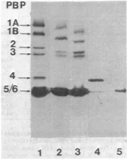
FIG. 1. PBPpatterns of P. vulgaris P18
(tracks
1 and2) and its stable L-forms(track 3). Analysesof the purified PBP4 (track 4) and PBP5 (track 5) isolated fromnormalcells. The cell envelopes of the normal bacteria were prepared viaspheroplast
formation (track 1) or by using the Ribi press (track 2). The isolated PBP4 was watersoluble. Theisolated PBP5 wasinGenapol. Forexperimental conditions,seethe text.member of this
taxonomic
group
(20).
PBP4
wasespecially loosely bound
tothe isolated cell
envelopes
from
which
it
wasselectively
released
by
treatmentwith
1 M
NaCl. Moreover,
PBP4-free
cell
envelopes
wereobtained
by submitting
the
bacteria
tothe modified
Nossal-Heppel
pro-cedure,
in
which
casePBP4
wasrecovered in
both
fractions S3 and
S4,
orby
disrupting
the
bacteria
with the Ribi press
(Fig.
1,
track
2),
in
which case PBP4
wasrecovered
in the
superna-tant
fraction.
The stable
L-forms
weredisrupted
under
those
conditions which had
permitted
isolation
from the
normal bacteria
of
cell
envelopes
with
afull
assortmentof
PBPs.
Analysis of
the stable
L-form
cell envelopes
(Fig. 1,
track
3;
Tables
1,
2, and 3) showed
that
PBP1A, 1B, 2,
3,
and 5/6
were present
and had
the
sameproperties
(rela-tive
abundance,
affinity
for
1-lactam antibiotics,
thermostabiity,
and
stability
of
theadducts
formed with
[14C]benzylpenicillin)
asthose
of
the
normal
bacteria.
In
someexperiments
(Fig.
1) the PBP1A and
lB
of the stable L-forms
had,
somehow,
reduced apparent molecular
weights,
but
this was not
always
the
case.PBP4,
howev-er,
wasalways
absent. Further
analysis
showed
that
the stable L-forms had
notlost their
ability
to
synthesize PBP4,
but had lost their
ability
toanchor
it in their
cell
envelope.
During growth,
PBP4
wastherefore secreted
in the culture medi-umfrom which
itcould
be isolated.DD- and LD-peptidase activities ofP. vulgaris P18 and its stable L-forms. The DD- and
LD-peptidase activities of
P. vulgaris P18 and itsstable
L-forms
are summarized in Table4. Cellenvelopes
possessing
afull
assortment of PBPs(i.e.,
preparedfrom
normalbacterial viasphero-plast formation) catalyzed
bothDD-carboxypep-tidase
and DD-transpeptidase activities. Thespe-cific
activities given in
Table 4were determinedon
reaction mixtures containing
Triton X-100ata
1% (vol/vol) final
concentration, a conditionthat increased
enzyme activity 10-fold. Theef-fects of increasing
concentrations ofbenzylpeni-cillin showed
that a major part (-80%) of thetotal DD-carboxypeptidase
activity was highlysensitive
tothe
antibiotic(±ID50s
0.02to0.06jxM), and that
aminorpart(=20%)wasattribut-able
toan enzymeof
moderate penicillinsensi-tivity (±ID50
2to6,uM).
Cell
envelopes lacking
PBP4 (i.e., preparedfrom normal bacteria
after disruption with theRibi
press orfrom the
stable L-forms)werenotcompetent
in catalyzing
transpeptidationreac-tions
and had only
alow
level ofDD-carboxy-peptidase activity of
moderate benzylpenicillinsensitivity. This DD-peptidase
activity was notenhanced
by the
presenceof
Triton X-100. It wasisolated
(in thepresenceof GenapolX-100)and identified
asPBP5asdescribed above (Fig.1,
track
5).Both the
supernatantfraction
obtained afterdisruption of the
normal bacteria with the Ribipress
and the culture filtrate
of the growingL-forms
contained
the highly penicillin-sensitive
DD-carboxypeptidase-transpeptidase. This
DD-peptidase
wasisolated
andidentified
asPBP4asdescribed above (Fig.
1,track 4). An antiserumprepared
against the DD-peptidasePBP4isolated TABLE 1. Relative abundance of the PBPs present inthe cellenvelopes of P.vulgarisP18and its stable L-forms asrevealedbyreaction withasaturatingconcentration(0.2mM) of
["4C]benzylpenicillin
Relative abundance(%)of PBPsfrom:
Normalbacteriaa
PBP
Spheroplast Ribi press L-forms
prepn prepn 1A 14 11 12 1B 15 13 12 2 4 4 12 3 3 5 8 4 5 0 0 5/6 59 67 56
a The cell envelopes of the normal bacteria were
prepared via spheroplastformation or by disruption with aRibi press.
152,
1. .Rl': -1:z' -,i:l.!O':
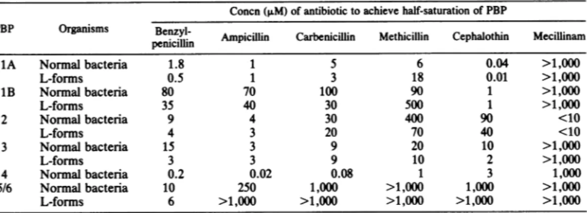
TABLE 2. Concentrationof
3-lactam
antibiotics necessarytoachieve half-saturation of the PBPs of P. vulgaris P18 and its stable L-formsConcn(,uM) of antibiotictoachieve half-saturation of PBP
PBP Organisms Benzyl- Ampicilin Carbenicillin Methicillin Cephalothin Mecillinam
penicillin 1A Normal bacteria 1.8 1 5 6 0.04 >1,000 L-forms O.5 1 3 18 0.01 >1,000 1B Normal bacteria 80 70 100 90 1 >1,000 L-forms 35 40 30 500 1 >1,000 2 Normal bacteria 9 4 30 400 90 <10 L-forms 4 3 20 70 40 <10 3 Normal bacteria 15 3 9 20 10 >1,000 L-forms 3 3 9 10 2 >1,000 4 Normalbacteria 0.2 0.02 0.08 1 3 1,000 5/6 Normal bacteria 10 250 1,000 >1,000 1,000 >1,000 L-forms 6 >1,000 >1,000 >1,000 >1,000 >1,000
from
P.
vulgaris
P18
(specific carboxypeptidase
activity, 4
U/mg of
protein) maximally
inhibited
the
activities of
the enzyme
by
80%. It had
exactly
the
sameeffect
onthe
corresponding
DD-peptidase-PBP4
of the stable L-forms.
P.
vulgaris
P18 and its stable L-forms also
synthesized
apenicillin-resistant (and
therefore
not
detectable
as aPBP)
meso-diaminopimelyl-(L)-D-alanine-cleaving
peptidase
(equivalent
tothe
carboxypeptidase 2 of Escherichia
coli
[7]).
This
LD-peptidase
wasfound
tobe
periplasmic
in the normal
bacteria
and
wasselectively
recov-ered in
fractions Si
and S2 when the
Nossal-Heppel technique
wasemployed.
The
sameLD-peptidase
wassecreted
in the culture medium
by
the
stable
L-forms
during growth (Table 4).
The
LD-peptidase
waspartially
purified
asdescribed
above.
Properties of the isolated
DD-and
LD-peptidases
of
P.vulgaris
P18 and
itsstable
L-forms.
(i)
Highly
penicillin-sensitive
DD-carboxypeptidase-transpeptidase PBP4. The water-soluble
DD-pep-tidase PBP4 had
anapparent molecular
weight
of about
46,000 (on
the
basis
of its
migration
by
sodium
dodecyl
sulfate-polyacrylamide gel
elec-trophoresis)
or
49,000
(on
the basis of its
Kd
value
by
Ultrogel
filtration).
It
wasanionic
atpH
8.3.
With the
cosubstrates,
freepentapeptide (the
TABLE 3. Stability of the complexes formed between
["4C]benzylpenicillin
and the PBPs ofP.vulgaris P18 and its stable L-forms
Half-life (min) of the complexes
PBP
Normalbacteria L-forms
IA 300 320 1B 240 220 2 120 110 3 180 155 4 180 Absent 5/6 10 12
carbonyl
donor) and amidated tetrapeptide (the
amino acceptor), and at the concentrations given
above, the DD-peptidase PBP4 concomitantly
catalyzed both hydrolysis of the pentapeptide
(Hy) and transpeptidation (T), with a Hy/T ratio
of about 10 (under conditions where less than
25%
of the radioactive pentapeptide was
uti-lized).
Optimal pH for enzyme activity was 8.8
in 0.25 M (or less)
Tris-hydrochloride
buffer.
The addition of 0.2 M NaCl to the same buffer
inhibited
the
enzyme activity by 70%.
UDP-N-acetylmuramyl-pentapeptide was not used as
carbonyl
donor for the transpeptidation reaction
although it was a substrate (Km
=0.6 mM) for
the carboxypeptidase activity.
The DD-peptidase PBP4 exhibited wide
varia-tions
in its sensitivity to
P-lactam
antibiotics
(Table 5). The ID50 values thus obtained related
well with the corresponding values of Table 2,
except
for benzylpenicillin and
mecillinam.
Somehow,
these two antibiotics had much
high-er
affinities
for the water-soluble PBP4 than for
the
membrane-bound PBP4. On the basis of the
rates
of enzyme recovery (3), the adducts
formed
between the DD-peptidase and
benzyl-penicillin
and ampicillin had half-lives of 240 and
170 min,
respectively. The inhibition of the
DD-peptidase by
carbenicillin was of the competitive
type. The
Km
value for the substrate
(UDP-N-acetylmuramyl-pentapeptide) varied depending
on
the carbenicillin concentrations used (Fig. 2).
This
observation was at variance with that made
previously with the corresponding
DD-carboxy-peptidase-transpeptidase
PBP4 of P. mirabilis,
in which case the inhibition appeared to be
clearly
noncompetitive (11).
(il)
Moderately
penicillin-sensitive
DD-carboxy-peptidase PBP5. DD-CarboxyDD-carboxy-peptidase PBP5 did
not
catalyze
transpeptidation reaction with the
above system of cosubstrates. It functioned
solely
as a
carboxypeptidase on either
UDP-N-acetylmuramyl-pentapeptide or the free
penta-peptide.
Itwas
half
inhibited by a 1 ,uM
benzyl-STABLE
L-FORMS OF P. VULGARIS1047
TABLE 4. Specific activitiesofDD- and LD-peptidases of P. vulgaris and its stable L-forms
Spact(mU/mg) ofpeptidasea
Organisms Prepn
DDCarboxy-
DD-Trans-LD-Carboxy-peptidase peptidase peptidase
Normal bacteria Cell envelopes prepared via 10 (H andM)C 1 (H) NDd spheroplastformationb
Cellenvelopes disrupted in Ribi 0.5(M) ND ND
press
Supernatantafter disruption 5 (H) 0.5 (H) 2.4 (R)
with Ribi press
L-forms Cellenvelopes 0.7(M) ND ND
Culture filtrate 5.7(H) 0.4(H) 2.5 (R)
aTheletters within parentheses indicate sensitivity to benzylpenicillin as follows: H, highly sensitive (ID50,
-0.01
to0.06ILM);
M, moderately sensitive(ID5o,
=2to 6,uM); R, resistant.bAsdetermined in reaction mixtures containing 1% Triton X-100. The presence of the detergent caused an approximately
10-fold
increase in enzyme activity as compared with reaction mixtures containing the same cell envelopeswithout Triton X-100. The addition of Triton X-100 had little or no effect in all other cases.cThe inhibitory effect exerted by increasing concentrations ofbenzylpenicillinwas biphasic, showing that, under the assay conditions used, the moderatelypenicillin-sensitivepeptidase activity represented 10 to20oof the total activity.
dND, Not detected.
penicillin concentration. The removal of
Genapol
X-100
caused irreversible enzyme
de-naturation.
(ill) Penicillin-insensitive
LD-carboxypeptidase.
Filtration of water-soluble LD-carboxypeptidase
on
Ultrogel
AcA4-4
indicated
amolecular
weight of 32,000. The enzyme activity increased
as
the
pH of
the
reaction mixture increased from
pH 7.5
(Tris-hydrochloride
buffer) to
11(Gly-cine-NaOH buffer).
At
pH 8.9,
variations of the
molarity of
the
Tris-hydrochloride
buffer from
0.02
to0.22
werewithout any effect,
and theKm
value
for the
hydrolysis of
UDP-N-acetylmura-myl-tetrapeptide
was
0.4 mM. The free
tetrapep-tide and the amidated
tetrapeptide
had
equiva-lent substrate activities.
DISCUSSION
The present
investigation confirms
the results
previously obtained
by Martin et al. (13) on the
L-form
strains
of
P.
mirabilis LVI and LD52
(8)
and
expands
them
by
showing
that the stable
L-TABLE 5. Antibioticconcentrations required to inhibit by
50%o
the purifiedDD-carboxypeptidase-transpeptidasePBP41 ID5o Antibiotic value (>LM)
Benzylpenicillin
... 0.01Ampicillin
... 0.006Carbenicillin
... 0.08 Methicillin... 1Cephalothin
...1
Mecillinam
... 2aThe enzymeused had a specific activity of 4 U/mg ofprotein (carboxypeptidaseassay).
form derived
from
P.
vulgaris
P18 haskept
theability
of the
parental
strain to
synthesize
all
of
the PBPs and the
DD-and LD-peptidases known
to
be
involved
in
peptidoglycan cross-linking
and
its
further
maturation during
the
bacterial
life cycle.
However,
both
the
DD-carboxypepti-dase-transpeptidase PBP4,
which in the
parental
strain
is
loosely bound
to
the outer surface
of
the
plasma
membrane,
and
the
LD-carboxypepti-dase, which is located
in. the
periplasmic region,
are
secreted
in
the
culture medium during
growth by the
protoplast-like,
stable L-forms.
Mutants
of
E.coli
lacking
PBP4
(15)
grow
nor-mally under
awide range of laboratory
condi-500200
0100
0)~~~~~
E 020~~~~~~
0~~~~~~~~
E I-. 1 2 3 4 51/[S](mM1)
FIG. 2. Inhibition of the
DD-carboxypeptidase
ac-tivity of the isolated PBP4 by
increasing
concentra-tions of
carbenicillin
(0, 100, 200, and 500nM).
Lineweaver-Burkplot. VOL.
152,
1982tions, strongly
suggesting
that this PBP is
dis-pensable. On this
basis,
alack of
integration
of
PBP4 within the plasma membrane is
probably
not at least the
main defect
for the
hereditary
and permanent
inability of
the
stable L-forms
tosynthesize wall
peptidoglycan.
This
inability
may be due to
defects in the
early
stages of
peptidoglycan
synthesis
or,
morelikely,
tode-fects in the subsequent
lipid cycle
possibly
caused
by the
physical
and
chemical changes
in
the
plasma membrane which
permitted
transi-tion of Proteus mirabilis to life in the form of
anenvelopeless, osmotically stable L-form (6).
ACKNOWLEDGMENTS
The work has been supported in part by grants from an
Actionconcertee with the Belgian Government(convention
79/84-I1), the Fonds de la Recherche Scientifique M6dicale, Brussels, Belgium (contract 3.4501.79), and the National Institutes of Health (Public Health Service grant 5RO1 AI-13364-05).
LITERATURE CITED
1. Fleck, J.1963. Etude de la croissance en milieuliquide
d'une souchedeProteus et de sa formeL.C.R. Soc. Biol.157:183-185.
2. Frere,J. M., M.Leyh-Bouille,J. M. Ghuysen, M. Nieto, andH. R. Perkins. 1976. Exocellular
DD-carboxypepti-dases-transpeptidases from Streptomyces. Methods En-zymol. 45:610-636.
3. Frere,J. M., M. Leyh-Bouille, J. M. Ghuysen, and H. R. Perkins. 1974. Interaction between ,B-lactam antibiotics
and exocellular DD-carboxypeptidase-transpeptidase of Streptomyces R61. Eur. J. Biochem.50:203-214. 4. Georgopapadakou, N. H., and F. M. Liu. 1980.
Penicillin-binding proteins in bacteria. Antimicrob. Agents
Che-mother.18:148-157.
5.Ghuysen,J. M. 1980.Antibioticsandpeptidoglycan
me-tabolism, p. 9-117. In P.G. Sammes (ed.), Topics in antibiotic chemistry, vol. 5. Ellis Horwood, Chichester. 6. Gmeiner, J., and H. H. Martin. 1976.Phospholipid and
lipopolysaccharide in Proteus mirabilis and its stable
protoplast L-form. Difference in content and fatty acid composition. Eur. J.Biochem.67:487-494.
7. Izaki, K., and J. L. Strominger. 1968. Biosynthesis of peptidoglycan ofbacterial cell walls. XIV. Purification
andproperties oftwo D-alaninecarboxypeptidases from Escherichiacoli. J. Biol.Chem. 243:3193-3201. 8. Kandler, O., and G. Kandler. 1956. Trennung and
Charak-terisierung verschiedener L-Phasen-Typen von Proteus mirabilis. Z. Naturforsch. 11:252-259.
9. Kohn, A. 1960. Lysis of frozen and thawed cells of
Escherichia coli by lysozyme and their conversion into spheroplasts. J. Bacteriol. 79:697-706.
10. Lowry, 0. H., N. J. Rosenbrough, A. L. Farr, and R.J. Randall. 1951. Protein measurement with the Folin phenol reagent. J.Biol. Chem. 193:265-275.
11. Martin, H.H., C. Maskos, and R.Burger.1975. D-alanyl-D-alaninecarboxypeptidase in the bacterial form and L-forms of Proteus mirabilis. Eur. J. Biochem. 55:465-473. 12. Martin, H. H., W.Schilf, and C. Maskos. 1976. Purifica-tionofthemembrane-boundDD-carboxypeptidaseof the unstable spheroplast L-form of Proteus mirabilis by affini-tychromatography. Eur. J. Biochem. 71:585-593. 13. Martin, H. H., W. Schilf, and H. G. Schieffer. 1980.
Differentiationofmycoplasmatalesfrom bacterial proto-plast L-forms by assay for penicillin-binding proteins. Arch. Microbiol. 27:297-299.
14. Martin, H. H., M. Tonn-Ehlers, and W. SchiU. 1980.
Cooperation of benzylpenicillinand cefoxitin in bacterial growthinhibition. Phil. Trans. R. Soc. London B 289:365-367.
15. Matsuhashi, M., Y. Takagaki, I. N. Maruyama, S. Ta-maki, Y. Nishimura, H. Suzuki, U. Ogino, and Y. Hirota. 1977. Mutants of Escherichiacolilacking in highly
peni-cillin-sensitiveD-alanine carboxypeptidase activity. Proc. Natl. Acad. Sci. U.S.A. 74:2976-2979.
16. Medill-Brown, M., W. G. Hutchinson, and E. Cocklin. 1960. The L-forms of Proteus mirabilis. Ann. N.Y. Acad. Sci. 79:372-379.
17. Minck, R., A.Kirn, and M. Galleron. 1957. Recherches sur la transformation L en milieux synthetiques d'une souche de Proteus. Ann. Inst. Pasteur 92:138-141. 18. Nguyen-Dlst6che, M., J. M. Ghuysen, J. J.Polock,P. E.
Reynolds, H. R. Perkins, J. Coyette, and M. R. J. Salton. 1974. Enzymes involved in wall peptide crosslinking in
Escherichiacoli K12 strain 44. Eur. J. Biochem. 41:447-455.
19.Nossal, N. G., and L. A. Heppel. 1966. The release of enzymes by osmotic shock from Escherichia coli in
exponentialphase. J. Biol. Chem. 241:3055-3062.
20. Ohya, S., M.Yamazaki, S.Sugawara,and M.Matsuhashi. 1979. Penicillin-binding proteins in Proteus species. J.
Bacteriol.137:474-479.
21. Ribi, E., T. Perrine, R. List, W. Brown, and G. Goode. 1959. Use of pressure cell to prepare cell walls from
Mycobacteria.Proc. Soc. Exp. Biol. Med. 100:647-649. 22. Schilf, W., and H. H. Martin. 1980. Purification of two
DD-carboxypeptidases-transpeptidases with different
penicillin sensitivities from Proteus mirabilis. Eur. J.
Biochem.105:361-370.
23. Shepherd,S.T., H. A. Chase, and P. E. Reynolds. 1977. Theseparation and properties oftwopenicillin-binding proteins fromSalmonella typhimurium. Eur. J. Biochem.
78:521-523.
24. Spratt, B. G. 1977. Properties of the penicillin-binding proteins of Escherichia coli K12. Eur. J. Biochem. 72:341-352.
25. Tulasne, R. 1949. Existence of L-forms in common
bacte-ria and their possible importance. Nature (London) 164:876-877.