CONSTRUCTION AND CHARACTERIZATION OF
OC125 SINGLE-CHAIN ANTIBODY
By
HUISHAO
DEPARTEMENT DE MICROBIOLOGIE ET D'INFECTIOLOGIE
FA CUL TE DE NIEDECINE
UNIVERSITE DE SHERBROOKE
SHERBROOKE, QUEBEC
A MEMOIR SUBMITTED TO THE FACUL TY OF MEDICINE
OFTHEREQUIRENIENTSFORDEGREEOF
MASTER OF SCIENCE (M.Sc.)
FEBRUARY 2002
l+I
National Libraryof Canada Bibliothèque nationale du Canada Acquisitions and Acquisitions et
Bibliographie Services services bibliographiques 395 Wellington Street
OttawaON K1AON4
Canada
395, rue WeUington
Ottawa ON K1A ON4
Canada
The author bas granted a
non-exclusive licence allowing the
National Library of Canada to
reproduce, loan, distribute or sell
copies of this thesis in microfonn,
paper or electronic formats.
The author retains ownership of the
copyright
in
this thesis. Neither the
thesis nor substantial extracts from it
may
be printed or otherwise
reproduced without the author' s
penmss1on.
L'auteur a accordé une licence non
exclusive permettant
à
la
Bibliothèque nationale du Canada de
reproduire, prêter, distribuer ou
vendre des copies de cette thèse sous
la
forme de microfiche/film, de
reproduction sur papier ou sur format
électronique.
L'auteur conserve la propriété du
droit d'auteur
qui
protège cette thèse.
Ni la thèse
ni
des extraits substantiels
de celle-ci ne doivent être imprimés
ou autrement reproduits sans son
autorisation.
1. TABLE OF CONTENTS
1. Table of contents ...•...•...•..•... 2
II. Abstract (French) ...•..•... 5
m.
Abstract (English) ...•... 7IV. Glossary of abbreviations ...•... 9
V. List of tables and figures •••..•...••...•.•...•...•.... 12
Chapter 1 Introduction ... 13
1.1 Human ovarian cancer ... .13
~) 1.2 CA125 tumor antigen ... 17
1.3 Strategies to be used to study the function of proteins ... 24
1.4 Structure and properties of ScFvs ... 25
1.4.1 Monoclonal antibodies ... 25
1.4.2 Shortcomings of monoclonal antibodies ... 28
1.4.3 Single-chain antibodies (ScFv) ... 29
CHAPTER 2 Materials and Methods ... 32
2.1 Derivation of ScFv constructs ... 32
2.1.1 mRNA isolation and purification ... 32
2.1.2 First-strand cDNA synthesis ... .34
2.1.3 Primary PCR amplification ... 34
2.1.4 Purification ofprimary PCR products ... .35
2.1.5 Assembly and fill-in reactions ... .35
2.1.6 Second PCR amplification and purification ... .36
2.3 Preparation of ScFv cDNA library ... .40 2.4 CA125 antigen ... 41 2.5 Phage display ... 41 2.5.1 Transformation ... 41 2.5.2 Phage rescue ... 42 2.5.3 Panning ... 43
2.5.4 Reinfection ofE.coli with enriched phage clones ... .43
2.5.5 Screening for phage antibodies in enriched clones ... .44
2.5.6 Screening for soluble antibodies in enriched clones ... .45
2.5.7 Binding analysis by ELISA ... .45
2.6 Colony-li:ft assay ... 46 2.6.l Transformation ... 46 2.6.2 Colony lift ... 4 7 2.6.3 Periplasmic Extract ... .49 2.7 Cell lines ... 49 2.8 Transient transfection ... 50 2.9 Immunoblot analysis ... 51 2.10 Sequence analysis of ScFvs ... 51 I -) 2.11 Immunoprecipitation ... 52
CHAPTER 3 Results ... 53
3.1 Construction of the OC125 ScFv cDNA library ... 53
3.2 ScFv cDNA library screening ... 53
3.2.1 Phage display ... 55
3.2.2 Colony lift assay ... 56
3.3 Antigen binding assay using periplasmic extracts ... 56
3 .4 Expression of ScFvs in periplasmic extracts ... 60
3.5 Confirmation ofScFv cDNA ... 60
3.6 ScFv sequence analysis ... 63
3.7 Intracellular expression of the OC125 ScFvs in eukaryotic cells ... 65
CHAPTER 4 Discussion ...•... 72
Conclusions ...•.•...•....•...•...••...•... 81
References ...•...•...•...•...•...•.•..•...•... 82
•
II.
ABSTRACT
Le cancer ovanen est la principale cause de la mortalité attribuable aux cancers gynécologiques dans les pays occidentaux. De plus, pour la majorité des patientes (>70%), la maladie en diagnostiqué en phase tardive. Ces diagnostiques tardifs conduisent à un taux de survie de 15 à 30 pour cent après cinq ans. Dans le but d'accroître la qualité et la rapidité du dépistage, la découverte et l'utilisation de marqueurs spécifiques sont d'ungrand intérêt. Pour les cancers ovariens, la progression clinique de la maladie est en corrélation avec l'expression de l'antigène tumoral CA125, ce qui en fait un marqueur de choix. Cet antigène est actuellement utilisé en tant qu'indice de récurrence clinique des cancers ovariens. Aussi, l'antigène CA125 est employé pour vérifier l'efficacité des traitements de chimiothérapie pour ces cancers. Des études antérieures ont montré que CA 125 est une glycoprotéine de haut poids moléculaire avec des propriétés semblable à une molécule de mucine (mucin-like ). La nature biochimique et moléculaire de cet antigène ainsi que sa fonction demeure peu ou pas connu. Dans cette étude, nous avons développé une approche basée sur l'expression d'anticorps à chaîne unique (ScFv) pour abroger sélectivement CA125 afin d'étudier son rôle dans les cancers ovariens chez l'humain.
L'ADNc qui code pour l'anticorps ScFv a été produit à partir des ARNm extraient de !'hybridome OC125. Cet hybridome exprime l'anticorps monoclonal spécifique à la partie extracellulaire de CA 125. Les gènes codant pour la partie variable des chaînes lourdes (VH) et légères (VL), séparées par la séquence codant pour le peptide (Gly4Ser)3, ont été clonés dans le même vecteur. L série de gènes résultants, contenant les ScFvs, a
été clonée dans le vecteur d'expression procaryote pCANT AB 5E. Cette banque d' ADNc a été criblée par les essais de "phage-display" et "colony-lift". La capacité des ScFvs de lier CA 125 a été validée par ELISA. Six ScFvs positifs ont été choisis parmi 176 candidats. La séquences en acides nucléiques montre que la région VL et la majeure partie des peptide de liaison ont été éliminées dans l'anticorps ScFv #148. Cet anticorps a démontré la meilleure affinité pour CA 125 en ELISA. Par la suite, celui ci a été sous-cloné dans le vecteur d'expression eucaryote pSTCF.KDEL contenant le peptide signal pour le transport de la protéine au réticulum endoplasmique. Un système d'expression transitoire de la protéine encodé par le plasmide pSTCF.KDEL dans des lignées cellulaires d'ovaires humains a été utilisé comme modèle. Nous avons ainsi démontré une diminution significative de # 148 ScFv dans les cellules d'une lignée co-exprimant l'antigè CA125 (OVCAR3: cellules tumorales d'ovaries humains) par rapport à une lignée n'exprimant pas CA125 (PA-1). Par contre, si l'on compare avec d'autres ScFvs ne liant pas CA125 en ELISA, dans ces deux mêmes lignées cellulaires, leurs concentrations demeurent équivalentes. Ces résultats suggèrent que la protéine ScFv #148 peut lier CA125 dans le réticulum endoplasmique des cellules OVCAR3. Néanmoins, des expériences de l'immunoprecipitation n'ont pas démontré une interaction directe entre la protéine de ScFv #148 et CA125.
En conclusion, dans cette étude nous avons construit un ScFv contre l'antigène CA 125. Cet anticorps lorsque exprimé chez E. coli est soluble et reconnaît spécifiquement la protéine CA125 en ELISA. Lorsque le ScFv fut sous-cloné dans le vecteur eucaryote pSTCF.KDEL et ensuite transfecté dans des cellules ovariennes humaines, un niveau d'expression élevé a été obtenu.
1 -)
III.
ABSTRACT
Ovarian cancer is the leading cause of death from gynecological cancers in W estem countries. Over 70% of the patients present with late stage disease, the :five-year survival rate for these patients remains at best of 15-30%. Tumor antigen CA125 expression has been shown to correlate with the clinical course of the disease, and CA 125 is currently used as a predictor of clinical recurrence in ovarian cancer and to monitor response to chemotherapy treatment.
Previous studies showed that CA 125 is a high molecular weight glycoprotein having properties of a mucin-type molecule. The biochemical and molecular nature and the function of this antigen are poorly understood. In this study, we have developed an approach based upon intracellular expression of single-chain antibodies (ScFvs) to achieve selective abrogation of CA125, thus to study the role of CA125 in human ovarian cancer cells.
The cDNA coding ScFv was generated from mRNA extracted from the hybridoma cell line OC125, which expresses monoclonal antibodies that specifically recognize an epitope located in the extracellular portion of CA125. Genes coding for the VH and VL were cloned into the same vector, separated by a linker sequence that coded the peptide (Gly4Ser)3. The constructed ScFv library was cloned into the procaryotic expression vector pCANTAB 5E and the cDNA library was screened by phage-display and colony-lift assay. CA125 antigen-binding activity of ScFvs was tested by ELISA, and six positive ScFvs were selected among 176 candidates. Sequencing analysis showed that the VLregion and most of the linker were deleted from CA125-positve #148 ScFv, which
has the strongest binding activity in ELISA. After subcloning #148 OC125 ScFv into the ER-targeting eukaryotic expression vector pSTCF.KDEL, in which the protein was targeted into ER, and using a transient expression system, we showed that the expression level of #148 ScFv was significantly lower in the human ovarian cancer cell line OVCAR3, which expresses CA125, than in the cell line PA-1, which does not express CA125. Meanwhile, the control ScFv proteins, which did not bind to CA125 in ELISA, displayed equal expression levels in both cell lines. These results suggest that # 148 OC125 ScFv protein may bind to CA125 in the ER of OVCAR3 cells. However, immunoprecipitation experiments did not demonstrate a direct interaction between # 148 ScFv and the CA125 protein.
In conclusion, the study shows that a ScFv against the CA 125 antigen was constructed, and that the soluble antibodies bind to CA125 protein in ELISA when expressed in
E.coli. When the ScFv was subcloned into the eukaryotic vector pSTCF.KDE, which
targets cloned genes to the ER, high levels of expression were obtained in mammalian cells. An interaction between OC125 ScFv and CA125 protein was not demonstrated in this study.
IV. GLOSSARY OF ABBREVIATIONS
2xYT-AG: 2xYT medium containing lOOµg/ml ampicillin and 2% (v/v) glucose 2xYT-AK: 2xYT medium containing lOOµg/ml ampicillin and 50µg/ml kanamycin 2xYT-AI: 2xYT medium containing lOOµg/ml ampicillin and lmM lPTG
4-CN: 4-chloro-1-naphthol AA: amino acid
ATCC: American Type Culture Collection bp: base pair
kb: kilo-base pair
CA125: cancer antigen 125
cAMP: cyclic adenosine monophosphate cDNA: complementary deoxyribonucleic acid CDR: complementary determining region CH: heavy chain constant region
-) CL: light chain constant region CMV: cytomegalovirus
dNTP: deoxyribonucleic triphosphate
DMEM: dulbecco's modified eagle's medium
E.coli: Escherichia coli
EDTA: ethylene-diaminetetra-acetic acid disodium sait EGFR: epidermal growth factor receptor
ELISA: enzyme-linked immunosorbent assay EOC: epithelial ovarian cancer
ER: endoplasmic reticulum Fab: fragment of antibody FBS: fetal bovine serum Fv: fragment of variable region FW: framework
g: gram
,
-
)•
µg: microgram ng: nanogram pg: p1cogram
g3p: gene
m
minor coat protein HRP: horsradish peroxidase Ig: immunoglobulin lPTG: isopropyl-1-thio-13-D-galactopyranoside kDa: kilodalton kV: kilovolt L: liter ml: milliliter µl: microliterLMP 1: latent membrane protein 1 M:mole
mM: millimole
MAb: monoclonal antibody NFDM: non-fat dry milk
PBS: phosphate-buffered saline PCR: polymerase chain reaction
RT PCR: reverse transcription polymerase chain reaction P .E.: periplasmic extraction
PEG: polyethylene glycol pfu: plaque-forming unit
PVDF: polyvinylidene difluoride RNA: ribonucleic acid
mRNA: messenger ribonucleic acid rpm: revolutions per minute
RS primer: restriction site primer
ScFv: single-chain variable region fragment
1 • )
1
TGF-a: transforming growth factor alpha U: unit
µF: microfarad
VH: heavy chain variable region V L: light chain variable region
V. LIST OF TABLES AND FIGURES
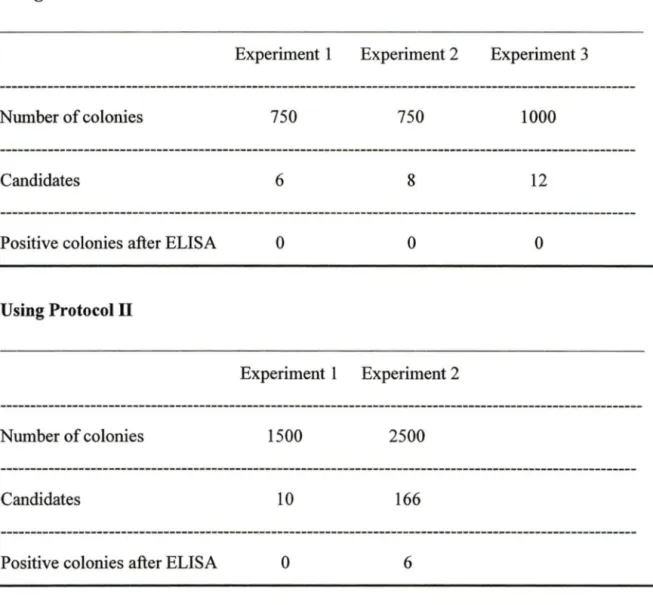
Table 1: Detection ofCA125 antigen in serwn Table 2: Summary ofresults of colony-lift assay
Figure 1: 35 of 55 patients had a clinical recurrence a:fter a second-look operation Figure 2: Primary tumor growth rate is reduced in Muc-1 deficient mice
Figure 3: Structure of a monoclonal antibody and the derived single-chain antibody Figure 4: Derivation of ScFv constructs
Figure 5: pCANTAB SE vector showing control regions
Figure 6: pSTCF.KDEL eukaryotic vector expressing ScFv genes Figure 7: ScFv Iibrary screening using Colon y-lift assay
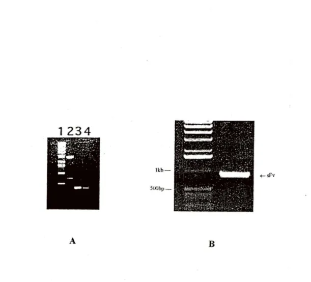
. ) Figure 8: Validation of amplified Vtt, VL and assembled ScFv
Figure 9: Expression and antigen binding activities of ScFvs in periplasmic extracts Figure 10: Immunoblot assay to detect the expression of ScFvs in periplasmic extracts Figure 11: pCANTAB.OC125 ScFvs digested with
SfiJ. /
NotlFigure 12: Sequence analysis of0Cl25 ScFvs
Figure 13: Expression of ScFvs in trasient transfected ovarian cancer ce Ils Figure 14: Strategy of immunoprecipitation
Figure 15: ScFv transfected OVCAR3 cell lysates immunoprecipitated with
anti-myc
antibodyCHAPTER 1 INTRODUCTION
The objective of this study is to identify the role of tumor antigen CA125 in human ovarian cancer cells using ScFv. This study includes three major parts: construction and screening of a OC125 ScFv, expression of the recombinant ScFv plasmid and analysis of ScFv protein's binding activity to the CA125 tumor antigen. The introduction covers four parts: human ovarian cancer (section 1.1), CA125 (section 1.2), strategies to be used to study the fonction of protein (section 1.3) and structure of ScFv (section 1.4).
1.1 Human ovarian cancer
Ovarian cancer represents the fifth most signi:ficant cause of cancer-related death for women and is the most frequent cause of death from gynecological neoplasia in the Western world. Ovarian cancer causes more deaths than any other cancer of the female reproductive system. Most (80-90%) ovarian tumors are epithelial in origin and arise from the coelomic epithelium. The remainders arise from germ cell or sex cord/stromal cells. A hereditary component in the latter group is rare, but includes granulose-cell tumors in patients with Peutz-Jeghers syndrome (Ferry, J.A. et al, 1994) and autosomal dominant inheritance of small-cell carcinoma of the ovary (Lamovec, J., et al., 1995; Longy, M., et al., 1996).
r--'
)
Over 70% of ovarian cancer patients present with late stage disease, the majority of which cannot be completely resected by surgery. Chemotherapy bas become the primary adjunct to surgery in obtaining a clinical remission or enhancing disease free survival in ovarian cancer patients. Although response to initial chemotherapy treatment approaches 70%, most are transient and approximately 80% of patients will recur and eventually die of the disease. A variety of salvage agents and strategies have been investigated, but none have demonstrated long-term effectiveness. The five-year survival rate of the patients with a stage III epithelial ovarian cancer remains, at the best, at 30% (American Cancer Society Surveillance Research, 1997). This poor prognosis is largely due to the fact that 75% of cases present with extra-ovarian disease, which in turn, reflects the absence of symptoms in early-stage disease. Advanced stage ovarian cancer (stage IV) bas a five-year survival rate of approximately 10% whereas early stage (stage
n
ovarian cancer bas a five-year survival rate of at least 85%. These data suggest that there may be • a survival benefit from the detection of ovarian cancer at an early stage.To be able to develop appropriate screening strategies for ovarian cancer, it is necessary to understand the processes of carcinogenesis and tumor progression. More than 90% of epithelial ovarian cancers are clonai neoplasms that arise from the progeny of a single cell (Jacobs, I.J., et al, 1992). Given the clonality of most ovarian cancers, multiple genetic alterations must occur in the progeny of a single cell to permit progression from a normal epithelial phenotype to that of a malignant cell capable of uncontrolled proliferation, invasion, and metastasis. Somatic mutations have been found in sporadic ovarian cancers that activate oncogenes or that result in loss of tumor suppressor gene
(
.
)
function. Di:fferent ovarian cancers can also exhibit aberrant autocrine and/or paracrine growth regulation with alteration in the expression of growth factors and their receptors. Certain changes in oncogenes, tumor suppressor genes, growth factors, and their receptors occur in a significant :fraction of epithelial ovarian cancers, whereas others are uncommon.
Numerous of tumor suppressor genes have been identified in cancers and subsequently evaluated in ovarian cancers, including RB, VHL, WT, and p53 (Donehower, L.A., 1992). Tumor suppressor genes normally function to inhibit or to slow down the cell growth and division cycle; they function to prevent the development of neoplasia. Mutations in tumor suppressor genes cause the cell to ignore one or more of the components of the network of inhibitory signais, removing the brakes from the cell cycle and resulting in a higher rate of uncontrolled growth of cancer. Loss of p53 function is observed in more than 50% of advanced ovarian cancers, but in only 15% of stage 1
lesions (Marks, J.R., et al., 1991). Mutation of p53 is only occasionally observed in ovarian cancers with low malignant potential and is rarely detected in benign ovarian tumors. Consequently, abnormalities of p53 have been considered a "late change" in tumor progression, associated with the acquisition of metastatic potential.
Activation of several oncogenes has been reported in subsets of ovarian cancers, including erb-B2 (Bergmann, C., et al., 1986; Berchuck, A., et al., 1990; Felip, E., et al., 1995), cyclin D-1 (Baserga, R., 1994), and EGFR (epidermal growth factor receptor) (Warenius, H.M., et al., 1996; Hochhauser, D., et al., 1996). Oncogenes are mutated
(__-)
forms of proto-oncogenes whose functions are to encourage and promote the normal growth and division of cells. Deregulation of proto-oncogenes may result in overproduction of growth factors; flooding of the cell with replication signais; uncontrolled stimulation in the intermediary pathways; and/or unrestrained cell growth driven by elevated levels of transcription factors. Normal ovarian surface epithelium expresses EGFR detected by immunohistochemical techniques and this expression is lost in approximately 30% of ovarian cancers, associated with a slightly better prognosis (Berchuck, A., et al, 1991). Activation of EGFR can occur through truncation of its extracellular domain and this variant has been observed in some ovarian cancers (Huang, H.J.S., et al, 1997; Hekis, J.V., et al, 1997).
Growth of ovarian cancers can be stimulated by several peptide and lipid growth factors. Peptide ligands that bind to the EGFR are produced by ovarian cancers including EGF, transforming growth factor alpha (TGF-a), and amphiregulin (Stromberg, K., et al, 1994). Antibodies against TGF-a can inhibit the growth of ovarian cancer cell lines that continue to express EGFR, consistent with autocrine growth stimulation (Stromberg, K., et al, 1992). Paradoxically, it has been dif:ficult to document activation of EGFR in ovarian cancer cell lines, challenging the functional importance of this particular receptor for autocrine growth stimulation (Ottensmeier, C., et al, 1996).
, .. --... 1.2 CA125 tumor antigen
Tumor markers are used for multiple purposes in clinical care, including screemng asymptomatic subjects, differential diagnosis of symptomatic patients, treatment planning, prognosis during and immediately following treatment, and monitoring for recurrence. CA125 is the most widely used ovarian tumor marker, and is currently approved in the United States and Canada for monitoring of disease to determine if second-look surgery is required. The main concem about using CA125 as a first-line test for ovarian cancer is its apparent Jack of sensitivity for early-stage disease (Helzlsouer, K.J., et al, 1993). Only 50% of stage 1 ovarian cancer-patients have elevated CA125 preoperative serum levels. The traditional cutoff level for a positive CA125 test is 30 or 35U/ml. It is important to note that this level was established for patients with clinically established disease (Bast, R.C., et al, 1983), and was not recommended as the appropriate cutoff level for screening asymptomatic populations.
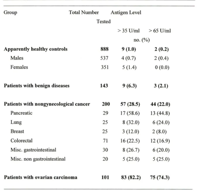
CA125 is a mullerian duct differentiation antigen that is overexpressed in epithelial ovarian cancer cells and released into the blood, although its expression is not entirely con:fined to ovarian cancer. Ovarian cancer CA125 antigen was first detected by Bast and Knapp in 1981, using the monoclonal antibody OC125 that was raised against the human ovarian carcinoma cell line OV433 (Bast, R.C., et al, 1981). These investigators subsequently developed a radioimmunoassay for the antigen and showed that serum CA125 levels are elevated in about 80% of patients with epithelial ovarian cancer (EOC) but in Jess than 1 % ofhealthy women (Table 1) (Bast, R.C., et al, 1983). The usefulness
(
'-(
_)
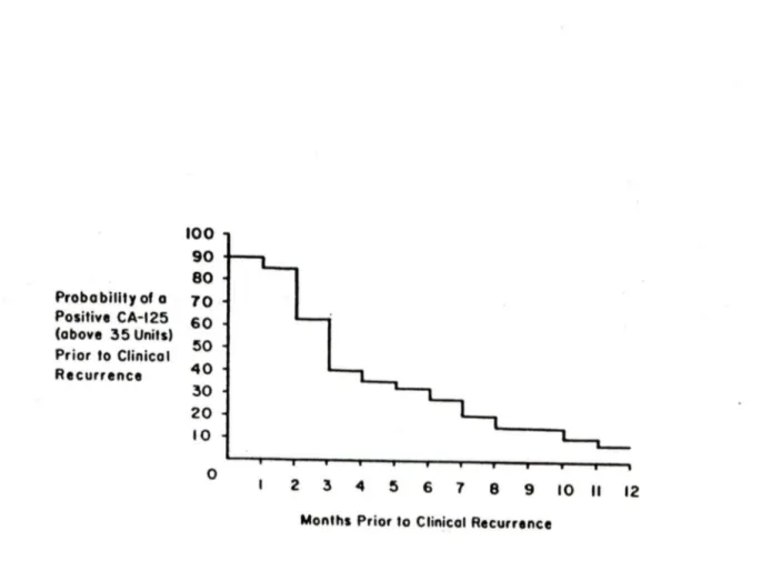
of CA125 levels in monitoring the progress of patients with EOC has been proved by a variety of studies. Most reports indicate that a rise in CA125 levels precedes clinical detection by about 3 months. During chemotherapy, changes in serum CA125 levels correlate with the course of the disease. lt is why today the CA125 assay is the most useful diagnostic test in the management of ovarian cancer. It is currently used as a predictor of clinical recurrence in ovarian cancer and to monitor response to chemotherapy treatment (Figure 1) (Niloff, J.M., et al., 1986; Fish, R.G., et al., 1987;
Makar, A.P., et al., 1993; Rustin, G.J.S., et al., 1996).
Despite the widespread use of CA125 as a clinical marker of ovarian cancer, the biochemical and molecular nature and the function of this antigen is poorly understood. At the time we started this study, the gene had not been cloned and its sequence was therefore unknown. Just before 1 finished my project, Dr. Kenneth Lloyd, who discovered CA125, recently reported the isolation of a long, but partial, cDNA that corresponds to the CA125 antigen. A rabbit polyclonal antibody produced to purified CA125 antigen was used to screen a /...ZAP cDNA library from OVCAR-3 cells in E.coli. The longest insert from the 54 positive isolated clones had a 5965 bp sequence containing a stop codon and a poly A sequence but no clear 5' initiation sequence. Northem blotting showed that the level of MUC16 mRNA correlated with the expression of CA125 in a panel of cell lines. But since it has an incomplete structure, it only can be considered as the best candidate for CA 125.
(
)
(
)
Group Total Number Antigen Level
Tested
> 35 U/ml > 65 U/ml no.(%)
Apparently healthy controls 888 9 (1.0) 2 (0.2)
Males 537 4 (0.7) 2 (0.4)
Females 351 5 (1.4) 0 (0.0)
Patients with benign diseases 143 9 (6.3) 3 (2.1)
Patients with nongynecological cancer 200 57 (28.5) 44 (22.0)
Pancreatic 29 17 (58.6) 13 (44.8)
Lung 25 8 (32.0) 6 (24.0)
Breast 25 3 (12.0) 2 (8.0)
Colorectal 71 16 (22.5) 12 (16.9)
Mise. gastrointestinal 30 8 (26.7) 6 (20.0)
Mise. non gastrointestinal 20 5 (25.0) 5 (25.0)
Patients with ovarian carcinoma 101 83 (82.2) 75 (74.3)
Table 1. Detection of CA125 antigen in serum: serum CA125 levels are elevated in about 80% of patients with epithelial ovarian cancer (EOC) but in less than 1 % of healthy women.
100 90 80 Probo billty of o 10 Positive CA-125 60 (above 35 Units) 50 Prior to Clinicat Recurrence 40 30 20 10 0 2 3 4 5 6 7 8 9 10 Il 12
(
-)
Monlhs Prior to Clinicat RecurrenceFigure 1. 35 of 55 patients had a clinical recurrence after a second-look operation. Serial CA125 levels rose to >35 U/ml in 94% of these patients before their recurrences became clinically detectable. The median leadtime before clinical recurrence was 3 -months, and in over 90% of patients was at least 2 months. Among the 20 of 55 patients who did not recur clinically, CA125 levels were always <35 U/ml in serum.
--Niloff, J.M. et al. (1986) American Journal of Obstetrics and Gynecology, 155:56-60
) Previous studies showed that the CA125 epitope is carried on a large glycoprotein with a
molecular weight in the range of 200-2000 kDa, while other studies have reported that CA125 consists of many subunits of 50-200 kDa (De Los Frailes, M.T., et al., 1993; Kobayashi, K., et al., 1993; Matsuoka, Y., et al., 1987; Nagata, A., et al., 1991; Yu, Y.H., et al., 1991; Zurawski, V.R., et al., 1988). The study of Lloyd et al. has shown that CA125 is a high molecular weight glycoprotein having properties of a mucin-type molecule (Lloyd, K.O., et al., 1997). Mucins, or mucin-type glycoproteins, can be defined as large extended molecules with a high percentage (50%-90%) of their molecular mass made up of carbohydrate that is attached via 0-glycosidic linkage through N-acetylgalactosamine to serine and/or threonine. They can be subdivided into the classical secretory or soluble mucins and the membrane-associated mucins(Spicer, A.P., et al, 1995). In this regard, it has been suggested that mucin-like proteins could play a role in the increased invasiveness of tumor cells and cell/cell-matrix adhesion (Guddo, F., et al., 1998; Yonezawa, S. et al., 1997). For instance, the Muc-1 mucin protein, one of the most characterized mucins, has been found to be expressed in all invasive types of pancreas, bile duct and breast carcinomas but were not frequently detected in the non-invasive types (Suwa, T., et al., 1998). In addition, the growth rate of primary breast tumors was found to be significantly reduced in Muc-1 de:ficient mice, and the percentage of animais with metastases was lower in Muc-1 de:ficient mice than in Muc-1 positive mice (Figure 2), suggesting that Muc-1 plays an important role in the progression ofmammary carcinoma (Spicer, A.P., et al., 1995).
(
-)
l )
4 100*
~ ~ 80 3 U) Cl> U) 24135 !!J üi'°
1: O>'°
60 ai ·a; E 43181 3: 2 5 0 -~ E U) :J êO ... E ë <{ 20 0 0 69 76 83 90 97 104 111 118 125 Muc-1 - /- Muc-1 +/+Mouse Age (days)
Figure 2. Primary tumor growth rate is reduced in Muc-1 deficient mice. A, graph
showing growth rate of polyoma middle T-induced mammary tumors in Muc-1 -/- (filled square) and Muc-1 +/+ (open square) mice. At 104 days, Muc-1 -/- mice had significantly smaller tumors than did Muc-1 +/+ mice (p<0.05). By the 124-day end point, differences in tumor size were highly significant (p<0.001). Asterisks indicate statistical significance. B, graph showing the percentage of Muc-1 +/+ and -/- mice with _ metastatic lesions in the lung at 124 days. The trend toward decreased rates of tumor metastasis in Muc-1 -/- mice suggested that the lack ofMuc-1 was showing some effects. The sample size was not sufficiently large to reach statistical significance.
) CA125 expression has been detected in over 80-90% of ovarian cancer (Bast, R.C., et al., 1983), but has never been found in the ovary during development or in the adult. Because CA125 seems to be a mucin-like protein, it might play an adhesive role, similar to MUC-1, by presenting carbohydrates as ligands for selectin-like molecules and thus aiding metastatic dissemination. In addition, evidence show that CA125 production and/or release can be regulated by several hormones (Bischof, P., et al., 1986; Karlan B.Y., et al., 1988; Brumsted J.R., et al., 1990), growth factors (Marth, C., et al., 1990) and cAMP(Ishiwata I., et al., 1986). Taken together, these data suggest that the deregulation of CA125 expression may play arole in the pathogenesis of ovarian cancer.
n
1.3 Strategies to be used to study the function of proteinsA variety of genetic strategies have been used to study the :function of a protein in transformation, these methods usually involved either gain or loss of protein :function.
In the first instance, studies to define the role of a gene in the transformation process were done using single gene transfer experiments. In this context, the cDNA coding the protein of interest is transfected into selected cells in order to analyze any new phenotypes associated with overexpression of this protein. One obvious limitation of this approach is the necessity of cloning the target gene prior to conducting gene transfer experiments.
As an alternative to the gain-of-:function studies, the use of gene therapy strategies to specifically knockout a protein of interest in transformed cells off ers new means to define its :function. In this regard, loss of a critical protein in a transformed cell may be more relevant to study its mediated pathway. The most common method is the use of antisense molecules. Thése molecules are designed specifically to target sense ( coding) sequences to block the transcription and translation of the encoded genetic information (Gura, T., 1995). Thus, this method relies on the prior knowledge of the coding sequence of the target gene. In addition, severe limitations, including non-specific effects, instability, difficulty to achieve adequate intracellular levels, have precluded widespread use of antisense molecules (Gewirtz, A.M., et al., 1996; Stein, T.A., 1995).
!,....-,)
Ribozymes are RNA molecules that have catalytic activities. They function by binding to a specific RNA target through antisense and inactivate it by cleaving the phosphodiester backbone at a specific site (James, H.A., et al., 1998). Although ribozymes have been used to inhibit a variety of viral and cellular genes, the method also relies on the prior knowledge of the coding sequence of the target molecule.(
.
)
One obvious common limitation to these approaches is the necessity of cloning the target gene prior to conducting the gain or loss of function experiments. Thus, these current strategies are of limited use to study the function of CA125 since the gene is not cloned and its sequence remains unknown.
1.4 Structure and properties of ScFvs
1.4.1 Monoclonal antibodies
Antibodies have long been used in biomedical science as m vitro tools for the identification, purification and functional manipulation of target antigens; they have been exploited in vivo for diagnostic and therapeutic applications as well. Recent advances in antibody engineering have allowed the genes encoding antibodies to be manipulated so that the antigen-binding domain can be expressed intracellularly. The specific and high-a:ffinity binding properties of antibodies, combined with their ability to be stably expressed in precise intracellular sites inside mammalian cells, has provided a powerful new family of molecules for cancer gene therapy applications.
·)
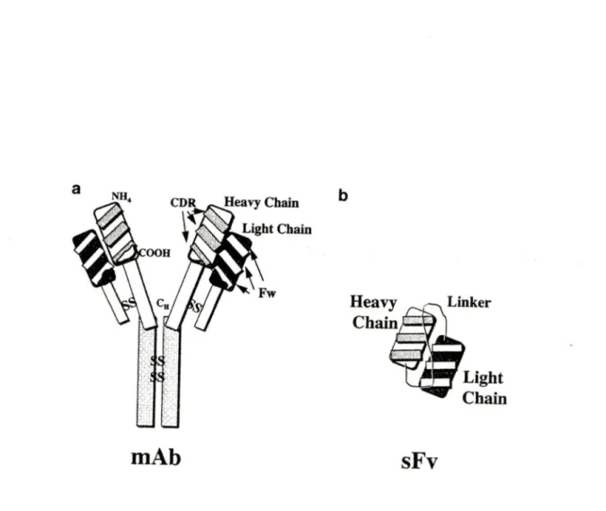
Typical monoclonal antibodies (Figure 3A) have a common structure consisting of two
identical heavy and light chain polypeptides held together by disul:fide bridges and non-covalent bonds. The DNA and amino acid sequence of the C region is relatively conserved within a given animal species while the V region sequence is antigen-dependent. Pairing of V regions of heavy and light chain creates an antigen-binding site (paratope ), which recognizes the epitope of an antigen. Most protein antigens contain many potential epitopes, which are recognized by a correspondingly large number of specific antibodies that comprise a polyclonal antibody population.
The constant (C) region is located at the carboxy-terminus of the antibody chain. In the mouse, there are five classes of constant heavy (CH) chain genes (a, ô, B, y, µ)and two
classes of constant light (CL) chain genes (K and Â.). DNA and amino acid sequences are relatively conserved within each class. Antibodies are grouped into :five major classes according to their CH region: lgA, IgD, IgE, lgG and IgM. An antibody is further distinguished by the class of light chain ( K or Â.) paired with its heavy chain. The class of each chain can be identi:fied immunologically or isotyped with commercially available reagents which are animal-species-speci:fic.
b
(
-)
mAb
sFv
Figure 3. Structure of a monoclonal antibody and the single-chain antibody derived from it. The single-chain antibody is constructed by linking the variable heavy and light regions of the antibody using a small flexible peptide liker. The variable heavy and light regions are derived by PCR (polymerase chain reaction). CDR, complementary -detennining region; FW, framework; MAb, monoclonal antibody; Ctt. heavy chain constant region; SS, disul:fide cross-links; ScFv, single-chain antibody.
--Piché, A. and Rancourt, C. (1999) Gene Therapy, 6:1202-1209
(
-)
The variable (V) region is located at the amino-terminus of the antibody chain. It consists of altemating framework (FW) and hypervariable, or complementarity-determining regions (CDRs). The greatest sequence diversity occurs in the CDRs, while the FW region sequences are more conserved. The J region (heavy and light chains) and the D region (heavy chain only) lie immediately upstream from the C region. The variability in the variable regions of both light and heavy chains is for the most part restricted to three small hypervariable regions in each chain. The remaining parts of the variable region, framework, are relatively constant. These findings led to the prediction that only the 5 to 10 amino acids in each hypervariable region form the antigen-binding site (Capra, J.D. et al., 1977; Wu, T.T. et al., 1970). Sequence variations within the J and D regions further influence the conformation of the site. The cumulative effect of the variations in these regions determines the antigenic affinity and specificity of an antibody, as well as its idiotype.
1.4.2 Sbortcomings of monoclonal antibodies
One of the major failures of conventional cancer therapy has been the inability to achieve a sufficient differential in toxicity between normal and neoplastic tissues. Since hybridoma technology was established, monoclonal antibodies have been raised against a variety of tumor-associated antigens to overcome the lack of specificity. Unfortunately, MAbs have for the most part, failed to fulfill the expectations ofbeing the desired "magic bullets" capable of directing cytotoxic effects strictly to malignant cells. The notable exception has been in the treatment of B-cell lymphomas that have exhibited an exciting
i
· ) response rate when treated with unconjugated (Brown, L., et al, 1989) and radiolabeled (Kaminski, M.S., et al, 1993) MAbs. However, similar progress has not been observed in the treatment of ovarian carcinoma and other solid malignancies. These tumors contain disordered vasculature and lack draining lymphatics, resulting in elevated interstitial pressure. In this environment, the diffusion of a relatively large, 150 kDa IgG molecule is limited to lmm in 2-3 day. In contrast, smaller molecules such as 50 kDa Fab fragments exhibit faster movement through the tumor interstitium, on the order of 1 to 2 mm per day (Jain, P .K., 1989).
1.4.3 Single-chain antibodies (ScFvs)
Using recombinant techniques, we can obtain Fab (:fragment of antibody), Fv (fragment of variable region) and ScFv (single chain variable region fragment) (Figure 3B). Pastan et al. have developed methods to derive cDNAs, which encode the variable regions of specific immunoglobulins (Pastan, 1., et al., 1993). Specific primers to the :framework 1 and 4 of the variable regions of an lgG gene are used to amplify the variable heavy (V H) and light (VL) chains of an antibody, the two regions are artificially joined together with a neutral linker and expressed as a single polypeptide chain. This enables the Vtt and VL regions to associate intramolecularly and stabilizes variable domain combinations that internet weakly. Most ScFv contain a single 15-amino acid linker (Gly4Ser)3. This construction facilitates chain pairing and minimizes refolding and aggregation problems encountered when the two chains are expressed individually. The affinity and stability of ScFv antibodies containing the (Gly4Ser)3 residues are generally comparable to those of
.-~~) the native antibody (Huston, J.S. et al, 1988; Whitlow, M. and Filpula, D., 1991;
Takkinen, K. et al, 1991).
The recombinant ScFv retains the tumor specificity of its original mAb with little or no decrement in binding affi.nity (Huston, J.S. et al, 1988). ScFv with their small size of approximately 30-35 kDa should exhibit an excellent penetration into solid tumors. These molecules present the functional monovalent antigen-binding site of an antibody containing six antigen-binding loops, or complementary determining regions (CDRs), :flanked by the supporting framework regions. Furthermore, ScFv consistently exhibit highly specific tumor retention and diffuse tumor penetration in their terminal distribution phase in immunodeficient mice bearing subcutaneous human tumor xenografts. However, the small size of these molecules also allows rapid elimination through the kidneys (Gregory, P.A. and Robert S., 2000). These factors, coupled with the monovalent nature of the ScFv's interaction with its target antigen lead to significantly lower quantitative tumor retention than is achievable with the parent MAbs.
The intracellular antibody is a strategy to obtain phenotypic knock-out of selected gene products in mammalian cells (Biocca, S. et al, 1990; Cattaneo, A. et al, 1997; Richardson, J.H and Marasco, W.A., 1995). This technology uses the ectopie expression of recombinant antibodies targeted to different intracellular compartments in order to neutralize intracellular antigens. The function of many antigens has been success:fully inhibited by expressing antibodies in the cytoplasm, the nucleus and the secretory
)
.)
pathway of animal and plant cells (Biocca, S. et al, 1993; Biocca, S. et al, 1994; Tavladoraki, P. et al, 1993; Duan, L. et al, 1994; Mhashilkar, A.M. et al, 1995; Marasco, W.A. et al, 1993). Although some ScFvs are soluble cytoplasmic proteins, others are highly concentrated in granular structures whose number, shape and size vary for each ScFv and are typical for each antibody (Cardinale, A. et al, 2001). The construction of a ScFv does not require cloning of the gene encoding the target protein, and derivation of a ScFv relies only on the availability of a hybridoma cell line producing the parental antibody. Thus, the rationale for using ScFvs to achieve selective abrogation of a protein has been established. Intracellular ScFvs can be employed to sequester a target protein in a specific subcellular compartment, to decrease the availability of protein for receptor/ligand binding at the cell surface and to inhibit the function of an oncogene. It has been recently shown in our lab that the abrogation of cell surface expression of the latent membrane protein 1 (LMPl) of Epstein-Barr virus, a transmembrane protein, was mediated by an ER-targeted anti-LMPl ScFv (Piché, A., et al., 1998). These findings suggest that intracellular ScFvs can be employed to selectively abrogate the function of a membrane-associated protein, such as CA125, thereby enabling definition of its role in human ovarian cancer cells. It is thus our hypothesis that a ScFv derived from the OC125 monoclonal antibody can be constructed and used intracellularly in human ovarian cancer cells to achieve the phenotypic knockout of the CA125 tumor antigen in order to identify the role ofthis antigen in ovarian cancer.
)
CHAPTER 2 MATERIALS AND
METHODS
2.1 Derivation of ScFv constructs
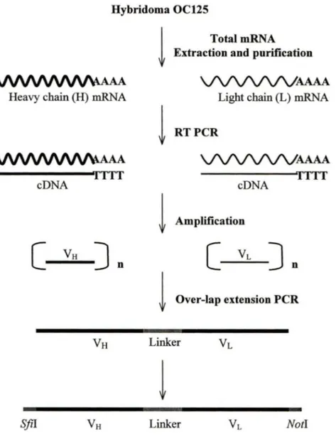
The mRNA extracted from the murine hybridoma cell line OC125 was used to generate ScFv cDNA, and the ScFv constructs were constructed with the Recombinant Phage Antibody System (Amersham Pharmacia Biotech) according to the instructions provided by the manufacturer (Figure 4).
2.1.1 mRNA isolation and purification
-) The QuickPrep
mRNA
Purification Kit (Amersham Pharmacia Biotech) was used to isolate and purify hybridoma mRNA.Approximately 5x10⁶ OC125 hybridoma cells were pelleted by spin for 5 minutes at 1 OOOg, then l .5ml of Extraction Buffer was added to resuspend the cell pellet and the cells were lysed by gently vortexing. Three ml ofElution Buffer was added to the extract and mixed by vortexing, then after centrifugation at full speed for 10 minutes at room temperature, 4ml of the supematant was transferred onto the surface of the resin of a Oligo ( dT)-Cellulose Spun Column. The column was mixed for 10 minutes by gently inverting, then the resin was separated from the suspension by spin at 350g for 2 minutes. Tuen the supematant was discarded and 3ml of High-Salt Buffer was used to resuspend the matrix. The matrix was pelleted by spin at 350g for 2 minutes.
n
Hybridoma OC125!
TotalmRNAExtraction and purification
VVVV\/\M/\4.AAA
VV\/VVVAAAA
Heavy chain (H) mRNA Light chain (L) mRNA
!
RTPCRVVVV\/\M/\4.AAA
VV\/VVVAAAA
TTT TTT cDNA cDNA!
Amplification[
VH
] n[
VL
] n!
Over-lap extension PCR(
__)
VH
LinkerVL
!
S.fil
VH
LinkerVL
NotlFigure 4. Derivation of ScFv constructs: The murine hybridoma cell line OC125 was
used to generate cDNA from purified mRNA by reverse transcription PCR. The V 8 and V L chains were amplified from the cDNA by PCR using mouse variable region primers. The V8 and
VL
DNA fragments were linked together by overlap extension PCR using a (Gly4Ser)3 linker to generate a 750-bp ScFv construct with flanking S.fil and Not! restriction sites.) The matrix was washed two more tiines, then resuspended using 3ml ofLow-Salt Buffer. Spinning at 350g for 2 minutes and discarding the supematant, 0.75ml of 65°C Blution Buffer was used to elute the bound poly (A/RNA by centrifuge at 350g for 2 minutes. The mRNA (750µ1 total volume) was ethanol-precipitated by addingl5µ1 of Glycogen Solution (lOmg/ml), 1/10 volume of 2.5M potassium acetate (pH5.0) and 2.5 volumes of -20°C 95% ethanol. Spinning at full speed for 10 minutes at room temperature, the precipitate was washed twice with cold 95% ethanol, dried and then resuspended in 20µ1 ofRNase-:free water for storage at-70°C.
2.1.2 First-strand cDNA synthesis
~ _ ~) M-MuLV reverse transcriptase was used in the first-strand cDNA reaction to generate cDNA :from the mRNA (:from section 2.1.1). The mRNA was heated at 65°C for 15 minutes and chilled iinmediately on ice. For each sample, the reaction was prepared in duplicate. One tube was labeled as light chain and the second tube as heavy chain. Bach reaction contained 10µ1 of mRNA, 11µ1 of RNase-free Water, 11µ1 of Primed First-Strand Mix and lµl ofDTI Solution. The mixture was incubated for 1 hour at 37°C.
2.1.3 Primary PCR amplification
The first-strand antibody cDNA (from section 2.1.2) was used as a template for PCR amplification to generate suitable quantities of antibody heavy and light chain DNA for cloning. Bach PCR reaction contained 33µ1 of First-strand reaction, 2µ1 ofLight Primer
:) Mix, and 64µ1 of Sterile distilled water (or 33µ1 of First-strand reaction, 2µ1 of Heavy Primer 1, 2µ1 ofHeavy Primer 2, and 62µ1 of Sterile distilled water). After 5 minutes of denaturation at 95°C, 1 µl of AmpliTaq DNA polymerase was added, followed by 30 cycles of 1 minute at 94°C, 2 minutes at 55°C, and 2 minutes at 72°C, and at the end of cycling an incubation of20 minutes at 72°C.
2.1.4 Purification of primary PCR products
Prior to performing the assembly reaction, the amplified heavy and light chain PCR fragments were isolated from the other reaction components by electrophoresis on 0.8% agarose gel. The 340bp heavy chain and 325bp light chain fragments were eut out from ( ) the gel, from which the amplified cDNA was extracted using Gel Extraction Kit
(QIAGEN), according to the instructions provided by the manufacturer.
(
2.1.5 Assembly and fill-in reactions
The ScFv cDNAs were generated by assembly of purified Vtt, the (Gly4Ser)3 linker, and
VL fragment. A mixture containing 50ng of heavy chain product, 50ng of light chain product, 4µ1 ofLinker-primer Mix, 5µ1 of IOxPCR Buffer, 2.5µ1 of dNTP Mix (20mM of each dNTP), and 5µ1 of25mM MgC12, was made. Sterile distilled water was added to a total volume to 49µ1, after 5 minutes of denaturation at 94°C, 1µ1 (SU) of AmpliTaq DNA polymerase was added, followed by 7 cycles of 1 minute at 94°C, 4 minutes at 63°C.
:-) 2.1.6 Second PCR amplification and purification
In the second PCR, the assembled ScFv DNA was amplified and restriction sites were added. A primer mixture was made, which containing lµl (SU) of AmpliTaq DNA polymerase, S µ1 of 1 OxPCR Buffer, 1 µ1 of dNTP Mix (20mM of each dNTP), 4µ1 of RS Primer Mix (Restriction Site Primers, which contain either Sfi I or Not I restriction sites. Its sequence is not given in the manufacturer's instruction.), and 39µ1 of sterile distilled water. The primer mixture was mixed briefly and added to the assembly reaction (from section 2.1.S), followed by 30 cycles of 1 minute at 94°C, 2 minutes at SS°C, and 2 minutes at 72°C. The reaction was hold at 4°C ifnecessary.
The second PCR product was transferred to the top of the compacted resin of a prepared (
~)
MicroSpin Columns, which was put in a sterile l .Sml microcentrifuge tube, and spun at 73Sg for 2 minutes. The column was discarded. The effluent in the tube contained the purified assembled ScFv DNA that was ready for quantitation on an agarose gel alongside the ScFv Marker.The ScFv genes were digested with Sfi I and Not 1, agarose gel-purified, and then ligated into the prokaryotic expression vector pCANTAB SE (Pharmacia Biotech) that had been eut with the same restriction enzymes. Screening of recombinant clones expressing a ScFv against the CA12S protein was accomplished by phage-display and colony-lift assay, as will be described in section 2.S and 2.6.
' ) 2.2 Plasmids
The phagemid pCANTAB SE (Figure 5) which contains the OC12S ScFv DNA under the control of the IPTG inducible and glucose repressible lac promoter; a g3p leader sequence that directs transport of the protein to the inner membrane/periplasm of E.co/i where the main g3p domain attaches the fusion protein to the tip of the assembling phage. pCANTAB SE also contains a sequence encoding an E-tag, which allows easy immunological detection of ScFv protein expression, followed by an amber translational stop codon at the junction between the cloned ScFv and the sequence for the g3p. When a suppressor strain of E.coli, such as DHllS, is transformed with this recombinant vector, translation continues through the amber stop codon to produce the ScFv-g3p !' fusion protein which can be displayed on the phage tip. In nonsuppressor strains, such as
'.)
HB21 S l, the stop codon is recognized, protein synthesis is aborted at the stop codon, the g3p fusion protein is not made, and the resulting ScFv protein is transported and accumulated into the periplasmic space.
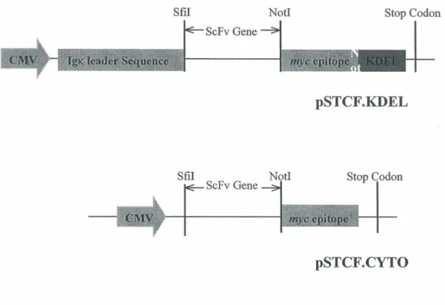
The endoplasmic reticulum (ER)-targeting eukaryotic vector pSTCF. KDEL (Figure 6)
contains a CMV promoter; an IgK leader sequence that directs the ScFv protein into ER, a mye epitope coding an easily detected mye tag, while the KDEL sequence located at the C-terminus causes the protein to be retained in the ER. The pSTCF.cyto vector (Figure 6), which directs cytoplasm expression, only has a CMV promoter and a mye tag sequence. In most cases, ScFv expressed in a cytoplasm vector are not stable (Willianms, G.T. et al, 1990), and in this study it is used as a negative control for ScFv expression.
)
E tag (2380-2418)
Amber Stop Codon (24251
pCANTAB 5 E Nde 1 (3512) 4522 bp
Figure 5. pCANT AB SE vector showing control regions. This prokaryotic vector is
designed such that ScFv genes can be cloned between the leader sequence and the main body of the M13 gene 3. The g3p leader sequence directs transport of the protein to the -periplasm of E.coli. The ScFv is expressed as a fusion protein with the E-tag peptide to
allow easy detection. The vector also contains an amber translational stop codon at the junction between the cloned ScFv and the sequence for the g3p.
)
Sfil Notl Stop Codon ScFv Gene
pSTCF.KDEL
Sfil Notl
ScFv Gene Stop Codon
pSTCF.CYTO
()
Figure 6. pSTCF.KDEL eukaryotic vector expressing ScFv genes. Expression of the ScFv protein is driven by the CMV promoter. The ScFv cDNA is introduced between the Sfil and Notl restriction sites. The lgx leader sequence directs the ScFv protein to the ER, and the KDEL signal at the COOH terminus leads to retention of the fusion protein in this cellular compartment. The ScFv open reading frame is also fused with a c-myc epitope to allow easy detection by Western-Blot. In pSTCF.cyto vector, the ScFv protein expression is targeted to the cytoplasm.
--Piché, A., Grim, J., Curiel, D.T. et al. (1998) Cancer Research, 58:2134-2140
--- - 2.3 Preparation of the ScFv cDNA library
1
~)
One µlof ligated pCANTAB 5 vector was transfonned into 40µ1 of competent E.coli XLI-Blue by electroporation. After incubating at 37°C for I ho
ur with shaking at 250rpm, the transfonned cells were plated onto a LB-1.5% agar (For IL: lOg Bacto-tryptone, 5g Yeast extract, 5g NaCl and I5g agar) plate containing lOOµg/ml of ampicillin, and incubated overnight at 37°C. The next day, 5ml of LB medium containing IOOµg/ml of ampicillin was used to suspend all of the colonies from the plate. This suspension was incubated for 6 hours at 37°C with shaking at 250rpm. A miniprep was made with 2.5ml of the culture using the QIAprep Spin Plasmid Miniprep kit (QIAGEN) and the remaining 2.5ml of culture was kept at -80°C as a frozen stock.
PCR was perfonned to confirm that the insert was present in the recombinant plasmid library, and was run on PTC-200 Peltier Thermal Cycler (MJ Research). One µl of I :500 diluted pCANTAB.OCI25 ScFv library (-5ng ofDNA) was mixed with 5µ1 of lOxPCR buffer, Iµl of dNTP Mix (20mM of each dNTP), 4µ1 of RS Primer Mix, and 38µ1 of sterile distilled water. A:fter 4 minutes of denaturation at 94°C, Iµl (SU) of AmpliTaq DNA polymerase was added, followed by 30 cycles of 1 minute at 94°C, 2 minutes at 55°C, and 2 minutes at 72°C. Tuen the PCR procuct was loaded on 0.8% agarose gel, alongside with DNA marker and ScFv marker.
:'} 2.4 CA125 antigen
(_)
Tue CA12S antigen was obtained from 10-fold concentrated conditioned media ofhuman ovarian cancer OVCAR3 cells. Fifteen ml of media was centrifuged at lOOOrpm in a Millipore BioMax 100 Unit (Millipore), until the volume was reduced to about 7SOµl. This concentrated media was removed and the BioMax unit was washed with 7SOµl of PBS. Tue concentrated media and the washing buffer were transferred and combined together in a l .Sml microcentrifuge tube. The protein concentration was 2S.2Smg/ml.
2.5 Phage display
2.5.1 Transformation
Tue library ofpCANTAB SE vector containing the OC12S ScFv insert was transformed into competent E.coli DH11S by heat-shock. Plasmid pUC19 (Amersham Pharmacia Biotech) was used as a control plasmid to determine the transformation efficiency. One µlof 1:8000 diluted miniprep DNA extracted from the ScFv cDNA library was mixed with 100µ1 of competent cells, the mixture was placed on ice for 4S minutes, incubated in a 42°C water bath for 2 minutes, and then chilled briefly on ice for 90 seconds. Tue mixture was diluted with 900µ1 of room temperature SOC (lOg Bacto-tryptone, 2.Sg yeast extract, lml SM NaCl, l.2Sml lM KCl, lOml lM MgS04, Sml lM MgCh, 2.Sml 20% glucose, for SOOml) medium and incubated for one hour at 37°C with shaking at
) cens were plated onto a 2xYT-AG plate (17g Bacto-tryptone, lOg Yeast extract, 5g NaCl, and 15g agar, for lL, containing lOOµg/ml ampicillin and 2% glucose) to determine the transformation efficiency. The plate was incubated ovemight at 37°C.
2.5.2 Phage rescue
The remaining 900µ1 ofundiluted transformed cens (section 2.5.1) was added to 9.lml of 2xYT-AG medium, and incubated at 37°C with shaking at 250rpm, until an 00600 of0.5 was reached. At this point, lml of the culture was kept to make a frozen stock. Tuen, 4x10¹⁰ pfu (plaque forming unit) of M13K07 helper phage (Amersham Pharmacia Biotech) was added to the 9ml of culture. After another one hour of incubation, the cens • ) were spun for 15 minutes at 3500 rpm. The penet was gently resuspended with lOml of 2xYT-AK medium (17g Bacto-tryptone, lOg Yeast extract, and 5g NaCl, for lL), which contained lOOµg/ml ampicillin (to select for cens with phagemid) and 50µg/ml kanamycin (to select for cens infected with M13K07). The suspension was incubated ovemight at 30°C with shaking at 250rpm. The cens were peneted by spinning at 2500rpm for 20 minutes at 4°C. The supematant, which contained the recombinant phage, was used for the panning reaction.
,~---) 2.5.3 Panning
The library was subjected to five rounds of panning. The 1 Oml supematant (:from section 2.5.2), which contained recombinant plasmid in phage particles having the ScFv expressed on their surface, was mixed with 2ml of ice-cold PEG/NaCI (200g Polyethylene glycol 8000, 146.lg NaCI, for IL), and incubated on ice for 60 minutes. The suspension was centrifuged at 12,000rpm in a Beckman SS-34 rotor for 45 minutes at 4 °C, and the pellet containing the phage was resuspended with 16ml of 2x YT medium.
A 25cm2 tissue culture flask (FALCON) was coated overnight at 4 °C with 5ml of CA125 antigen (2mg of protein) diluted in 0.05M Na2C03 (pH9.6), and blocked by filling the flask with 4% NFDM (Non-fat Dry Milk) (BioRad) in lxPBS for 1 hour at 37°C. The 16ml of PEG-precipitated recombinant phage was diluted with 14ml of blocking buffer (containing 0.01% sodium azide) and incubated at room temperature for
15 minutes. Twenty ml of the diluted recombinant phage was added to the 4% NFDM-blocked flask, and the flask was incubated for 2 hours at 37°C with rocking. The flask was washed 20 times with lxPBS/0.1 % Tween and 20 times with PBS only.
2.5.4 Reinfection of E.coli with enriched phage clones
Ten milliliters oflog-phase DHl lS cells were added to the flask (:from section 2.5.3), and incubated for 1 hour at 37°C. Tuen 100µ1 of cell suspension was removed to prepare ten-fold dilutions in 2xYT medium (1:10, 1:100, 1:1000). One hundred µl ofundiluted cells
r--.
and 100µ1 of each dilution were plated onto separate SOBAG plates (For IL, add 20g'
)
Bacto-tryptone, 5g Yeast extract, 0.5g NaCl, lOml lM MgC12, 55.6ml 2M glucose, and 15g agar, containing 20mg/ml ampicillin). After incubating overnight at 30°C, individual well-isolated colonies were transferred to separate tubes containing 400µ1 of 2xYT-AG medium, and were incubated overnight at 30°C with shaking at 250 rpm. These recombinant phage-infected cells contain the pCANTAB 5E phagemid with the ScFv gene insert, and do not contain complete, infectious phage genomes.
2.5.5 Screening for recombinant phage antibodies in enriched clones
Forty µl of overnight culture (from section 2.5.4) was transferred to 400µ1 of 2xYT-AG
(_ ~ medium containing 2x10⁸ pfu of M13K07 helper phage, and incubated for 2 hours at 37°C with shaking at 150 rpm. The culture was centrifuged for 20 minutes at 2500rpm at room temperature, and the pellet was resuspended with 400µ1 of 2xYT-AK medium (no glucose). The suspension was incubated overnight at 37°C with shaking at 250 rpm. The overnight culture was centrifuged as described above, and 320µ1 of supernatant (which contained recombinant plasmid in phage particles expressing the ScFv) was mixed with 80µ1 of 4% NFDM in lxPBS. The mixture was incubated for 10 minutes at room temperature. An ELISA was performed as described in section 2.5.7 using mouse anti-Ml3 antibody (Amersham Pharmacia) to detect the phage which display antibodies on its tips.
: ,-) 2.5.6 Screening for expression of soluble antibodies in enriched clones
Forty µlof overnight culture (of section 2.5.4) was transferred to a tube containing 400µ1 of 2xYT-AG medium, and incubated for 2 hours at 30°C with shaking at 250rpm. The culture was centrifuged at 2500rpm for 20 minutes at room temperature, and the pellet was resuspended with 400µ1 of 2xYT-AI medium (containing lmM IPTG). The cell suspension was incubated for 3 hours at 30°C with shaking at 250rpm. The suspension was centrifuged as described above, and 320µ1 of supematant (which contained soluble recombinant antibodies) was mixed with 80µ1of4% NFDM in lxPBS. The mixture was incubated for 10 minutes at room temperature. An ELISA was performed as described in section 2.5.7 using anti-E tag antibody (Amersham Pharmacia Biotech).
2.5. 7 Binding analysis by ELISA
An ELISA was performed to detect ScFv either expressed on the surface of phage
.
particles or :free in bacterial extracts. The CA125 antigen was applied to a 96-well microtiter plates (SARSTEDT) at a concentration of 5µg/ml in 0.05M Na2C03 (pH9.6) at 4°C ovemight. A:fter being washed 6 times with lxPBS-0.l %Tween, the wells were blocked with 4% NFDM in lxPBS for 1 hour at 37°C. Tuen, 50µ1/well ScFv phage extract (from section 2.5.5) or soluble ScFv extract (from section 2.5.6) was applied and incubated for 1 hour at 37°C. A:fter washing, phage that carried ScFv on their surface were detected by adding 50µ1 of horseradish peroxidase (HRP)-conjugated mouse anti-M13 (Amersham Pharmacia) 1:10,000 diluted in blocking buffer and incubating for 45
)
~ anti-E tag lg 1:5,000 diluted in blocking buffer was added and incubated for 45 minutes
at 37°C. For revelation, 100µ1/well of TMB substrate (Pierce) was used, and the absorbance was read at 450nm after incubation for 20 minutes at room temperature in the dark.
Once the CA125-positive clones had been identified by ELISA, 2µ1 of phage supematant (of section 2.5.5), which contained positive recombinant antibodies, was used to infect 400µ1 of log phase E.coli HB2151. The culture was incubated for 30 minutes at 37°C
with shaking. SOBAG plates were inoculated with a loopful of the infected culture and incubated overnight at 30°C. Individual colonies were transferred into l Sml centrifuge tubes, which contained Sml of 2xYT-AG medium, for production of soluble antibodies as ( ) described in section 2.6.3.
2.6 Colony-lift assay
2.6.1 Transformation
The ligated pCANTAB SE vector containing the OC125 ScFv insert was transformed into competent E.co/i HB2151 by electroporation: 40µ1 of the cells were transferred to a
pre-chilled 0.2cm cuvette, 1 µl of salt-free DNA containing 20ng pCANTAB/ScFv cDNA library was added and shaken to the bottom of the cuvette, which was then placed on ice for 1 minute. The electroporator (BioRad) was programed to give 25µF, 2.SkV at 200 ohms. After electroporation, lml of SOC medium was immediately added to resuspend the cells, and the suspension was transferred to a l Sml tube and incubated for 1 hour at
~ 37°C with shaking at 250rpm. The cells were centrifuged for 5 minutes at 3000rpm, and
lml of 2xYT-G medium was used to resuspend the cell pellet. Each 100µ1 ofthis lml
(_)
)
·'
suspension was used to plate a 2xYT-AG plate, then these 10 plates were incubated overnight at 30°C.
2.6.2 Colony lift
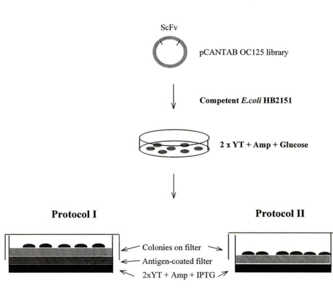
A piece of dry Butterfly-Membrane (Schleicher & Schuell) was used to lift the well-isolated colonies (-300/plate) in each plate. After lifting, the membrane with colony-side face up was either put on the top of 2xYT-AI plate (containing lOOµg/ml ampicillin and lmM IPTG) with (1 st protocol) or without (2nd protocol) a CA125 antigen-coated membrane beneath (Figure 7). The plate was incubated overnight at 30°C. The second day, after washing with lxPBS/0.1 %Tween, the antigen-coated membrane (of the 1 st protocol) or the colony-membrane (of the 2nd protocol) was blocked with 4% NFDM in lxPBS for 1 hour at room temperature with rocking. The membranes were probed with anti-E Tag 1: 1000 diluted in blocking buffer for 2 hours at room temperature with rocking. After washing, the same membrane was probed with HRP-conjugated anti-mouse Ig (Sigma) 1 :10,000 diluted in blocking buffer for 45 minutes at room temperature with rocking. For detection, 8ml/membrane of 4-CN (4-chloro-1-naphthol) substrate (Sigma) was added and incubated for - 30 minutes with rocking in the dark. When blue dots (- lmm diameter) were detected on the membrane, the development was stopped with water. The corresponding colony was picked :from the original plate to inoculate Sml of2xYT-AG medium and incubated overnight at 30°C with shaking at 250rpm.
0
(__)
Protocol 1
' ' ::;. ' ' ' ;~ ~~~~~~~~~~ -- - - --ScFvpCANTAB OC125 library
Competent E.coli HB2151
~
2 x YT + Amp +Glucose~
~ Colonies on filter ~ ~ Antigen-coated filter ~ 2xYT+Amp + IPTG ~Protocol II
Figure 7. ScFv library screening using Colony-lift assay. ScFv cDNA library was transformed into competent E.coli HB2151 and grown on 2xYT-AG plate. A butterfly-membrane was used to lift the colony. Protocol 1: a CA125 antigen-coated butterfly-membrane was put on the 2xYT-AI agar surface, and the colony-lift membrane was put on this antigen-coated membrane with the colony-side face up. Protocol II: the colony-lift membrane was put directly on top of 2xYT-AI agar, with the colony-side face up. For both protocols, the membrane was developed using 4-CN substrate. Colonies corresponding to the blue dots were picked and grown in liquid medium.
)
2.6.3 Periplasmic ExtractFive ml of overnight culture (from section 2.6.2) was added to 15ml of 2xYT-AG medium, and incubated for 1 hour at 30°C with shaking at 250rpm. The culture was centrifuged for 15 minutes at 3,500rpm at 4°C, and the pellet was resuspended with 30ml of2xYT-AI (without glucose) and incubated for 3 hours at 30°C with shaking at 250rpm. After centrifuging the culture for 20 minutes at 4,000rpm at 4°C, 750µ1 of ice-cold 0.2xTES buffer (40mM Tris-HCI pH8.0, O.lmM EDTA, and O.lM sucrose, filter sterilized) was used to resuspend the pellet by vortexing. The suspension was incubated on ice for 30 minutes, and then the contents were centrifuged at full speed for 10 minutes at 4 °C. The supernatant, which contains the soluble antibodies from the periplasm, was
:~-
-) transferred to a clean tube and stored at-20°C until needed.2. 7 Cell lin es
The human ovarian cancer cell line PA-1 and OVCAR3 were obtained from the ATCC (American Type Culture Collection). PA-1 cells were maintained in DMEMIF12 medium (Bio Media) supplemented with 10% (v/v) fetal bovine serum (FBS) (Hyclone Laboratories), 1 % (v/v) L-glutamine, 1 % antibody and 1 % fungizone (Gibco). OVCAR3 cells were maintained in RPJ\.11-1640 medium (Bio Media) supplemented with 20% (v/v) FBS, 1 % (v/v) L-glutamine, 1 % antibody, 1 % fungizone and 0.0 lmg/ml bovine insulin.
·-...) 2.8 Transient transfection
PA-1 or OVCAR3 cells (·--300,000/well) were plated in 6-well plates (FALCON) 24 hour prior to transfection in 3ml of growing medium, and the cells were incubated in the incubator with 5% C02 at 37°C. For transient transfection experiments, cells were transfected with plasmid DNA with the liposomal transfection reagent SuperFect (QIAGEN). Plain cell growth medium (no supplements) was used to dilute 2µg ofDNA (dissolved in TE buffer, pH7.4) to a total volume of 100µ1. A:fter mixing the diluted DNA, 10µ1 of SuperFect Transfection Reagent was added to the DNA, and mixed by pipetting up and down 5 tiines. After incubating the sample for 10 minutes at room temperature to allow complex formation, 0.6ml of complete growth medium was added
·) to the 100µ1 of complex. During the complex formation, the cells were washed once with PBS. The total volume of the reaction was transferred to the cells in the 6-well plate, and the cells were incubated with the complexes for 5 hours with 5% C02 at 37°C. A:fter removing the medium and washing the cells with PBS, 3ml of fresh cell growth medium was added to the cells.
A:fter variable times of incubation, cells were lysed usmg Promega lysis buffer (Promega). The protein concentration of the lysates was measured by the Bradford method with BioRad Protein Assay according to the manufacturer's instruction (BioRad).
2.9 Immunoblot analysis
Equal amounts ofprotein (,..,35µg) were loaded and separated by SDS-PAGE (12%) with a stacking gel concentration of 4%. After transfer onto Hybond-P PVDF membranes (Amersham Pharmacia Biotech) (300mA, 90 minutes, 4°C) and blocking with 3% NFDM, the membranes were probed with either 1:1,000 anti-E Tag antibody or1:2,000
anti-c-myc
antibody (Invitrogen). After washing with lxPBS-0.1%Tween, 1:10,000 diluted (in blocking buffer) HRP-conjugated rabbit anti-mouse antibody was used. The immunoblots were developed using ECL or ECL PLUS system (Amersham Pharmacia Biotech) according to the manufacturer's instructions. To ensure equivalent protein loading, the membrane was also eut and probed with 1 :7 ,000 anti-tubulin antibody< __
J
(Sigma).)
2.10 Sequence analysis of ScFvs
The nucleotide sequences of the ScFvs having different expression levels and antigen binding activities were determined using DNA Symo Sequencing technology and run on a Li-Cor sequencer by BioS&T Inc. (Montreal, Que bec). Samples were prepared with QIAprep Miniprep Kit (QIAGEN). The pCANTABS-Sl and S6 primers were used: pCANTABS-Sl primer: CAACGTGAAAAAATTATTATTCGC