Université de Montréal
Feedback regulation of Gab2-dependent signaling by the
Ras/MAPK pathway
par Xiaocui Zhang
Programmes de Biologie Moléculaire Faculté de Médecine
Thèse présentée à la Faculté de Médecine
en vue de l’obtention du grade de Philosophiae Doctor (Ph.D.) en Biologie Moléculaire
option Biologie des Systèmes
Dec., 2014
i
Résumé
La voie de signalisation des Récepteurs Tyrosine Kinase (RTK) occupe un rôle central dans la régulation de la croissance cellulaire, la prolifération, la différentiation et la motilité. Une régulation anormale des RTKs mène à plusieurs maladies humaines telles que le cancer du sein, la seconde cause de mortalité chez les femmes à cause de l’amplification et la mutation fréquente de la protéine tyrosine kinase HER2 (ERBB2). Grb2-associated binder (Gab) 2 est une protéine adaptatrice qui agit en aval de plusieurs RTKs, y compris HER2, pour réguler de multiples voies de signalisation. En réponse à la stimulation par de nombreux facteurs de croissances et cytokines, Gab2 est recruté à la membrane plasmique où il potentialise l’activation des voies de signalisation Ras/mitogen-activated protein kinase (MAPK) et PI3K (phosphatidylinositol-3-kinase)/Akt (protein kinase B). En plus d’occuper un rôle essentiel durant le développement du système hématopoïétique, Gab2 est souvent amplifié dans les cancers, notamment le cancer du sein et les mélanomes. Cependant, les mécanismes moléculaires qui régulent le fonctionnement de Gab2 sont peu connus.
Il est établi que lors de l’activation des RTKs, Gab2 est phosphorylé au niveau de plusieurs résidus Tyrosine, menant à l’association de différentes protéines comme p85 et Shp2. En plus de la phosphorylation en Tyrosine, notre laboratoire ainsi que d’autres groupes de recherche avons identifié que Gab2 est aussi phosphorylé au niveau de résidus Ser/Thr suite à l’activation de la voie de signalisation MAPK. Cependant, la régulation des fonctions de Gab2 par ces modifications post-traductionnelles est encore peu connue. Dans le but de comprendre comment Gab2 est régulé par la voie de signalisation MAPK, nous avons utilisé différentes approches. Dans la première partie de ma thèse, nous avons déterminé un nouveau mécanisme démontrant que la voie de signalisation Ras/MAPK, par le biais des protéines kinases RSK (p90 ribosomal S6 kinase), phosphoryle Gab2. Ce phénomène se produit à la fois in vivo et in
vitro au niveau de trois résidus Ser/Thr conservés. Des mutations au niveau de ces sites de
phosphorylation entrainent le recrutement de Shp2 menant à l’augmentation de la motilité cellulaire, ce qui suggère que les protéines RSK restreignent les fonctions dépendantes de Gab2. Ce phénomène est le résultat de la participation de RSK dans la boucle de rétroaction négative de la voie de signalisation MAPK. Dans la seconde partie de ma thèse, nous avons démontré que les protéines ERK1/2 phosphorylent Gab2 au niveau de plusieurs résidus
ii
pS/T-P à la fois in vitro et in vivo, entrainant l’inhibition du recrutement de p85. De plus, nous avons établi pour la première fois que Gab2 interagit physiquement avec ERK1/2 dans des cellules lors de l’activation de la voie de signalisation MAPK. Par ailleurs, nous avons montré un nouveau domaine d’attache du module ERK1/2 sur Gab2. Des mutations sur les résidus essentiels de ce domaine d’attache n’entrainent pas seulement la dissociation de ERK1/2 avec Gab2, mais diminuent également la phosphorylation dépendante de ERK1/2 sur Gab2.
Ainsi, nos données montrent que la voie de signalisation MAPK régule les fonctions de la protéine Gab2 par le biais des kinases RSK et ERK1/2. Cette thèse élabore par ailleurs un schéma complet des fonctions de Gab2 dépendantes de la voie de signalisation MAPK dans le développement de nombreux cancers.
iii
Abstract
Receptor tyrosine kinase (RTK) signaling plays an essential role in modulating cell growth, proliferation, differentiation and motility. Abnormal regulation of RTKs results in many human diseases, including breast cancer, the second leading cause of cancer mortality in women by the frequent amplification and mutation of the HER2 (ERBB2) tyrosine kinase. The Grb2-associated binder (Gab) 2 is an adaptor protein that acts downstream of several RTKs, including HER2, to regulate multiple signaling pathways. In response to the stimulation of a number of growth factors and cytokines, Gab2 is recruited to the plasma membrane, where it potentiates the activation of the Ras/mitogen-activated protein kinase (MAPK) and PI3K (phosphatidylinositol-3-kinase)/Akt (protein kinase B) pathways. In addition to playing an important role in the hematopoietic system during development, GAB2 is often amplified in cancers including breast cancer and melanoma, however, little is known about the molecular mechanisms that regulate Gab2 function.
It has been well established that upon RTKs activation, Gab2 becomes phosphorylated on several Tyr residues leading to diverse adaptor protein associations, such as p85 and Shp2. Aside from the tyrosine phosphorylation, our lab and other groups noticed that Gab2 is also phosphorylated on Ser/Thr residues upon activation of MAPK signaling. However, less is known about this post-translational modification in the regulation of Gab2 functions. In order to understand how Gab2 is regulated by the MAPK pathway, we used different approaches. In the first part of my thesis, we determined a new mechanism by which the Ras/MAPK pathway through RSK (p90 ribosomal S6 kinase) phosphorylated Gab2 on three conserved Ser/Thr residues, both in vivo and in vitro. Mutation of these phosphorylation sites promoted Shp2 recruitment leading to increased cell motility, suggesting that RSK restricts Gab2-dependent functions as a result of participation in the negative feedback loop of MAPK signaling. In the second part of the thesis, we found that ERK1/2 phosphorylated Gab2 on several potential pS/T-P residues, both in vivo and in vitro, resulting in inhibited p85 recruitment. In addition, to the best of our knowledge, we established for the first time that Gab2 physically interacted with ERK1/2 in cells upon activation of the MAPK pathway. Furthermore, we revealed a novel ERK1/2 docking domain in Gab2. Mutation of the essential residues in this docking domain not only disrupted ERK1/2-Gab2 interaction, but also
iv
diminished ERK1/2-dependent phosphorylation on Gab2.
Taken together, our data showed that the MAPK pathway regulates Gab2 functions through both RSK- and ERK1/2-dependent manners. Given that Gab2 is overexpressed in several cancers, this thesis decodes a complete figure of modulating actions of Gab2 by MAPK signaling in cancer development.
v
Table of Contents
Résumé ... i Abstract ... iii Table of Contents ... v List of Tables ... ix List of Figures ... xList of Abbreviations ... xii
Acknowledgements ... xix
Contribution of Co-authors ... xxi
Chapter 1 Introduction and Literature Review ... 1
1.0 General Introduction ... 2
1.1 Characteristics of carcinogenesis ... 3
1.1.1 Sustaining proliferation and evading growth suppressors ... 4
1.1.2 Angiogenesis formation in the primary tumor site ... 5
1.1.3 Invasion and metastasis ... 6
1.2 Breast cancer and receptor tyrosine kinase ... 8
1.2.1 Grades, stages and types of breast cancer ... 8
1.2.2 Treatments of breast cancer ... 11
1.2.3 ErbB2/HER2 in breast cancer ... 13
1.2.3.1 EGFR family ... 13
1.2.3.2 Activation mechanisms of HER2 receptor ... 15
1.3 The Ras/MAPK/RSK signaling pathway ... 17
1.3.1 Background: Mechanisms of MAPK cascade activation ... 17
1.3.2 The ERK1/2 module ... 19
1.3.2.1 Identification of ERK1/2 and their physiological functions ... 19
1.3.2.2 Activation mechanisms and inhibitors of ERK1/2 ... 20
1.3.2.3 Docking interactions from ERK1/2 to substrates ... 22
1.3.2.4 Subcellular localization of the ERK1/2 cascade and its biological functions ... 25
1.3.2.4.1 ERK1/2 in the nucleus and cell proliferation ... 26
1.3.2.4.2 ERK1/2 in the plasma membrane and regulation of cytoskeletal elements ... 27
vi
1.3.2.4.4 ERK1/2 in other cellular compartments ... 31
1.3.2.5 Regulation of the ERK1/2 cascade by feedback loops ... 32
1.3.3 MAPK–activated protein kinase RSK (90 kDa ribosomal S6 kinase) ... 37
1.3.3.1 Discovery of RSKs, their tissue expression and physiological functions ... 37
1.3.3.2 Structural features of RSKs ... 38
1.3.3.3 Activation mechanisms and inhibitors of RSK ... 39
1.3.3.4 Subcellular localization and RSK-mediated biological functions ... 42
1.3.3.4.1 Regulation of transcription factor activities ... 43
1.3.3.4.2 Regulation of cell cycle progression and cell proliferation ... 44
1.3.3.4.3 Regulation of protein synthesis and cell growth ... 47
1.3.3.4.4 Regulation of cell survival ... 48
1.3.3.4.5 Regulation of cell migration ... 49
1.3.3.4.6 Other substrates and functions ... 50
1.4 The Gab/Dos family of docking proteins ... 51
1.4.1 Discovery of Gab proteins and their biological functions in mice ... 51
1.4.2 Activation mechanisms of Gab protein-dependent signaling ... 52
1.4.2.1 Structural features of Gab proteins ... 52
1.4.2.2 Role of the PH domain ... 53
1.4.2.3 Role of the proline-rich domains in Gab2 ... 54
1.4.2.4 Tyrosine phosphorylated Gab2 mediates Gab2-dependent complex formation and sustains its downstream signaling ... 55
1.4.2.5 Other binding partners to Gab2 ... 58
1.4.3 Role of Gab2 in tumorigenesis ... 59
1.4.3.1 Gab2 in breast cancer ... 61
1.4.3.2 Gab2 in melanoma ... 63
1.4.3.3 Gab2 in leukemia ... 63
1.4.4 Feedback regulation of Gab2 function ... 64
1.5 Rationale and objectives ... 69
1.5.1 Rationale ... 69
1.5.2 Objectives ... 69
vii
Chapter 2 Gab2 phosphorylation by RSK inhibits Shp2 recruitment and cell
motility ... 71
ABSTRACT ... 73
INTRODUCTION ... 74
MATERIALS AND METHODS ... 76
RESULTS ... 82
ACKNOWLEDGMENTS ... 93
REFERENCES ... 94
FIGURES ... 99
Preface to Chapter 3 ... 110
Chapter 3 ERK1/2 participate in a negative feedback loop that limits Gab2 function in response to growth factors ... 111
INTRODUCTION ... 114
MATERIALS AND METHODS ... 117
RESULTS ... 119
DISCUSSION ... 125
ACKNOWLEDGMENTS ... 128
REFERENCES ... 129
FIGURES ... 133
Chapter 4 General Discussion ... 142
4.1 Regulation of Gab2 by RSK kinases ... 143
4.1.1 Phosphorylation of Gab2 by RSK impacts the recruitment of its binding partners ... 143
4.1.2 Potential roles of phosphorylated Gab2 by RSK in signal transduction ... 144
4.1.3 The potential role of Gab2 phosphorylation by RSK in primary mast cells and the allergic response ... 145
4.2 Regulation of Gab2 functions by MAP kinases ... 146
4.2.1 ERK1/2 bind to Gab2 and further regulate its phosphorylation ... 146
4.2.2 Potential functions of ERK1/2-dependent phosphorylation sites in Gab2 ... 147
Is p85 a negative or positive regulator in the PI3K/Akt pathway? ... 148
4.3 Other potential mechanisms of Gab2-mediated cell motility ... 150 4.4 Deciphering the role of the MAPK pathway in the regulation of Gab2-dependent
viii
function ... 151 Chapter 5 Conclusions and Perspectives ... 153 Bibliography ... 156
ix
List of Tables
Table I Molecular subtypes of breast cancer ... 10 Table II Drugs for targeted therapy in breast cancers ... 12
x
List of Figures
Chapter 1
Figure 1.1 Cross-cancer alteration summary for the ERBB2 gene. ... 14
Figure 1.2 Schematic representation of HER receptors-mediated downstream signaling pathways. ... 15
Figure 1.3 Schematic representation of two classifications of MAPKs: conventional MAPKs and atypical MAPKs. ... 17
Figure 1.4 Activation of the conventional MAPK-mediated signaling pathways results in the phosphorylation of MAPKAPKs. ... 18
Figure 1.5 Alignment of MAPK-binding domains in substrates and molecular model of Lig-D bound to ERK2. ... 25
Figure 1.6 Simplified overview of phosphorylated ERK1/2 (p-ERK1/2) and their substrates localization in distinct cellular compartments. ... 30
Figure 1.7 Schematic representation of the cross-talk between the Ras/MAPK and PI3K/mTOR signaling pathways. ... 34
Figure 1.8 Schematic representation of structural features in RSK isoforms. ... 39
Figure 1.9 Schematic representation of human RSK1 kinase domains and phosphorylation sites. ... 40
Figure 1.10 Schematic representation of structural features of Gab proteins. ... 53
Figure 1.11 Schematic representation of various binding proteins recruited to Gab2. ... 58
Figure 1.12 Cross-cancer alteration summary for the GAB2 gene. ... 60
Figure 1.13 Schematic representation of feedback regulation of Gab2-dependent signaling. . 66
Chapter 2
Figure 2.1 Identification of Gab2 as a target of Ras/MAPK signaling. ... 99Figure 2.2 Activation of the Ras/MAPK pathway induces RSK-dependent phosphorylation of Gab2. ... 100
Figure 2.3 Identification of Ser160, Ser211, and Ser620 as being regulated by Ras/MAPK signaling. ... 101
xi
Figure 2.4 RSK predominantly phosphorylates Ser160, Ser211, and Ser620 in cells and in
vitro. ... 103
Figure 2.5 The Ras/MAPK pathway modulates Shp2 recruitment in an RSK-dependent manner. ... 104
Figure 2.6 A Gab2 mutant that cannot be phosphorylated on Ser160/211/620 promotes Shp2 recruitment. ... 105
Figure 2.7 Stable expression of the Gab2 S3A mutant alters MCF-10A cell morphology. .... 107
Figure 2.8 Stable expression of the Gab2 S3A mutant promotes MCF-10A cell migration. . 108
Figure 2.9 Schematic representation of Gab2 signaling and its negative regulation by RSK. 109
Chapter 3
Figure 3.1 Gab2 is phosphorylated in response to agonists of the Ras/MAPK pathway. ... 133Figure 3.2 ERK1/2 phosphorylate Gab2 in vitro and in cells. ... 134
Figure 3.3 ERK1/2 interact with Gab2 in a MEK1/2-dependent manner. ... 135
Figure 3.4 Identification of a D-domain in Gab2 involved in ERK1/2 binding. ... 136
Figure 3.5 Identification of ERK1/2 phosphorylation sites in Gab2. ... 138
Figure 3.6 ERK1/2 negatively regulate Gab2-dependent signaling by reducing p85 recruitment. ... 140
xii
List of Abbreviations
5-FU fluorouracil
AGC family protein kinases A, G and C
Akt protein kinase B
AMPK AMP-activated protein kinase
Apaf-1 apoptotic protease activating factor-1
APC anaphase-promoting complex
Arf6 ADP-ribosylation factor 6
BAC bacterial artificial chromosome array Bad Bcl-2-associated death promoter Bim BCL2-like 11, apoptosis facilitator BMMCs bone marrow-derived mast cells BRCA1 breast cancer gene 1
Bub1 budding uninhibited by benzimidazoles 1 CAMK Ca2+ /calmodulin-dependent protein kinase
CB cysteine cathepsin B
CBP CREB binding protein
CD domain common docking domain
CD36 a member of the class B scavenger receptor family Cdc25 cell division cycle 25
CDKs cyclin-dependent kinases
CGH comparative genomic hybridization
Chk1 checkpoint kinase 1
CKI cyclin-dependent kinase inhibitor
CLS Coffin-Lowry syndrome
c-Met hepatocyte growth factor receptor CREB cAMP response element-binding protein
xiii
Csw Corkscrew; Shp-2 ortholog in Drosophila CTKD carboxyl-terminal kinase domain
CXCL1/2 C-X-C motif chemokine 1/2
DAPK death-associated protein kinase DCIS ductal carcinomas in situ
DEF domain docking site for ERK, FXF
E2F E2 factor family of transcription factors
ECM extracellular matrix
EGFR epithermal growth factor receptor eIF2 eukaryotic initiation factor 2
eIF4E eukaryotic translation initiation factor 4E ELISA enzyme-linked immunosorbent assay
ELK1 ETS domain-containing protein
Emi2 early mitotic inhibitor 2
EMT epithelial-mesenchymal transition
ER estrogen receptor
ERK extracellular signal-regulated kinase
ERα estrogen receptor α
ES embryonic stem
EST expressed sequence tag
FcεRI lgE receptor
FGF fibroblast growth factor
FN1 fibronectin 1
FOXO3a forkhead box O 3a
Gab2 Grb2-associated binder 2
Gadd45 growth arrest and DNA-damage inducible
xiv
Grb2 growth factor receptor-bound protein 2 HIF-α hypoxia-inducible factor 1-alpha
HRG heregulin
IBC invasive breast carcinoma
IEG immediate early genes
IGF-1 insulin-like growth factor
IHC immunohistochemical
IL interleukin
JIP1 JNK-interaction protein 1
JNK c-Jun N-terminal kinases Ki67 a marker for proliferation rate KIBRA kidney and brain expressed protein
KIM kinase interaction motif
KSR suppressor of Ras
Lyn a non-receptor tyrosine-protein kinase Mad1 mitotic arrest-deficient protein
MAP2 microtubule-associated protein-2 MAPK mitogen-activated protein kinase MAPKAPKs MAPK activated protein kinases
MARK MAP-regulation kinase/microtubule affinity regulating kinase
MBD c-Met-binding domain
Mcl1 myeloid cell leukemia sequence 1 MDCK Madin-Darby canine kidney
MET mesenchymal-epithelial transition
Mi microphthalmia
mIMCD-3 mouse inner medullary collecting duct
xv
MK2/3 MAPK-activated protein kinase 2/3
MKP MAP kinase phosphatase
MLC myosin light chains
MLCK myosin light chain kinase
MMP-12 macrophage metalloelastase MMP-9 matrix metallopeptidase 9
MNKs MAPK-interacting kinases
MP1 MEK partner1
MSKs mitogen- and stress-activated kinases
MSP macrophage-stimulating protein
MST1R macrophage-stimulating protein receptor NES nuclear export signals
NLK Nemo-like kinase
NLS nuclear localization sequences nNOS neuronal nitric oxide synthase
NPC nuclear pore complex
NTKD amino-terminal kinase domain NTS nuclear transport signal
P phosphate
p21 WAF1/CIP1
p85 regulatory subunit of PI3K
PAK1 p21-activated kinase-1
PDCD4 programmed cell death protein 4 PDGF platelet-derived growth factor
PDK1 3-phosphoinositide-dependent protein kinase-1
PFS progression-free survival
xvi
PI phosphatidylinositol
PI3K phosphatidylinositol-3-kinase
PIP3 phosphatidylinositol (3,4,5)-trisphosphate
PKA protein kinase A
PLC phospholipase C
PP protein phosphatase
PP2A protein phosphatase-2A
PR progesterone receptor
PSPL positional scanning peptide library
PTB phosphotyrosine binding
PTEN phosphatase and tensin homolog
PTP protein-tyrosine phosphatase
PUMA p53 upregulated modulator of apoptosis
RA rheumatoid arthritis
Raf regulator of α-fetoprotein RANK receptor activation of NF-κB
Raptor regulatory associated protein of mTOR
RB1 retinoblastoma-associated
RBL rat basophilic leukemia
Rheb Ras homolog enriched in brain ROCK II Rho kinase II
Ron recepteur d'origine nantais
RSK p90 ribosomal S6 kinase
RTK receptor tyrosine kinase S100A8/9 protein S100-A8/A9
S6K S6 kinase
xvii SDF-1 stromal cell-derived factor-1
SGK serum-and glucocorticoid-induced protein kinase
SH Src-homology
Shc Src homology and collagen
Shp2 SH2 domain-containing protein tyrosine phosphatase SNP single nucleotide polymorphism
SOS guanylnucleotide exchange factor
SRF serum response factor
STAT3 signal transducer and activator of transcription 3 Stk/Ron receptor tyrosine kinase
TGFβ transforming growth factor beta TKIs tyrosine kinase inhibitors
TLR toll-like receptor
TNF tumor necrosis factor
TSP-1 thrombospondin-1
VASP vasodilator-stimulated phospho-protein VEGF vascular endothelial growth factor
WT wild-type
xviii
xix
Acknowledgements
There are many people who have accompanied and assisted me along the way, including families, friends and colleagues. To them, I owe a big debt of gratitude. Firstly, I would like to express my foremost and deepest sincere gratitude to my supervisor Dr. Philippe Roux, with whom I started my scientific career. Thanks for his recruitment and giving me an opportunity to do excellent cutting edge research. Having him on my side is a very rewarding experience. Throughout my Ph.D. studies, he provided unremitting support, guidance and encouragement. I am also especially thankful to him for his friendly attitude and help which make me overcome all difficulties throughout these years. As a mentor, he is a wise man and always gave me valuable suggestions and guidance; as a person, he is a kindly human being, and always cares about my life in Canada since I am far away from my country. I feel proud and grateful to work in the Roux lab and I will cherish this tremendous experience throughout my life.
I would also like to thank the members of my advisory committee, Drs. Sylvain Meloche and Marc Servant, for their support and invaluable comments throughout the past five years. Moreover, I would like to thank Dr. Sébastien Carréno for his helpful technical advice and support.
I would also like to thank all the members of the Roux lab, past and present, Drs. Joseph Tcherkezian, Jacob Galan and Yves Romeo for their invaluable advice, support and comments. I especially feel indebted to thank Dr. Geneviève Lavoie, who assisted me throughout these years. In addition, I am also grateful to Louis-André Julien, Marie Cargnello, Jérome Roger, Antoine Méant, Justine Paradis, Roméo Blanc, Farah Dandachi and Yasaman Nouhi for support and help in the lab.
My gratitude to the Institute for Research in Immunology and Cancer (IRIC) for providing me with such an excellent platforms to support cutting-edge scientific research. Also, I would like to thank the Molecular Biology Program, including Pascale LeThérizien and Vivianne Jodoin for supporting my studies, and giving me many chances to present my work in conferences. I am also very grateful to the China Scholarship Council (CSC) for
xx
sponsoring my Ph.D. studies abroad. Thanks to the Canadian Institutes of Health Research (CIHR) and other sources of funding for supporting my work.
Lastly, I would like to thank my husband Junkai for his love, patience and support throughout my Ph.D. studies. Thanks to my dear friends, Ting Jin, Yi Fang, Yang Liu, Ming Sun, Peng Wang, Wei Yuan, Yifei Yan and Jayshree Khanikar for all their friendship during my studies here. Most importantly, I would like to express my gratitude to my father, mother and brother. Without their unconditional love and support, this thesis would not have been possible.
xxi
Contribution of Co-authors
Chapter 2-Gab2 phosphorylation by RSK inhibits Shp2 recruitment and cell motility. Zhang X, Lavoie G, Fort L, Huttlin EL, Tcherkezian J, Galan JA, Gu H, Gygi SP, Carreno S, Roux PP.
(Mol Cell Biol. 2013 Apr; 33(8):1657-70.)
Zhang X. participated in the conception of the project, experiment design, analysis of results and preparation of the manuscript. Lavoie G. participated in the analysis of the interaction between phosphorylated Gab2 by RSK and Shp2 (Figure 2.6A and F). Lavoie G. and Fort L. participated in the analysis of cell migration (Figure 2.8A). Huttlin EL. and Gygi SP. performed the mass spectrometric analysis (Figure 2.3E-F). Tcherkezian J. participated in the generation and quantification of confocal images (Figure 2.7A and C). Gu H. provided the Gab2-/- MEF cells (Figure 2.6F). Carreno S. participated in the data analysis of cell motility (Figure 2.8D). Galan JA. generated data that was not include in the manuscript. Roux PP. supervised the project, analyzed the results and prepared the article.
Chapter 3-ERK1/2 participate in a negative feedback loop that limits Gab2 function in response to growth factors.
Zhang X, Lavoie G, Meant A, Cargnello M, Roux PP. (The manuscript is in preparation)
Zhang X. participated in the conception of the project, experiment design, analysis of the results and preparation of the manuscript. Lavoie G. participated in the in vitro kinase assay of Gab2 by ERK1, measuring the interaction between Gab2 and ERK1 CD-domain mutant as well as phosphorylation level of different Gab2 mutants (Figure 3.2F, 3.4A and 3.5B). Meant A. participated in the analysis of the interaction between Gab2 and p85 upon stimulation of MAPK signaling (Figure 3.6A). Cargnello M. participated in the λ-phosphatase experiment and preparation of article (Figure 3.1B). Roux PP. supervised the project, analyzed the results and prepared the article.
1
2
1.0 General Introduction
Breast cancer is the second leading cause of cancer mortality in women and accounts for 6.8% of all cancer deaths in 2014 (data from American Cancer Society). In most breast cancers, ductal carcinomas in situ (DCIS) are precursors, and their progression to invasive breast carcinoma (IBC) is a representative feature of tumor aggressiveness (Tamimi et al., 2008). Classifications of tumor grades, stages and molecular subtypes of breast cancers are helpful to define the cancer-related types and target diagnosis. In general, by using several immunohistochemical (IHC) markers, including the estrogen (ER)/progesterone(PR) receptor, HER2/neu (a member of the epidermal growth factor receptor family) and Ki67 (a marker for proliferation rate), breast cancers are predominately classified into four subtypes, including luminal A, luminal B, the triple negative basal-like and HER2 types (Carey et al., 2006). To date, around 10~15% of breast cancer patients have been determined with aberrant regulation of HER2 by overexpression or mutation, which leads to the upregulation of multiple intracellular signaling pathways, such as MAPK (mitogen-activated protein kinase) signaling (Kurokawa et al., 2000).
Aside from being involved in breast cancers, MAPK signaling has also been implicated in the development of several human diseases, including inflammatory diseases and severe bone disorders (Lawrence et al., 2008). Given that the MAPK pathway, through specific downstream effectors, leads to diverse biological events such as cell survival, proliferation and motility, the determination of novel substrates of MAPK signaling, likely substrates of ERK (extracellular signal-regulated kinase) and RSK (p90 ribosomal S6 kinase), will reveal new therapeutic targets for the treatment of human diseases. While ERK and RSK often co-regulate common protein substrates, they both phosphorylate specific sequences within target proteins. Whereas ERK phosphorylates substrates on the Ser/Thr-Pro residues that often require protein-protein interactions, RSK is a basophilic kinase that phosphorylates substrates on Arg-Xxx-Xxx-pSer/Thr consensus sequences (Cargnello and Roux, 2011).
Gab2 (Grb2-associated-binding protein 2) has been identified as an “amplifier” in signal transduction from receptors to multiple downstream signaling pathways, including MAPK and PI3K (phosphatidylinositol-3-kinase) signaling, resulting in increased cell proliferation and migration (Liu and Rohrschneider, 2002). Intriguingly, Gab2 in turn can act as a substrate
3
of the MAPK and PI3K pathways, indicating that Gab2 is involved in feedback loops within these signaling pathways (Lynch and Daly, 2002; Arnaud et al., 2004). In many instances, MAPK and PI3K signaling-mediated Gab2 phosphorylation on Ser/Thr residues result in altered cell proliferation and migration (Lynch and Daly, 2002; Arnaud et al., 2004). Given that the Gab2 gene is located on chromosome 11q14.1, a region frequently amplified in breast cancer (Bocanegra et al., 2009), the understanding of how Gab2 is regulated at the molecular level will be useful for the treatment of breast cancer.
1.1 Characteristics of carcinogenesis
The behavior of a normal cell is governed by precise molecular mechanisms that respond to the external environment. Changes in the extracellular environment are often transduced to intracellular signaling cascades through specific receptor systems, such as ion channel-linked receptors, enzyme-linked receptors and G protein-coupled receptors. These receptor systems stimulate many different signaling pathways that are tightly regulated and act as an integrated network, driving cells to divide, differentiate, proliferate, undergo apoptosis or migrate (Hille, 1994; Delcourt et al., 2007). During these processes, monitoring programs, such as cell cycle checkpoints, are executed to ensure cells are behaving properly (Topham and Taylor, 2013). However, a series of diseases such as cancer can occur when the monitoring programs in an individual cell are deregulated, and the cell behaves inappropriately. Cancer often originates from heritable genetic alterations or epigenetic changes within normal tissue, which results in the accumulation of genomic alterations that convert the cell’s behavior from normal to malignant. For instance, gene silencing, gene amplification and gene mutation affect the structure or function of the genome, and are possible inducers of cancer.
Intriguingly, based on the common characteristics of carcinogenesis, Hanahan and Weinberg in 2011 proposed a paradigm in which a normal cell acquires six hallmark capabilities to become malignant, fostered by genome instability (Hanahan and Weinberg, 2011). These six hallmarks include: (I) sustaining growth-stimulatory signals; (II) downregulating growth-inhibitory signals; (III) resisting cell death; (IV) enabling replicative immortality; (V) inducing angiogenesis; and (VI) activating invasion and metastasis (Hanahan and Weinberg, 2011). This description provides a solid foundation for a better understanding
4
of the molecular mechanisms related to cancer biology. In the following sections, the basic characteristics of the tumor are summarized.
1.1.1 Sustaining proliferation and evading growth suppressors
Normal tissues properly secrete and release growth-stimulatory signal molecules inducing cell proliferation. Tumor cells often upregulate these growth-promoting signals, which sustain chronic proliferation. Several mechanisms underlie the upregulation of these enabling signals. Firstly, aberrant regulation of receptor proteins on the cell surface by overexpression or mutation promotes cell proliferation. It may either allow a cell to hyper-respond to the low concentration of circulating growth factors or cause ligand-independent receptor activation by spontaneous receptor dimerization, such as occurs with the members of the receptor tyrosine kinase family (Foley et al., 2010). In addition, autocrine or paracrine signaling can affect cancer cell proliferation. In the former pathway, a cancer cell produces its own growth factors, such as interleukin (IL)-6, to stimulate its own proliferation (Gao et al., 2007); in the latter pathway, cell growth is dependent on cross-communication between different cell types. For instance, stromal cells adjacent to carcinoma tissue secrete stromal cell-derived factor-1 (SDF-1) for cancer cell proliferation (Kojima et al., 2010). Moreover, alteration of components of intracellular signaling cascades, such as MAPK and PI3K signaling, is another mechanism underlying cancer cell proliferation. For instance, either Ras/Raf (regulator of α-fetoprotein) mutation or PTEN (phosphatase and tensin homolog) deletion leading to the upregulation of Ras/MAPK (mitogen-activated protein kinase) and PI3K (phosphatidylinositol-3-kinase)/Akt (protein kinase B) signaling have been observed in certain human tumors (Hollestelle et al., 2007; Hollander et al., 2011).
Aside from sustaining growth-promoting signaling, tumor cells also have to circumvent powerful monitoring programs that negatively regulate cell proliferation, such as tumor suppressors. Two of the famous tumor suppressors are RB1 (retinoblastoma-associated) and
p53, which act as central nodes in regulation of cell proliferation, senescence and apoptosis
(Sherr and McCormick, 2002). The first tumor suppressor gene, RB1, was identified in 1988 by studies of retinoblastoma, a rare childhood eye tumor (Dunn et al., 1988). When receiving physiological growth-inhibitory signals from the outside of a cell, such as transforming growth factor beta (TGF-β) or contact inhibition, pRb, the phosphorylation product of Rb,
5
interacts with E2F (the E2 factor family of transcription factors) leading to the inhibition of the cell cycle-related gene expressions, such as cyclinB (Zhang et al., 1999). Moreover, oncogene expression, DNA damage, or cellular stress can induce p53 stabilization and activation. If the DNA is excessively damaged, or if the levels of growth-stimulatory signals, glucose or oxygenation tend to be high, p53 arrests cell at different phases of the cell cycle until these abnormal events have been successfully addressed (Zilfou and Lowe, 2009). Otherwise, the p53 pathway promotes cellular apoptosis, through activation of the caspase proteases, such as caspase 9, or release apoptogenic factors such as pro-apoptotic proteins of the Bcl-2 family from mitochondria (Schuler and Green, 2001). In order to circumvent these anti-growth factors, cancer cells become insensitive to these factors by defection or the lack of some growth-inhibitory proteins related to the cell cycle or cell survival, such as p21Cip1, p27Kip1, p16INK4A, pRb, Smad3, or p53 (Grady, 2005; Schwartz and Shah, 2005).
1.1.2 Angiogenesis formation in the primary tumor site
Angiogenesis is a normal physiological process in which new blood vessels sprout from pre-existing vessels. The function of angiogenesis is to provide sustenance to support the active cells. Whereas the development of the vasculature in embryogenesis involves vasculogenesis and angiogenesis, the normal vasculature is quiescent in healthy adults (Patan, 2004). On certain physiological conditions, angiogenesis is transiently “turned on”, such as tissue regeneration. However, in cancer development, an “angiogenic switch” always remains on and is hyperactivated leading to a continued sprouting of new vessels to supply nutrients for the neoplastic growth. In carcinogenesis, angiogenesis leads to excessive vessel branching in solid tumors. Because these blood vessels have an erratic blood flow, they become distorted and enlarged with endothelial cell fast proliferation and resistance to apoptosis (Ucuzian et al., 2010).
Angiogenesis is a complex process that is modulated either by angiogenic stimulatory factors or inhibitory factors. One such angiogenic inducer is the vascular endothelial growth factor (VEGF) (Shweiki et al., 1992). Hypoxia and oncogene-induced signals, such as Ras proto-oncogenes, drive angiogenesis leading to VEGF secretion (Rak et al., 1995; Forsythe et
al., 1996). Upon VEGF binding to its specific cell-surface receptor, the VEGFR-mediated
6 proliferation and migration.
Angiogenesis is also a well organized stepwise process beginning with extracellular matrix (ECM) degradation by ECM degrading proteases, such as matrix metallopeptidase 9 (MMP-9), which are secreted and released from VEGF-induced endothelial cells (Ghosh et al., 2012). Next, the migrated endothelial cells undergo proliferation and differentiation to form new blood vessels; Finally, endothelial cells form a new basement membrane and produce growth factors such as platelet-derived growth factor (PDGF) to ensure the stability of the new blood vessel (Appelmann et al., 2009). In contrast, the angiogenesis process is counterbalanced by angiogenic inhibitory factors. For instance, thrombospondin-1 (TSP-1) is a critical antagonistic molecule of VEGF that blocks cell survival signals and drives cell apoptosis (Kaur et al., 2010). It has been shown that TSP-1 regulates cell survival by directly modulating the formation of the CD36 (a member of the class B scavenger receptor family)/CD47/intergrins/VEGFR complex (Neufeld et al., 1999; Rodriguez-Manzaneque et
al., 2001; Lawler and Lawler, 2012). Together, these findings suggest that the understanding
of angiogenic stimulatory and inhibitory factors may pave the way for developing novel therapeutic approaches and anti-cancer drug innovations.
1.1.3 Invasion and metastasis
Aside from uncontrolled proliferation, cancer cells progressing from epithelial tissues to higher malignancy carcinomas require alterations of cell shape and de-attachment from the adjacent cells or the extracellular matrix (ECM). In addition, cancer metastases formation in the distant organs away from the primary site represents aggressive cancers and accounts for 90% of cancer deaths (Mehlen and Puisieux, 2006). Metastasis is a well-organized process and consists of several consecutive steps: (I) local invasion by carcinomas; (II) migration into adjacent blood or lymphatic vessels; (III) trafficking in the lymphatic and hematogenous systems; and (IV) colonization of distant tissue.
The epithelial-mesenchymal transition (EMT) enables cancer cells to become invasive
One of the acquisitions of tumor cell invasion is the EMT program execution, which allows cells to survive, invade and disseminate. Activation of the EMT program is either transient or permanent depending on the cancer cell type and stage.
7
The EMT is induced by several signals, such as Wnt signaling and TGF-β signals, correlating with the alteration of adherence molecules (Taki et al., 2003; Xu et al., 2009). Among these adherence molecules, E-cadherin plays the predominant role in EMT progression, being responsible for maintaining cells in a proper position and assembling epithelial cell sheets. Decreased E-cadherin expression promotes cell migration and invasion, whereas increased expression of E-cadherin allows cells to resist metastasis (Onder et al., 2008). Several transcription factors, such as Snail, Slug, Twist and Zeb1/2, affect EMT program execution (Cano et al., 2000; Bolos et al., 2003; Vesuna et al., 2008; Lau et al., 2013). For example, Snail, Slug, Twist and Zeb1/2 negatively regulate E-cadherin expression in several cancer cell lines, resulting in reduced adherence junctions and alterations of cell morphologies from polygonal/epithelial to spindly/fibroblastic.
Paracrine signaling from stromal cells promote tumor invasion and metastasis
Many studies indicate that tumorigenesis is not solely a cell-autonomous behavior, and cannot be understood simply by analysis of the genomes of tumor cells. Alterations in the microenvironment, such as the cross-communication between stromal and tumor tissues (referred to as paracrine signaling) is required for cell invasion and metastasis. The stromal cell-derived factors influence the adjacent target cancer cell. For instance, macrophages in the tumor periphery produce matrix-degrading proteases such as the macrophage metalloelastase (MMP-12) and the cysteine cathepsin B (CB), leading to cancer cell invasion by degrading the cell matrix membrane (Dong et al., 1997; Vu and Werb, 2000; Vasiljeva et al., 2006). Notably, paracrine signaling is different from endocrine signaling. In the former, the secreted molecules cannot be diffused too far, and only reach the adjacent target cells; in the latter, the secreted hormones are distributed widely throughout the body by the blood circulation (Molecular Biology of The cell. 4th edition).
In addition to tumor metastasis, chemoresistance which causes the failure of cancer treatment is affected by paracrine signaling. A network of endothelial-carcinoma-myeloid signaling interactions provides a mechanism linking chemoresistance and metastasis (Acharyya et al., 2012). Although chemotherapeutic agents kill cancer cells, under certain conditions they also induce TNF-α production by endothelial cells and other stromal cells. In turn, TNF-α acts on cancer cells and increases NF-κB activity, leading to the
8
chemotherapeutic resistance of cancer cells by upregulating of the CXCL1/2 (C-X-C motif chemokine 1/2)-S100A8/9 (protein S100-A8/A9) loop (Acharyya et al., 2012).
Formation of metastatic colonies in anatomical sites
Cancer metastasis consists of two steps: (I) tumor cells migrate from a primary site to distant organs and; (II) they settle down in foreign tissues. Colonization is a complex process and also a challenging step of tumor metastasis. Indeed, some disseminated cancer cells hardly survive in an external microenvironment. Hence, each disseminated cancer cell needs its own set of solutions for colonizing a particular organ (Gupta et al., 2005). On the one hand, disseminated cancer cells colonize foreign tissues through self-seeding, which are supported by stromal cells adjacent to the primary tumor. On the other hand, after residing in the distant tissue, cancer cells no longer benefit from EMT-inducing signals from primary tumors. In the absence of such signals, cancer cells may make this new home a noninvasive stage, and undergo the mesenchymal to epithelial (MET) process, which is the reverse of EMT, leading to new cancer colonies similar to the primary tumor (Elshamy and Duhe, 2013). Although these findings suggest the possible mechanisms regarding the metastases, the explanations likely remain simplistic. The precise mechanisms by which disseminated cancer cells settle down in secondary sites require in-depth analysis.
1.2 Breast cancer and receptor tyrosine kinase
1.2.1 Grades, stages and types of breast cancer
Tumor grade is a classification based on the morphological similarities between tumor and normal cells. In general, the more the morphological similarities between tumor cells and normal cells, the lower the tumor grade tends to be; the faster the growth rate of tumor cells, the higher the tumor grade tends to be. Based on these two factors, there are three tumor grades: (I) Grade 1 or low grade: the tumor cells look the most similar to normal cells, high differentiation and are slow-growing; (II) Grade 2 or intermediate/moderate grade: the tumor cells look different from normal cells and grow faster; and (III) Grade 3 or high grade: the tumor cells look very abnormal, poorly-differentiated and are fast-growing (http://ww5.komen.org/).
In addition to tumor grade, staging of cancer is another key indicator in cancer prognosis. Cancer stage is different from tumor grade because it refers to many factors, such as a primary
9
tumor location, a tumor size, lymph node involvement, and metastasis formation in a secondary organ (Gannon et al., 2013). Among these, the high capabilities of invasion and metastasis are the most harmful factors in breast cancer prognosis. While colon carcinomas are likely to invade the liver, breast cancers promiscuously form metastases in many organs throughout the body, including lungs, bones, liver, and the brain. In the invasion-metastasis cascade of breast cancer, invasive ductal cancer begins in the milk ducts, spreads into the surrounding breast tissue, and invades the rest of the body through the lymph nodes and blood circulation. Given that the ability of cancer cells invasion from the primary tumor into surrounding tissues and blood vessels is different, ductal carcinoma in situ (DCIS, intraductal carcinoma) is described as non-invasive breast cancer (stage 0), and invasion of tumor cells into distant lymph nodes and other organs is classified as stage IV. In general, the higher the stage of invasive breast cancer, the poorer is the prognosis of survival.
The classification of breast cancer characteristics can be helpful to define the cancer-related types and diagnosis in the future. By using molecular and genetic tools, a complete profile of each subtype of tumor cell can be determined. Hormone receptor status (estrogen receptor or progesterone receptor), HER2/neu status and proliferation rate (Ki67) are the three major testing standards. Based on these factors, breast cancers are classified into four major molecular subtypes: (1) luminal A; (2) luminal B; (3) triple negative basal-like type and; (4) HER2 type (Tamimi et al., 2008). Additional details of the four cancer subtypes are shown in the Table 1. Notably, not all breast cancer types are included in this classification, such as apocrine molecular type (looks like normal breast cells but characterized by androgen receptor expression) and claudin-low type (Prat et al., 2010), indicating the complexity of breast cancer types.
10
Luminal tumor cells. Most breast cancers belong to luminal tumors, which normally start in the inner (luminal) cells of the mammary ducts. Luminal tumors consist of two types: luminal A and luminal B, both of which tend to be hormone receptors-positive (ER+/PR+). In some cases, luminal A tumors also tend to be HER2/neu-negative (HER2-) with low expression of Ki67, indicating that this type of cancer has a lower-growth rate (Sun et al., 2014). Most of the luminal A tumor cells are classified as tumor grade 1 or 2, and likely to have a good prognosis with high survival-rates. However, approximately 15% of luminal A tumors have p53 mutations leading to a poor prognosis (Carey et al., 2006). Unlike luminal A-type, luminal B-type tumors are positive for HER2+ and/or Ki67 and 30% of them have p53 mutations. The survival rates of luminal B patients are fairly high but not as good as those with luminal A
tumors.
Triple negative/basal–like tumor cells. This subtype of tumor cells is hormone receptors negative (ER-, PR-), and HER2 negative (HER2-). Most triple negative tumors are also basal-like tumors because of a similar feature to the outer cells of the mammary ducts. In patients, about 15~20% of breast cancer are triple negative or basal-like tumors, and most basal-like tumors have p53 mutations (Badve et al., 2010). Compared to luminal cancer subtypes, the triple negative and the basal-like tumors are often more aggressive and have a
Table I Molecular subtypes of breast cancer Subtype Molecular marker Prevalence
(approximate) Luminal A ER+ and/or PR+, HER2-,
low Ki67 40%
Luminal B ER+ and/or PR+, HER2+ (or HER2- with high Ki67)
20%
Triple negative/basal-like
ER-, PR-, HER2- 15-20%
HER2 type ER-, PR-, HER2+ ( HER2-)
10-15%
Note: ER: estrogen receptor; PR: progesterone receptor; HER2: neu receptor; Ki67: a cellular marker for proliferation
11
poor prognosis. The BRCA1 (breast cancer gene 1) gene is the most studied gene in triple negative cancer. BRCA1 encodes a tumor suppressor protein BRCA1, which is involved in DNA damage repair and transcriptional response to DNA damage by regulating the transcription of several genes, including p21(WAF1/CIP1), Gadd45 (growth arrest and DNA-damage inducible), Cyclin B1, XPC (xeroderma pigmentosum C), 14-3-3σ (Somasundaram, 2003). It has been shown that BRCA1 mutations lead to carcinogenesis by inducing DNA damage and genome instability (Foulkes and Shuen, 2013). However, the mechanisms by which BRCA1 mutations are likely to promote carcinogenesis in estrogen-responsive tissues, such as ovary and breast, remain elusive (Wang and Di, 2014).
HER2 type. Around 10~15% of breast cancers fit this molecular profile, and 75% of HER2-type cancers have the p53 gene mutations. However, the HER2 type does not completely have HER2+ status because 30% of these cancers are also HER negative (HER2-). There are four common characteristics in HER2 type cancers, including ER-, PR-, lymph node involvement and higher tumor grade (Kim et al., 2006). The classification of breast tumor types helps to estimate the diagnosis and plan the best treatment for patients, and also provides information for developing novel therapies.
1.2.2 Treatments of breast cancer
Based on patients’ conditions and stages of cancer progression, different treatments are available. Local and systemic therapies are the two major ways for treating cancers. The former therapy is to remove tumors at the original site without affecting the rest of the body, including surgery and radiation therapies. The latter treatment involves administering drugs via the bloodstream and lymph cycles, including chemotherapy, hormone therapy, and targeted therapy (http://www.cancer.org/).
Chemotherapy is a treatment with drugs, which can reach cancer cells in most part of the body via the bloodstream. Given that cancer cells divide more quickly than normal cells, the purpose of these drugs is to attack cells with higher division rates. However, some cells in the body such as bone marrow cells also divide quickly. Hence, chemo drugs are prone to attack these normal cells as well, leading to severe side effects (Crozier et al., 2014). The most common used chemotherapy drugs are anthracyclines, taxanes, and the combination of fluorouracil (5-FU) and cyclophosphamide.
12
Hormone therapy. Around 67% of breast cancers are the hormone receptor-positive type, which contain hormone receptors for estrogen and/or progesterone. The aim of hormone therapy is to inhibit tumor growth by interrupting the estrogen-mediated pathway. However, this kind of treatment only works in hormone receptor-positive breast cancers (ER+/PR+) rather than hormone receptor-negative tumors (Mustonen et al., 2014). Tamoxifen, fulvestrant, and toremifene are common drugs for this treatment.
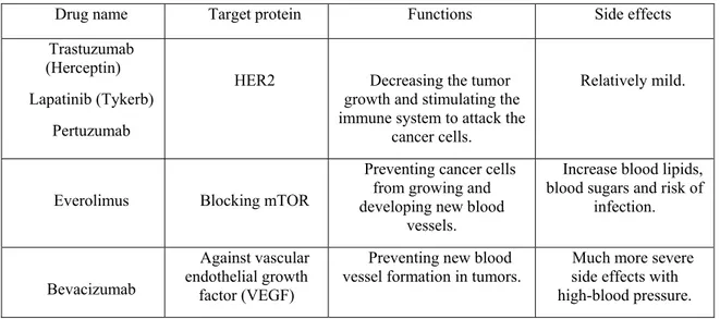
Targeted therapy. Since alterations of some related genes in tumorigenesis have been explored, scientists have been able to develop specific drugs based on their target proteins. Unlike chemo drugs, these drugs individually act on target proteins and have fewer side effects. One of the representatives is HER2 inhibitors (Li and Li, 2014). HER2/neu is overexpressed in 10~15% of breast cancer cell surface and makes them more aggressive and highly metastatic. Alternatively, mTOR inhibitor and vascular endothelial growth factor (VEGF) inhibitor are also used in cancer treatment. Examples regarding targeted therapy are shown in Table II.
Table II Drugs for targeted therapy in breast cancers
Drug name Target protein Functions Side effects Trastuzumab
(Herceptin) Lapatinib (Tykerb)
Pertuzumab
HER2 Decreasing the tumor growth and stimulating the immune system to attack the
cancer cells.
Relatively mild.
Everolimus Blocking mTOR
Preventing cancer cells from growing and developing new blood
vessels.
Increase blood lipids, blood sugars and risk of
infection.
Bevacizumab
Against vascular endothelial growth
factor (VEGF)
Preventing new blood
vessel formation in tumors. side effects with Much more severe high-blood pressure.
13 1.2.3 ErbB2/HER2 in breast cancer 1.2.3.1 EGFR family
The epithermal growth factor receptor (EGFR) family is a major class of the receptor tyrosine kinase (RTK) superfamily, consisting of four members: EGFR (HER1), HER2 (ErbB2), HER3, and HER4 (Foley et al., 2010). HER receptors are transmembrane glycoproteins containing a highly conserved extracellular domain, and an intracellular ATP-binding kinase domain (Figure 1.1) (Yarden, 2001). To date, several ligands binding to different HER receptors (HER1, HER3, HER4) have been identified. Upon binding to ligands, HER receptors form dimers followed by intracellular kinase domain phosphorylation and adaptor protein recruitments, leading to the activation of several downstream signaling pathways. Unlike other isoforms, HER2 is the only isoform of the EGFR family whose ligand is still unknown. However, HER2/HER3 heterodimerization, leading to the auto-phosphorylation of HER2, is the most relevant to HER2-dependent carcinogenesis. Abnormal regulation of HER2 by overexpression and mutation has been indicated in around 10~15% of breast cancers.
14
Figure 1.1 Cross-cancer alteration summary for the ERBB2 gene.
It has been found that the ERBB2 gene is located on human chromosome 17 (17q12), which is amplified in multiple cancers. Amplification and mutation of ERBB2 are the two major causes in ErbB2/HER2-induced carcinogenesis. In breast cancer, the ERBB2 gene set is amplified in 13.5% of 482 cases. In addition, 6.3% of 127 cases in bladder urothelial carcinoma carried mutations in the ERBB2 gene set, indicating that the differences of ERBB2 gene alterations depend on the cancer types. (Adapted from http://www.cbioportal.org, Cerami et al., Cancer
15
1.2.3.2 Activation mechanisms of HER2 receptor
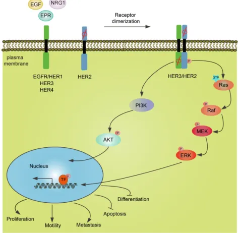
As indicated in Figure 1.2, the alternation of the ERBB2/HER2 gene set occurs not only in breast cancer but also in other cancers, such as bladder cancer. De-regulation of the
Figure 1.2 Schematic representation of HER receptors-mediated downstream signaling pathways.
Upon ligands binding, HER receptors dimerize in the plasma membrane. Notably, among these receptors, no ligands of HER2 have been determined, and the intracellular kinase domain of HER3 is impaired. But the dimerization of receptors (HER2/HER3) could induce phosphorylation of tyrosine residues in the cytoplasmic domain and thereby drives amplification of the MAPK and the PI3K pathways. Diverse signaling pathways converge in the nucleus. For instance, ERK1/2 translocate into the nucleus and phosphorylate some transcription factors, leading to cell proliferation, motility, and metastasis. Alternatively, cell apoptosis and cell differentiation are inhibited.
16
ErbB2/HER2 receptor by overexpression or mutation induces hyper-activation of the ligand-independent receptor through spontaneous receptor dimerization or allow a cell to hyper-respond to a low concentration of circulating growth factors (Yarden, 2001). Being upstream (inducer) of the RTK signaling cascade, activated HER2 receptor drives several downstream effector activations and amplifies signal transduction, leading cells to proliferate, migrate and invade. Based on the crucial function of HER2 in tumorigenesis, the understanding of the mechanisms of how HER2 functions is essential for cancer therapy innovation.
After receptor dimerization, tyrosine residues of RTKs in the cytoplasmic domain become auto-phosphorylated leading to HER2-mediated complex formation. Src-homology (SH) 2 domains present in binding partners play crucial roles in HER2 association. One such example is p85, which binds to the pYXXM motif of HER2, through its SH2 domain. Moreover, Grb2 and phospholipase C (PLC) through their SH2 domains associate with specific phospho-tyrosine sites in HER2 (Yamazaki et al., 2002). Phosphotyrosine binding (PTB) domains presented in some adaptor proteins are also responsible for HER2 interactions. For instance, Shc through PTB domains interacts with the NPXpY motif in HER2 (Batzer et
al., 1995).
Even though the HER2-associated adaptor proteins Grb2 and p85 lack catalytic activities, they can still amplify the activation of HER2-mediated signaling cascades through binding to diverse enzymes such as the guanylnucleotide exchange factor (SOS) and the phosphatidylinositol-4,5-bisphosphate 3-kinase (p110), separately. It has been shown that Grb2 through its SH3 domains constitutively binds to SOS. Upon recruitment to the activated receptor, Grb2 acts as a bridge linking activated receptor to the downstream signaling apparatus (Margolis and Skolnik, 1994). Alternatively, activated HER2 induces PI3K/Akt signaling activation through p85 recruitment (De Luca et al., 2012). p85 is a regulatory subunit of PI3K, which constitutively binds to the catalytic subunit of PI3K (p110). Upon binding to the receptor, p85 PI3K recruits p110 PI3K to the plasma membrane where p110 promotes phosphatidylinositol (3,4,5)-trisphosphate (PIP3) production leading to the activation of Akt (Hu et al., 1992).
17
1.3 The Ras/MAPK/RSK signaling pathway
1.3.1 Background: Mechanisms of MAPK cascade activation
Mitogen-activated protein kinases (MAPKs) are Ser/Thr kinases that transmit extracellular ligand-binding events to intracellular signaling transduction cascades, resulting in diverse cellular functions, such as cell growth, proliferation, survival and differentiation. In mammals, 14 MAPKs have been identified and divided into 2 types: conventional MAPKs and atypical MAPKs (Figure 1.3). Conventional MAPKs consist of four groups: the extracellular signal-regulated kinase1/2 (ERK1/2), p38 isoforms (α, β, γ and δ), c-Jun N-terminal kinases1/2/3 (JNK1/2/3), and ERK5 (Cargnello and Roux, 2011). Atypical MAPKs comprise ERK3/4, ERK7, and Nemo-like kinase (NLK), which have not been well documented.
Figure 1.3 Schematic representation of two classifications of MAPKs: conventional MAPKs and atypical MAPKs.
All MAPKs contain the kinase domain in the middle, as shown in blue. Additional regions as indicated: TAD: a transactivation domain; C34: a region conserved in ERK3 and ERK4; AHQr: a domain rich in Ala, His, and Glu. Notably, two putative nuclear localization sequences (NLS) are present in ERK5 and ERK7/8. (Adapted from Microbio.l Mol. Biol. Rev., 2011
18
Each conventional MAPK cascade consists of at least three conserved, successively acting kinases: a MAP kinase kinase kinase (MAP3K or MEKK), a MAP kinase kinase (MAP2K or MEK) and a MAP kinase (MAPK). The MAP3K is often activated by small GTP-binding proteins of the Ras/Rho family, such as Ras or Rap (Chong et al., 2003). Phosphorylated MAP3K emits a signal to MAP2K, a dual-specificity kinase, which phosphorylates both Thr and Tyr residues present in the activation loop of most MAPKs. Once phosphorylated and activated, the MAPK phosphorylates numerous substrates either in the cytoplasm or the nucleus (Deschenes-Simard et al., 2014).
In some respects, atypical MAPKs behave differently from conventional MAPKs. Compared to the three-tiered conventional MAPK signaling cascades, it seems that atypical
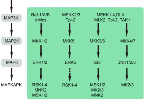
Figure 1.4 Activation of the conventional MAPK-mediated signaling pathways results in the phosphorylation of MAPKAPKs.
MAPK signaling cascades are triggered upon stimulation by the diverse growth factors, cytokines and cellular stress. MAPKAPKs include five subgroups: RSK, MNK, MSK, MK2/3 and MK5. Each MAPKAPK is phosphorylated by its respective upstream kinase. (Adapted from Journal of Molecular
19
MAPKs are organized into two-tiered kinase cascades. In addition, atypical MAPKs have no conserved T-X-Y motif in their activation loop (Figure 1.3). It appears that these atypical MAPKs have only one residue in the kinase domain to be phosphorylated. For instance, ERK3 and ERK4 have an S-E-G motif, while NLK contains a T-Q-E motif (Deleris et al., 2010; Ishitani et al., 2010). Although ERK7 has a T-X-Y motif in its kinase domain, it is
auto-phosphorylated by ERK7 rather than an upstream MAP2K (Abe et al., 2001). Accumulating evidence has shown the involvement of scaffold proteins, which hold the
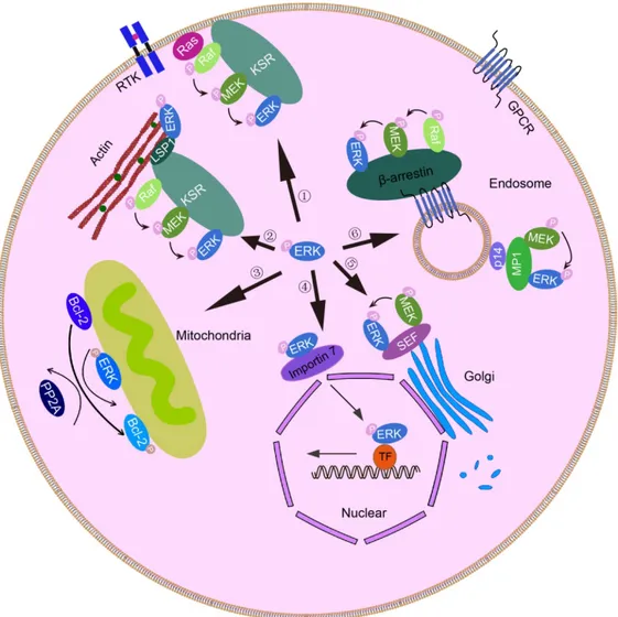
components of three-tiered kinase cascades together and keep the kinase close to its substrate, which is essential for maintaining the magnitude and duration in the conventional MAPKs signaling pathway. For example, in the ERK1/2 signaling cascade, the kinase suppressor of Ras (KSR) is responsible for the assembly of Raf, MEK1/2 and ERK1/2, and the MEK partner1 (MP1) holds MEK1/2 and ERK1/2 together (Therrien et al., 1995; Sharma et al., 2005). In JNK signaling, the JNK-interaction protein 1 (JIP1) holds MLK, MKK7 and JNK1-3 together (Yasuda et al., 1999). In addition, β–arrestin 2 serves as a scaffold protein for both the ERK1/2 and JNK pathways. In the former signaling pathway, β–arrestin 2 directly interacts with Raf and ERK1/2 but indirectly binds to MEK1/2; in the latter signaling cascade, β–arrestin 2 assembles ASK1, MKK4 and JNK3 together (Tohgo et al., 2002; Guo and Whitmarsh, 2008).
Once phosphorylated and activated, MAP kinases phosphorylate substrates on Ser/Thr-Pro residues, which is a common characteristic of conventional and atypical MAPKs. The diverse consequences of MAPK activation in an individual cell are eventually determined by the specific substrates involved in the different cellular processes. Most notably, among these substrates, phosphorylation of the MAPK activated protein kinases (MAPKAPKs) is the additional enzymatic and amplification step of MAPK signaling. As indicated in Figure 1.4, MAPKAPKs include the p90 ribosomal S6 kinase (RSKs), mitogen- and stress-activated kinase (MSKs), MAPK-interacting kinases (MNKs), MAPK-activated protein kinase 2/3 (MK2/3), and MK5 (Chung et al., 1991; Cargnello and Roux, 2011).
1.3.2 The ERK1/2 module
20
The cDNAs of ERK1 and ERK2 were cloned in the early 1990s, and their products were originally found to be phosphorylated on Tyr and Thr residues upon growth factor stimulation (Boulton et al., 1990; Boulton et al., 1991). The amino acid identity between human ERK1 and ERK2 is 84%. Given the high similarity of sequences between ERK1 and ERK2, these two proteins are traditionally designated as ERK1/2. ERK1 and ERK2 are co-expressed widely in many cells and tissues, with high levels in the brain, skeletal muscle, thymus and heart (Boulton et al., 1991). Whereas the activation of ERK1 and ERK2 are induced in parallel upon stimulation, few reports indicated that each ERK isoform could also be selectively phosphorylated (Papkoff et al., 1994). Moreover, other studies have shown that activation of ERK1 and ERK2 result in different biological events. For instance, in MCF-10A cells, Shin et al. observed that ERK2 (not ERK1) was required in the Ras-induced EMT process (Shin et al., 2010). Consistent with these results, it was also found that erk1-knockout mice (erk1-/-) could survive with a defect in thymocyte development, whereas erk2-null mice (erk2-/-) were embryonic lethal due to the severely impaired development of the placental vasculature (Pages et al., 1999). Why did erk1-/- mice and erk2-/- mice have such different phenotypes? Were they due to the different isoform functions or expression levels? These were addressed by Lefloch et al. in a series of gene ablation experiments (Lefloch et al., 2008). It was shown that only a single erk allele expressed in mouse, regardless of erk1 or
erk2, resulted in a lethal phenotype rather than the mice with two single alleles separate from erk1 and erk2, indicating that a particular dosage of erk is necessary for mouse survival.
Given that ERK2 expression is higher than ERK1 in most cells, the unfavorable outcome of
erk2 knockout mice may be due to the diminished total ERK content instead of its unique
biological functions. Even though several studies have reported different pathological roles of ERK1 and ERK2, these two isoforms are still considered to possess the same functions in most biological processes.
1.3.2.2 Activation mechanisms and inhibitors of ERK1/2
ERK1/2 are core components in the Ras/MAPK pathway, which regulate multiple biological processes such as cell proliferation, differentiation and migration. ERK1/2 are activated in response to different growth factors, cytokines, osmotic stress, hormones and microtubule disorganization (Xing et al., 1998; Conway et al., 1999; Naor et al., 2000; Komis
21
et al., 2011). The activation of ERK1/2 is caused mainly by a receptor on the plasma
membrane, such as receptor tyrosine kinases (RTKs) or heterotrimeric G protein-coupled receptors (Porter and Vaillancourt, 1998). For instance, upon a ligand binding, tyrosine kinase receptors (RTKs) are activated by trans-phosphorylation on tyrosine residues in the intracellular kinase domain. The subsequent trans-phosphorylation of RTKs leads to the recruitment of various adaptor proteins, such as Src homology and collagen (Shc), growth factor receptor-bound protein 2 (Grb2) and phospholipase C-γ (PLCγ), which contain Src homology 2 (SH2) domains or phosphotyrosine-binding (PTB) domains. One of the well-characterized mechanisms of RTK-induced Ras activation is mediated by the Grb2-SOS complex. Upon RTK activation, the guanine-exchange factor SOS is recruited to the plasma membrane from the cytosol by constitutive association with Grb2 (Pruett et al., 1995). Membrane-bound SOS promotes Ras activity by catalyzing GDP/GTP exchange. Once bound to GTP and activated, two “switch domains” in Ras shift enabling Ras to interact with several downstream effectors, such as Raf. Furthermore, this physical association leads to Raf phosphorylation. Activated Raf transmits signals by phosphorylating the downstream effectors MEK1/2. MEK1/2 are dual-specificity protein kinases since they can phosphorylate substrates on both Thr and Tyr residues. As the only substrate of MEK1/2, ERK1/2 are phosphorylated by MEK1/2 on the Thr-Glu-Tyr motif, which is required for ERK1/2 activation. This phosphorylation also induces an allosteric conformational change in ERK1/2, leading to MEK1/2 dissociation. Once phosphorylated and activated, ERK1/2 either translocate into the nucleus or stay in the cytoplasm, where they can phosphorylate many substrates such as transcription factors or cytoskeletal proteins.
Appropriate regulation of Ras/Raf activity is essential for the proper maintenance of cell proliferation. Abnormal regulation of Ras/Raf leads to the hyperactivation of the MAPK pathway and cause many human diseases such as cancer. Many studies have already shown that mutation of KRAS or BRAF occurs in a wide range of cancers, including pancreatic, colorectal cancer, biliary cancer, melanoma, thyroid cancer and ovarian cancer (Brose et al., 2002; Lee et al., 2003). Given its diverse modes of action in cancer development, a specific blockade of the ERK1/2 cascade by inhibitors represents an attractive target in cancer therapy. Since ERK1/2 are the only substrates of MEK1/2, MEK1/2 inhibitors are used to characterize
22
ERK1/2 function in a wide range of biological contexts. Unlike other kinase inhibitors, most of the MEK1/2 inhibitors are non-competitive ATP molecules such as PD184352 and U0126, which interact more strongly with an inactive form of MEK rather than an active form (Favata
et al., 1998; Allen et al., 2003). This interaction blocks conformational transition of MEK1/2
and thus prevents their enzymatic activities.
One of the conundrums in cancer treatment is that a few cancer cells often survive and are resistant to chemotherapy, leading to a failure of the treatments. For example, B-Raf inhibitors can cause drug resistance in patients by re-activating MAPK signaling. Therefore, the combination of B-Raf and MEK inhibitors in cancer treatment could be more efficient than either alone. For instance, co-treatment of trametinib (the first MEK inhibitor in clinical treatment) and dabrafenib (a BRAFV600E inhibitor with ATP competitor), which was approved by the FDA in January of 2014, sustains melanoma patients progression-free survival (PFS) up to 9.4 months, whereas the average PFS of only trametinib-treated patients is 5.8 months (Menzies and Long, 2014).
1.3.2.3 Docking interactions from ERK1/2 to substrates
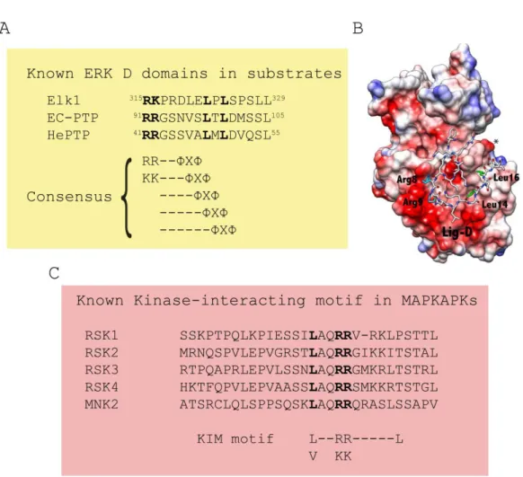
In an individual cell, various molecules, independently or coordinately, fulfill their specific actions at a particular time and place. To avoid unfavorable outcomes, these molecules must be precisely arranged and regulated. As such, appropriate partner selection becomes one of the fundamental mechanisms to keep the high efficiency and fidelity of signal transduction. To date, several mechanisms underlying the precise signal transmission in the ERK-MAPK cascade have been determined. One such example is the docking interaction, which is achieved by the common docking domain (the CD domain) in ERK1/2 (Tanoue et al., 2001). It has been shown that the CD domain, which locates outside of the catalytic region of MAPKs and consists of negatively charged (acidic residues) and hydrophobic amino acids, is crucial for forming electrostatic and hydrophobic interactions with substrates (Tanoue et al., 2001). In addition, it has been found that the ED/TT motif in ERK1/2 forms a docking grove with the CD domain, and thus participates in the association with partner molecules (Tanoue
et al., 2001).