Annu. Rev. Microbial. 1991. 45:37�7
Copyright © 1991 by Annual Reviews Inc. All rights reserved
SERINE j3-LACTAMASES AND
PENICILLIN-BINDING PROTEINS
lean-Marie GhuysenCentre d'Ingenierie des Proteines, Universite de Liege, Institut de Chimie, B6, B-4000 Sart Tilman (Liege I), Belgium
KEY WORDS: bacterial wall peptidoglycan, {3-lactam antibiotics, emergence of resistance, enzyme catalysis, serine peptidascs, divergent evolution, molecular modeling, protein engineering
CONTENTS
The Penicillin-Interactive, Active-Site Serine Protein Family . . . .... . . . . ... . . 37
The Low-Mr PBPs . . . .. . . .... . . .... . . .. . . .. . . 45
The (3-Lactamases and the Penicillin Sensory-Transducer BLAR ... . ... 47
The Atomic-Level Enzyme-Ligand Interactions . . . ... . . ... . . .. . . .. . . 50
The High-Mr PBPs of Class A and B ... .. . . ... . . . .... . . .. . ... . . 54
Intrinsic Resistance by Emergence of Altered High-Mr PBPs . ... . . ... . . 58
The Penicillin-Interactive, Active-Site Serine Protein Family In the bacterial world, hundreds, perhaps thousands, of distinct proteins catalyze rupture of the lactam amide bond of penicillin and transfer the penicilloyl moiety to an essential serine, forming a serine ester-linked acyl derivative (Figure 1). The reaction is analogous to the catalyzed rupture of a peptide bond by the serine peptidases of the trypsin and subtilisin families. It 37 0066-4227/91/ 100 1-003 7$02.00
Annu. Rev. Microbiol. 1991.45:37-67. Downloaded from www.annualreviews.org
by UNIVERSITE DE LIEGE on 01/06/11. For personal use only.
Quick links to online content
Further
ANNUAL REVIEWS
r---Acyl
High k.l�
rapid breakdown jl-Lactamases k+2 ... k+l • E-D --r-- E + P H20 protein� �
Low k+l very s low or (omplete breakdown inerfnessPeni (illi n-bi nd in9-proteins
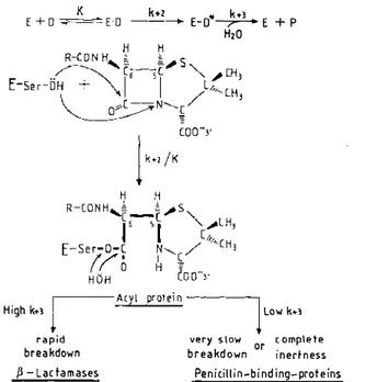
Figure 1 The penicillin-interactive, active-site serine proteins. Reactions with 13-lactam car bonyl donors . E = protein; D = ,B-Iactam carbonyl donor; E·D = Michaelis complex; E-D* = acyl protein; P = reaction product (penicilloate); K = dissociation constant; k+2 and k+3 '" first-order rate constants. The acyl enzyme mechanism was proposed in 1965 (112). Involvement of an essential serine was demonstrated in 1976 for reaction with the Streptomyces R61 penicillin binding protein (36) and in 1979-1981 for reaction with several j3-lactamases (17, 20, 35, 71). The concept of a penicillin-interactive, active-site serine protein family was put forward in 1988 (66).
involves initial binding of the ligand to the enzyme (K), enzyme acylation (k+2) and, in a third step, enzyme deacylation (k+3)' A common substrate requirement is that a negative charge must occur at the 3' position in the penicillins and the 4' position in the cephalosporins (this group is usually a carboxylate, but a sulfonate is found in the monobactams), and the acyl side chain borne by the f3-lactam ring must be on the f3 face in a cisoid position relative to the thiazolidine or dihydrothiazine ring. As a result of these requirements, the scissile amide bond in the 13-lactam ring is equivalent to a carboxy-terminal peptide bond extending between two carbon atoms with the D configuration
Given the endocyclic nature of the scissile bond, acylation of the active-site serine generates a bulky leaving group that remains part of the acyl enzyme through the C5-C6 bond (Figure 1) and thus cannot diffuse away. As a result of this occupancy, only water has access to the active site and can attack the acyl enzyme, release the penicilloyl moiety, and concomitantly recover the
Annu. Rev. Microbiol. 1991.45:37-67. Downloaded from www.annualreviews.org
J3-LACT AMASES AND PBPs 39 enzyme. This mechanism is general. However, with the f3-lactamases, water has a facile access to the active site and is an excellent attacking nucleophile. The f3-lactamases tum over rapidly and hydrolyze effectively the f3-lactam substrates into biologically inactive metabolites. With the penicillin-binding proteins (PBPs), the acyl enzyme is almost hydrolytically inert. The higher the k+2/K and the smaller the k+3 are, the lower the antibiotic concentration required to immobilize completely a given PBP as acyl enzyme at the steady-state of the reaction (46, 78).
The fact that the acyl enzymes are sufficiently stable to be analyzed with SDS gel electrophoresis has led to the development of a convenient procedure for the detection of the individual PBPs as radioactively labeled proteins (104). All the bacteria possess multiple PBPs. Each bacterial species has its own assortment of PBPs. The PBPs occur in a small number of copies per cell, from a few hundred to one or two thousand. Their affinities for penicillin and other f3-lactam antibiotics vary widely, and their molecular masses range from 25,000 to 100,000.
The penicillin-interactive proteins fall into three groups: the f3-lactamases, the low-Mr PBPs, and the high-Mr PBPs. The f3-lactamases and the low-Mr PBPs are monofunctional catalytic entities. The active-site serine resides close to the amino terminus of the protein. The high-Mr PBPs possess a several-hundred-amino acid domain that is fused to the amino terminus of the penicillin-binding domain per se, which is assumed to start 60 residues upstream from the essential serine. Pair-wise comparison of amino acid sequences leads to the conclusion that each of these groups falls into several classes (Figure 2). Members of a given class are related in the primary structure. However, proteins that lack relatedness may fulfill similar func tions. Whether they belong to class A, C, or D, the f3-1actamases are f3-lactam antibiotic-hydrolyzing enzymes. Conversely, similarity in the primary struc ture does not necessarily reflect similarity in the physiological function. The
Streptomyces Kl5 low-Mr PBP of class A is related to the staphylococcal f3-lactamase of class A, and the Streptomyces R61 low-Mr PBP of class B is related to the Citrobacter freundii f3-lactamase of class C by a low but significant similarity index of about 7. The carboxy-terminal, penicillin binding domain of the class C high-Mr PBP BLAR of Bacillus Iicheniformis
is related to the Oxa-2 f3-lactamase of class D by a high-similarity index of 40. Divergent evolution implies that proteins of the same family evolved from a common ancestor. Depending on the evolutionary distance, they may have acquired very different amino acid sequences and distinct functionalities and specificities while conserving the same polypeptide scaffolding. The case of a divergent evolutionary relationship between the penicillin-interactive, active site serine proteins and domains is supported by a bulk of experimental data and predictional studies.
The three-dimensional structures are known for the class A f3-lactamases of
Annu. Rev. Microbiol. 1991.45:37-67. Downloaded from www.annualreviews.org
Low Nr PBPs N
%I�e�bri;;�
(
(lass A; E. (oli 5*1; e. subtilis S!*I
{j-Iat tama""s
(lass A: e.licheniformis Streptomyces albus G. Streptomyces K1S .--- ---� S. aureus
Class B. 5treptomyc� Ral .. - - - .. Class C : t. freundii
Class C. E tOli 4; Streptomytes R39 Class 0: S. typhimurium (Oxas; PSEsl t High Nr PBPs pea
I
C'--_--'r
' //// " 0//.�
/ //m;n;br�n�
i NClass A: E.toli lA, lB Class e: E. toli 2.3, S.aureus 2'
S. pneumoniae 2x, 2b N. gonorrhoeae 2 E. hirae 3r, 5
.01
(lass ( : BLAR : B. lithenifo,mis penicillin sensory-tranSducer ({l-Iactamase ind"tlionl
Figure 2 The penicillin-interactive, active-site serine proteins. Groups, classes, and membrane topology. PBPJPBD, penicillin-binding protein/domain; (*), membrane-bound; PHP, penicillin hydrolyzing protein. Many f3-lactamases other than those listed are of known primary structure. - indicates similarity in the primary structure. The algorithm used for homology searches (66) generates the best alignment for each pair of amino acid sequences and expresses the extent of similarity by a score. This score is the sum of the individual scores obtained for each pair of amino acids minus a penalty of 8 for each gap introduced. Pair-wise comparison of members of a gi�en class generates scores that are at least 5 but may be as high as 50 standard deviations above that expected from a run of 20 randomized pairs of proteins with the same amino acid composi tions as the two proteins under consideration. This standard deviation defines a similarity index. Figure 7 shows pair-wise comparison of the amino·terminal and carboxy-terminal domains of the high-Mr PBPs of class A and B.
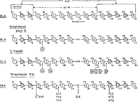
Staphylococcus aureus (56), Streptomyces albus G (25, 62), and B. licheni formis (82); the class C f3-lactamase of C. freundii (89); and the class B low-Mr PBP of Streptomyces R61 (70, 72) . They have a unique signature in the form of several amino acid groupings that occur along their amino acid sequences (66, 103) (Figure 3): the tetrad active-site serine-X-X-Iysine (S*XXK), where X is a variable amino acid; the triad serine or tyrosine-X asparagine [S(Y)XN]; a peptide segment (DIE) that contains two dicarboxylic amino acids; and the triad lysine or histidine-threonine or serine-glycine [K(H)T(S)GJ. As a result of the folding of the polypeptide chain, these four
Annu. Rev. Microbiol. 1991.45:37-67. Downloaded from www.annualreviews.org
� A B ( A ( 0 � S C � E.(gli 5 B.s.ubtilis 5 Streptomy,es K15 Streptomyces R61 [,oli 4 Atfinomadura R39 2-l.,t.mases S. aureus . Streptomy,es .Ibus G B.lictieniformis C. freundii Ox. -2 High-Mr PBPs E.coli lA E.coli 1 a E.coti 2 E:,oli 3 S.aureus 2' N.gonorrhoeae 2 S. pneumonia. 2)( S. pneumoni.e 2 b E. hiru 5 B.litheniformis BlAR .!:!!:'.i 030 ? V30 A32 A21 R50 K 25 G40··547 53S .. E45 A 21 0.23 HI MI HI HI MI HI Ml Ml HI HI f-,'XXK -42 ,13LTK 62 ? S36HTK 63 3i S64TTK 51 60 593VTK 93 40 S�2TllK 240 41 598NMK 245 37 S 63TSK 54 41 S89VFK 60 40 ,B6TlK 54 42 584VSK B2 44 56BTFK 14 463 5465N1K 55 50B 5S10LAK 5B 3 2B S330TVK 53 305 s30lTVK 46 403 540STlK $ 306 S31OA!K 48 335 S337TMK 54 3a3 S3BSVVX 53 420 S4Z11TFK 54 400 51,OZTYK 70
/3-LACTAMASES AND paps 4 1
,\YlXN - DIE -KIHITtS1G :0- (00- Ref. 5139 GN 41 0183.0204 31 K242TG lS8 1,03 IS SmOAN 43 E141,. 0179 47 Kl11TG 181 412 115 S115GC 41 0169. am 69 K14zTG 46 291 (01 YI905N 63 025� STEe 69 H319 TG 14 406 30 S306DN 38 D347.0355 61 KQ7 TG 57 477 83 S347NN 44 039� 0411 46 K458TG 11 53B tol 51210N 33 Ei51 TELN 65 K225 SG 53 261 19 SI530N 35 E·,89PELN 65 K257 TG 52 312 23 SI"ON 35 E",ao.PELN 61 K24B TG S6 307 87 Y170AN 64 0211AEAY 65 K335 TG 45 3Bl 19 Y1Io6GN 12 E161 4B K210 TG 62 275 Z2 5524KN 32 £559 156 K716 TG 131 BSO 14 5512MN 26 0601. H2O 77 K69BTG IJ,3 844 14 S3B1 AD 34 E424,Ol,l,l 96 K544SG B6 633 5 S359SN 34 E396.0409 64 K49HG 91 56B 66 S4640N 25 E492,0511 87 K5995G 68 670 5362SN 34 E399, E414 6 2 K497 TG 81 581 100 Sl95 SN 34 EIJ2,044 106 K547 SG 200 150 74 S442�N 32 f415. E463 13O X614 TG 62 679 28 54800N 25 ESOB. E542 14 K617TG 58· 616 33 Y416 GN 11 0490 48 K539 TG 59 601 13
Figure 3 The penicillin-interactive, active-site serine proteins. Location of the motifs S*XXK, S(Y)XN, DIE, and K(H)T(S)G along the amino acid sequences. The amino acid numbering refers to the protein precursors. The low-Mr PBPs and the f3-lactamases are manufactured with a cleavable signal peptide. Cleavage may produce a mature protein that has a ragged amino terminus (NH3 +). The amino acid sequence of the Streptomyces a/bus G f3-lactamase (23) has been corrected by deleting Glyl28 and substituting the dyad Gln1 4 1 -Leu142 by Met (4, 67). In the ABL numbering scheme for the f3-lactamases of class A (4), S* of the S*XXK motif is at position 70, S of the SON motif is at position 130, E* of the EID motif is at position 166, and K of the KT(S)G motif is at position 234. The E. coli PBPI B is a mixture of two major proteins resulting from the use of alternative sites for the initiation of translation. The numbering refers to the largest form. (*), Unpublished data (see text). Reference 86 should read 85.
motifs are brought close to each other , generating an active-site at the junction between an all-a domain and an a-f3 domain whose five-stranded f3-sheet is protected by additional a-helices on both faces (Figure 4) . In this structure, the serine of the S *XXK motif is at the amino terminus of helix a2 of the all-a domain and occupies a central position in the cavity. The S(Y)XN motif connects helices a4 and a5 of the all-a domain and forms one side of the cavity. The DIE motif resides at the entrance of the cavity. The K(H)T(S)G motif on the innermost f33 strand of the f3-sheet forms the other side of the cavity. Using the S. aureus f3-lactamase as reference, one finds that the differences in length in the S. albus G f3-lactamase, the C. freundii f3-lactamase , and the Streptomyces R6 1 PBP are associated with surface loops, small helices, and f3-strands that occur away from the active site and that connect conserved structures (Figure 5). In this proposed scheme, the con served secondary structures have the same numbering.
Superimpositiori experiments lead to the result that the f33 strand is
sim-Annu. Rev. Microbiol. 1991.45:37-67. Downloaded from www.annualreviews.org
Figure 4 Structure of the Streptomyces albus G (3-lactamase of class A. Polypeptide folding (A) and disposition of the conserved motifs (B)". The active site is indicated by the white arrow in A and is threefold enlarged in B. Adapted from References 25, 62, 67.
ilarly situated with respect to the S*XXK motif in the three-dimensional structures . However, the yeA or S)N motif in the 13-lactamase of class C and the PBP is about 4- 5
A
farther away from the K(H)T(S)G motif than the corresponding SDN motif in the 13-lactamases of class A. As a consequence of this displacement, the phenolic oxygen of the tyrosine of the yeA or S)N motif and the 1'-OH of the serine of the SDN motif are similarly disposed with respect to the 1'-OH of the active-site serine of the S*XXK motif . Superimposition experiments also highlight noticeable differences at the level of the DIE motif . Site-directed mutagenesis (49) and molecular modeling (J.Lamotte-Brasseur , G. Dive, O. Dideberg, J.-M. Frere, & J.-M . Ghuysen, in preparation) support the view that the glutamic acid E* of the sequence E*XELN (Figures 3 and 4) of the class A ,B-Iactamases is essential and that a water molecule bound to this glutamic acid acts as proton abstractor-donor in catalysis. The corresponding sequences DAEA Y in the C. freundii f3-lactamase and DSTEQ in the Streptomyces R61 PBP (Figure 3) have a spatial
Annu. Rev. Microbiol. 1991.45:37-67. Downloaded from www.annualreviews.org
� l' " Streptomyces albus G. " " " 91 'n BlA '0 10 19 C . freundii ., '" BLA ,) \I. 'S Streptomyces R 61. PBP '" 'SS '" In '8)
I3-LACT AMASES AND PBPs 43
'0' ", '" -(211-,0, m '" 'Sl '" ,eo '" I'9t ,91 KSG DIE KTG HTG
Figure 5 Occurrence of the conserved motifs and secondary structures along the amino acid sequences of the S. aureus /3-lactamase of class A (56), the Streptomyces albus G /3-lactamase of class A (25, 62, 67), the C. freundii /3-1actamase uf class C (89), ami the Streptomyces R61 PBP (70, 72). Circles, a-helix; pointing boxes, /3-strand. BLA = /3-lactamase. The amino acid numbering is that of the protein precursors.
disposition that is not compatible with such a role in catalysis . Yet alterations of the DIE motif of the C. freundii ,8-lactamase [DAEA Y -> DAKA Y and DAEA Y -> (E , K or T)AEA Y] modify the specificity profile of the enzyme and extend its hydrolytic activity to oxyimino-cephalosporins (117, 118).
All the ,8-lactamases, the low-Mr PBPs, and the pencillin-binding domains of the high-Mr PBPs possess, along their amino acid sequences, the same four motifs as those found in the penicillin-interactive proteins of known three dimensional structure (Figure 3). Identification of the S*XXK, S(Y)XN, and K(H/R)T(S)G motifs (66, 103) is not, most often, an issue of controversy [HTG occurs in the Streptomyces R61 PBP (30) and RSG in the PSE-4 carbenicillinase (10)], and analysis, at the molecular level, of protein mutants (see below) leads to the conclusion that these motifs are elements of the active sites . Given that several dicarboxylic amino acids occur approximately at the expected distances in the primary structures, identification of the DIE motif is hypothetical unless the importance of the predicted motif is demonstrated
Annu. Rev. Microbiol. 1991.45:37-67. Downloaded from www.annualreviews.org
experimentally. As shown by site-directed mutagenesis, Asp444 in the high Mr PBP2 of Escherichia coli is probably the homolog of the glutamic acid E* of the ,B-lactamases of class A (H. Adachi, M. Ishiguro, S. Imajo, T. Ohta, &
H. Matsuzawa, personal communication).
The penicillin-interactive, active-site serine proteins and domains exhibit endless variations. Predictional studies suggest that the 275 amino acid ,B-lactamases of class D, which are the smallest known ,B-Iactamases, have a shorter connection between the YDN and KTG motifs and that the homologous 477-amino acid PBP4 of E. coli (83) and 538-amino acid PBP of Actinomadura R39 (B. Granier, C. Duez, S. Lepage, 1. Van Beeumen, S. Englebert, et ai, in preparation), which are the largest known low-Mr PBPsl DD-peptidases, have an additional 180- to 190--amino acid insert located between the S*XXK and SDN motifs and occurring on the surface of the protein. Deletion by genetic engineering of a substantial part of this insert from PBP4 produces a truncated protein that still binds penicillin (H. Mottl & W. Keck, personal communication). Several ,B-lactamases and low-Mr PBPs are susceptible to thiol reagents. The Streptomyces Kl5 PBP has two cys teines. One of them occurs in the active-site as a result of the replacement of the usual S(Y)XN motif by a SDC motif, and the other occurs at the tenth position downstream from the lysine of the KTG motif, i.e. probably on strand,B4 (P. Palomeque-Messia, M. Leyh-Bouille, M. Nguyen-Disteche, C. Duez, S. Houba, et ai, in preparation). Derivatization of one of these cys teines by para-chloromercuribenzoate greatly decreases but does not abolish the enzymatic activities of the PBP (77).
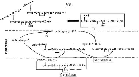
The wall peptidoglycan is an essential polymer of the bacterial cell. Its structure and biosynthesis are well-known (Figure 6). The peptidoglycan precursors are lipid-transported disaccharide (N-acetylglucosaminyl-N acetylmuramyl)-L-Ala-Y-D-Glu-L-Xaa-D-Ala-D-Ala pentapeptide units. De pending on the bacterial species, L-Xaa is a diamino acid whose w amino
group is either free or substituted by additional amino acids. These pre cursors, the end products of the cytoplasmic and membrane cycles of the biosynthetic pathway, are transported onto the outer face of the plasma membrane for final peptidoglycan assembly, a process that requires a glycan chain elongation and interpeptide crosslinking machinery.
The penicillin-interactive, active-site serine proteins fulfill multiple func tions. The low-Mr PBPs and the high-Mr PBPs of class A and B are involved, in one way or another, in peptidoglycan assembly. They are the targets of penicillin action. The ,B-lactamases are defensive enzymes that the bacteria synthesize to protect their PBPs from the deleterious effects of penicillin. ,B-Lactamase synthesis is not always constitutive. The high-Mr PBP BLAR of class C acts as a penicillin sensory-transducer involved in the specific in ducibility of ,B-Iactamase synthesis.
Annu. Rev. Microbiol. 1991.45:37-67. Downloaded from www.annualreviews.org
.B-LACTAMASES AND PBPs 45 ,G/M
,
/ G' H/ L-Ala-o-�
L-xn-o-Ala.·· Wall ,M/G"I
Iw
7-GL AI 0 GI LX
- a- • E.J
- I!a-0 AI (0 -a-
- . NH ....- L-Ala-O-Glu rL-Xaa-O-Ala-O-Ala L-J 101NHI NHI
----�
Undecaprenyl-P-P-�
f --
- - - ----� Undecaprenyi·P
"\
� Lipid-P-P-H Lipid-P-P-H-G
..c I I
�
L-Ala-O-t:J
L-Xaa-O-Ala-O-Ala L-Ala-D-tJ
L-xaa-O-Ala -a·Ala- - - - --- --�-=::::==---":::::"---, '::::"'---""=--...., ---
-
---Cytoplasm
Figure 6 Bacterial wall peptidoglycan assembly from lipid-transported disaccharide pentapeptide units. The final steps (heavy arrow) are made by transglycosylation and peptide crosslinking.
The
Low-Mr PBPs
The low-Mr PBPs are manufactured with a cleavable signal peptide. Some low-Mr PBPs do not possess transmembrane segments. The E. coli PBP4 and the Streptomyces Kl5 PBP somehow interact with the membrane surface and are isolated as membrane components. Overproducing strains produce a high proportion of the PBPs in a water-soluble form. The PBP of Streptomyces
R61 and that of Actinomadura R39 are excreted in the medium during growth. When compared to the excreted protein, the 406-amino acid precursor of the
Streptomyces R61 PBP possesses both a cleavable peptide signal and a cleavable 26-amino acid carboxy-terminal extension (30). Should it not be removed during protein maturation, this carboxy-terminal extension might function as a stop transfer sequence.
All the other known low-Mr PBPs are membrane-bound (97) (Figure 2). The polypeptide chain does not terminate approximately 60 residues down stream from the KT(S)G motif as observed with the soluble PBPs, but has an -50- to lOO-amino acid carboxy-terminal extension (Figure 3), the end of which contains a signal-like peptide segment that serves as membrane anchor. As a consequence, the bulk of the protein is on the outer face of the membrane. Catalytically active, water-soluble derivatives that lack the mem brane anchor can be produced by proteolytic treatment of isolated membranes (33, 123) and by engineering the encoding genes (34, 61).
The low-Mr PBPs are DD-peptidases. They catalyze acyl transfer reactions
Annu. Rev. Microbiol. 1991.45:37-67. Downloaded from www.annualreviews.org
from o-alanyl-o-alanine-terminated peptides and depsipeptide analogs. Thus, rupture of the carboxy-terminal peptide (or ester) bond of acetylz-L-Lys-o Ala-o-Ala (or Acz-L-Lys-o-Ala-o-Iactate) involves transfer of the ACz-L-Lys o-alanyl moiety to the active-site serine with formation of a serine-ester linked acyl (Acz-L-Lys-o-alanyl) enzyme, and then to an exogenous acceptor (45,
46, 88). Because the scissile bond is acyclic, the leaving group o-Ala (or o-lactate) does not remain part of the acyl enzyme and, as a corollary, the acyl enzyme is short-lived. The efficacy with which the low-Mr PBPs/oo peptidases perform hydrolysis vs transpeptidation of the carbonyl donor depends on the efficacy with which water, the leaving group o-Ala, and an exogenous, suitably structured peptide H2N-X can attack the acyl enzyme (Figure 7). This picture, however, is an oversimplification (40, 44, 47, 48, 88, 94). Hydrolysis and transpeptidation do not proceed through strictly identical pathways. The amino acceptor does not behave as a simple alternate attacking nucleophile of the acyl enzyme. It also influences both the initial binding of the o-alanyl-o-alanine-terminated carbonyl donor to the enzyme and the ensuing enzyme acylation step. An excess of amino acceptor can inhibit the DD-peptidase activity.
Most of the known low-Mr PBPs/DD-peptidases act mainly as DD carboxypeptidases. The Streptomyces K l 5 PBP/DD-peptidase, however, is peculiar in that the relative acceptor activity for attack of the acyl enzyme is H20 « D-Ala « H2N-X (88). As a consequence, hydrolysis of ACz-L Lys-D-Ala-D-Ala, in the absence of H2N-X, is negligible because breakdown of the acyl enzyme by the leaving group D-Ala regenerates the original tripeptide. The enzyme turns over but is, seemingly, silent. In the presence of an amino compound H2N-X of high acceptor activity, AC2-L-Lys-D-Ala-o-Ala
E-'''-�
Donor Ac1- L-Lys-D-Ala-
�
-NH-D-Ala k·2/K1
0A"i .. "m. A'r
?i
b
�l
Acceptor H26 HI f-i-x HI N-D-Ala
t t + t
E -Ser-OH R-D-Ala-(OOH R-D-Ala- (ON H-X R-D-Ala - (ONH- D-Ala Hydrolysis Transpe ptidation Original donor
Figure 7 DD-peptidase-catalyzed reactions with ACrL-Lys-D-Ala-D-Ala carbonyl donor. E-Ser OH: enzyme and the active-site serine.
Annu. Rev. Microbiol. 1991.45:37-67. Downloaded from www.annualreviews.org
{3-LACT AMASES AND PBPs 47 is quantitatively converted into the transpeptidated product ACrL-Lys-o-Ala CONH-X. This PBP functions as a strict transpeptidase .
AC2-L-Lys-o-Ala-o-Ala and J3-lactam antibiotics are competing substrates of the low-Mr PBPs/DD-peptidases. The J3-lactam antibiotics, however, are suicide substrates or mechanism-based inactivators: they immobilize the es sential serine in the form of a stable ester-linked acyl enzyme (Figure 1). Yet the inertness of the acyl (benzylpenicilloyl) PBP is not absolute . Slow attack of the penicilloyl moiety by water generates penicilloate, which is the degra dation product of J3-lactamase action on penicillin. Alternatively, a slow and rate-limiting intramolecular rearrangement of the penicilloyl moiety can rup ture the bond between C5 and C6 (Figure 1) and release the leaving group (3, 37-39,80, 121) . As a result of the vacancy thus created in the active site, the newly formed acyl (phenylacetylglycyl) enzyme undergoes immediate break down . Attack by water gives rise to phenylacetylglycine; attack by an amino compound NHi-X gives rise to the transpeptidated product phenyl acetylglycyl-CONH-X.
AC2-L-Lys-D-Ala-D-Ala is an analog of the carboxy-terminal tripeptide moiety of the peptidoglycan precursors (Figure 6). As with the reactions that they catalyze , the low-Mr PBPs/oo-carboxypeptidases help control the extent of peptidoglycan crosslinking (8a) . They hydrolyze the carboxy-terminal o-alanyl-o-alanine peptide bond of peptidoglycan precursors and , in those bacterial species where peptidoglycan crosslinking is mediated by a carboxy terminal o-alanyl-(o )-meso-diaminopimelic acid linkage, they hydrolyze bonds previously made through transpeptidation. All the bacteria possess one or several low-Mr PBPs. Streptococcus pneumoniae PBP3-negative mutants are severely affected in cell division and show highly altered morphology (99a) . A lO-fold overproduction of the E. coli PBP5 causes conversion of the rod-shaped cells into round cells (106) . The sporulation-specific PBP5a is temporally and spatially regulated in B . subtilis. It is detected in large amounts only during spore peptidoglycan synthesis from stage II to stage V
(16, 113) .
The [3-Lactamases and the Penicillin Sensory-Transducer BLAR
The J3-lactamases are secretory proteins. They are excreted in the growth medium (gram-positive bacteria) or they accumulate in the periplasm (gram negative bacteria) . The J3-lactamase III of Bacillus cereus and that of B . licheniformis contain a diacylglyceride that i s thioether-linked to the amino terminal cysteine and a fatty acid that is amide-linked to the same cysteine (57). This hydrophobic moiety functions as the anchor of the protein to the plasma membrane. In fact , these J3-lactamases have membrane-bound and secretory forms.
Annu. Rev. Microbiol. 1991.45:37-67. Downloaded from www.annualreviews.org
The only known function of the ,8-lactamases is hydrolysis of the ,8-lactam antibiotics. ,B-Lactamases show hysteretic kinetics (99). A unique conforma tion is induced in the enzymes by each of several closely related ,8-lactam substrates, and the enzymes can adjust to unfavorable modifications in the substrate.
The ,8-lactamases lack oo-peptidase activity and thus do not interfere with wall peptidoglycan metabolism. However, they catalyze acyl transfer reac tions on noncyclic depsipeptides (2, 91-93) with, for some of them, complex steady-state kinetics comparable to those shown by the low-Mr PBPS/OD peptidases. Like the PBPs, the ,8-lactamases may give rise to long-lived acyl enzymes (42) and long-lived acyl enzymes may undergo various types of intramolecular rearrangements (18). Rupture of the bond between SI and C5 in the acyl enzyme formed by reaction with the penam sulfones and 6-,8-bromo(iodo)penicillanate or rupture of the bond between 01 and C5 in clavulanate is an integral part of the ,8-lactamase inactivation process. The established common feature is that of a branched pathway in which ,8-elimination leads to a hydrolytically inert acyl enzyme and, sometimes, to further modification of some residues of the active site.
The ,8-lactamases are dispensable enzymes, while the low-Mr PBPS/OD peptidases fulfill functions in wall peptidoglycan metabolism. A possible mechanism that may explain the emergence of ,8-lactamases is the excretion of one or several low-Mr PBPs/DD-peptidases by bacteria exposed to lactam antibiotics. Further improvement of this detoxication mechanism results in the conversion of these water-soluble PBPs into ,8-lactam-hydrolyzing enzymes (69). Hence, the presumed features of evolution are catalysis of deacylation of the acyl enzyme formed with cyclic amide compounds, i.e. the ,8-lactam antibiotics, and loss of productive binding of planar acyclic amides, i.e. loss of DD-peptidase activity. Productive binding of planar acyclic esters has no physiological significance; it is conserved to varying extent.
Emergence of new f3-lactamases can be achieved by altering a limited number of amino acids in the direct environment of the active site. A key mutation in the decreased cephalosporinase activity of the S. albus G ,8-lactamase of class A is the conversion of the SDN motif into SDS (62, 63). This single change has little effect on the hydrolytic activity towards good penicillin substrates but results in a drastically decreased hydrolytic activity towards the cephalosporins. Yet the presence of a SDN or SDS motif in the ,8-lactamases of class A does not, by itself, determine the specificity profile of the enzyme. The f3-lactamase III of B . cereus possesses a SDS motif (57). It does not exhibit as strong a preference for penicillins as does the S. albus G
,8-lactamase SDS mutant.
A key step in the increased cephalosporinase activity of the R-TEM ,8-lactamases of class A is alteration of the ,83 strand. The first mutant of this
Annu. Rev. Microbiol. 1991.45:37-67. Downloaded from www.annualreviews.org
/3-LACTAMASES AND PBPs 49 type, obtained through selective pressure on the E. coli host cells (51), has one single change just after the KSG motif (KSGA - > KSGT) (66). R-TEM f3-lactamase mutants from clinical isolates exhibiting high hydrolyzing capac ity towards third-generation cephalosporins have more than one alteration (21, 101). Thus, for example, the TEM-5 enzyme, which confers resistance to ceftazidime, has two changes in the f33 strand (KSGAGE - > KSGTGK) and one additional change upstream from the DIE motif (DRWE* - > DSWE*, where E* is the essential glutamic acid). The TEM-4 enzyme, which confers resistance to both cefotaxime and ceftazidime, has one change in the f33 strand (KSGAG -> KSGAS), one change at position 26 upstream from the SDN motif (VEY - > VKY; note that the 25-amino acid stretch that extends between this tyrosine and the SSN motif controls the specificity profile of some high-Mr PBPs, see below) and two other changes close to the amino (L19 -> F) and carboxy (T261 -> M) terminus of the mature protein.
Some f3-lactamases are produced in maximal amounts only in the presence of J3-lactam antibiotics. The locus bEaR, required for the induction of 13-lactamase synthesis in B. licheniformis, encodes the 60 I-amino acid BLAR protein (125). This high-Mr PBP of class C (Figure 2) acts as penicillin sensory-transducer. A likely organization of BLAR (68) is that the 346-amino acid amino-terminal region, i.e. the transducer, has the same membrane topology as that found in some chemotactic transducers, with an extracellular domain responsible for signal reception and a cytosolic domain responsible for the generation of an intracellular signal. The 255-amino acid carboxy terminal region of BLAR, i.e. the penicillin sensor, extends on the outer face of the plasma membrane and is fused to the transducer by means of an additional transmembrane segment; this fusion is a direct consequence of the absence of periplasm in B . licheniformis.
The penicillin sensor of BLAR can be produced in E. coli as a periplasmic, stable water-soluble low-Mr PBP (68). This PBP, which now has the amino acid numbering M l ... R255, possesses the four markers S*56TTK, Y130GN, D144, and K193TG. It is very similar, in the primary structure, to the J3-lactamases of class D, in particular to the Salmonella typhimurium
Oxa-2 J3-lactamase (homology index > 40). It lacks detectable J3-lactamase, Do-peptidase, or esterase activity. Its only function is to bind penicillin. In all likelihood, conformational changes induced by penicilloylation of the active site serine give rise to a signal that, in the intact BLAR, is transmitted to the extracellular domain of the transducer and, from this, to the intracellular domain.
J3-Lactamase inducibility is a more refined defensive mechanism than J3-lactamase constitutivity. The BLAR penicillin sensor might have evolved from a class-D J3-lactamase. The presumed features are loss of deacylation of
Annu. Rev. Microbiol. 1991.45:37-67. Downloaded from www.annualreviews.org
acyl enzyme, no acquisition of oo-peptidase activity, and fusion to a trans membrane transducer.
The Atomic-Level Enzyme-Ligand Interactions
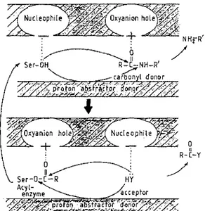
Serine peptidase--catalyzed rupture of a peptide bond is carried out by several enzyme reagents that fulfill the required functions of an electrophile or oxyanion hole, a proton abstractor-donor, and a nucleophile (Figure 8). In each of the serine peptidases of the trypsin and subtilisin families, two backbone NH groups, one of which belongs to the active-site serine, create the oxyimion hole environment; a histidine adjacent to the essential serine (and in interaction with a buried aspartic acid) is involved in proton abstrac tion-donation; and one side of the active site generates a nucleophilic suction pump in the immediate vicinity of the essential serine (75). Reaction-rate enhancement, when compared to that of the analogous uncatalyzed reaction, depends on the goodness of fit of the ligand in the active site and on its correct positioning with respect to the enzyme functional groups.
Geometry optimization, superimposition searches, and conformational analysis of lactam compounds belonging to different chemical families (peni cillins, 6-epoxy-spiropenicillins, cephalosporins, thienamycin, y-Iactams,
6 ______________ : U� �. Ser-O-(-R HY Acyl enzyme �""-"=,,,, .. NHrR'
/
o 11 R-C-YFigure 8 Serine peptidase-catalyzed rupture of a peptide bond. The oxyanion hole polarizes the C = 0 bond; the nucleophile enhances the reactivity of the serine hydroxyl or the acceptor HY: and the proton abstractor-donor achieves nucleophilic attack.
Annu. Rev. Microbiol. 1991.45:37-67. Downloaded from www.annualreviews.org
I3-LACT AMASES AND PBPs 51
Figure 9 Coplanarity of the binding entity of benzylpenicillin (A) and the most stable, extended conformer of ACrL-Lys-D-Ala-D-Ala (B). Electrostatic potential isocontours at -10 kcallmol. I, COOH; 2, CO of the scissile CONH bond (i.e. the electrophilic center); 3, CO of the exocyciic carbonyl (penicillin) or the L-Lys-D-Ala peptide bond (tripeptide). Distances and dihedral angles are given in Reference 76. The increased volume of well 2 of penicillin when compared to well 2 of AC2-L-Lys-D-Ala-D-Ala expresses the increased intrinsic reactivity of a f3-lactam amide bond when compared to an acyclic peptide bond. The two additional wells seen in the tripeptide are generated by the acetyl carbonyl groups that substitute the a and f lateral chains of lysine. The views are shown along the plane of the scissile amide bond. The a and f side chains of lysine are above and below the reference plane, respectively.
lactivicin) have led to the concept of a productive binding entity (76). To confer activity to these compounds (either as substrates of the (3-lactamases or inactivators of the PBPs), the triad formed by the carbon atom of the carbox ylate, the carbonyl of the scissile amide bond (i.e. the electrophilic center C =
0) and the oxygen atom of the exocyc1ic carbonyl (or COH in thienamycin) must fulfill rather strict spatial requirements-Dne of which is an almost coplanar disposition of these four atoms (Figure 9A). In the most stable extended conformer of AC2-L-Lys-o-Ala-o-Ala, the triad formed by the car boxylate, the C = 0 of the scissile o-Ala-o-Ala peptide bond, and the C = 0
of the L-Lys-o-Ala peptide bond has a spatial disposition comparable to that of the equivalent triad of the biologically active lactam compounds (Figure 9B). These functional groups generate a typical electronic property, and this electronic distribution gives rise to an electrostatic potential of defined shape and volume. Isosterism, however, is partial. The ACrL-Lys moiety of ACrL Lys-D-Ala-D-Ala and the side-chain of the lactam ring in the antibiotics are oriented differently (79). The spatial disposition adopted by the ACrL-Lys
Annu. Rev. Microbiol. 1991.45:37-67. Downloaded from www.annualreviews.org
moiety relative to the o-alanyl-o-alanine dipeptide in the tripeptide can be visualized using the negative electrostatic wells generated by the carbonyl groups of the two acetyl substituents (Figure 9B).
The active site of the f3-lactamases of class A and C and that of the Streptomyces R61 PBP/oo-peptidase are dense hydrogen bonding networks (Figure lOA). One important feature of the enzyme-ligand interactions (lac tam compound-f3-lactamase; lac tam compound-PBP; ACrL-LYS-D-Ala-o-Ala PBP) is an antiparallel hydrogen bonding between the innermost 133 strand of the j3-sheet and the binding entity of the ligand (Figure lOB) (25, 56, 62, 67, 70, 72, 82, 89). The carbonyl oxygen of the scissile bond forms hydrogen bonds to the backbone NH group of the active-site serine itself and to the backbone NH of the alanine (the class A j3-lactamase), serine (the class C
j3-lactamase), or threonine (the PBP) immediately downstream from the K(H)TG motif of the j3-strand, in a way reminiscent of the oxyanion-hole hydrogen bonds found in the peptidases of the trypsin family. The NH and CO of the exocyclic amide bond borne by the lactam ring or the equivalent groupings of the L-Lys-o-Ala peptide bond of ACz-L-Lys-o-Ala-o-Ala are in hydrogen bond interactions with the backbone carbonyl of the same alanine, serine, or threonine of the j3-strand and with the side chain of the asparagine of the S(Y)XN motif, respectively. The carboxylate (sulfonate in the mono bactams) head of the ligand interacts with the OH group of the threonine of the K(H)TG motif and is oriented towards the lysine or histidine of the same motif.
Optimal positioning of the electrophilic center of the ligand relative to the '}I-OH of the active-site serine also depends much on the appendages borne by the binding entity. Structural variations in the acyl side chain of the lactam ring, or the R-L-Xaa moiety of the R-L-Xaa-o-alanyl-o-alanine-terminated peptides, cause wide variations in the value of the second-order rate constant (k+zIK) of protein acylation. These effects are both protein and ligand specific (41,45). In the emerging picture, the Streptomyces R61 PBP possess es a subsite that specifically interacts with the side chain borne by the lactam ring and another subsite that specifically interacts with the € side chain of the
L-Iysine residue of the tripeptide ACz-L-Lys-o-Ala-o-Ala. As a corollary, these side-chains are not interchangeable (9). Also according to the picture, the a side chain of ACz-L-Lys-o-Ala-o-Ala extends to the exterior of the
enzyme cavity and, thereby, binding of glycan-substituted pentapeptides to the active-site would be mediated mainly by the C-terminal tripeptide moiety.
Though we are learning much, microbiologists still seek answers to ques tions regarding the distinction between the PBPs and the f3-lactamases in terms of stability vs lability of the acyl enzymes formed by reaction with the lactam antibiotics, the inability of the f3-lactamases to perform acyl transfer reactions on acyclic carbonyl donor peptides, and the mechanism of
Annu. Rev. Microbiol. 1991.45:37-67. Downloaded from www.annualreviews.org
J3-LACT AMASES AND PBPs 53
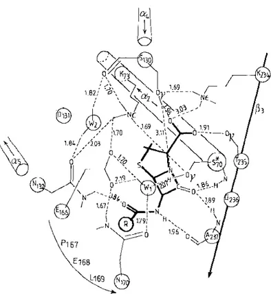
Figure 10 Hydrogen bonding interactions between penicillin and the active site of the Strep tomyces albus G J3-lactamase of class A (25, 62, 67; J. Lamotte-Brasseur, G. Dive,
O.
Dideberg, J.-M. Frere, J. M. Ghuysen, in preparation). S*70 = active-site serine. The amino acid numbering follows the ABL scheme (4). S*70K73 is S89K92 in the precursor (Figure 3). Similarly, S 1 30DN1 32 is S153DN155; E166PELN170 is EI89PELNI93; K234TGA2375 is K257TGA260. Two water molecules WI and W2 are shown . Distances are given in A. The acyl side chain of penicillin is represented byCO-®.
WI, W2, E 166, and S I30 would be involved in proton abstraction-donation. Attack of the carbonyl carbon of the {3-lactam scissile bond by theOr
of S*70 occurs on the well-exposed a face . Similar hydrogen bonding networks are proposed for the S. aureus (56) and B. licheniformis (82) J3-lactamases of class A, the C. freundii J3-lactamase of class C (89), and the Streptomyces R6 1 PBP (70, 72).transpeptidation by the PBPs/oo-peptidases. Recent advances, however, have shed light on the mechanism of proton abstraction-donation.
Site-direCted mutagenesis (49) and molecular modeling (J. Lamotte Brasseur, G. Dive, O. Dideberg, 1.-M. Frere, & 1. M. Ghuysen, in prepara tion) support the view that the essential glutamic acid E* of the E*XELN sequence, i.e. the E/D motif, activates, via a bound water molecule, the essential serine of the class A f3-lactamases (Figure 10) and that the serine of the SDN motif may be involved in proton shuttle. This serine has a homolog
Annu. Rev. Microbiol. 1991.45:37-67. Downloaded from www.annualreviews.org
in the tyrosine of the Y AN motif in the 13-lactamases of class C, but the spatial
disposition of the D/E motif in these 13-lactamases is not compatible with the role of a general base in catalysis. Superimposition of class C C. freundii {3-1actamase onto trypsin, using the active-site serine and the oxyanion hole as reference points, results in the phenolic oxygen of the tyrosine of the Y AN
motif in the {3-lactamase being < 0.5
A
from the Ne2 position of the essentialhistidine in trypsin, indicating that the tyrosine, as its anion, might act in a
way similar to the essential histidine in trypsin (89). The constellation of two
positively charged lysines around this tyrosine would sufficiently lower its
pKa value.
The High-Mr PBPs of Class
Aand B
The high-Mr PBPs of class A and B have two functional domains that are fused to each other and that operate in a concerted manner (Figure 2) . An
amino terminal domain several hundred amino acids long is linked on its carbonyl side to the penicillin-binding domain per se. The penicillin-binding domain is assumed to start 60 residues upstream from the S*XXK motif and to terminate 60 residues downstream from the KT(S)G motif. In most high Mr PBPs, but not in all, this domain bears a tail in the form of a - IOO-amino acid carboxy terminal extension (Figure 3).
The high-Mr PBPs are not synthesized as preproteins but have a highly hydrophobic reglon close to the amino terminus that acts as a noncleaved signal-like segment, both to translocate the bulk of the protein to the peri plasm and to anchor the protein in the plasma membrane (1, 6, 1 2, 31, 32,
96). The E. coli PBP3 has been suggested to be a lipoprotein (84) because the sequence L26LCGC30 is similar to the consensus sequence for the modifica tion and processing of bacterial lipoproteins. However, only a small fraction of PBP3 appears to be lipid-modified, at least under conditions of protein overexpression. The PBP3 undergoes maturation by eliminating a cleavable lO-amino acid carboxy-terminal stretch, from 1578 to S588 , in a way reminis cent of that observed with the low-Mr PBP of Streptomyces R61. Short hydrophobic regions upstream from Leu558 in the carboxy terminal tail are important for the functioning and/or stability of the protein (52, 84).
The simple mode of membrane insertion of the high-Mr PBPs allows the production of water-soluble forms through proteolytic treatment of isolated membranes (32, 96). Genetic constructs aimed at removing the membrane anchor from high-Mr PBPs also yield truncated proteins that are sufficiently
stable to be isolated ( 1 , 6, 31). They may require high salt concentrations to
remain soluble and to retain their penicillin-binding capacity. A gene fusion that removes the amino terminal domain of the E. coli PBP3 and links the carboxy-terminal domain to the amino-terminal of the {3-galactosidase leads
to a chimeric protein that binds penicillin but is unstable (55) . Expression of
that portion of the gene that encodes only the carboxy-terminal domain of the
Annu. Rev. Microbiol. 1991.45:37-67. Downloaded from www.annualreviews.org
f3-LACT AMASES AND PBPs 55
Amino t e rmina l doma ins
I
E. hira e5 1
-400
30
1
I E. COli l A
1
-�I
S. C!ureus 2'1 �I
N.gonorrhoeae 2 1-�041
1
I S. pneumoniae 2x l-\lJl � 1·3�
I'E-.-co-'I'-i -l-s'l � I E. col i 2
1
( - 1851\5 � I E. coli 3I
E. (oli 1 AI
..-101/6
-181!
10
\
E. coli 18I
S. pneum oniae 2bI
Pen i c il l i n - binding dQmain�E . hira e 5
I
S. aureus 2' ,,-I06/� ' \
N. gonorrhoeae2\
-114
20
1
�
\341\1
-690
511
�
__..,_../
S. pneumoniae 2)(\
""'-=:2001\2
, I
E.(oli2 1 ..
-162 / 8. \
E.(oli31J
�I
S p""m oo; " 1b1 -'"'''
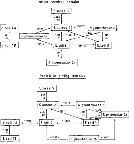
Figure 11 Search of homology between the amino acid sequences of the amino-terminal domains and penicillin-binding, carboxy-terminal domains of the high-Mr PBPs of class A and B . Comparison scores (negative values) and significance in standard deviations (positive values) are shown. For more details, see the legend of Figure 2. The E. coli PBPs lA and IB are of class A. The other PBPs are of class B .
PBP yields the expected polypeptide as evidenced by immunological tests and penicillin binding, but this polypeptide is very unstable. Similar observations have been made with other high-Mr PBPs. In addition, no simple in vitro assays are available that would allow one to monitor the enzymatic activities related to peptidoglycan synthesis of the high-Mr PBPs. To all appearances, all high-Mr PBPs lack activity on AC2-L-Lys-o-Ala-o-Ala, the standard sub strate analog of the low-Mr PBPs.
The distinction between the two classes A and B (Figures 2 and 1 1) derives from homology searches. The E. coli PBPs l A and I B of class A and the E.
coli PBPs 2 and 3 of class B have received much attention.
The 85G-amino acid PBP I A (encoded by ponA in the 73.5-min region of
Annu. Rev. Microbiol. 1991.45:37-67. Downloaded from www.annualreviews.org
the chromosome map) and the 799- to 844-amino acid PBP I B (encoded by ponB in the 3 . 3-min region of the map) are bifunctional, peptidoglycan
synthesizing enzymes (59, 85 , 1 07 , 108). Their amino terminal domain is a
penicillin-insensitive transglycosylase that catalyzes glycan chain elongation, and their penicillin-binding carboxy-terminal domain is a transpeptidase that catalyzes peptidoglycan crosslinking. Their bifunctionality is revealed by the reaction products that they generate upon incubation with the lipid-linked precursor N-acetylglucosaminyl-N-acetylmuramyl (o-alanyl-o-alanine-ter minated pentapeptide) diphosphoryl-undecaprenol. The overall reaction, however, proceeds with a low turnover number, suggesting that the in vitro conditions poorly mimic the in vivo situation. PBPIA and PBP I B show differences in reaction conditions for optimal activity. They may compensate for each other according to the cell culture conditions. Selective inactivation of the transpeptidase domain of both PBPIA and PBP l B by derivatization of the essential serine by ,B-lactam antibiotics causes cell death by cell lysis. Deletion of both ponA and ponE genes has also that effect.
The interplay between the two catalytic domains of PBPl B is well documented. Penicillin inhibits peptide crosslinking and, under certain con ditions, greatly increases formation of uncrosslinked peptidoglycan, suggest ing that the uncoupling of the two reactions stimulates the transglycosylase activity. Moenomycin, which inhibits the transglycosylase ( 1 20), strongly
inhibits or prevents peptide crosslinking, suggesting that transpeptidation requires prior or concomitant glycan chain elongation. Studies with monoclo nal antibodies also suggest that transpeptidation depends on the product of transglycosylation and that PBPl B has only one type of pentapeptide (donor)! tetrapeptide (acceptor) transpeptidase activity (24). Membrane components act as effectors of the catalyzed reaction and strongly stimulate the polymerization reaction.
The 633-amino acid PBP2 (encoded by pbpA at the 14-min region of the map) and the 588-amino acid PBP3 [encoded by pbpB (also calledftsI) at the 2-min regionJ of class B fulfill essential functions in cell morphogenesis, in connection with other membrane proteins. The 14-min region contains, in addition to the PBP2-encoding pbpA gene; other genes required for cell-wall elongation and maintenance of the rod shape of the cell. In particular, rodA
encodes the 370-amino acid integral membrane protein RodA (81), and dacA
encodes the low-Mr PBP5 (15). Inactivation of dacA does not cause growth defects, but a l O-fold overproduction of PBPS results in spherical cells. Inactivation of PBP2 by the amidinopenicillin mecillinam causes E. coli to grow and divide as round-coccal cells. Mutations in either pbpA or rodA also have that effect and confer resistance to mecillinam.
The 2-min region of the chromosome map contains, in addition to the PBP3-encoding pbpB gene, other genes required for the synthesis of the
Annu. Rev. Microbiol. 1991.45:37-67. Downloaded from www.annualreviews.org
I3-LACT AMASES AND PBPs 57 peptidoglycan precursors (murE, murF, murG, murC, ddl) and for cell division and septum formation (jtsW, JtsQ, JtsA, JtsZ) (109). Selective in activation of PBP3 either by mutation of pbpB or by reaction with, for example, the monobactam aztreonam, leads to filamentous growth of the cells. FtsW is an intrinsic membrane protein very similar to RodA (similarity index: 40). Mutations in FtsW also cause defects in cell division (58). The 420-amino acid FtsA protein is though to participate in the construction of the septum (98, 1 16). It must be synthesized during a short period just before division for cell division to be completed. High-level expression of FtsA inhibits cell septation. The 383-amino acid FtsZ protein ( 1 24) probably regulates the frequency of cell division. It is the target of the SOS-induced inhibitor SfiA (64). Overexpression of FtsZ leads to a hyperdivision activity displayed as the minicell phenotype ( 122). The relative levels of FtsZ and trigger factor, an abundant cytosolic 543-amino acid protein that might act as a chaperone, probably control initiation of cell division (50).
The pair RodA-PBP2 is involved in formation of the rod-shape of the cell (8 1). The pair FtsW-PBP3 may be involved in cell division (58), but interac tion between RodA and PBP3 has also been suggested (8). The complex RodA-PBP2 appears to be linked to the ribosomes via the lav gene product and to the cyclic AMP (cAMP)-receptor protein (CAP) complex via the adenylate cyclase-encoding cya gene and the CAP-encoding crp gene (11 , 90). The cAMP-CAP complex is involved in the transcriptional regulation of many operons. Mutations in either cya or crp confer high-level resistance to mecillinam, associated with a slow growth rate. All these data suggest that the intrinsic membrane proteins RodA and FtsW (and perhaps FtsA and FtsZ) link PBP2 and PBP3 at the surface of the cell to structures, chaperones, and/or enzymes in the cytosol, and that correct functioning of these networks is essential for cell morphogenesis.
A connection between PBPs and intracellular components is a feature that is not unique to E. coli. In the course of sporulation, the PBP profile of
Bacillus subtilis undergoes profound remodeling with a sporulation-specific increase in vegetative PBPs and a de novo synthesis of PBPs (114). SpoVE, which is extremely similar in the primary structure to the pair RodA-FtsW of
E. coli (58, 65), seems to be involved in this process. Mutations in the spaVE
locus cause a blockage at stage V of sporulation (95), and spaVE mutants are grossly deficient in cortex, which consists of a sporulation-specific peptido glycan. B . subtilis also contains homologs of the E. coli cell division genes
JtsA and JtsZ. Expression of these B. subtilis homologs in E. coli results in filamentation and cell death (7).
The use of E. coli strains overproducing PBP2 and RodA (60) or PBP3 and FtsW (M. Matsuhashi, personal communication) supports the view that these PBPs catalyze transpeptidation reactions. Assignment of a peptidoglycan
Annu. Rev. Microbiol. 1991.45:37-67. Downloaded from www.annualreviews.org
transpeptidase activity to the penicillin-binding, carboxy-terminal domain of the high-Mr PBPs is also supported by the study, at the molecular level, of the resistance to f3-lactam antibiotics acquired through emergence of altered PBPs with a much decreased affinity for the drugs (see below). A remodeling of the penicillin-binding-transpeptidase site that results in a decreased affinity for the drug should presumably affect the specificity profile of the enzyme for the carbonyl donor and/or amino acceptor peptides involved in transpeptidation and, thereby, should cause structural modifications of the wall peptidoglycan. The analysis of the peptidoglycans of peniCillin-sensitive and penkillin resistant pneumococci has confirmed the prediction (43).
The question of which enzymatic function the amino terminal domains of the high-Mr PBPs of class B perform is still a matter of controversy. These domains (at least some of them) possess several conserved amino acid group ings not found in the transglycosylasc domain of the E. coli PBPs l A and IB of class A (96) . These groupings might be the signature of an active site
whose function would be different from that of a transglycosylase. One should remember that PBPs are involved, directly or indirectly, in the average glycan chain length of the peptidoglycan, in the extent of peptidoglycan a-acetylation, and in the amounts of peptidoglycan-attached lipoprotein. None of these observations has received a satisfactory explanation.
Intrinsic Resistance by Emergence of Altered High-Mr PBPs
Assuming that a given high-Mr PBP is the most sensitive killing target of a lactam antibiotic, conversion of this PBP into an altered PBP whose penicillin-binding domain has acquired a decreased affinity for the drug, but has retained the capacity of performing its physiological (transpeptidase) function, causes an increased resistance of the bacterial cell to this particular antibiotic.
Alterations affecting amino acids located away from the active site may be neutral or they may stabilize the protein in response to changes occurring within or close to the active site. Study of an altered PBP2X of a S.
pneumoniae laboratory mutant with a decreased affinity for cefotaxime shows that such distal alterations may be of real significance (74). By reference to the PBP2X of the cefotaxime-sensitive strain, the two mutations G597 -> D and G601 -> V, occurring about 50 residues downstream from the KTG motif, are responsible for the first level of resistance to the drug. One may hypothesize that these amino acid alterations cause changes in the conforma tion and thereby, changes in the specificity profile of the penicillin-binding domain.
The acquisition of resistance through alterations affecting the active site is well documented. Many (3-lactamases of class A have a tyrosine
approximate-Annu. Rev. Microbiol. 1991.45:37-67. Downloaded from www.annualreviews.org
/3-LACTAMASES AND PBPs 59 ly 25 residues upstream from the SDN motif (4). Alterations of the equivalent peptide stretches Tyr334-S359SN of the E. coli PBP3, Tyr337-S362SN of the
Neisseria gonorrhoeae PBP2, and Tyr41 9-S447SN of the S. pneumoniae
PBP2B profoundly modify the specificity profile of these PBPs. Cephalexin, at the minimum inhibitory concentration (MIC), kills E. coli by inactivating
PBP3. E. coli laboratory mutants have been obtained whose PBP3 has a much decreased affinity for cephalexin and other cephalosporins but not for penicil lins and aztreonam (53 , 54). These PBP3 mutants have either a Va1344 - > Gly substitution (occurring approximately in the middle of the Tyr334-S359SN sequence) or an Asn361 - > Ser substitution (converting the SSN motif into a SSS motif). This latter mutant has some degree of functional impairment that causes the formation of E. coli cells with pointed polar caps ( 1 10) . Maximal level of resistance to cephalexin, however, is achieved by combining four changes: the conversion SSN -> SSS, the substitution Glu349 -> Lys (which also occurs approximately in the middle of the Tyr -> SSS stretch), and the substitutions Val530 -> lIe and TyrS4 1 -> Ser (about 50 to 40 residues upstream from the carboxy terminus of the 388-amino acid protein). Penicillin kills N. gonorrhoeae by inactivating PBP2. The PBP2 of clinical isolates that have a decreased affinity for penicillin differs from the PBP2 of the penicillin-sensitive strains only in the insertion of an additional aspartic acid (Asp345A) between Arg345 and Asp346, thus affecting the Tyr337-S362SN sequence ( 13). However, a penA gene contain ing only the Asp345A codon insertion does not transform a penicillin sensitive gonococcus to as high a level of resistance to penicillin as the penA R genes from other non-Iactamase-producing, chromosomally mediated resis tant gonococcal strains. The PBP2 of these strains contains not only the inserted Asp345A but numerous other amino acid changes in the penicillin binding domain. Similarly, a s. pneumoniae PBP2B mutant obtained from clinical isolates exhibiting a > lOOO-fold increased resistance to penicillin has 1 7 amino acid substitutions (27). Seven of these changes are within the Tyr23 1-S249SN sequence and another change affects the amino acid im mediately downstream from the SSN motif.
Acquired resistance may emerge through recombinational events that re cruit portions of homologous genes from related species and result in the creation of mosaic genes encoding hybrid PBPs (26, 29, 102, 105). Such horizontal gene transfers occur between species that are naturally transform able such as Neisseria and Streptococcus spp. The gene encoding the penicil lin-resistant PBP2 of Neisseria meningitidis consists of regions almost iden tical to the corresponding regions of the penicillin-sensitive strains and of two regions that are very different from them. These two blocks of altered sequence have resulted from the replacement of meningococcal sequences with the corresponding sequences of the penA gene of Neisseria flavescens.
Annu. Rev. Microbiol. 1991.45:37-67. Downloaded from www.annualreviews.org
The resulting altered form of PBP2', when compared to that of the penicillin sensitive N. meningitidis strain, contains 44 amino acid substitutions and 1 amino acid insertion. N. gonorrhoeae also produces altered forms of PBP2, whose part of the penA gene has been replaced by the corresponding region of the N. flavescens gene.
Methicillin-resistant S. aureus strains have imported an inducible high-Mr PBP2 ' of low affinity for the drug ( 1 00). The PBP2' -encoding mecA gene may have evolved through recombination of a PBP gene of some bacterium and a {3-lactamase inducible gene. The promoter and close upstream region of
mecA are similar to those of the staphylococcal f3-lactamase inducible gene, and the mecR region has two main open reading frames, one of which encodes a putative protein that has strong similarity in the primary structure to the penicillin sensory-transducer BLAR of B . licheniformis ( I l l ; B . Berger Bachi, personal communication). The mecA gene is linked to the aadD gene that encodes the 4' ,4" adenyltransferase responsible for tobramycine resis tance ( 1 1 9). The two linked genes are carried on a moving element and may have emerged in S. aureus through transposition.
The staphylococcal PBP2 ' has two homologs, PBP5 (32) and PBP3R (96), in the penicillin-resistant Enterococcus hirae strains. Complete derivatization of these PBPs by penicillin or cefotaxime requires conditions under which serum albumin is partially acylated. These PBPs can take over the functions needed for wall peptidoglycan assembly under conditions. in which all the other PBPs are inactivated. Though they have conserved the characteristic motifs of the penicillin-interactive proteins, their active site has acquired a configuration that almost completely discriminates between penicillin and D-alanyl-D-alanine-terminated peptides, the ultimate achievement in intrinsic penicillin resistance.
ACKNOWLEDGMENTS
It gives me great pleasure to acknowledge the contribution of the colleagues who, at the University of Liege, carried out the work described here: Drs. P. Charlier, J. Coyette, D. Dehareng, O. Dideberg, G. Dive, C. Duez, J. Dusart, J . -M. Frere, B . Joris, J. Lamotte-Brasseur, M. Leyh-Bouille, M. Nguyen-Disteche, and C. Piron-Fraipont. 1 also thank Dr. J . Van Beeumen of the University of Ghent, Belgium, and Drs. J . Kelly and J . Knox of the University of Connecticut, Storrs, USA. The work at Liege has been sup ported by the Fonds de la Recherche Scientifique Medicale, the Fonds National de la Recherche Scientifique, the Belgian Government, the Govern ment of the Walloon Region, and the Fonds de Recherche de La Faculte de Medecine, Universite de Liege.
Annu. Rev. Microbiol. 1991.45:37-67. Downloaded from www.annualreviews.org
Literature Cited
1 . Adachi, R., Ohta, T., Matsuzawa, R. 1987 . A water-soluble form o f penicil lin-binding protein 2 of Escherichia coli
constructed by site-directed mutagene sis. FEBS Lett. 226:150-54
2. Adam, M . , Damblon, C . , Plaiton, B . , Christiaens, L . , Frere, J . M. 1990. Chromogenic depsipeptide substrates for j3-lactamases and penicillin-sensitive DD-peptidases. Biochern. J. 270:525-29
3. Adriaens, P . , Meesschaert, B . , Frere, J.-M . , Vanderhaege, H . , Degelaen, J . , e t al. 1978. Stability of D-5,5-dimethyl .6?-thiazolidine-4-carboxylic acid in
relation to its possible occurrence as a degradation product of penicillin by the exocellular DD-carboxypeptidase-trans peptidase from Streptomyces R61 and the membrane-bound nn-carboxypepti dase from Bacillus stearotherrnophilus.
J. BioI. Chern. 253:3660-65
4. Ambler, R. P . , Frere, J.-M . , Ghuysen, J.-M . , Jaurin, B . , Levesque, R. C . , et al. 1 99 1 . A standard numbering scheme for the class A j3-lactamases. Biochern.
J. In press
5. Asoh, S . , Matsuzawa, R . , Ishino, F., Strominger, J . L. , Matsuhashi, M . , et al. 1986. Nucleotide sequence of the
pbpA gene and characteristics of the deduced amino acid sequence of penicil lin-binding protein 2 of Escherichia
coli K 1 2 . Eur. J. Biochern. 160:231-38
6. Bartholome-De Belder, 1 . , Nguyen Disteche, M . , Houba-Herin, N . , Ghuysen, 1.-M. , Maruyama, 1 . N . , et al. 1988. Overexpression, solubilization and refolding of a genetically engineered derivative of the penicillin-binding pro tein 3 of Escherichia coli K 1 2 . Mol. Microbiol. 2:51 9-25
7 . Beall, B . , Lowe, M . , Lutkenhaus, J . 1 988. Cloning and characterization of
Bacillus subtilis homologs of Es
cherichia coli cell division genes jtsZ
and jtsA. J. Bacteriol. 1 70:4855-64 8. Begg, K . J . , Spratt, B . G . , Donachie,
W. D . 1986. Interaction between mem brane proteins PBP3 and RodA is re quired for normal cell shape and division
in Escherichia coli. J. Bacterial. 167: 1 004-8
8a. Begg, K. J . , Takasuga, A . , Edwards, D. R . , Dewar, S. J . , Spratt, B. G . , et al. 1990. The balance between different peptidoglycan precursors determines
/3-LACTAMASES AND PBPs 61
whether Escherichia coli will elongate or divide. J. Bacteriol. 172:6697-6703 9. Bentley, P. H . , Stachulski, A. V. 1983. Synthesis and biological activity of some fused j3-lactam peptidoglycan an alogues. J. Chern. Soc. Perkin Trans.
1 : 1 1 87-92
1 0 . Boissinot, M . , Levesque, R. C. 1990. Nucleotide sequence of the PSE-4 carbenicillinase gene and correlations with the Staphylococcus aureus PCI /3-1actamase crystal structure. J. BioI.
Chern. 265 : 1225-30
1 1 . Bouloc, P. , Jaffe, A . , D'Ari, R. 1989. The Escherichia coli lov gene product connects peptidoglycan synthesis, ribo somes and growth rate. EMBO 1. 8:3 1 7-23
1 2 . Bowler, L. D . , Spratt, G. D. 1989. Membrane topology of penicillin binding protein 3 of Escherichia coli. Mol. Microbial. 3 : 1 277-86
1 3 . Brannigan, J. A . , Tirodimos, I. A . , Zhang, Q.-Y . , Dawson, C. G . , Spratt, B . G. 1990. Insertion of an extra amino acid is the main cause of the low affinity of penicillin-binding protein 2 in penicil lin-resistant strains of Neisseria gonor rhoeae. Mol. Microbiol. 4:913-19
1 4 . Broome-Smith, J . K . , Edelman, A . , Yousif, S . , Spratt, B . G. 1985. The nu cleotide sequences of the ponA and ponB genes encoding penicillin-binding proteins I A and 1 B of Escherichia coli. Eur. J. Biochem. 147 :437-46 1 5 . Broome-Smith, J. K .. Ioannidis. I.,
Edelman, A . , Spratt, B . G . 1988. Nu cleotide sequences of the penicillin binding protein 5 and 6 genes of Es cherichia coli. Nucleic Acids Res. 16: 1 6 1 7
1 6 . Buchanan, C. E . , Neyman, S . L. 1986.
Correlation of penicillin-binding protein composition with different functions of two membranes in Bacillus subtilis fore spores. J. Bacteriol. 165 :498-503 1 7 . Cartwright, S. J . , Coulson, A. E. W.
1980. Active-site of staphylococcal 13-lactamase. Phil. Trans. R. Soc. London B289:370-72
1 8 . Cartwright, S. J . , Waley, S. G. 1983. j3-Lactamase inhibitors. Med. Res. Rev. 3:34 1-82
19. Chan, P. T. 1986. Nucleotide sequence of the Staphylococcus aureus PC1 /3-lactamase gene. Nucleic Acids Res. 14:5940
20. Cohen, S. A., Pratt, R. F. 1980.
In-Annu. Rev. Microbiol. 1991.45:37-67. Downloaded from www.annualreviews.org
