CD40 engagement enhances eosinophil survival through induction of
cellular inhibitor of apoptosis protein 2 expression: Possible involvement in
allergic inflammation
Fabrice Bureau, DVM, PhD,a* Gregory Seumois, BSc,a* Fabrice Jaspar, BSc,a Alain Vanderplasschen, DVM, PhD,b Bruno Detry, BSc,b Paul-Pierre Pastoret, DVM, PhD,b Renaud Louis, MD, PhD,c and Pierre Lekeux, DVM, PhDa
From the Departments of aPhysiology and bImmunology/Vaccinology, Faculty of Veterinary Medicine, and cthe Department of Pneumology,
Faculty of Medicine, University of Liège, Liège. *These authors contributed equally to this work.
Abstract
Background: CD40 engagement enhances eosinophil survival, suggesting a role for this receptor in the development of eosinophilia.
Objective: We examined whether CD40 enhances eosinophil survival by inducing the expression of antiapoptotic proteins. Three members of the inhibitor of apoptosis protein (IAP) family, namely cellular (c)-IAPl, C-IAP2, and XIAP, and 2 antiapoptotic proteins of the Bcl-2 family, namely Bcl-xL and Bfl-1/A1, were
investigated.
Methods: Blood and sputum were obtained from healthy subjects and atopic asthmatic patients. Blood eosinophils were isolated by means of magnetic selection. Expression of CD40, IAPs, and Bcl-2 proteins was investigated by using flow cytometry, immunoblotting, or both. CD40 stimulation was achieved with agonistic antibodies or soluble ligands. Apoptosis was assessed by staining with propidium iodide and FITC-conjugated annexin-V. C-IAP2 expression was inhibited with anti-sense oligonucleotides.
Results: Freshly isolated eosinophils from healthy and asthmatic patients did not express CD40. Conversely, eosinophils expressed CD40 spontaneously when cultured for 48 hours. At this time point, CD40 stimulation significantly delayed eosinophil apoptosis. Inhibition of eosinophil apoptosis was accompanied by induction of C-IAP2 but not c-IAPl, XIAP, Bcl-xL, or Bfl-1/A1 expression. Antisense knockdown of c-iap2 abolished
CD40-induced enhancement of eosinophil survival. Sputum cells from asthmatic patients, unlike those from healthy subjects, substantially expressed CD40 and C-IAP2. Moreover, a strong correlation was found between the percentage of eosinophils in the sputum from asthmatic patients and the sputum level of CD40 and C-IAP2 expression.
Conclusion: The results demonstrate that CD40 engagement enhances eosinophil survival through induction of C-IAP2 expression and suggest a role for this mechanism in allergic inflammation.
Key words: Allergy, apoptosis, asthma, atopy, CD40, eosinophils, granulocytes, inflammation, neutrophils Abbreviations used: AS: Antisense; CD40L: CD40 ligand; DMRIE-C: 1,2-Dimyristyloxypropyl-3-dimethyl-hydroxy ethyl ammonium bromide-cholesterol; IAP: Inhibitor of apoptosis protein; Ic: Immune complexes ODN: Oligodeoxyribonucleotide; rhCD40LT: Trimeric recombinant human CD40L
INTRODUCTION
Allergy is characterized by the accumulation of activated eosinophils at the site of inflammation. Inhibition of eosinophil apoptosis has been proposed as a key mechanism for the development of tissue eosinophilia in allergic disorders.1 IL-5, IL-3, and GM-CSF, 3 TH2 cell-derived cytokines, dramatically increase the life span of
purified eosinophils by inhibiting their spontaneous apoptosis in vitro.2-4 Moreover, these cytokines are over-expressed in inflamed allergic tissue5 and have been shown to increase eosinophil survival in vivo, thereby crucially contributing to the accumulation of eosinophils in allergic inflammation.6,7
CD40, a 50-kd member of the TNF receptor super-family, is expressed on many different cell types, including B cells, macrophages, monocytes, dendritic cells, endothelial cells, epithelial cells, fibroblasts, and carcinoma cells.8 CD40 ligand (CD40L, also known as gp39 and CD154) is a protein with homology to TNF.8 CD40L is expressed on many cell types, such as activated T cells, dendritic cells, natural killer cells, monocytes, mast cells, and eosinophils, and can engage CD40 as a membrane-bound or soluble ligand.8 CD40-CD40L interaction
provides critical signals regulating diverse cellular responses, ranging from proliferation and differentiation to growth suppression and cell death.8 Although CD40 induces apoptosis in some cells, such as hepatocytes and carcinoma cells,9,10 it conveys antiapoptotic signals in other cells, including B cells.11 CD40 ligation rescues B cells from IgM- or Fas-induced apoptosis by means of upregulating expression of Bcl-xL and Bfl-1/A1,12-14 2
antiapoptotic members of the Bcl-2 family of proteins. In B cells CD40 stimulation also triggers expression of the cellular inhibitor of apoptosis protein 2 (c-IAP2, also referred to as HIAP-1 and MIHC),15 a member of the inhibitor of apoptosis (IAP) family. However, whether CD40-induced C-IAP2 expression is sufficient to protect B cells from apoptosis has never been demonstrated.
A few years ago, Ohkawara et al16 reported that eosinophils isolated from the blood or tissue of allergic subjects express CD40 and that CD40 stimulation results in enhanced survival of cultured blood eosinophils. These findings suggest that CD40 ligation, like IL-5, IL-3, and GM-CSF stimulation, might enhance eosinophil survival in allergic disorders and therefore participate in the development of eosinophilia. However, although potentially relevant in the pathophysiology of allergy, CD40-mediated delay of eosinophil apoptosis has not been further investigated.
Here we confirm that eosinophils may express CD40 and that CD40 engagement delays eosinophil apoptosis. Furthermore, we show that CD40-induced enhancement of eosinophil survival results from induction of C-IAP2 expression and provide in vivo evidence that this mechanism might be involved in allergic inflammation. METHODS
Subjects
Fifteen patients with atopic asthma (age, 35 ± 18 years) and 10 healthy subjects (age, 42 ± 19 years) participated in the study. All subjects were steroid naive. Asthma was diagnosed on the basis of a clinical history of recurrent wheeze, breathlessness, or cough associated with either significant reversibility of FEV1 (>15% from baseline
and >200 mL) after inhalation of 400 µg of salbutamol when baseline FEV1 was less than 80% of predicted
value or bronchial hyperrespon-siveness to methacholine (methacholine dose producing a 20% fall in FEV1
[PC20M]<16 mg/mL when baseline FEV1 was >70% of predicted value). Atopy was defined by positive skin
prick test responses (wheal diameter of <3 mm when compared with that produced with control saline) to common aeroallergens (Stallergènes). The protocol of the study was approved by the local ethical committee. Sputum induction and processing
Sputum induction and processing were performed as previously described.17 Sputum cell counts were performed on cytospin preparations stained with Diff-Quick (Dade Berhing). In all cases 500 nonsquamous nucleated cells were counted by an experienced observer blind to the clinical details.
Cell sorting and culture
Neutrophils were isolated from the blood of healthy subjects. Granulocytes were separated from mononuclear cells by means of density centrifugation (Histopaque, Sigma). Contaminating erythrocytes were removed from the granulocyte fraction by means of hypotonic lysis. The resulting granulocyte population contained mainly neutrophils (>95%), as determined by counting of cytospin preparations stained with Diff-Quick. Eosinophils were isolated from the blood of healthy and asthmatic subjects by incubating the granulocyte population with microbeads coated with anti-CD16 and anti-CD3 antibodies (CD16 and CD3 MicroBeads, Miltenyi Biotec). CD16-positive neutrophils and residual CD3-positive T cells were depleted by passing the cells through a magnetic cell-separation system (VarioMacs, Miltenyi Biotec). CD16-negative eosinophils were collected, and eosinophil purity was controlled by staining with Diff-Quick. The resulting cell population always contained at least 99% eosinophils. Blood neutrophils and eosinophils were cultured at 2 × 10⁶/mL in RPMI-1640 medium supplemented with 1% glutamine, 10% FCS, 50 µg/mL streptomycin, and 50 IU/mL penicillin (all from GIBCO BRL).
Antibodies and cytokines
In flow cytometric experiments FITC-conjugated anti-CD40 (Serotec) and anti-CD40L (Calbiochem) mAbs and isotype-matched control antibodies conjugated to FITC (Serotec) were used. In immunoblot experiments anti-CD40, anti-c-IAPl, anti-C-IAP2, anti-Bfl-1/Al (all from Santa Cruz Biotechnology), and anti-XIAP (R&D Systems) polyclonal antibodies; anti-Bcl-xL (Transduction Laboratories) and anti-α-tubulin (Santa Cruz Biotech)
mAbs; and control rabbit serum (Serotec) were used. CD40 stimulation was obtained by using agonistic anti-CD40 mAbs (Becton & Dickinson Biosciences) or trimeric recombinant human anti-CD40L (rhanti-CD40LT; Bender
MedSystems). CD40L inhibition was obtained with neutralizing anti-CD40L mAbs (Becton & Dickinson Biosciences). Recombinant human GM-CSF and IFN-γ were purchased from Roche. IgA-immune complexes (IgA-Ic) were obtained by incubating 15 µg of human IgA (Sigma) with 20 µg of anti-human IgA (Sigma) per milliliter of culture medium.
Analysis of CD40 and CD40L protein expression by means of flow cytometry
Cells were washed, stained with FITC-conjugated anti-CD40 (1:10 dilution) or anti-CD40L (1:50 dilution) antibodies for 30 minutes at 4°C, washed again, and assessed for CD40 or CD40L expression by means of flow cytometry with a FACStar Plus (Becton & Dickinson).
Apoptosis-necrosis assays
Apoptosis and necrosis were assessed by staining with annexin-V-FITC and propidium iodide with the Annexin-V-FLUOS staining kit (Roche), according to the recommendations of the manufacturer. Flow cytometric analyses were performed with a FACStar Plus (Becton & Dickinson).
Immunoblots
Immunoblots were performed as previously described.18 The membranes were incubated for 1 hour with the first antibody (1:200 dilution) and then incubated for 45 minutes with peroxidase-conju-gated goat anti-rabbit IgG (1:5000 dilution, Kirkegaard & Perry Laboratories) or peroxidase-conjugated rabbit anti-mouse IgG for Bcl-xL
and α-tubulin (1:1000 dilution, Dako). The intensity of specific signals for C-IAP2 and CD40 was assessed by using photoden-sitometry of the autoradiography (Gel Doc 2000, Bio-Rad). Equal loading of proteins on the gels was always confirmed by probing the blots for α-tubulin (data not shown).
RT-PCR
RNA extraction, reverse transcription, and amplification were performed as previously described.18 cDNA products were amplified by using primers specific for cd40 (5' primer, CTG TTT GCC ATC CTC TTG GT; 3' primer, CGA CTC TCT TTG CCA TCC TC) and the gene encoding glyceraldehyde 3-phosphate dehydrogenase (5' primer, ACT GGC ATG GCC TTC CGT GT; 3' primer, TTA CTC CTT GGA GGC CAT GT).
Antisense knockdown of CIAP2
Antisense phosphorothioate oligodeoxyribonucleotides (AS ODNs) directed to c-iap2 messenger (m)RNA and negative control ODNs have been designed and manufactured by BIOGNOSTIK (Germany). Their sequences were as follows: c-iap2 AS ODNs, 5'-TAT GTT CAT ATG GTT TAG-3'; control ODNs, 5'-ATA TCC TTC CAG TAC AG-3'. The ODNs were delivered to the cells in the form of complexes with a liposome formulation of the cationic lipid l,2-dimyristyloxypropyl-3-dimethyl-hydroxy ethyl ammonium bromide (DMRIE) and cholesterol (DMRIE-C Reagent, Gibco BRL). One milliliter of OPTI-MEM I Reduced Serum Medium (Gibco BRL) containing 24 µL of DMRIE-C and 1 mL of OPTI-MEM I containing 8 µg of DNA were mixed and allowed to complex for 45 minutes at room temperature. Eosinophils (2 × 10⁶) from healthy patients cultured for 40 hours were then suspended in 0.25 mL of serum-free medium, added to the transfection medium, and incubated for 4 hours. Afterward, 2 mL of growth medium containing 20% FCS was added, and the cells were cultured for another 4 hours before stimulation with rhCD40LT or agonistic anti-CD40 antibodies. Forty-eight
hours later, eosinophils were assessed for c-IAP2 expression and viability. Statistical analysis
Standard least-square linear regressions were carried out to study the correlation between the amount of C-IAP2 and CD40, as assessed by means of photodensitometry, and the percentage of eosinophils in sputum from asthmatic patients. Coefficients of correlation (r) were presented as measures of linear association for regression relationships. Other numeric data, except sputum cell counts, were presented as means ± SDs. The differences between mean values were estimated by using a Student t test for unpaired data. Sputum cell counts were expressed as medians and compared between atopic asthmatic patients and healthy subjects by using the Mann-Whitney test. In any statistical analysis, P values of less than .05 were considered significant.
RESULTS
Eosinophils spontaneously express CD40 when cultured
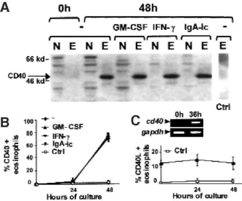
Blood neutrophils and eosinophils from healthy subjects were purified and cultured for 0, 24, or 48 hours before being assayed for CD40 expression by using immunoblot-ting (Fig 1, A, data obtained at 24 hours are not shown) and flow cytometry (Fig 1, B, data obtained for neutrophils are not shown) to examine the potential of granulocytes to express CD40. Freshly isolated blood granulocytes did not express CD40. Conversely, CD40 was detectable in a few eosinophils cultured for 24 hours and was strongly expressed in eosinophils cultured for 48 hours. CD40 was undetectable in neutrophils cultured for 24 or 48 hours.
RT-PCR analyses were performed to determine whether induction of CD40 expression in cultured eosinophils is due to transactivation of the cd40 gene (Fig 1, C). Freshly isolated eosinophils expressed only low levels of cd40 mRNA. These levels were dramatically increased in eosinophils cultured for 36 hours, indicating that increased CD40 expression in these cells is due to cd40 upregulation.
IFN-γ, GM-CSF, and IgA-Ic have been reported to induce or enhance CD40 expression in certain cell types.16, 19,
20 Blood eosinophils and neutrophils have been cultured in the presence of these compounds for 24 or 48 hours
before being assayed for CD40 expression to determine whether they affect CD40 expression in granulocytes. Neither IFN-γ (10 µg/mL), GM-CSF (500 U/mL), nor IgA-Ic induced CD40 expression in neutrophils, as determined by means of immunoblotting (Fig 1, A) and flow cytometry (data not shown). Similarly, these compounds were unable to enhance spontaneous expression of CD40 in eosinophils (Fig 1, A and B). Blood eosinophils have been demonstrated to consti-tutively bear CD40L.21 In our study the percentage of freshly isolated blood eosinophils expressing CD40L was 12.9% ± 2.1% (Fig 1, C). This percentage remained constant over a culture period of at least 48 hours (Fig 1, C). Freshly isolated and cultured blood neutrophils did not express CD40L (data not shown).
FIG 1: Eosinophils spontaneously express CD40 when cultured. A, Blood eosinophils (E) and neutrophils (N) from healthy subjects were cultured in the presence or absence of IFN-γ, GM-CSF, or IgA-Ic for 0, 24, 36, or 48 hours. Both cell types were assayed for CD40 expression by means of immunoblotting. Ctrl, Immunoblot performed with a control rabbit serum. B, Eosinophils were assayed for CD40 or CD40L expression by using flow cytometry (B; Ctrl, results obtained with isotype-matched control antibodies) and analyzed by means of RT-PCR for cd40 expression (C; as a control for quantification, glyceraldehyde 3-phosphate dehydrogenase [gapdh] was also amplified).
CD40 ligation potently enhances survival of cultured blood eosinophils
We next investigated the effects of CD40 stimulation on the survival of cultured blood eosinophils. Blood eosinophils from healthy donors cultured for 48 hours were treated with rhCD40LT or agonistic anti-CD40
antibodies. Forty-eight hours later, eosinophil viability was evaluated by using dual-color
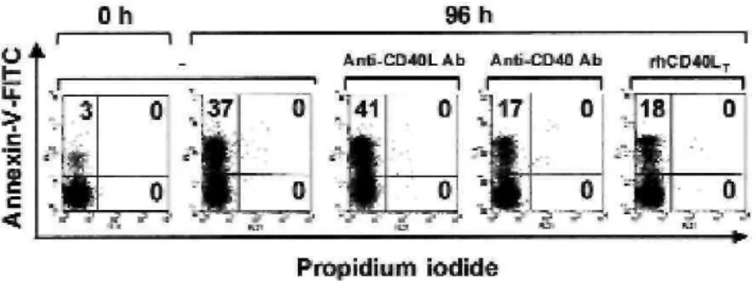
annexin-V-FITC/propidium iodide staining and flow cytometric analyses. Because eosinophils themselves express CD40L, we also examined whether CD40-CD40L interactions involved in autocrine or paracrine modes of stimulation might modulate eosinophil viability. In this case CD40L inhibition was obtained with neutralizing anti-CD40L antibodies.
CD40L neutralization did not affect the rate of eosinophil death (Fig 2). Conversely, the percentage of apoptosis in CD40-activated eosinophils was significantly reduced (16% ± 3% in eosinophils treated with anti-CD40 antibodies and 19% ± 4% in eosinophils treated with rhCD40LT) compared with the percentage of apoptosis in
untreated eosinophils (39% ± 7%, Fig 2). Further experiments showed that rhCD40LT and anti-CD40 antibodies
inhibited eosinophil apoptosis in a dose-dependent manner, with peak effects reached at 4 µg/mL rhCD40LT and
1:200 vol/vol anti-CD40 antibodies, and that their prosur-vival effects were still detectable 5 days after treatment (as in eosinophils cultured for 7 days, data not shown).
Freshly isolated blood eosinophils were also cultured for 96 hours in the presence of 125 U/mL GM-CSF, a potent inhibitor of spontaneous granulocytic apoptosis.2 At 96 hours, GM-CSF-stimulated eosinophils showed a significant decrease in apoptosis (14% ± 4%) compared with that seen in untreated cells (data not shown). However, at this time point, the rate of apoptosis in GM-CSF-treated and CD40-activated eosinophils was comparable, indicating that CD40 activation is as potent as GM-CSF stimulation in protecting eosinophils from spontaneous apoptosis.
FIG 2: CD40 ligation enhances survival of cultured blood eosinophils. Blood eosinophils from healthy subjects were cultured for 48 hours before treatment with either neutralizing anti-CD40L antibodies (1:200 dilution), agonistic anti-CD40 antibodies (1:200 dilution), or rhCD40LT (2 µg/mL). Forty-eight hours later, eosinophils were assayed for apoptosis by using flow cytometry.
CD40 engagement induces C-IAP2 expression in cultured blood eosinophils
The expression levels of Bcl-xL and Bfl-1/A1 and 3 representative members of the IAP family, namely XIAP,
c-IAP1, and C-IAP2, were assessed by means of immunoblotting in untreated eosinophils and in eosinophils treated with agonistic anti-CD40 antibodies or rhCD40LT to address the question of whether CD40 stimulation
protects eosinophils from apoptosis through the control of Bcl-2 family proteins or IAPs.
XIAP, c-IAP1, and C-IAP2 (Fig 3) were undetectable in freshly isolated eosinophils and in eosinophils cultured for 96 hours, whereas Bcl-xL and Bfl-l/Al were weakly expressed in these cells (data not shown). Neither XIAP,
c-IAP1, Bcl-xL, nor Bfl-1/Al protein levels were modified after CD40 engagement (data not shown).
Conversely, both anti-CD40 antibodies and rhCD40LT induced c-IAP2 expression in cultured eosinophils (Fig
FIG 3: CD40 engagement induces C-IAP2 expression in cultured blood eosinophils. Blood eosinophils from healthy subjects were cultured for 48 hours before treatment with either agonistic anti-CD40 antibodies (1:200 dilution) or rhCD40LT (2 µg/mL). Forty-eight hours
later, the cells were assayed for C-IAP2 expression by using immunoblotting. Ctrl, Immunoblot performed with a control rabbit serum.
Induction of C-IAP2 expression contributes to the reduction in eosinophil apoptosis after CD40 stimulation
To confirm that induction of C-IAP2 expression observed in CD40-activated eosinophils contributes to the delayed apoptosis in these cells, we determined the effects of selectively inhibiting C-IAP2 expression after CD40 stimulation on eosinophil longevity. c-iap2 knockdown was obtained by using c-iap2 AS ODNs. Exposure of blood eosinophils from healthy subjects cultured for 40 hours to an optimal dose of
DMRIE-C/c-iap2 AS ODN complexes for 8 hours before CD40 stimulation with rhCD40LT or anti-CD40 antibodies resulted
in an approximately 4-fold decrease in C-IAP2 expression, as determined by means of immunoblotting (Fig 4, A) and photodensitometry (data not shown) performed 48 hours after stimulation. Conversely, DMRIE-C alone and DMRIE-C/control ODN complexes had no effect on CD40-induced C-IAP2 expression (Fig 4, A).
The ability of c-iap2 AS ODNs to specifically down-regulate C-IAP2 allowed exploration of its role in enhanced eosinophil survival after CD40 engagement. Apoptosis assays were performed on eosinophils cultured for 40 hours that were first treated for 8 hours with DMRIE-C alone or in combination with c-iap2 AS or control ODNs and then treated for 48 hours with rhCD40LT (Fig 4, B) or anti-CD40 antibodies (data not shown). Inhibition of
C-IAP2 expression by c-iap2 AS ODNs significantly increased the rate of apoptosis in CD40-activated eosinophils (Fig 4, B). Indeed, although DMRIE-C alone induced eosinophil apoptosis (compare Fig 4, B, with Fig 2), the percentage of apoptosis was greater in eosinophils treated with AS ODNs (87% ± 6%) than in eosinophils treated with control ODNs or with DMRIE-C alone (60% ± 8% and 65% ± 6%, respectively). Evidence for expression of CD40 and C-IAP2 in sputum eosinophils from patients with atopic asthma Blood and sputum samples were taken from patients with atopic asthma to determine whether eosinophils from allergic patients express CD40 and C-IAP2. When compared with healthy subjects, atopic asthmatic patients had significantly increased sputum relative eosinophil counts (25% vs 0%) and reduced sputum relative macrophage counts (30% vs 58%).
FIG 4: AS knockdown of c-iap2 increases the rate of apoptosis in CD40-activated eosinophils. Blood eosinophils from healthy subjects were cultured for 40 hours and then treated for 8 hours with DMRIE-C alone or combined with c-iap2 AS or control ODNs before
stimulation with rhCD40LT (2 µg/mL). Forty-eight hours later, the cells were assayed for C-IAP2 expression by using immunoblotting (A) or
for apoptosis by using flow cytometry (B).
FIG 5: Evidence for CD40 and C-IAP2 expression in sputum eosinophils from atopic asthmatic patients. A, Blood eosinophils and sputum cells from healthy subjects (lanes 1 and 2, respectively) and atopic asthmatic patients (lanes 3and 4, respectively) were isolated and assayed for CD40 and C-IAP2 expression by means of immunoblotting. B, Relationships between levels of CD40 and C-IAP2 expression in sputum cells from asthmatic patients, as assessed by means of photodensitometry, and sputum relative eosinophil counts.
Freshly purified blood eosinophils from atopic asthmatic patients neither expressed CD40 nor C-IAP2 (Fig 5, A, lane 3). However, when cultured, blood eosinophils from asthmatic patients, like blood eosinophils from healthy subjects, spontaneously expressed CD40. Moreover, CD40 stimulation also induced C-IAP2 expression in these cells (data not shown).
Sputum cells from asthmatic patients substantially expressed both CD40 and C-IAP2 (Fig 5, A, lane 4), whereas these proteins were undetectable in sputum cells from healthy subjects (Fig 5, A, lane 2). The levels of c-IAP2 and CD40 expression in sputum cells from asthmatic patients, as assessed by means of photodensitome-try, were strongly correlated with sputum relative eosinophil counts, with very important levels of CD40 and C-IAP2 expression in sputum samples containing more than 40% eosinophils (Fig 5, B). Altogether, these results suggest that CD40 and C-IAP2 are expressed in eosinophils present at the site of allergic inflammation.
DISCUSSION
Enhanced eosinophil survival crucially contributes to the accumulation of these effector cells at the site of inflammation in allergic diseases.1,6,7 Despite their important role in the pathophysiology of allergy, the precise
mechanisms by which eosinophils escape spontaneous apoptosis in allergic disorders have not been completely elucidated. In the present study we confirmed a previous report by Ohkawara et al16 that eosinophils may express CD40 and that CD40 activation delays eosinophil apoptosis as potently as GM-CSF stimulation. We also demonstrated that CD40 mediates enhancement of eosinophil survival at least partly through induction of C-IAP2 expression and provided in vivo evidence that CD40-mediated C-C-IAP2 expression occurs in eosinophils at the site of allergic inflammation.
Ohkawara et al16 reported that freshly isolated blood eosinophils from allergic patients express CD40 and that IgA-Ic enhance the basal expression of CD40 in these cells. Regarding these specific results, our data contrast with those of Ohkawara et al. First, in our study CD40 protein was undetectable in blood eosinophils from both healthy and allergic subjects, as demonstrated by means of immunobloting and flow cytometric experiments. Although Ohkawara et al clearly showed that blood eosinophils from allergic patients express cd40 mRNA, their flow cytometric analyses indicated that CD40 protein is hardly detectable at the surface of these cells. It is therefore possible that cd40 transcription occurs at low levels in blood eosinophils but that posttranscriptional events prevent CD40 protein expression in these cells. Second, our data show that IgA-Ic are unable to affect CD40 expression in blood eosinophils. In the study of Ohkawara et al,16 CD40 expression was substantially increased in blood eosinophils cultured for 18 hours in the presence of IgA-Ic when compared with freshly isolated eosinophils; however, comparison with control eosinophils cultured for 18 hours in the absence of IgA-Ic was not made. Our data indicate that cultured blood eosinophils spontaneously express CD40 and that IgA-IgA-Ic are unable to modulate this expression. Together, these observations suggest that spontaneous CD40 expression, rather than IgA-Ic-mediated CD40 expression, might have occurred in the study of Ohkawara et al.
IFN-γ and GM-CSF are potent inducers of CD40 expression in certain cell types.19,20 Eosinophils bear receptors for IFN-γ and GM-CSF22,23 However, these cytokines failed to enhance CD40 expression in eosinophils, suggesting that the molecular mechanisms by which IFN-γ and GM-CSF mediate CD40 expression are deficient in eosinophils. An interesting question therefore concerns the stimuli capable of inducing CD40 expression in eosinophils. Our findings, in addition to those of Ohkawara et al,16 clearly indicate that such stimuli are present in the medium of cultured eosinophils and at the site of allergic inflammation. Further studies are needed to identify them.
The data presented herein confirm previous findings of Ohkawara et al16 that CD40 engagement induces delayed apoptosis in eosinophils. Interestingly, inhibition of eosinophil CD40L had no effect on eosinophil life span, indicating that the interaction between eosinophil CD40 and CD40L does not contribute to enhanced eosinophil survival. CD40 stimulation has been demonstrated to enhance B-cell survival through induction of several antiapoptotic proteins, including Bcl-xL and Bfl-1/A1.12-14 In B cells CD40 activation also results in c-IAP2
expression.15 However, whether induction of c-IAP2 expression participates in CD40-induced enhancement of B-cell survival has not been established. IAP family members may suppress apoptosis induced by a variety of stimuli in different cell types.24 These proteins appear to interfere with cell death, triggering cascade at different levels. For example, c-IAP1 and C-IAP2 can bind to the TNF receptor-associated factor 2, which is essential for the activation of nuclear factor KB,a transcription factor that induces the expression of genes that counteract apoptotic signals and prevent cell death.25 Moreover, c-IAP1 and C-IAP2 have also been shown to be direct inhibitors of the terminal effector caspases 3 and 7.26 Here, using immunoblotting and AS knockdown of c-iap2, we unambiguously showed that CD40 engagement protects eosinophils from apoptosis at least partly by inducing C-IAP2 expression. Moreover, by showing a convincing correlation between C-IAP2 expression and
sputum eosinophil percentage, we provide strong evidence that C-IAP2 is expressed in eosinophils present at the site of inflammation and that C-IAP2 might contribute to the persistence of eosinophilic inflammation.
The contribution of CD40-CD40L interactions to allergen-induced airway inflammation has been investigated in mice lacking CD40 or CD40L.27-29 These studies generated contradictory conclusions. Indeed, although Lei et al28 concluded that CD40-CD40L interactions play a crucial role in the development of allergic inflammation, Hogan et al27 and Mehlop et al29 stated that CD40-CD40L interactions are not required for allergen-induced inflammatory response. It is possible that the discrepancy between these 2 conclusions might have arisen from protocol differences. Hogan et al27 and Mehlop et al29 have assessed the magnitude of tissue eosinophilia within 1 day of allergen exposure. At this time point, CD40-/- and CD40L-/- mice displayed high levels of lung
eosinophilia similar to that observed in wild-type mice. Conversely, Lei et al28 measured the percentage of lung eosinophils 72 hours after allergen challenge. At this time point, CD40L-deficient mice exhibited a significant decrease in the percentage of lung eosinophils when compared with wild-type mice. Because CD40 engagement enhances eosinophil survival, as demonstrated in the present study and in the previous report of Ohkawara et al,16 the results obtained in CD40 and CD40L knockout mice are all consistent with a model in which CD40-CD40L interactions do not interfere with the early development of tissue eosinophilia after allergen exposure but are rather critically required for the maintenance of eosinophilic inflammation.
It has been established that GM-CSF-, IL-5-, and IL-3-mediated enhancement of eosinophil survival is a key contributory event leading to the accumulation of eosinophils at the site of inflammation in allergic disorders.6,7 Thus it was assumed that therapeutic inhibition of this mechanism could be useful in the resolution of allergic inflammation.1,30 Similarly, our data, in addition to the finding that CD40 ligation is essential for Ig isotype switching to IgE in B cells,31 prompt us to postulate that CD40 and CD40L, as well as C-IAP2, could be important pharmacologic targets in controlling allergic inflammation.
ACKNOWLEDGEMENTS
We thank Drs Vincent Bours, Didier Cataldo, Pierre Châtelain, Bruno Fuks, and Marie-Paule Merville for advice and Monique Henket, Martine Leblond, Fabienne Pyr, Ilham Sbaï, and Jocelyne Sele for excellent technical and secretarial assistance.
Supported in part by a grant from the “Union Chimique Belge Pharma” (UCB Pharma, Belgium). F.B. is Research Assistant and A.V. is Research Associate at the National Fund for Scientific Research (Belgium). REFERENCES
1. Simon HU, Blaser K. Inhibition of programmed eosinophil death: a key pathogenic event for eosinophilia? Immunol Today 1996;16:53-5. 2. Owen WF Jr, Rothenberg ME, Silberstain DS, Gasson JC, Stevens RL, Austen KF, et al. Regulation of human eosinophil viability, density, and function by granulocyte/macrophage colony-stimulating factor in the presence of 3T3 fibroblasts. J Exp Med 1987;166:129-41. 3. Rothenberg ME, Owen WF Jr, Silberstein DS, Woods J, Sobermen RJ, Austen KF, et al. Human eosinophils have prolonged survival, enhanced functional properties, and become hypodense when exposed to human interleukin 3. J Clin Invest 1988;81:1986-92.
4. Rothenberg ME, Petersen J, Stevens RL, Silberstein DS, McKenzie DT, Austen KF, et al. Il-5-dependent conversion of normodense human eosinophils to the hypodense phenotype uses 3T3 fibroblasts for enhanced viability, accelerated hypodensity, and sustained antibody-dependent cytotoxicity. J Immunol 1989;143:2311-6.
5. Kay AB, Ying S, Varney V, Gaga, Durham SR, Moqbel R, et al. Messenger RNA espression of the cytokine gene cluster, interleukin 3 (IL-3), IL-4, IL-5, and granulocyte/macrophage colony-stimulating factor, in allergen-induced late-phase cutaneous reactions in atopic subjects. J Exp Med 1991;173:775-8.
6. Simon HU, Yousefi S, Schranz C, Schpowal A, Bachert C, Blaser K. Direct demonstration of delayed eosinophil apoptosis as a mechanism causing tissue eosinophilia. J Immunol 1997;158:3902-8.
7. Walker C, Virchow JC, Bruijnzeel PL, Blaser K. T cell subsets and their soluble products regulate eosinophilia in allergic and nonallergic asthma. J Immunol 1991;146:1829-35.
9. Afford SC, Randhawa S, Eliopoulos AG, Hubscher SG, Young LS, Adams DH. CD40 activation induces apoptosis in cultured human hepatocytes via induction of cell surface fas ligand expression and amplifies fas-mediated hepatocyte death during allograft rejection. J Exp Med 1999;189:441-6.
10. Eliopoulos AG, Davies C, Knox PG, Gallagher NJ, Afford SC, Adams DH, et al. CD40 induces apoptosis in carcinoma cells through activation of cytotoxic ligands of the tumor necrosis factor superfamily. Mol Cell Biol 2000;20:5503-15.
11. Gordon J. CD40 and its ligand: central players in B lymphocyte survival, growth, and differentiation. Blood Rev 1995;9:53-6. 12. Merino R, Grillot DA, Simonian PL, Muthukkumar S, Fanslow WC, Bondada S, et al. Modulation of anti-IgM-induced B cell apoptosis by Bcl-xL and CD40 in WEHI-231 cells. Dissociation from cell cycle arrest and dependence on the avidity of the antibody-IgM receptor interaction. J Immunol 1995;155:3830-8.
13. Lee HH, Dadgostar H, Cheng Q, Shu J, Cheng G. NF-kaρρaB-mediated up-regulation of Bcl-x and Bfl-1/A1 is required for CD40 survival signaling in B lymphocytes. Proc Natl Acad Sci U S A 1999;96:9136-41.
14. Kuss AW, Knodel M, Berberich-Siebelt F, Lindemann D, Schimpl A, Berberich I. Al expression is stimulated by CD40 in B cells and rescues WEHI 231 cells from anti-IgM-induced cell death. Eur J Immunol 1999;29:3077-88.
15. Craxton A, Shu G, Graves JD, Saklatvala J, Krebs EG, Clark EA. P38 MAPK is required for CD40-induced gene expression and proliferation in B lymphocytes. J Immunol 1998;161:3225-36.
16. Ohkawara Y, Lim KG, Xing Z, Glibetic M, Nakano K, Dolovich J, et al. CD40 expression by human peripheral blood eosinophils. J Clin Invest 1996;97:1761-6.
17. Cataldo D, Munaut C, Noël A, Bartsch P, Foidart JM, Louis R. MMP-2-and MMP-9-linked gelatinolytic activity in the sputum from patients with asthma and chronic obstructive pulmonary disease. Int Arch Allergy Immunol 2000;123:259-67.
18. Bureau F, Vanderplasschen A, Jaspar F, Minner F, Pastoret PP, Merville MP, et al. Constitutive nuclear factor-?B activity preserves homeostasis of quiescent mature lymphocytes and granulocytes by controlling the expression of distinct Bcl-2 family proteins. Blood 2002;99:3683-91.
19. Van Kooten C, Banchereau J. Functions of CD40 on B cells, dendritic cells and other cells. Curr Opin Immunol 1997;9:330-7. 20. Alderson MR, Armitage RJ, Tough TW, Strockbine L, Fanslow WC, Spriggs MK. CD40 expression by human monocytes: regulation by cytokines and activation of monocytes by the ligand for CD40. J Exp Med 1993;178:669-74.
21. Gauchat JF, Henchoz S, Fattah D, Mazzei G, Aubry JP, Jomotte T, et al. CD40 ligand is functionally expressed on human eosinophils. Eur J Immunol 1995;25:863-5.
22. Ishihara C, Ochiai K, Kagami M, Takashahi H, Matsuyama G, Yoshida S, et al. Human peripheral eosinophils express functional interferon-gamma receptors (IFN-gammaR). Clin Exp Immunol 1997;110:524-9.
23. DiPersio J, Billing P, Kaufman S, Eghtesady P, Williams RE, Gasson JC. Characterization of the human granulocyte-macrophage colony-stimulating factor receptor. J Biol Chem 1988;263:1834-41.
24. Clem RJ, Duckett CS. The iap genes: unique arbitrors of cell death. Trends Cell Biol 1997;7:337-9.
25. Rothe M, Pan MG, Henzel WJ, Ayres TM, Goeddel DV. The TNFR2-TRAF signaling complex contains two novel proteins related to bac-uloviral inhibitor of apoptosis proteins. Cell 1995;83:1243-52.
26. Roy N, Deveraux QL, Takahashi R, Salvesen GS, Reed JC. The c-IAP-1 and c-IAP-2 proteins are direct inhibitors of specific caspases. EMBO J 1997;16:6914-25.
27. Hogan SP, Mould A, Kikutani H, Ramsay AJ, Foster PS. Aeroallergen-induced eosinophilic inflammation, lung damage, and airways hyperreactivity in mice can occur independently of IL-4 and allergen-specific immunoglobulins. J Clin Invest 1997;99:1329-39. 28. Lei XF, Ohkawara Y, Stampfli MR, Mastruzzo C, Marr RA, Snider D, et al. Disruption of antigen-induced inflammatory responses in CD40 ligand knockout mice. J Clin Invest 1998;101:1342-53.
29. Mehlhop PD, van de Rijn M, Brewer JP, Kisselgof AB, Geha RS, Oettgen C, et al. CD40L, but not CD40, is required for allergen-induced bronchial hyperresponsiveness in mice. Am J Respir Cell Mol Biol 2000;23:646-51.
30. Ward I, Dransfield I, Chilvers ER, Haslett I, Rossi AG. Pharmacological manipulation of granulocyte apoptosis: potential therapeutic targets. Trends Pharmacol Sci 1999;20:503-9.
31. Jabara HH, Fu SM, Geha RS, Vercelli D. CD40 and IgE: synergism between anti-cD40 monoclonal antibody and interleukin 4 in the induction of IgE synthesis by highly purified human B cells. J Exp Med 1990;172:1861-4.