concussed athletes
Par Luke C. Henry
Département de psychologie Faculté des Arts et Sciences
Thèse présentée à la faculté des études supérieures en vue de l’obtention du grade de Philosophiae Doctor (Ph.D.)
en neuropsychologie clinique option: recherche et intervention
25 juin 2011
Microstructural and metabolic changes in the brains of
concussed athletes
présentée par : Luke C. Henry
a été évaluée par un jury composé des personnes suivantes :
Dr. Franco Lepore, président-rapporteur Dr. Maryse Lassonde, directrice de recherche Dr. Dave Ellemberg, co-directeur de recherche
Dr. Sarah Lippé, membre du jury Dr. J. Scott Delaney, examinateur externe Dr. Gilles Bibeau, représentant du doyen de la FES
Résumé
Les commotions cérébrales ont longtemps été considérées comme une blessure ne comportant que peu ou pas de conséquences. Cependant, la mise à la retraite forcée de plusieurs athlètes de haut niveau, liée au fait d'avoir subi des commotions cérébrales multiples, a porté cette question au premier plan de la culture scientifique et sportive. Malgré la sensibilisation croissante du public et la compréhension scientifique accrue des commotions cérébrales, il reste encore beaucoup d’inconnus au sujet de ces blessures. En effet, il est difficile de comprendre comment cette atteinte peut avoir des effets si profonds malgré le fait qu’elle n’entraîne apparemment pas de conséquences physiques apparentes lorsque les techniques traditionnelles d’imagerie cérébrale sont utilisées.
Les techniques de neuroimagerie fonctionnelle ont cependant contribué à répondre aux nombreuses questions entourant les conséquences des commotions cérébrales ainsi qu'à accroître la compréhension générale de la physiopathologie de commotions cérébrales. Bien que les techniques de base telles que l'imagerie structurelle comme les scans TC et IRM soient incapables de détecter des changements structurels dans la grande majorité des cas (Ellemberg, Henry, Macciocchi, Guskiewicz, & Broglio, 2009; Johnston, Ptito, Chankowsky, & Chen, 2001), d'autres techniques plus précises et plus sensibles ont été en mesure de détecter avec succès des changements dans le cerveau commotionné. Des études d’IRM fonctionelle ont entre autres établi une solide relation entre les altérations fonctionnelles et les symptômes post-commotionels (Chen, Johnston, Collie, McCrory, & Ptito, 2007; Chen et al., 2004; Chen, Johnston, Petrides, & Ptito,
2008; Fazio, Lovell, Pardini, & Collins, 2007). Les mesures électrophysiologiques telles que les potentiels évoqués cognitifs (ERP) (Gaetz, Goodman, & Weinberg, 2000; Gaetz & Weinberg, 2000; Theriault, De Beaumont, Gosselin, Filipinni, & Lassonde, 2009; Theriault, De Beaumont, Tremblay, Lassonde, & Jolicoeur, 2010) et la stimulation magnétique transcrânienne ou SMT (De Beaumont, Brisson, Lassonde, & Jolicoeur, 2007; De Beaumont, Lassonde, Leclerc, & Theoret, 2007; De Beaumont et al., 2009) ont systématiquement démontré des altérations fonctionnelles chez les athlètes commotionnés. Cependant, très peu de recherches ont tenté d'explorer davantage certaines conséquences spécifiques des commotions cérébrales, entre autres sur les plans structural et métabolique.
La première étude de cette thèse a évalué les changements structurels chez les athlètes commotionnés à l’aide de l'imagerie en tenseur de diffusion (DTI) qui mesure la diffusion de l'eau dans la matière blanche, permettant ainsi de visualiser des altérations des fibres nerveuses. Nous avons comparé les athlètes commotionnés à des athlètes de contrôle non-commotionnés quelques jours après la commotion et de nouveau six mois plus tard. Nos résultats indiquent un patron constant de diffusion accrue le long des voies cortico-spinales et dans la partie du corps calleux reliant les régions motrices. De plus, ces changements étaient encore présents six mois après la commotion, ce qui suggère que les effets de la commotion cérébrale persistent bien après la phase aiguë.
Les deuxième et troisième études ont employé la spectroscopie par résonance magnétique afin d'étudier les changements neurométaboliques qui se produisent dans le
cerveau commotionné. La première de ces études a évalué les changements neurométaboliques, les aspects neuropsychologiques, et la symptomatologie dans la phase aiguë post-commotion. Bien que les tests neuropsychologiques aient été incapables de démontrer des différences entre les athlètes commotionnés et non-commotionnés, des altérations neurométaboliques ont été notées dans le cortex préfrontal dorsolatéral ainsi que dans le cortex moteur primaire, lesquelles se sont avérées corréler avec les symptômes rapportés. La deuxième de ces études a comparé les changements neurométaboliques immédiatement après une commotion cérébrale et de nouveau six mois après l’atteinte. Les résultats ont démontré des altérations dans le cortex préfrontal dorsolatéral et moteur primaire dans la phase aiguë post-traumatique, mais seules les altérations du cortex moteur primaire ont persisté six mois après la commotion.
Ces résultats indiquent que les commotions cérébrales peuvent affecter les propriétés physiques du cerveau, spécialement au niveau moteur. Il importe donc de mener davantage de recherches afin de mieux caractériser les effets moteurs des commotions cérébrales sur le plan fonctionnel.
Abstract
Concussions had long been considered an injury of little to no consequence. However, the forced retirement of several high profile athletes due to the impact of having suffered multiple concussions has pushed the issue to the forefront of scientific and sports culture alike. Despite the growing public awareness and the ever-expanding scientific understanding of concussions there is still much that remains unknown about these injuries. Indeed, understanding how an injury can have such profound effects, though mostly transient, without any apparent physical consequence continues to confound how concussions are conceptualized in research.
Neuroimaging techniques have helped answer many of the questions surrounding the physical consequences of concussions on the brain as well as increasing the general understanding of the pathophysiology of concussions. While basic structural imaging techniques such as CT scans and MRI are unable to detect any structural changes in the vast majority of cases (Ellemberg, et al., 2009; Johnston, et al., 2001), other more precise and sensitive techniques have been able to successfully detect changes in the concussed brain. Functional MRI studies have further established a strong relationship between functional alterations and post-concussion symptoms (Chen, et al., 2007; Chen, et al., 2004; Chen, et al., 2008; Fazio, et al., 2007). Electrophysiological measures such as ERP (Gaetz, et al., 2000; Gaetz & Weinberg, 2000; Theriault, et al., 2009; Theriault, et al., 2010) and TMS (De Beaumont, Brisson, et al., 2007; De Beaumont, Lassonde, et al., 2007; De Beaumont, et al., 2009) have consistently demonstrated alterations in concussed
athletes. However, there has been very little research that has attempted to further explore the specific structural and metabolic aspects of concussion.
The first study assessed structural changes in concussed athletes using diffusion tensor imaging which measures water diffusion in white matter. We compared concussed athletes with non-concussed control athletes in the days immediately after injury and again six months later. Our results indicated a consistent pattern of increased diffusion along neural tracts of the cortical spinal tract and in the corpus callosum underlying motor cortex. Furthermore, these changes were still present six months after injury suggesting that the effects of concussion are persistent past the acute phase.
The second and third studies employed magnetic resonance spectroscopy as a means of investigating the neurometabolic changes that occur in the concussed brain. The first of these studies investigated the neurometabolic changes, neuropsychological aspects, and symptomatology in the acute post-injury phase. While neuropsychological testing was unable to show differences between concussed and non-concussed athletes, neurometabolic alterations were noted in the dorsal lateral prefrontal cortex as well as in primary motor cortex which correlated with reported symptoms. The second study investigated neurometabolic changes immediately after concussion and again six months after injury. Results indicated alterations in the dorsolateral prefrontal and primary motor cortices in the acute post-injury phase, but only those in primary motor cortex persisted to the six month time point.
Table of Contents
Résumé ... 3
Abstract ... 6
Table of Contents ... 8
List of Figures ... 11
List of Symbols and Abbreviations... 12
Dedication ... 15
Acknowledgements ... 16
General Introduction ... 17
Theoretical Considerations ... 18
Historical perspectives, Definition, and Symptomatology ... 18
Epidemiology... 24
Pathophysiology theories ... 29
Biomechanics ... 35
Neuroimaging ... 41
Functional and Hemodynamic Changes ... 45
Microfunctional and Neurometabolic Changes... 49
Article #1 ... 62
Acute and Chronic Changes in Diffusivity Measures after Sports Concussion ... 63
Abstract ... 63
Introduction ... 64
Methods ... 69
Participants ... 69
Image Preprocessing and Registration ... 72
Voxelwise statistics ... 73
Results ... 74
Discussion ... 77
Figure Legends ... 95
Article #2 ... 105
Neurometabolic changes in the acute phase following sports concussions correlate with symptom severity ... 106 Abstract ... 106 Introduction ... 107 Methods ... 113 Participants ... 113 Neuropsychological Testing ... 115 Neuroimaging ... 115 MR Imaging ... 115 MR Spectroscopy ... 116 Statistics ... 117 Results ... 118 Neuropsychological Testing ... 118
Magnetic Resonance Spectroscopy ... 119
Discussion ... 121
References ... 129
Figure Legends ... 139
Article #3 ... 147
Metabolic changes in concussed American football players during the acute and chronic post-injury phases ... 148
Abstract ... 149 Background ... 150 Methods ... 155 Participants ... 155 Neuroimaging ... 156 MR Imaging ... 156 MR Spectroscopy ... 157
Statistics ... 158 Results ... 159 Discussion ... 161 Conclusions ... 167 References ... 170 Figure Legends ... 179 General Discussion ... 184 General Discussion ... 185
Summary and Implications... 185
Limitations ... 191
Future Directions ... 194
List of Figures
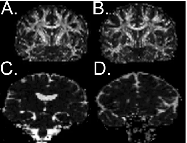
Article #1Figure 1: Example FA and MD maps ... 98
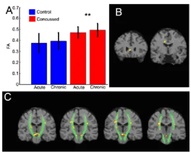
Figure 2: FA Alterations ... 99
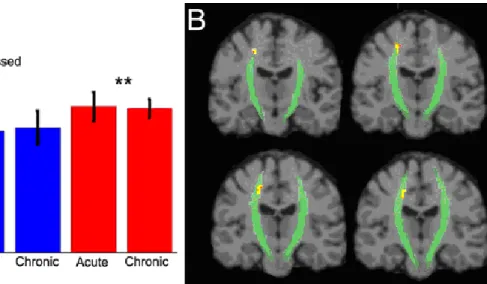
Figure 3: AD Alterations ... 100
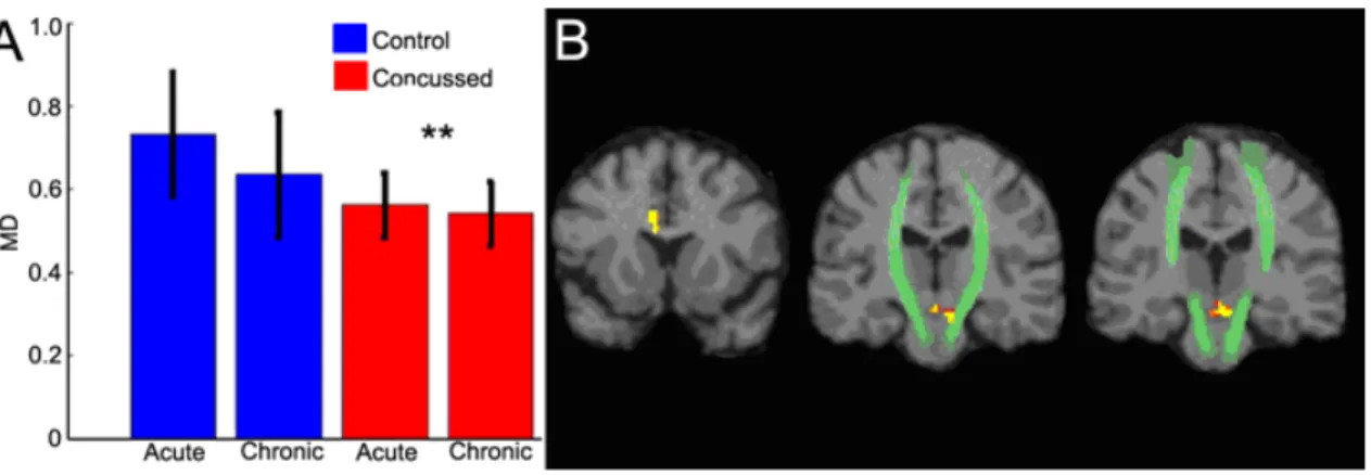
Figure 4: MD Alterations ... 101
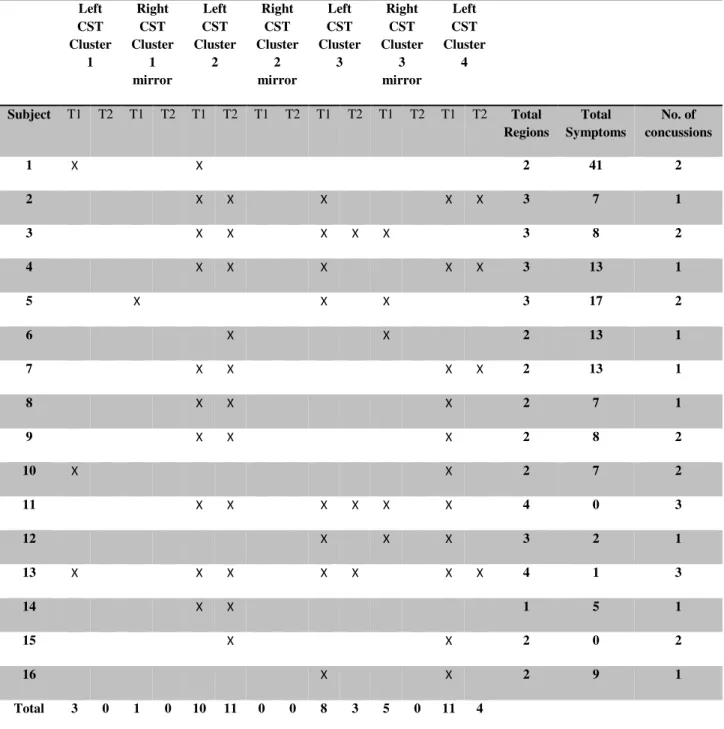
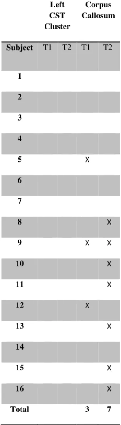
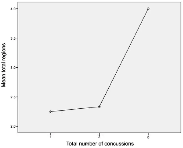
Figure 5: Altered regions and number of concussions... 104
Article #2 Figure 1: Regions of Interest ... 143
Figure 2: Typical spectrum from M1 ... 144
Figure 3: Averaged metabolite ratios in each of the 3 regions of interest ... 144
Figure 4: Cranial Symptom Cluster Correlations ... 145
Article #3 Figure 1: Typical spectra and regions of interest ... 181
Figure 2: Metabolic changes in the DLPFC ... 182
List of Symbols and Abbreviations
AD: axial diffusivityALS: amyotrophic lateral sclerosis
AMPA: 2-amino-3-5-methyl-3-oxo-1,2- oxazol-4-yl propanoic acid AQ: absolute quantitation
ARAS: ascending reticular activating system Asp-NAT: Aspartate N-acetyltransferase ATP: adenosine triphosphate
BOLD: blood oxygen level dependent BSRF: brainstem reticular formation Ca2+: calcium
CBF: cerebral blood flow CK: creatine kinase Cl-: chloride
CoA: coenzyme A Cr: creatine
CSP: cortical silent period CST: cortical spinal tract CT: computer tomography
CTBI: chronic traumatic brain injury CTE: chronic traumatic encephalopathy DAI: diffuse axonal injury
DTI: diffusion tensor imaging EAA: excitatory amino acids EEG: electroencephalography FA: fractional anisotropy
fMRI: functional magnetic resonance imaging GABA: γ-Aminobutyric acid
GCS: Glasgow coma scale Glu: glutamate
GOAT: Galveston orientation and amnesia test H+: Hydrogen
LoC: loss of consciousness M1: primary motor cortex MD: mean diffusivity Mg2+: Magnesium mI: myo-inositol
MR: magnetic resonance
MRI: magnetic resonance imaging MRS: magnetic resonance spectroscopy MTBI: mild traumatic brain injury NAA: N-acetyl aspartate
NAAG: N-Acetylaspartylglutamic acid NFL: National Football League
NMDA: N-methyl D-aspartate
NMDAr: N-methyl D-aspartate receptor NSE: neuron-specific enolase
PCS: post-concussion syndrome PCr: phosphocreatine
PET: positron emission tomography rCBF: regional cerebral blood flow RQ: relative quantitation
SPECT: single photon emission tomography TAI: traumatic axonal injury
TBI: traumatic brain injury
Dedication
For my mother, my brother and my sister- Thank you from the very marrow of my bones.
For my Uncle Bill- your resolve reminds me to never shrug.
And for my father- If you could see me now…
Acknowledgements
My first and primary thanks go to my supervisors Dr. Maryse Lassonde and Dr. Dave Ellemberg. From my arrival to Montréal, Maryse warmly welcomed me into her laboratory and every day since then has shown me great kindness and generosity. I cannot express the depths of my gratitude for her mentorship and support. Dave’s open door and willingness to spend time with me waxing philosophic over the finer points of research inspired me to think differently and I am profoundly grateful for his support.
I am also indebted to Sébastien Tremblay, my collaborator and part-time French teacher. Our lengthy conversations and many laughs helped make the long hours at the scanner pass by so quickly. Patrice Voss, my friend and co-conspirator, wanna see a movie this week? Thanks for all the distraction in the name of good mental health.
To my Montréal mom, Maria van der Knaap. Your frankness and openness, not to mention your overt Dutch-favoritism made walking the long hallway to my office a welcome diversion on the tougher days. I will truly miss you. Of course to Stéphane Denis, for your sense of humour, light-heartedness, and willingness to speak a little bit slower for une tête carré.
To all my friends and family (especially Blair, Joseph, Peter, Cam and Al) back home in Alberta- yeah, I just did that. To Cam and Cory—thanks for getting me started despite your better instincts. A final note to Luke Benry- Your ridiculous name allowed me to win every scholarship you deserved.
Theoretical Considerations
Historical perspectives, Definition, and Symptomatology
Concussions 1 have been described and documented throughout history, spanning across many cultures (see Echemendia, 2006; Shaw, 2002; Verjaal & van 'T Hooft, 1975 for a complete review). The term itself which means “to shake violently,” was not used to refer to brain injury until the 16th century. The earliest descriptions of concussive injuries date back as far as 3000 B.C.E. The Edwin Smith papyrus, 1700 B.C.E., catalogs 10 cases of traumatic brain injury, nine of which detail penetrating or fracturing head injuries; however, one case depicts a patient who received a blow to the head and suffered subsequent neurological change without visible injury (Verjaal & van 'T Hooft, 1975). The author of the ancient text explained that,
“It can hardly be devoted that the items of the diagnosis are indications of causes evidently concerning the shoulder, the hand, and the foot of the side affected. Apparently the condition of the patient is similar to that resulting from some other injury or disease as the surgeon is warned against false conclusions arising from this similarity” (as cited in Verjaal & van 'T Hooft, 1975).
Biblical texts recount an apparent concussion in the fabled story of David and Goliath. In Greek texts Hippocrates wrote of a “shaking or concussion of the brain produced by any cause [that] inevitably leaves the patient with an instantaneous loss of voice
1
Officially according to the CDC and WHO the term “concussion” has been replaced by the term mild traumatic brain injury (MTBI), but “concussion” is still used ubiquitously in the sports concussion literature. Consequently, the current thesis uses the term concussion where it is understood to be a specific kind of MTBI (Gerberding & Binder, 2003; Holm, Cassidy, Carroll, & Borg, 2005).
(unconsciousness)” dating back as far as 415 B.C.E. (Verjaal & van 'T Hooft, 1975). Some 1200 years later (832-929 C.E.), Rhazes, a Persian physician, observed that brain injury could occur without skull fracture or gross pathology, a fact that still confounds the general understanding of concussion.
The Renaissance brought several physicians who further described concussion including Lafranchi of Milan (1315) who taught that concussive blows jolt and agitate the brain, and Jacopo Berengario da Carpi (1470-1550) who penned one of the original pathophysiological theories of concussion claiming that small intracerebral hemorrhages caused the loss of consciousness (LoC) (Muller, 1975). A contemporary of da Carpi, the French physician Ambroise Paré (1510-1590), is credited with popularizing the term “concussion” when he wrote about the “commotion or shaking of the brain” in injured soldiers (Verjaal & van 'T Hooft, 1975). A century later Jean-Louis Petit (1674-1750) distinguished between the immediate post-concussion changes in arousal and sensibility and the slower, gradual decline associated with hemorrhage. He also acknowledged that there are “degrees” of concussion, and they are not simply all-or-none (as cited in Verjaal & van 'T Hooft, 1975). Alexis Littré performed the first documented post-mortem on a closed-head injury patient, a prisoner who was sentenced to be broken on the wheel. To escape the horrific torture to which he had been sentenced, the man rushed head-long into a wall (Feinsod, 2002). Upon examination, Littré found no visible pathology or injury, consistent with Paré’s original assertion that concussions create a functional rather than a structural deficit such as a lesion (Muller, 1975; Shaw, 2002).
Reconciling the neural effects of concussion with the apparent absence of visible damage was a point of emphasis in the 19th century. The French military surgeon Jean-Pierre Gama proffered in 1835 that the “delicate” nature of neural tissue left it particularly vulnerable to stretching and breaking when the head is injured (Strich, 1970), a phenomenon now understood to be diffuse axonal injury, though the term traumatic
axonal injury is now more favored (see section on Biomechanics). The autonomic
nervous system symptom cluster of concussion including headache, nausea, and dizziness was described by Victor Von Bruns (1811-1883) and Ernst Von Bergmann (1836-1907) (Muller, 1975). The 20th century was dominated by the introduction of experimental animal models of concussion, understanding the biomechanical mechanisms that precipitate concussion, and pathophysiology theories that will be reviewed later.
Defining concussion is complex and fraught with controversy. There remains much contention over which symptoms are necessary for the diagnosis and the relative weight of each as they pertain to severity. Benjamin Bell (1749-1806) defined concussion in 1787 as
“every affection of the head attended with stupefaction, when it appears as the immediate consequence of external violence, and when no mark or injury is discovered, is in general supposed to proceed from commotion or concussion of the brain, by which is meant such a derangement of this organ as obstructs its usual functions to render it capable of having its real nature ascertained by dissection” (as cited in Peerless & Rewcastle, 1967).
This definition, though pioneering, is inadequate. It does key in on the presence of a confused state (stupefaction) and that there is an injury for which there is no discernable
outward sign, but misses out on several other signs and symptoms. Granted, it was an important step in even acknowledging that there was indeed an injury, but it had little value otherwise. Furthermore, this and other contemporary definitions of concussion fail to account for the long term consequences and the effects of multiple blows to the head. While historically LoC was the defining characteristic (Muller, 1975; Ward, 1964) and is still considered by some to be indicative of severity and integral to diagnosis (Ropper & Gorson, 2007), all of the most recent consensus statements overwhelmingly state that it is not necessary to define or diagnose concussion (Aubry et al., 2002; Cantu, 2001; McCrory et al., 2005; McCrory et al., 2009; Webbe & Barth, 2003), nor is it associated with injury severity (Henry & Lassonde, 2009; McCrory, et al., 2009). The transient nature of the LoC associated with concussion also led to speculation that there was no structural damage because concussions were purely physiological events and therefore without physical consequence (Denny-Brown & Russell, 1940). Holbourn contested the notion that concussions were not accompanied by physical damage though he lacked the tools to prove his assertion (1943, 1945). Peerless and Rewcastle (1967) further specified the nature of these physical injuries as being shear strain to white matter or traumatic axonal injury (see Biomechanics) (Gentry, 1994). While gross imaging techniques rarely reveal structural damage to the concussed brain, it is now largely understood that concussions do result in at least transient structural damage (Gentry, 1994; Johnston, et al., 2001) and that physiological and morphological changes may continue for days or weeks (Giza & Hovda, 2001). Considering all these issues, concussion will be defined in the current thesis as a closed head injury due to a direct blow to the head or shaking of
the head from an impulsive force resulting in a transient alteration in mental status and brain processes that may include loss of consciousness, memory dysfunction (retrograde or anterograde amnesia), impairment of reflex activity, and/or disorientation. Although for the majority of cases most symptoms resolve within 7-10 days, physical symptoms such as headache, cognitive symptoms including memory dysfunction, and neurological
disturbances like sleep disturbance may persist indefinitely (Binder, 1986; Echemendia,
2006; Quality Standards Subcommittee & Neurology, 1997) and may be worsened when
multiple impacts are sustained (De Beaumont, Lassonde, et al., 2007; Guskiewicz et al.,
2003; Theriault, et al., 2010).
Concussions can vary in severity across a number of different factors including the force-mass combination, force vector, and individual differences. Because of this variability, categorizing all concussions as a unitary phenomenon is impractical and incorrect. Concussions had been categorized as either simple or complex based on the amount of time it takes a patient to recover (McCrory, et al., 2005): Simple (type 1) concussions resolve without complication 7-10 days after the injury whereas Complex concussions (type 2) include all cases where the patient suffers persistent symptoms, prolonged LoC, or prolonged cognitive impairment and are typically incurred by people who have had multiple concussions, or have lowered impact tolerance (McCrory, et al., 2005). However, such a distinction is extremely limited both clinically and experimentally as it is a retrospective classification with no bearing on early management, only altering later treatment course. The most recent consensus statement has nixed this distinction with the retention of the basic concept that most concussions
resolve within 7-10 days in most people, somewhat longer in children and adolescents, and even more protracted in a minority of people (McCrory, et al., 2009). Many grading systems have been employed (see Cantu, 2001 for a thorough review), but the practice of grading concussions has been abandoned for the most part. Instead, the severity of the injury is often determined after all of the symptoms have resolved. Specifically, the severity relates to the nature, burden, and duration of symptoms (Cantu, 2001; McCrory, et al., 2009).
Persons suffering from a concussion often appear dazed, forgetful, display inappropriate affect, and report experiencing headache, nausea, double vision, increased emotionality, changes in sleep patterns, and express increased sensitivity to light and noise (McCrory, et al., 2009; Quality Standards Subcommittee & Neurology, 1997). Further, enduring problems typical of more severe concussions include headaches, irritability, noise and light sensitivity, fatigue, anxiety, and impaired concentration and memory dysfunction. The presence of one or more of the above mentioned symptoms after the occurrence of a head injury is termed Post Concussion Syndrome (PCS). These symptoms typically resolve within 30 days, but persist for some patients (Eyres, Carey, Gilworth, Neumann, & Tennant, 2005; King, 1997; King, 1996) in whom the remaining symptom cluster including physical, cognitive, and behavioral symptoms is referred to as persistent PCS which can last well past 30 days from months to years after the injury incident suggesting at least persistent functional alterations and likely damage (Ryan & Warden, 2003).
Epidemiology
The prevailing attitude in sports culture had been rather dismissive of concussions, where they once were considered as a minor injury without consequence, even comical in instances where a player struggled to regain his balance or could not find his way back to the bench (Echlin, 2010; Zillmer & Spiers, 2001). In the recent past, sportscasters humorously spoke of how a struck player was “seeing stars,” had his “bell rung,” or even how on particularly devastating hits may be unable to return to the bench or sideline (Echlin, 2010; Zillmer & Spiers, 2001). This has continued to a lesser degree as hard hits are still rewarded and reinforced within sports media. Athletes compound the indifference with bravado and an understandable, though misguided desire to continue playing. The seriousness of concussion is slowly coming to the forefront in the athletic community and the public at large as the documented short and long term symptoms/effects continue to be investigated due to the forced retirement of several high profile athletes including former NFL quarterbacks Steve Young and Troy Aikman and NHL star Eric Lindros to name a few. Of the 51 cases of neuropathologically confirmed chronic TBI (CTBI) which can only be done by autopsy, 90% have occurred in athletes. The gross pathology of CTBI is obviously not uniform across all cases. However, common elements include 1) reduced brain weight particularly in the frontal and temporal lobes that typically extends to other structures such as the hippocampus, entorhinal cortex and amygdala as severity increases, 2) enlarged lateral and third ventricles, 3) volume loss in the corpus callosum, 4) cavum septum pellucidum in almost 70% of cases and with fenestrations in nearly 50% of those cases, and 5) scarring and
neuronal loss of the cerebellar tonsils (Corsellis, Bruton, & Freeman-Browne, 1973). Other common gross features include pallor of the substantia nigra and locus coeruleus, atrophy of the olfactory bulbs, thalamus, mammillary bodies, brainstem, and cerebellum (Corsellis, et al., 1973). More recent examination of five case studies revealed very similar gross anatomical findings (McKee et al., 2009).
Given the controversy in defining concussions as well as the largely uninformed social attitudes toward them, diagnosing concussions is an even more imprecise and daunting task. It has been conservatively estimated that over 2 million sports related concussions occur annually in the United States, (Reddy, Collins, & Gioia, 2008). These figures are only estimations as they do not account for the large number of unreported and undiagnosed incidences, leaving a potentially large gap between the numbers of athletes who report having sustained a concussive injury and those who actually sustained a concussive injury. Indeed, recent work points to a lack of knowledge and education about concussion in coaches, trainers and athletes as a crucial factor in diagnosing and reporting such injuries (Cusimano, 2009; Echlin et al., 2010). Consequently, identifying a concussion is not simply a matter of refining diagnostic techniques and heightening clinical vigilance. It is also incumbent upon the athletes themselves to recognize and report post-concussion symptoms (Delaney, Lacroix, Leclerc, & Johnston, 2002; Echlin, 2010). A three-year prospective study among intercollegiateathletes reported that of all sports related injuries, 6.2% were concussive (Covassin, Swanik, & Sachs, 2003) with rates differing across sports where boxing leads the way by a significant amount (Echemendia & Julian, 2001). Further, concussion incidence increases from practices to
games in all sports (Echemendia, 2006). A systematic literature review spanning from 1985 to 2000 found the highest incidence of concussion for high school, college, and amateur athletes in hockey and rugby, while soccer had the lowest (Koh, Cassidy, & Watkinson, 2003). Annual football concussion rates including recreational, high school, collegiate, and professional levels indicate that players have an annual concussion incidence estimated to be 4–20% (Bailes & Cantu, 2001) and that they are three times more likely to sustain a second concussion in the same season (Guskiewicz, Weaver, Padua, & Garrett, 2000). However, Delaney et al. (2002) reported that 70.4% of football players and 62.7% of soccer players reported having experienced concussion symptoms during the previous year, but only 23.4% of concussed football players and 19.8% of concussed soccer players realized they had suffered a concussion.
The concussion rate is even more staggering when multiple concussions (84.6% for football players and 81.7% for soccer players) are considered. At the mild end of the spectrum, the harmful consequences of multiple concussions were originally introduced by Quigley in 1945 who anecdotally determined with his infamous three-strike rule that an athlete who had experienced three concussions in a season was to be held out for the remainder of that season. Multiple concussions were referenced just seven years later without any more rigorous research on the matter (Thorndike, 1952). Some two decades later, Gronwall described the cumulative effects of mild closed head injuries in a series of papers covering a range of neuropsychological domains including memory, information processing, perception, and motor skills (Gronwall & Wrightson, 1974, 1975, 1981; Gronwall, 1977). Though important in documenting the cumulative effects of multiple
mild head injuries, none of these studies were done with athletes where the nature of the injury and the resultant management can differ from a non-sports related injury, namely that athletes may tend to “fake good” in order to return to play before the signs and symptoms have resolved (Echemendia, 2006). At the other end of the spectrum, severe manifestations of the effects of repeated blows to the head in boxers were first documented and dubbed “punch drunk syndrome” by pathologist Harrison Martland (1928), and later termed Dementia Pugilistica due to its infamy amongst boxers (Millspaugh, 1937). The term chronic traumatic brain injury is now used to describe the condition which first begins as cognitive deteriorations, followed by degrading executive functions, motor impairments, and dementia in the bleakest of cases.
Elucidating the temporal effects of concussion is also important as this informs management and return to play decisions. The acute post-injury phase, approximately three months post-concussion, has been the most heavily documented in terms of the neuropsychological effects of concussion (Barth et al., 1989; Boutin, Lassonde, Robert, Vanassing, & Ellemberg, 2008; Chen, et al., 2007; Collins et al., 2003; Collins et al., 1999; Fazio, et al., 2007; Iverson, Brooks, Collins, & Lovell, 2006). The neuropsychological assessment of athletes outside of the acute post-injury phase has revealed mixed results. Iverson, Brooks, Lovell, and Collins (2006) found that high school athletes with a history of one or two prior concussions did not demonstrate any neuropsychological deficits six months after injury relative to athletes who had not suffered any concussions. Similarly, a study of collegiate athletes at least six months removed from their last injury did not demonstrate any detectable enduring
neuropsychological changes (Bruce & Echemendia, 2009). Understanding the similarities and differences between the acute and chronic post injury phases is thus crucial to determining how the brain changes, how it recovers, and/or how it remains altered due to a concussion.
The aggregate of the data detailing the effects of repeated blows to the head is beginning to paint a very complex, yet consistent picture. What seems apparent in understanding the neuropsychological sequelae of concussions, particularly after repeated injuries, is that the resultant consequences are not binary in nature. That is to say, they are not all or nothing, but rather they progress in a semi-stepwise manner. Indeed, the evidence suggests that there is a continuum of potential outcomes ranging from no consequences to a second or third injury (Iverson, Brooks, Lovell, et al., 2006; McCrory, 2001) to subclinical eletrophysiological changes (De Beaumont, Lassonde, et al., 2007; Gaetz, et al., 2000; Theriault, et al., 2009; Theriault, et al., 2010) to more subtle cognitive and motor changes (De Beaumont, et al., 2009), progressing toward mood and cognitive symptoms (Guskiewicz et al., 2005; Guskiewicz, Marshall, et al., 2007) and finally to severe injuries such as CTBI (Corsellis, et al., 1973; Heilbronner et al., 2009; Jordan, 1990; Roberts, Allsop, & Bruton, 1990) that have accompanying neurohistopathological correlates (Corsellis, et al., 1973; Geddes, Vowles, Nicoll, & Revesz, 1999; Geddes, Vowles, Robinson, & Sutcliffe, 1996; Hof et al., 1992; McKee, et al., 2009). Though these changes can take a career’s worth to accrue, rare though severe acute cases can result in immediate disability or even death in the uncommon second-impact syndrome (Bey & Ostick, 2009; Cantu, 1995; Cantu, 1996). Our clinical and scientific
understanding of the effects of sustaining impacts to the head has increased dramatically, but it is clear that there are many more questions than answers at the present time.
Pathophysiology theories
Currently there is no unified pathophysiology theory to explain the underlying mechanisms of concussion (McCrory, et al., 2005). Historically researchers focused theories around explaining loss of consciousness, usually considered to be exclusively a brainstem phenomenon, and memory dysfunction widely understood to be a cortical phenomenon. Though many theories have been put forward, current research in the field has generally shifted its attention from finding a mechanism toward describing and treating concussion. The five most historically prominent theories include the vascular theory, the centripetal theory, the reticular theory, the pontine cholinergic theory, and the convulsive theory.
The vascular theory, the oldest pathophysiology theory, is generally considered to be outdated and insufficient to explain concussion. Its basic assumption is that LoC and other transient neural dysfunctions are caused by brief episodes of cerebral ischemia (Nilsson & Ponten, 1977; Verjaal & van 'T Hooft, 1975). The cause of the ischemic events is attributed to a number of pathophysiologies including vasospasm, vasoparalysis, expulsion of blood from capillaries, reflex stimulation, and most prominently an obstruction of blood flow due to compression of the brain (Echemendia, 2006; Shaw, 2002). However, membrane potential and neuronal functioning are not lost immediately after brief ischemic events (hemorrhages, strokes, etc) (Lipton, 1999) as proposed by the vascular theory (Shaw, 2002). Theoretically, the vascular theory is logical in that brain
functions are disturbed after ischemic events, but the effects of such injuries are not as immediate and abrupt as they are in concussion. Experimental proofs against the vascular theory using a heartless frog model demonstrated that vascular involvement is not necessary for a concussion to occur (Denny-Brown & Russell, 1940); furthermore, the predicted energy production deficiency is not necessary to observe a concussed state (Nilsson & Ponten, 1977).
The centripetal theory was championed by Ommaya and Gennarelli (1974). According to this theory, concussions occur across a graded set of clinical syndromes caused by mechanical stresses and strains that inevitably affect the structure and function of the brain beginning at the surface (cortex) in less severe injuries extending to the diencephalic-mesencephalic juncture for the most severe of injuries (Ommaya & Gennarelli, 1974) where rotational force vectors (see Biomechanics) were shown to be responsible for the majority of concussions. The heuristic value of this theory, especially as it pertains to biomechanics is still valuable, though its bias toward LoC as being associated with more severe injuries proves to be its undoing as there are many clinical cases where patients sustained LoC without the predicted cognitive after-effects (Echemendia, 2006).
The reticular theory has easily been given the most precedence in understanding concussion as it explains a number of the symptoms associated with concussion. Nuclei within the brainstem are associated with the control of reflex activity and the maintenance of autonomic activity. Injury or disruption of these nuclei could account for
LoC, changes in blood pressure, slowing heart rate, altered cerebral blood flow, and the disruption of various reflexes, loss of equilibrium and muscle tone, respiratory arrest, nausea/vomiting, and pupil dilation (Shaw, 2002). Theoretically, the basic principle posits that a concussive blow temporarily disrupts (paralyzes/depresses) polysynaptic pathways within the brainstem reticular formation (BSRF)/ ascending reticular activating system (ARAS). Recovery within the BSRF stimulates the ARAS to become operational again thus allowing the cortex to be reactivated again via thalamocortical pathways prompting a quick recovery of consciousness. The source of brainstem injury is postulated to be the flexion of brainstem structures during the peak rotational acceleration/deceleration at the cervicomedullary junction causing stretching and possible shearing leading to massive functional failures (Shaw, 2002). Experimental work in rats (Povlishock, Becker, Cheng, & Vaughan, 1983), cats (Povlishock, Becker, Miller, Jenkins, & Dietrich, 1979; Povlishock, Becker, Sullivan, & Miller, 1978), and monkeys (Gurdjian, 1972; Jane, Steward, & Gennarelli, 1985) supports the reticular theory showing traumatic chromatolytic changes (disintegration of the granule Nissl bodies in a nerve cell body, usually occurring after exhaustion of the cell or damage to its peripheral process) to neurons in the BRSF. Clinical observations demonstrating injury to the brainstem further support reticular involvement (Oppenheimer, 1968).
The reticular theory has endured in large part due to the ambiguous nature of the evidence used to support it. While evoked potentials have been used to champion the theory, these results neither prove nor disprove its basic tenets. Similarly, the reticular theory predicts that when a concussive injury incapacitates the ARAS, the subsequent
cortical EEG recording will be of low frequency and high voltage (Shaw, 2002). In actuality, immediate experimental post-concussion EEG patterns fall into two categories: 1) an almost complete attenuation of the EEG or 2) a brief period of excitation marked by both high frequency and high amplitude spiking (epileptiform). While these two patterns are in conflict with each other, neither matches the prediction of depressed reticular activity posited by this theory theory (Hayes, Katayama, Young, & Dunbar, 1988; Shaw, 2002). The neuropathological data is the most convincing evidence in favor of the reticular theory. However, this data is limited in its applicability to a general pathophysiological understanding of concussion because experimentally it is specific to certain methodologies and selective in terms of the anecdotal accounts citing only those cases where the impact was to the back of the head (Shaw, 2002).
Disruption of BSRF activity explains muscle flaccidity and reflex suppression following cerebral concussion, but it cannot explain the convulsive movements occasionally seen experimentally in animal studies (Govons, Govons, VanHuss, & Heusner, 1972; Nilsson, Ponten, & Voigt, 1977; Nilsson et al., 1994) and anecdotally reported clinical accounts (Chadwick, 1997a, 1997b; McCrory & Berkovic, 1998, 2000; Perron, Brady, & Huff, 2001; Ropper & Gorson, 2007). Even Andy (1989) who reported a concussive injury to the brainstem with subsequent convulsions implicated reticular formation discharges, in stark contrast to the depressed activity necessary for the reticular theory to hold water. Perhaps most damning to the reticular theory is the absence of an understanding of how the reticular formation would come to be depressed by a concussive injury leading to questions of whether LoC would necessarily occur even if
the BSRF was disrupted (Shaw, 2002). Also missing is a comprehensive account of how reticular functioning is depressed by a concussive insult or whether LoC is the necessary consequence of disrupted BSRF activity (Shaw, 2002). Furthermore, this theory fails to account for traumatic memory loss, one of the most frequently observed symptoms in concussion that is also more predictive of severity than LoC (Shaw, 2002).
The pontine-cholinergic theory is a modern neurochemical variant of the reticular theory. Where the reticular theory posits that the brain stem is deactivated because of the injury, the pontine-cholinergic theory contends that the brain insult activates an inhibitory/depressive system (Barr, 2005). The concussive injury sets in motion a cascade of events that excite an inhibitory cholinergic system in the dorsal pontine tegmentum, again supposing a pre-eminent role for the brainstem. The inhibition of the cholinergic system then reduces consciousness to variable levels, depending on the level of excitation of the inhibitory system. While this theory accounts for altered consciousness and LoC, the neurochemistry does not add up. Studies using an acetylcholine blocker to circumvent the inhibition of the cholinergic system failed to prevent or reduce concussive symptoms (Lyeth et al., 1988).
The symptoms of concussion bear remarkable similarity to those of a generalized seizure (Shaw, 2002). Earl Walker and colleagues (1944), who originally proposed the convulsive theory of concussion based their theory in part on experimental observations of tonic-clonic movements in concussed animals (Walker, et al., 1944). From these observations they concluded that the shaking or vibration of the brain resulted in a short
lasting widespread neuronal discharge followed by a long lasting period of neuronal exhaustion and/or inhibition that manifests itself in muscle relaxation, behavioral stupor, and depressed cortical rhythm (Walker, et al., 1944). Both conditions can be marked by a near instantaneous, though transient (seconds or minutes) LoC and an equally sudden return to consciousness commonly accompanied by a period of depressed and disoriented function. Reflex suppression (stretch, withdrawal, and corneal) is also common to both concussion and seizure. Physiological changes including increased heart rate, blood pressure, and transitory respiratory arrest are also common to both (Shaw, 2002). Parallel disturbances in autonomic function include giddiness and nausea. Post-incident headaches are commonly reported as are both anterograde and retrograde memory dysfunction. Residual symptoms of seizure and concussion share similarities as well including: irritability, depression, sleep dysfunction, anxiety, fatigue, persistent headaches, restlessness, dizziness, emotional lability, and slowed cognitive processing (Shaw, 2002).
The convulsive theory is well supported by electrophysiological, neurochemical, and ultrastructural data, but it does not readily account for LoC. It is best explained by cortico-cortical and cortico-reticular theories where it is posited that hypersynchronous cortical epileptiform activity blocks sensory signals leaving the cerebral cortex functionally detached which renders the patient unresponsive and insensible. This account, though logical is only theoretical, thus leaving a significant knowledge gap in support of this theory. The most oft-cited criticism of the convulsive theory is the low frequency of tonic-clonic seizures in concussion, though many physicians do
acknowledge their rarefied occurrence resulting from concussion (Shaw, 2002). While the coupling of convulsions and concussions is rare, the primary issue is of mechanism, not manifestation. A convulsion is the shaking of the body due to rapid tonic-clonic muscle movements. A seizure on the other hand is the result of abnormal electrical activity in the brain that can result in a wide range of behavioral manifestations, including but not limited to convulsions. Indeed, there is an entire classification of seizures that are nonconvulsive (i.e. absence seizures), yet these seizures are equivalent to convulsive seizures in terms of what is happening at a neuronal level (Engel & Pedley, 1998). Therefore, the label “convulsive” is misleading as convulsive movements, if they occur, are a symptom not a mechanism. Rather, hyperexcitability and the ensuing excitotoxicity are the common mechanisms and should be reflected in the nomenclature of the pathophysiological theory. This is more than a trivial semantic argument as the basis chosen for comparison is reflected in the understanding of concussion which subsequently informs treatment and management.
Biomechanics
Despite the seeming lack of physical damage in concussive injuries, it is important to recognize that concussions are the result of the brain moving within the cranial vault, and that movement has physical consequences on the brain. Without the benefit of modern imaging technology, Brodie (1828) eloquently contended that, “If we consider that the ultimate structure of the brain is on so minute a scale that our senses are incapable of detecting it, it is evident that there may be changes and alterations of structure which our senses are incapable of detecting also.” At about the same time, the
aforementioned French military surgeon J-P Gama was conducting what are widely accepted to be the first biomechanical investigations of concussion. His model was composed of a gelatinous substance meant to simulate the brain suspended by thin wires in a long-neck glass flask. By striking the walls of the flask he was able to document oscillatory and vibratory movements of the wires. Though rudimentary by current standards, this was the first effort to document the biomechanics of concussive injuries in general, and of coup-contrecoup injuries in particular (Gama, 1835). Our understanding of the biomechanics of concussion is now more sophisticated, understanding that they are predicated on the physiological composition of the brain and its position within the skull. The physical properties of nervous tissue make the brain relatively incompressible, but readily distortable (Barr, 2005; Holbourn, 1943, 1945; Kandel, Schwartz, & Jessell, 2000). Anatomically, the brain is suspended inside the skull by cerebrospinal fluid in the subarachnoid space allowing the brain to move within the skull. The brain’s response to sudden changes in the velocity of the head is to vacillate, slide, or rotate within the skull (Holbourn, 1945; Shaw, 2002). The cerebrospinal fluid normally protects the brain from injury by cushioning it against the skull; however, when the velocity of the brain lags behind that of the skull (acceleration) or continues to move after the skull has stopped (deceleration), the brain comes into violent contact with the bones of the skull, particularly at the frontotemporal suture (Cantu, 1992). Compression of the skull without fracture may lead to a focal injury accompanied by an increase in intracranial volume as well as a probable increase in intracranial pressure (Gurdjian, 1972). The resulting pressure wave passes through the relatively closed system of the brain, meninges,
cerebrospinal fluid system, and vasculature causing general and local tissue deformation, thereby compromising the structural integrity of the neurons (Gurdjian, 1972).
Most concussions result from either a direct blow to the head or from an impulse insult to the head. Despite the absence of direct contact to the head, impulse injuries can be just as severe as impact injuries (Barth, Freeman, Broshek, & Varney, 2001). Regardless of the nature of the injury (direct or indirect), concussive effects are exerted via inertial loading (acceleration or deceleration) (Shaw, 2002). Impact injuries occur when an object of sufficient mass strikes the skull such that kinetic energy imparted to the skull is transferred to the brain (Echemendia, 2006). These types of injuries typically result in focal damage where the affected brain area is directly beneath the point on the skull where the impact occurred- a coup injury. When the acceleration of the brain is abruptly halted by the skull an injury opposite to the point of impact can occur: a
contre-coup injury (Bailes & Cantu, 2001; Cantu, 1992; Gama, 1835). Impulse injuries result in abrupt inertia changes of the head without direct impact.
Concussions occur along one of two force vectors: Linear (translational) or rotational (angular). Linearly applied force is simply inertial force applied in a straight line. Typically linear forces result in a coup injury leading to the compression and possible stretching of neural tissue (Echemendia, 2006). However, because not all combinations of force and mass produce the same effects, injury outcomes vary. For example, a small object moving at high velocity, like a bullet, is more likely to penetrate the skull and create local damage along its trajectory. Similarly, larger objects moving at
slower speeds may crush the skull, but avoid any acceleration/deceleration forces on the brain thereby not resulting in a concussion. Concussions are more likely to result when there is an intermediary combination of mass and force, which is characteristic of the vast majority of head injuries (Holm, et al., 2005).
Rotational injuries result from an inertial force that produces an angular acceleration of the head around the midline axis (Echemendia, 2006; Holbourn, 1945). Because of the interconnectivity between the bones, muscles, and connective tissue of the head, neck, and upper torso rotational injuries are more likely to result in a concussion when the force is directed laterally, rotating around the axis created around the midline of the neck up through the top of the skull (Echemendia, 2006) around the fronto-parietal junction (Bayly et al., 2005). These injuries are more complex in terms of the effects on neural tissue including shearing/tearing or stretching. Injuries resulting from angular acceleration are most detectable at gray-white matter junctures beginning at the cortical surface and less detectable descending toward the brain stem (Echemendia, 2006). Rotational acceleration is much more severe than linear acceleration, in large part because of the nature of brain tissue (Holbourn, 1945). When the skull is moved in a straight line the brain’s incompressible nature allows it to move as a whole thereby minimizing distortion. That is to say, the effects of linear force are negligible when compared with rotational forces (Holbourn, 1945). Conversely, when the skull is rotationally accelerated the brain’s lack of rigidity prevents it from moving as a whole thus distorting the brain considerably (Holbourn, 1945). However, a study investigating the impact of linear and rotational forces on symptom severity failed to find a clear
relationship (Guskiewicz, Mihalik, et al., 2007). That is to say, the role of linear and rotational force vectors may be clear in terms of brain deformation, but the role that force vectors in clinical outcome remains unclear at present.
Current biomechanical studies of brain deformation have been done using similar gel-filled skull models (Margulies, Thibault, & Gennarelli, 1990; Meaney et al., 1995) which are limited because they cannot account for the effects of heterogeneity, anisotropy, vasculature, meninges, and cerebrospinal fluid on brain deformation. High-speed x-ray on cadavers are also used (Hardy et al., 2001), but cannot account for differences in live subjects as well as spatial limitations. MR techniques in humans (Bayly, et al., 2005; Reese, Feinberg, Dou, & Wedeen, 2002; Sabet, Christoforou, Zatlin, Genin, & Bayly, 2008) on the other hand, provide a rich source of information pertaining to strain fields accompanied by strong spatial and temporal resolution, but are limited by the allowable forces that can be delivered to human subjects. MR studies are consistent in reporting compression in frontal regions and stretching in more posterior regions before subsequent concussion in occipital areas. Furthermore, radial-circumferential shear strains are observed, in kind with those observed in gel models where angular force vectors are applied. Unlike gel models, the MR studies appropriately account for the inherent heterogeneity, natural brain divisions (central fissure and central sulcus), and the brain’s suspension composed of the dura mater, falx cerebri, and tentorium membranes. What MR studies cannot account for due to technical limitations as well as ethical guidelines are the shear strains and subsequent axonal injuries.
Understanding the movement of the brain, though important, does not fully describe the nature of concussion. It is equally important to understand the effects of said movement, namely shear strains. Shear strain injuries are deformation of the axon perpendicular to the force vector and are most probable when the skull and brain are accelerated around the midline axis by an angular force (Ommaya & Gennarelli, 1974) though the pathophysiology that leads to traumatic axonal injury (TAI) in TBI is relatively unknown (Ducreux et al., 2005). The current view is that corpus callosum injury is caused by shear-strain rotational forces and/or impact against the free margin of the falx (partition-like fold of the dura that extends into the medial-longitudinal fissure). Ensuing tissue injury is marked by axonal stretching, disruption, and the eventual separation of nerve fibers (Huisman et al., 2004). Animal research (Liu et al., 2006) further suggests that TAI has downstream consequences that affect the integrity of the myelin sheath, the result of which would be significantly reduced neural conduction speed (Barr, 2005). TAI is more commonly seen at gray-white matter junctions (Ducreux, Huynh, et al., 2005; Ducreux, Nasser, Lacroix, Adams, & Lasjaunias, 2005; Huisman, et al., 2004; Le et al., 2005; Lee, Choi, & Chun, 2006; Okumura et al., 2005; Song et al., 2002) because the two tissue types differ in their respective rigidity (Holbourn, 1945) due to the myelin sheaths that surround white matter axons. While the myelin increases the speed of neural conduction in white matter, it also makes the tissue more fibrous (and therefore more rigid) perhaps explaining why TAI is more common in white matter.
Neuroimaging
Investigating how biomechanical forces impact the gross anatomy of the concussed brain has proven difficult. Computed Tomography (CT) and Magnetic Resonance Imaging (MRI) can provide information about anatomical and gross structural changes following a concussion. CT is an x-ray dependent technique that constructs a 3-D image of the brain. Clinically, CT scans can reveal brain lesions, contusions, fractures, and intracranial hemorrhaging. They are widely used in the management of closed head injuries, particularly within the first 24 hours post-injury, though it is generally only specified in cases for which there has been LOC (Livingston et al., 2000; Stein & Ross, 1992; Warren & Bailes, 1998) or persistent symptomatology (Rimal, Thapa, Munasinghe, & Errington, 2007).
Research on the utility of CT in sports concussion management is limited as the only studies available focus on boxers. Though there are instances where lesions are detected, the majority of concussed athletes have negative CT findings with weak predictable outcome validity (Jordan et al., 1992; Jordan & Zimmerman, 1988; Ross, Casson, Siegel, & Cole, 1987). The lack of consistent CT findings is quite common in the mTBI literature as well (Gentry, Godersky, Thompson, & Dunn, 1988; Groswasser, Reider-Groswasser, Soroker, & Machtey, 1987; Han et al., 1984; Newton et al., 1992; Zimmerman, Bilaniuk, Hackney, Goldberg, & Grossman, 1986). A large study investigating the prevalence of lesions in mTBI found that approximately only 16% of scans are positive (Iverson, Lovell, Smith, & Franzen, 2000), a rate that is considered quite high based on current clinical findings (Johnston, et al., 2001); moreover, patients
with positive scans share some common characteristics, chief among them being the presence of intracranial abnormalities, LOC, skull fractures, and lower Glasgow Coma Scale (GCS) and Galveston Orientation and Amnesia Test (GOAT) scores. The rates of positive CT findings is even further confounded when one considers that for some studies a GCS score of 13-15 is considered a mTBI (i.e. Iverson, et al., 2000), while for others, a score of only 15 (i.e. Audenaert et al., 2003) is considered a mTBI. Though they are still used in clinical and emergency settings because of their relative cost effectiveness and wide availability (Toga & Mazziotta, 2002), CT scans have fallen out of favour, particularly in research because of their limited ability to detect finite lesions and contusions, especially relative to MRI (Jordan & Zimmerman, 1990; Newton, et al., 1992; Snow, Zimmerman, Gandy, & Deck, 1986).
MRI is a more sensitive technique to investigate anatomical changes given its higher resolution, its capacity to image different planes, and because it provides better distinction between tissue types: gray matter, white matter, and cerebrospinal fluid. Typical injuries detected by MRI resulting from sport concussion include small cortical contusions or subdural hematomas and small white matter hemorrhages (Toga & Mazziotta, 2002) that are generally interpreted to be reflective of TAI (Bazarian, Blyth, & Cimpello, 2006). Though more effective at detecting abnormalities than CT (Jordan and Zimmerman, 1990; Newton et al., 1992), MRI has proven itself inconsistent with only moderate predictive validity in sports concussion (Jordan & Zimmerman, 1990; Newton, et al., 1992) and mTBI in general (Barr, 2005; Bazarian, et al., 2006; Toga & Mazziotta, 2002). Though its resolution is vastly improved over CT, MRI is still not able to detect
lesions in the majority of concussed athletes (Jordan, et al., 1992; Jordan & Zimmerman, 1990), limiting its clinical applicability. Moreover, CT and MRI are unable to provide information about functional alterations in brain function resulting from sports concussion.
Diffusion tensor imaging (DTI) provides information about brain microstructure by quantifying isotropic (typical of gray matter) and anisotropic (typical of white matter) water diffusion. DTI measurements are based on the fact that the network of fibers within the brain is composed of a distinct microstructure that inherently constrains the flow of water molecules. Diffusion tensors numerically model the direction of fastest diffusion in a pattern aligned with fiber orientation. DTI’s main utility in neurology is detecting the presence of axonal injury (Toga & Mazziotta, 2002). There is increasing evidence to suggest that TAI is present in TBI and that the extent of the damage is related to the severity of the injury as defined by initial GCS score (Huisman, et al., 2004). Similarly, Kraus et al. (2007) investigated TBI across the severity spectrum using DTI and found the degree of TAI to be related to the severity of the injury with severe TBI patients exhibiting the greatest extent of damage.
Several case studies using DTI on TBI patients from non-athletic populations have consistently noted reduced diffusion in the brain, especially around the splenium (thickened border at the posterior end of the corpus callosum) (Huisman, et al., 2004; Le, et al., 2005; Lee, et al., 2006). The observed axonal shearing in people with concussion has functional consequences. Lee and colleagues (2006) noted that brain areas affected
by axonal injury corresponded to the types of dysfunctions that patients exhibited. Similarly, Huisman and colleagues (2004) compared scores from the GCS with the observed amount of axonal shearing using DTI and found a positive correlation. One of the few studies to focus on mTBI found changes in white matter in the corpus callosum up to five years post injury (Inglese et al., 2005). While it is important to note the persistence of injury after such a long period of time, there are very few studies documenting TAI in the acute post injury phase in mTBI; however, studies suggest that damage can be detected within the first week of injury (Miles et al., 2008; Wilde et al., 2008).
There are several ways to analyze DTI data depending on the research or clinical question. Fractional anisotropy (FA) and mean diffusivity (MD) are the most often used measures of diffusivity (Toga & Mazziotta, 2002). FA is derived from three eigenvalues, the longest (λ1), middle (λ2), and shortest (λ3) distances around a given tensor which
correspond to linear (cigar-shaped) and planar (plate-shaped) anisotropic diffusion, and isotropic (spherical) diffusion respectively that define the shape of the tensor as an ellipse. Diffusion anisotropy can be modeled more completely using a set of three matrices where each plane is parameterized in a barycentric space with the three extremes at each corner (Toga & Mazziotta, 2002) making it possible to separate the eigenvalues in order to obtain values of axial diffusivity (AD) taken from the linear plane, and radial diffusivity (RD) derived by averaging the planar and spherical planes. Mean diffusivity is simply the average diffusivity along all three eigenvectors divided by three which gives the overall diffusion of water regardless of direction.
The presence of axonal shearing in people with TBI suggests that the concussed brain will undergo similar changes, though perhaps to a lesser extent when compared to brains that have been severely or moderately injured. Several studies to date have shown FA values to decrease in correlation with the severity of the TBI (Huisman, et al., 2004; Kraus, et al., 2007). Within adults and adolescents having suffered a MTBI, several studies have shown changes in FA in the acute post-injury phase (Miles, et al., 2008; Wilde, et al., 2008). In a pioneering study of concussed athletes Cubon and colleagues (2011) showed a similar decrease of FA in specific brain regions. We hypothesize that
traumatic axonal injury will be detected in concussed athletes and that TAI will be present, though to a lesser extent in the chronic post-injury phase. Specifically, fractional anisotropy and axial diffusivity will be decreased while mean diffusivity measures will show an increase.
Functional and Hemodynamic Changes
Functional MRI (fMRI) relies on the same principles as MRI, though instead of providing an anatomical picture based on hydrogen protons, it takes advantage of the magnetic properties of haemoglobin. While fMRI can track blood perfusion, the more common paradigm tracks blood oxygenation changes where the blood oxygenation level dependent (BOLD) signal is detected (Toga & Mazziotta, 2002). It is assumed that increased blood flow to a given brain area is related to the cognitive processing inherent to the task and the subsequent increased metabolic demand. Following injury, decreases in blood flow are therefore speculated to represent an impaired functional capacity.
Few studies have specifically targeted sports concussion using fMRI, but each reports dysfunction in concussed athletes (Chen, et al., 2007; Chen, et al., 2004; Chen, et al., 2008; Jantzen, Anderson, Steinberg, & Kelso, 2004; Lovell et al., 2007). Chen and colleagues (2007; 2004; 2008) report functional deficits in working memory in concussed athletes that manifest in reduced activation in dorsolateral prefrontal cortex while Jantzen et al. (2004) noted an atypical BOLD response on a finger tapping sequence in parietal and lateral frontal cortical areas. Furthermore, Chen et al. (2007; 2004; 2008) found that the detectibility of an atypical blood oxygen level dependent (BOLD) signal is directly related to the symptomatology experienced by the athlete. Though useful in understanding persistent symptomatology and its covariates (athletes were tested 1-14 months post concussion), the focus of the studies by Chen and colleagues does not address the acute phase during which deficits, transient though they may be, are most commonly seen in concussed athletes. Lovell and colleagues (2007) scanned concussed athletes in the acute post concussion phase (1 week post concussion) and again approximately one month after injury. Briefly, athletes who displayed hyperactivation on a cognitive task in the acute phase had prolonged recovery times relative to those athletes who demonstrated typical activation in the acute phase. The implication of the findings from Lovell et al. (2007) in concert with those of Chen and colleagues (2007; 2004; 2008) is that atypical activation in the acute phase is related to the presence or absence of symptoms, regardless of time post injury. Unfortunately, to date there has not been an fMRI study that has included other groups suffering from non-concussion related symptoms such as chronic fatigue or migraine thereby making it difficult to implicate the