Journal of Applied Biosciences 133: 13552 - 13560
ISSN 1997-5902Induction of somatic embryos of recalcitrant
genotypes of Theobroma cacao L.
Koné Daouda1, Kouassi Kan Modeste2, N’Nan Alla Oulo1, Koffi Kouablan Edmond3
1Université Félix Houphouët-Boigny, UFR Biosciences, Laboratoire de Génétique, 22 BP 582 Abidjan 22, Côte
d’Ivoire
2Université Félix Houphouët-Boigny, UFR Biosciences, Laboratoire de Physiologie Végétale, 22 BP 582 Abidjan 22,
Côte d’Ivoire
3Centre National de Recherche Agronomique (CNRA), Laboratoire Central de Biotechnologies (LCB), KM 17
Adiopodoumé, 01 BP 1740 Abidjan 01, Côte d’Ivoire
Corresponding author: Tél: (225)87504307 Email: [email protected]
Original submitted in on 5th October 2018. Published online at www.m.elewa.org on 31st January 2019
https://dx.doi.org/10.4314/jab.v133i1.7
ABSTRACT
Objective: In cocoa tree (Theobroma cacao L.), some elite genotypes have shown, in standard study conditions, an absence or a very weak response to induction of somatic embryos. This is the case of the original C8 genotype Trinidad. This study aims to improve the production of somatic embryos in this genotype
Methodology and results: To do this, staminodes and petals excised from immature buds of genotypes C1, C8 and C14 were used. Genotypes C1 and C14 are embryogenic under standard conditions. These floral explants were cultured on induction media differing by the type and concentration of auxins. Callus induction obtained in the three studied genotypes ranged from 80% to 90% with the petal explants and from 70% to 80% with the staminodes regardless of the type and concentration of auxins. Transfer of callogenic explants to DKW (Driver & Kuniyuki, 1984) medium supplemented with sucrose and glucose allowed the induction of somatic embryos at mean rates varying from 5% to 20% after only 84 days with petal explants and media supplemented with 2,4-dichlorophenoxyacetic acid (2,4-D) and 2, 4, 5-trichlorophenoxyacetic acid (2,4,5-T). No induction of somatic embryos was observed on the control medium with the C1, C8 and C14 genotypes. The C1 genotype induced somatic embryos in the presence of all concentrations of auxins. The highest rate of embryogenic callus and somatic embryo induction was obtained with 18 μM of 2, 4, 5-T in all C1 genotypes (39.29 ± 0.28; 17.98 ± 0.10), C8 (36.29% ± 0.26 and 15.01 ± 0.07) and C14 (33.92% ± 0.26 and 14.50 ± 0.16). The 2, 4, 5-trichlorophenoxyacetic acid (2, 4, 5-T). at 18 μM is the most appropriate auxin to remove recalcitrance in cocoa genotype C8. The use of higher 18 μM concentrations of 2, 4, 5-trichlorophenoxyacetic acid (2, 4, 5-T). and its application to other recalcitrant genotypes could confirm its beneficial effect on the removal of recalcitrance to somatic embryogenesis in cocoa.
Conclusion and application of results: 18 μM concentration of 2, 4, 5-trichlorophenoxyacetic acid (2, 4, 5-T) gave the best percentage of embryogenic explants and the highest average number of embryos in the three genotypes tested. Therefore, this protocol using 18 μM of 2, 4, trichlorophenoxyacetic acid (2, 4, 5-T) could be used to overcome recalcitrance to somatic embryogenesis in cocoa.
INTRODUCTION
The cocoa tree (Theobroma cacao L.) belonging to the family Malvaceae (Whitlock et al., 2001) is a tropical evergreen plant that is from Central and South America (Janny et al., 2003). Cocoa beans have nutritional, therapeutic and cosmetic interests and are therefore the main raw material for the chocolate industry. Indeed, it has been shown that cocoa beans stimulate the immune system, improve digestion and cellular detoxification. Cocoa products also have beneficial antimicrobial and antioxidant properties to human health (Arlorio et al., 2009, Sarmadi et al., 2011). Cocoa plays an important economic role as a source of foreign exchange for many tropical countries like Côte d'Ivoire. Cocoa farming also provides a substantial income for small holders in the tropics (Alemanno et al., 2003). Despite its importance, cocoa production cannot cover the global demand of beans for several reasons (International Cocoa Organization, 2014). Cocoa farming is affected by several pathogens, in addition to the aging of orchards, the high cost of inputs, and the unavailability and underutilization of improved plant material. To overcome these difficulties and increase production, elite, high-yielding varieties have been created for extension to allow the creation and renewal of plantations (Sonwa, 2002). In addition, cocoa is well known for its genetic variability due to its natural propagation system (allogamous), which generates a high degree of yield variation (Maximova et al., 2002). In order to reduce genetic variability, classical distribution techniques of plant material, such as buddings, cuttings and grafting, have been proposed. These horticultural vegetative propagation involve several drawbacks. Budding requires good technical skills and involves a risk of chupon growth from the rootstock. Plagiotropic cuttings have an unbalanced aerial system and, particularly, a horizontal running root system, giving them little drought resistance. Orthotropic cuttings develop a tap root, but their canopy height is lower compared to seedlings. Moreover, only a small number of orthotropic scions can be taken from a tree. This is why the development of a rapid, true-to-type mass
propagation system, such as somatic embryogenesis, continues to provide hope for cocoa genetic improvement (Alemmano et al., 1996). This in vitro multiplication method offers advantages over conventional methods for the large-scale propagation and production of woody plants while ensuring stability and genetic integrity (Maximova et al., 2002, Quainoo and Dwomo 2012). So called regenerants i.e. regenerated plants from somatic embryos behave like seedlings from seeds (Santos et al., 2002, Tan and Furtek, 2003). However, somatic embryo variation rate according to genotypes recalcitrance is a constraint that must be overcome. Overcoming this recalcitrance requires optimizing existing protocols or developing new ones. In Cocoa, somatic embryogenesis protocol developed by Li et al. (1998), while not exhaustive, highlighted a variation of responses of 19 cocoa genotypes to somatic embryogenesis. The responses of staminodes to callogenesis varied from 1% to 100% and the number of somatic embryos per explant ranged from 1% to 46%. Another protocol of somatic embryogenesis was developed by Lopez-Báez et al., (2001) using an induction medium comprising macro and microelements of Murashige and Skoog (1962) (MS) with different types and concentrations of phytohormones (acid 2, 4-D or 2, 4, 5-trichlorophenoxyacetic acid, and kinetin), out of 12 different genotypes. Once again, a variation of responses according to genotypes was observed. Elite genotypes are characterized by good genotypic performance (high productivity) and disease resistance. For example, the genotype coded C8 is an elite cocoa clone but its multiplication by somatic embryogenesis has given embryogenic callus at very low levels (Kouassi et al., 2017a, 2018). The objective of the present study was to overcome the recalcitrance of genotypes which are of undeniable interest for the cocoa culture. To this end, two types of donor explants (petals and staminodes) were cultivated in media with different variation of the auxins concentrations.
MATERIALS AND METHODS
Plant material: The plant material consisted of staminodes and petals from immature flower buds of the cocoa genotypes coded C1, C8 and C14. The flower buds were taken from the field of cocoa experimentation of the International Center for Agroforestry Research (ICAFR) of Abidjan (Côte d’Ivoire). Three cocoa genotypes were chosen according to their response to somatic embryogenesis. The genotype coded C8 is an elite cocoa genotype originated from Trinidad and Tobago that was recalcitrant to somatic embryogenesis. Genotypes coded C1 and C14 are from Côte d’Ivoire and responsive to somatic embryogenesis. But C14 was less responsive compared to C1 (Kouassi et al., 2017 a,b; 2018).
Methods
Collection and disinfection of flower buds: In the morning before 9 am, about 4 to 5 mm long flower buds were collected and placed in jars and stored in a cooler containing ice and sent to the laboratory. Then, buds were disinfected under a laminar flow hood in sterile conditions, first by soaking them in a 1% (w/v) calcium hypochlorite solution, followed by three rinses in sterile distilled water. After that, they were re-dipped in 70% alcohol solution for 30 seconds and rinsed thoroughly three times with sterile distilled water. Finally, they were immersed a second time in the same solution of calcium hypochlorite 1% (m/v) with three drops of Tween 20 for 10 min and then rinsed thoroughly three times with sterile distilled water.
Isolation and culture of explants: Petals and staminodes were isolated from disinfected flower buds after dissection with a scalpel blade. Petals and staminodes are explants that were placed on the callus induction medium at a number of 30 explants (15 petals and 15 staminodes) per petri dish under a laminar flow hood for sterile conditions.
Composition of culture media: The media used to induce callus (IC medium) and their development into somatic embryos (ED medium) are composed of macro and micro elements of DKW (Driver & Kuniyuki, 1984).
For testing their effect on callus induction and their ability to induce embryogenic callus, three auxins : picloram (Pic), 2,4-dichlorophenoxyacetic acid (2,4-D) and 2,4,5-trichlorophenoxyacetic acid (2,4,5-T) were added to DKW medium at different concentrations 4.5; 9 and 18 μM. Thus a total of nine induction media varying by the nature and concentration of auxins were prepared. Cytokinin used was kinetin at 1.162 μM. Control medium contained no auxin. Four (4) weeks after induction medium, explants were transferred to embryo development medium (ED medium) described by Li et al., (1998). Transfer of explants on this medium with no phytohormones was carried out every 28 days Culture conditions: The pH of the media was adjusted to 5.8 for IC medium and 5.7 for ED medium using 1N NaOH or HCl solutions. Media was solidified with 2 g/L phytagel before being sterilized by autoclaving for 20 min at 121 ° C and 1 bar. After sterilization, these culture media were dispensed according to 15 ml per sterile Petri dish of 90 mm diameter under a laminar flow hood. Cultures were incubated in continuous darkness in the culture chamber at 24 ± 1 ° C with a relative humidity of 70%. Petri dishes were arranged on rows according to a completely randomized design. Observed parameters and their evaluation: Twenty eight (28) days after induction, percentage of callogenic explants (PCE) was evaluated. Eighty four (84) days after induction, percentage of embryogenic calli (PEC) and mean number of somatic embryos (NSE) were evaluated on ED medium. These three parameters were calculated according to the following formulas: 1. induction evaluation was made on medium induction of callus (IC), after 28 days of culture;
2. Somatic embryos were assessed on ED medium by the percentage of embryogenic calli 84 days after explants induction. The percentage of callogenic explants (PCE), the percentage of embryogenic calli (PEC) and the mean number of somatic embryos (NSE) per explant were given respectively by the following formulas:
PCE = X 100 NSE =
PEC = X 100
Statistical analysis of the data: Results were subjected to analysis of variance (ANOVA) with Statistica 7.1 software. For unequal numbers, analysis
of variance across the generalized linear model (GLM) was adopted. When a significant difference was observed between averages, the Newman-Keuls Number of explants that induced calli
Total number of explants cultured
Number of callus that induced embryos Number of explants that induced callus
Number of induced embryos
range test at a 5% threshold was used to separate the
averages. Rate evaluation was based on a transformation Arc sin (p = proportion) before performing ANOVA tests. RESULTS
Percentage of callus induction from staminode and petal explants, after 28 days of culture, on different induction media showed significant differences in PCE depending on nature and concentration of auxins.
Percentage varied from 70% to 80% with staminodes and from 70% to 90% with petals. However, petals allowed obtaining callus at slightly higher rates than staminodes in all genotypes.
Table 1: Callus induction rate of staminode and petal explants as a function of auxin concentrations and types in genotypes C1, C8 and C14
Genotypes Auxins Concentration (µM) Percentage of Callogenic Explant (PCE) (%)
Staminodes Petals C1 Control 0 0.00±0.00 b 0.00±0.00 b Pic 4.5 86.33 ± 0.21a 95.34 ± 0,06 a 9 85.00 ± 0.19 a 96.33±0.15 a 18 74.33 ± 0.13 ab 90.66 ± 0.18 ab 2,4-D 4.5 72.00 ± 0.25 ab 92.00 ± 0.10 ab 9 86.66 ± 0.19 a 96.39 ± 0.14 a 18 87.68 ± 0.14 a 97.00 ± 0.13 a 2, 4,5-T 4.5 72.07 ± 0.09 ab 91.98 ± 0.10 ab 9 85.53 ± 0.11 a 95.00 ± 0.03 a 18 86.33 ± 0.16 a 96.67 ± 0.04 a C8 Control 0 0.00±0.00 b 0.00±0.00 b Pic 4.5 82.34 ± 0.03 a 90.66 ± 0.06 ab 9 80.33 ± 0.20 a 96.67 ± 0.05 a 18 73.00 ± 0.16 ab 89.86 ± 0 .23 ab 2,4-D 4.5 74 ± 0.19 ab 92.67 ± 0.32 ab 9 82.33 ± 0.17 a 96.33 ± 0.12 a 18 83.87 ± 0.14 a 96.00 ± 0.14 a 2, 4,5-T 4.5 75 ± 0.07 ab 92.90 ± 0.10 ab 9 80.33 ± 0.71 a 96.00 ± 0.04 a 18 84.67 ± 0.13 a 98.00 ± 0.05 a C14 Control 0 0.00±0.00 b 0.00±0.00 b Pic 4.5 85.33 ± 0.03 a 96.67 ± 0.04 a 9 80.67 ± 0.13 a 96 ± 0,06 a 18 72.33 ± 0.18 ab 91.67 ± 0.22 ab 2,4-D 4.5 73.67 ± 0.20 ab 92 ± 0.19 ab 9 83.53 ± 0.15 a 96.33 ± 0.16 a 18 84.63 ± 0.16 a 97.67 ± 0.17 a 2,4,5-T 4.5 75 ± 0.07 ab 92 ± 0.11 ab 9 84.67 ± 0.10 a 97.33 ± 0.03 a 18 85.33 ± 0.11 a 98.07 ± 0.04 a Statistical tests P <0.001 <0.001 F 6.86 2.02
In the same column, the averages followed by the same letter are statistically equal (test of Newman-Keuls to the threshold of 5 %); Average ± standard deviation; Pic :Picloram ;2,4-D : 2,4 Dichlorophenoxyacetique ; T : 2,4,5-Trichlorophenoxyacetique ; Observations were made after 28 days of culture.
No callus was obtained on the control medium with the
2, 4, 5-T.Induction rate of embryogenic calli and average number of embryos produced per explant showed that only petal explants induced somatic embryos. PEC ranged from 0% to 39.29% ± 0.28 and mean NSE from 0 to 17.98 ± 0.10 for C1 genotype.
These values ranged from 0% to 36.29% ± 0.26 (PEC) and from 0 to 15.01 ± 0.07 (mean NSE) in C8 genotype and from 0% to 33.92% ± 0.09 (PEC) and from 0 to 14.50 ± 0.16 (mean NSE) in genotype C14 (Table 2). Table 2: Somatic embryos induction as a function of auxin concentrations and types in genotypes C1, C8 and C14 Genotypes Auxins Concentration (µM) Somatic embryo induction by petal explants
Induction rate
embryogenic callus Mean number of somatic embryos per explant
C1 Control 0 00.00±00.00c 0.00±0.00 c Pic 4.5 18.82±0.06 b 7.06±0.2b 9 17.54±0.02b 8.30±0.34b 18 12.00±0.01bc 3.10±0.22bc 2,4-D 4.5 23.39±0.14 b 7.90±1.01b 9 35.29±0.27a 13.80±0.35a 18 37.29±0.26a 14.03 ±0.17a 2, 4,5-T 4.5 18.05±0.04 b 6.50±0.18b 9 26.77±0.03ab 11.50±0.35ab 18 39.29±0.28a 17.98 ±0.10a C8 Control 0 00.00±00.00c 0.00±0.00 c Pic 4.5 13.73±0.03 bc 3.80±0.16bc 9 14.50±90.04bc 4.70±0.21bc 18 00.00±00.00c 0.00±0.00 c 2,4-D 4.5 00.00±00.00 c 0.00±0.00 c 9 19.14±0.13b 7.70±0.08b 18 17.38±0.14b 4.40±1.0bc 2, 4,5-T 4.5 15.05±0.04 b 8.02±0.39b 9 16.77±0.03b 9.36±0.43b 18 36.29±0.26a 15.01 ±0.07a C14 Control 0 00.00±00.00c 0.00±0.00 c Pic 4.5 12.82±0.06 bc 5.09±0.2bc 9 15.54±0.02bc 6.40±0.34bc 18 00.00±00.00c 00.00±0.00 c 2,4-D 4.5 25.05±0.04ab 10.50±0.08ab 9 26.77±0.03ab 11.40±0.35ab 18 28.93±0.04ab 12.48±0.07ab 2, 4,5-T 4.5 12.05±0.02 bc 5.30±0.37bc 9 26.52±0.08ab 11.75±0.15ab 18 33.92±0.09a 14.50±0.16a Statistical tests P <0.001 <0.001 F 5.11 5.68
In the same column, the averages followed by the same letter are statistically equal (test of Newman-Keuls to the threshold of 5 %); Average ± standard deviation; Pic :Picloram ;2,4-D : 2,4-Dichlorophenoxyacétique ; 2,4,5-T : 2,4,5-Trichlorophenoxyacétique ; Observations were made after 84 days of culture
Lowest levels of PEC and average NSE were recorded in all genotypes tested in auxin-free control medium and in Pic-supplemented induction medium. No
embryogenic callus and no embryo were induced on the control medium in the C1, C18 and C14 genotypes. PEC and average NSE were higher in the presence of
9 μM 2,4-D (35.29% ± 0,29 and 13.80 ± 0.35), 18 μM of 2,4-D (37.29% ± 0.28 and 14.03 ± 0.17) and 18 μM 2,4,5-T (39.29% ± 0.28 and 17.98 ± 0.10) for C1 genotype and 18 μM of 2, 4.5-T for C8 (36.29% ± 0.26 and 15.01 ± 0.07) and C14 (33.92% ± 0.26 and 14.50 ± 0.16) genotypes. Thus, highest levels of PEC and average NSE were obtained with media supplemented
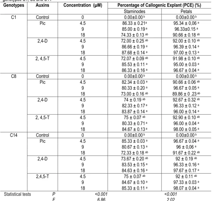
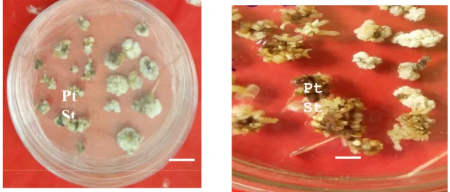
with 18 μM of 2, 4, 5-T in all genotypes: C1 (39.29% ± 0.28; 17.98 ± 0.10), C8 (36.29% ± 0.26 and 15.01 ± 0.07) and C14 (33.92% ± 0.26 and 14.50 ± 0.16). Figure 2 shows embryogenic calli developed by the C8 genotype petals induced on medium supplemented with 18 μM of 2,4,5-T
DISCUSSION
Responses of explants to somatic embryogenesis in cacao can be genotype dependent. In order to develop a reliable and applicable protocol for recalcitrant cocoa genotypes to somatic embryogenesis, three of them, from the most embryogenic to the least one, C1, C14 and C8 were tested on different media during this study. A total of 9 induction media, differing by three concentrations (4,5; 9 and 18 μM) of diverse auxins (Pic, 2,4-D and 2,4,5-T) were tested in order to overcome the recalcitrance. Petals and staminodes were used as explants. . Lowest levels of callogenic explants were obtained on control medium with explants derived from genotype C1, C8 and C14. This confirms the endogenous auxin content of the three genotypes (C1, C8 and C14) is probably lower than that of their exogenous auxin (Pic, 2, 4-D and 2, 4, 5-T). This also indicates that exogenous auxins stimulate elongation and cells division in addition to endogenous auxins activity. Embryogenic explants percentage and average number of embryos were observed only on petal explants derived calli of three cocoa genotypes C1, C8 and C14. This result shows that protocol developed in this study is more favorable to petal explants. These results are consistent with those of
Kouassi et al., (2017b) who revealed that petals of C1 and C14 genotypes were more responsive to somatic embryogenesis than staminodes. In contrast, Zuyasna et al., (2012) found that staminode explants produced more somatic embryos than petals. This could be explained by the fact that cocoa genotypes (North Aceh) used by this author are different from those assessed in the current study. Moreover, our results showed responses variation at genotypes studied level. Indeed, C1 genotype showed a good embryogenic response compared to C8 and C14, since it produced embryogenic calli and somatic embryos in presence of all of the three concentrations the auxins . This variability of response to somatic embryogenesis would therefore be attributed to the genotype. Genotype influence in response to somatic embryogenesis in cocoa was also reported by Alemanno et al., (1996), Issali et al., (2008a) and Kouassi et al., (2017a). Benson (2000) emphasized that phylogeny could have an immediate impact on the recalcitrance of some tissues and genotypes with respect to their totipotent power. Embryogenic callus and embryos development is a function of callus quality because callus formed during callogenesis consist of two types of callus: Figure 1: Calli from petal and staminode explants
cultured on an induction medium containing 18 μM of 2,4,5-T of the C8 genotype of Theobroma cacao. Petals; St- staminode; Bar: 0.5 cm
Figure 2 :Embryogenic calli obtained from the C8 genotype of Theobroma cacao on an induction medium supplemented with18 μM of 2, 4,5-T: embryos derived from petals 3 months after induction. Bar: 0.5 cm
Pt St
Pt
friable calli which lead to embryogenic callus and non-friable callus characterized by an absence of embryogenic callus production. No embryogenic callus and embryo production was obtained with 4.5 μM 2,4-D in the C8 genotype. This result is consistent with those from Kouassi et al., (2018) who used this same concentration for 2,4-D and 2,4,5-T. But a combination of 4.5 μM 2, 4-D with 20 μl of TDZ led to 2,20 of PEC in C8 genotype (Kouassi et al., 2017a). One possible explanation may be due to the cytokinin (TDZ) which is different to that evaluated in this study. At the opposite, somatic embryos were induced on calli derived from petals with 4.5 μM Pic and 2,4,5-T in all of the three genotypes. These results are contrary to those from Zuyasna et al. (2012) who reported that low
concentrations (less than 4.5 μM) of Pic were not able to induce somatic embryogenesis in the North Aceh cocoa genotype. This could probably be explained by difference in the genetic composition of genotypes studied. Among auxins tested, concentration of 2,4,5-T at 18 μM recorded the best PEC and average NSE between the three genotypes C1, C8 and C14 in comparison to the others concentrations. Thus, supplementation of 18 μM of 2,4,5-T in callus induction medium facilitated development of embryogenic calli into somatic embryos on embryos development medium. Such 2,4,5-T concentration (18 μM) could be an ideal one to overcome recalcitrance in cocoa genotypes.
CONCLUSION
Results of the current study showed that somatic embryogenesis in cocoa genotypes are genotype and explant dependent. This study set up an improved protocol compared to previous works in terms of embryo production in cocoa genotypes. It showed that the use of high concentrations (9 μM and 18 μM) of 2,4-D and 2,4,5-T had beneficial effects on embryogenic calli induction in cocoa, even in a recalcitrant genotype. 18 μM concentration of 2, 4, 5-T gave the best
percentage of embryogenic explants and the highest average number of embryos in the three genotypes tested. It is noticed that this concentration produced embryogenic calli in recalcitrant genotype C8 at the same level as responsive one C1. Therefore, this protocol using 18 μM of 2,4,5 T could be used to overcome recalcitrance to somatic embryogenesis in cocoa.
REFERENCES
Ajijah N, Hartati R S , Rubiyo R, Sukma D., and Sudarsono S, 2016. Effective cacao somatic embryo regeneration on kinetin supplemented DKW medium and somaclonal variation assessment using SSRS markers. Agrivita 38(1):80–92.
Alemanno L, Berthouly M, Michaux-Ferrière N, 1996.Somatic embryogenesis of cocoa from floral parts: Plantation, Recherche, Developpement. 3:234-237
Alemanno L, Ramos T, Gargadenec A, Andary C, Ferriere N, 2003. Localization and identification of phenolic compounds in Theobroma cacao L. somatic embryogenesis. An Bot. 92 (4): 613-623.
Benson E, 2000. Special symposium. In vitro plant recalcitrance, an introduction. In vitro cellular & Developmental Plant. 36: 141-148.
Dillinger T L, Barriga P, Escárcega S, Jimenez M, Lowe D S & Grivetti L E, 2000. Food of the Gods: Cure for humanity? A cultural history of the medicinal and ritual use of chocolate. Journal Nutrition, 130 (8): 2057-2072
Driver JA & Kuniyuki A H, 1984. In vitro propagation of paradox walnut root stock. HortScience, (19): 507-509
International Cocoa Organization, 2014. ICCO quartely bulletin of cocoa statistic XI (1) 2013/2014. Retrieved from http://www.i cco.org/about- us/icco-news/266-august-2014-quarterly-bulletin-of-cocoa-statis-tics.html.
Evans D A, Sharp W R & Flien C, 1981. Grouth and behaviour of cell cultures: embryogenesis and organogenesis in plant tissue culture, methods and application agricultures. Throupe TA Ed Academic press: 45-113.
Feguira A & Yannick J, 1993. Development of nucellar somatic embryo of Theobroma cacao L. Acta Horticulturae, 336: 231-236.
Franke R, Hermm M R, Denault J W, Ruegger M O, Humphrreys J M & Chapple C, 2002. Changes in secondary metabolism and deposition of an unusual lignin in the ref8 mutant of Arabidopsis. Plant Journal, 30: 47-59.
Gueye B, Herve J, Sané D, Borgel A, Aberlenc-Bertossi F & Verdeil, J.L, 2008. Les cellules végétales
compétentes à ce réactivées constituant des cibles privilégiées du 2,4-D. AUF Reinne (Résumé).
Issali A E, Traoré A, Kouablan K E, Kohi N A, Sangaré A, 2008a. Characterization of callogenic and embryogenic abilities of somme genotypes of cocoa (Theobroma cocoa L.) under selection in Côte d’Ivoire. Biotechnology. 7 (1): 51-58. Issali A E, Traoré A, Kouablan K E, Kohi N A, Sangaré
A, S ,2008b. Relationship between some phenological parameters and somatic embryogenesis in Theobroma cacao L. Journal Crop Sciences. Biotechnology 11 (1): 23-30.
Janny G M V, Barbara J R, Julie F, 2003. A la découverte du cacao, un guide pour la formation des facilitateurs. CABI Biosciences. 114p.
Kerns H R & Meyer M M J, 1986. Tissue Culture Propagation of Acer Freemanii Using Thidiazuron to Stimu- late Shoot Tip Proliferation. HortSciences, 21: 1209-1210. Li Z, Traoré A, Maximova S, Guiltinan M J ,1998
.Somatic embryogenesis and plant regeneration from floral explants of cacao (Theobroma cacao L.) using thidiazuron. In Vitro Cellular &Developmental Biol Plant. 34: 293-299.
Kouassi, K M, Manlé T E, Koné D, Soumahoro A B, Koné T, Kouablan K E, Koné M ,2017a .Effect of antioxidants on the callus induction and the development of somatic embryogenesis of cocoa [Theobroma cacao (L.)]. Australian Journal of Crop Science 11(1):25-31
Kouassi, K M, Kahia J, Kouame N C, Tahi G M, Kouablan K E, 2017b. Comparing the effect of plant growth regulators on callus and somatic embryogenesis induction in four elite Theobroma cacao L. Genotypes. Hortsciences., 52(1): 142–145.
Kouassi, K M, Kouablan K E, Silué O, Tahi G M, Touré M, Konan K P, 2018 .Comparison of systems combining auxins with thidiazuron or kinetin supplemented with polyvinylpirrolidone during embryogenic callus induction in three Theobroma cacao L. genotypes. Internatinal Journal Biological and Chemical Sciences. 12(2): 804-811, 2018.
López-Baez O, Moreno-Marginez L, Pacheco-Rodas S, 2001. Avances en propagación de cacao- Theobroma cacao-por embriogénesis
somática en México. International Workshop on new Technologies and Cacao Breeding. Malaysa; p. 163–176
Maximova S.N, Alemanno L, Young A, Ferrière N, Traoré A ,Guiltinan M J, 2002. Efficiency, Genotypic variability, and cellular origin of primary and secondary somatic embryogenesis of theobroma cacao L. In Vitro Cellular & Developmental. Biology-Plant 38: 252-259.
Minyaka E, Niemenak N, Fotso S N D & Omokolo N D ,2008. Effect of MgSO4 and K2SO4 on somatic
embryo differentiation in Theobroma cacao L. Plant cell Tissue and organ culture,94 (2): 149-160.
Murashige T & Skoog F, 1962. A revised medium for rapid growth and bioassays with tobacco tissue cultures. Physiology Plant. 15:473–497. Sonwa D J ,2002. Etude de cas d’aménagement
forestier exemplaire en Afrique centrale: Les systèmes agroforestiers cacaoyers Cameroun, 49p
Traoré A & Guiltinan MJ ,2006). Effects of carbon source and explants type on somatic embryogenesis of four cacao genotypes. HortSciences,41 (3): 753-758.
Thorpe T A, Harry IS & Kumar P P,1991. Application of micropropagation to forestry. In Micropropagation Eds Debergh, PC and Zimmerman, RHKluwer Academic Publishers, Dordrecht,The Netherlands,: P 311-336. Tan C L & Furtek D B, 2003 .Development of an in vitro
regeneration system for Theobroma cacao from mature tissues.Plant Sciences.164: 407-412.
Subhashini R, Rao U S M, Sumathi P, Gunalan G, 2010. A comparative phyto-chemical analysis of cocoa and green tea. Indian Journal of Science and Technology. 3(2): 188-192. Rusconi M, Conti A, 2010.Theobroma cacao L., the
Food of the Gods: A scientific approach beyond myths and claims. Pharmacological Research. 61 (1), p. 5-13.
Sarmadi B, Ismail I, Hamid M, 2011. Antioxidant and angiotensin converting enzyme (ACE) inhibitory activities of cocoa (Theobroma cacao L.) autolysates. Food Research International,Volume 44, Issue 1,P 290-296. Sarmadi B, Aminuddin F, Hamid M, Saari N,
effects of cocoa (Theobroma cacao L.) autolysates. Food Chemical 134 (2), 905-911. Quainoo A K, Dwomo B I, 2012. The effect of TDZ and
2, 4-D concentrations on the induction of somatic embryo and embryogenesis in different cocoa genotype. Journal. of Plant. Studies. Vol. 1, No. 1
Whitlock B, Bayer C & Baum D, 2001. Phylogenetic relationships and floral evolution of The Byttnerioidae (Sterculiaceae or Malvacecae). Based on sequences of the Chloroplast gene, ndhF. Systematic Botany, 26 (2): 420-437.