Functional Study of Ephrïns and Eph Receptors in the
Immune System
Guang Yu Département de Médecine Faculté de Médecine Université de MontréalThèse présentéei la Faculté des études supérieures en vue de l’obtention du grade de
Philosophiae Doctor (Ph.D.) en Sciences Biomédicales
September, 2004
Université de Montréal Faculté des études supérieures
U)
Li
o
\Ï
Université
de Montréal
Direction des bibliothèques
AVIS
L’auteur a autorisé l’Université de Montréal à reproduire et diffuser, en totalité ou en
partie,
par quelque moyen que ce soit et sur quelque support que ce soit, et exclusivement à des fins non lucratives d’enseignement et de recherche, des copies de ce mémoire ou de cette thèse.L’auteur et les coauteurs le cas échéant conservent la propriété du droit d’auteur et des droits moraux qui protègent ce document. Ni la thèse ou le mémoire, ni des extraits substantiels de ce document, ne doivent être imprimés ou autrement reproduits sans l’autorisation de l’auteur.
Afin de se conformer à la Loi canadienne sur la protection des
renseignements personnels, quelques formulaires secondaires, coordonnées ou signatures intégrées au texte ont pu être enlevés de ce document. Bien que cela ait pu affecter la pagination, il n’y a aucun contenu manquant.
NOTICE
The author of this thesis or dissertation has granted a nonexclusive license allowing Université de Montréal to reproduce and publish the document, in part or in whole, and in any format, solely for noncommercial educational and research purposes.
The author and co-authors if applicable tetain copyright ownership and moral rights in this document. Neither the whole thesis or dissertation, nor substantial extracts from it, may be printed or otherwise reproduced without the authot’s permission.
In compliance with the Canadian Privacy Act some supporting forms, contact information or signatures may have been removed from the document. While this may affect the document page count, it does flot represent any loss of content from the document
Cette thèse intitulée:
« Functiona] Study ofEphrins and Eph Receptors in the Immune System»
Présentée par:
Guang Yu
A été évaluée par un jury composé des personnes suivantes:
Dr. Yves Raymond
Président du jury
Jiangping Wu, M.D. Ph.D.
Directeur de recherche
Dr. Edward Bradley
Membre du jury
Dr. Mark featherstone
Examinateur externe
Dr. Yves Raymond
Représentant du doyen de la FES
SUMMARY
Ephrins (EFNs) are ccli surface ligands of Ephs, the largest family of celi-surface receptor tvrosine kinases. The function of EfNs in the immune system
has
flot been well studied, although some EfNsand
Ephs are expressed at high levels on certain leukocytes.The
data presented here indicate that the EFNB subciass (EfNBs)and
their receptors (EfNBRs) are expressed in peripherai lymphocytesand
monocytes/macrophages, with T celis being the dominant EFNBsand
EfNBRs ccli type. Solid phase EFNBs-Fc in the presence of suboptimai anti-CD3 crosslinking enhanced T-ce!! responses in terms of proliferation. activation marker expression, IfN-y production,and
cytotoxic T-cetl activity. Afier crosslinking, T ccli receptorand
EFNBRs congregated into aggregated rafts,and
this provided a morpho!ogical basis for interaction between TCRand
EFNBR signaling pathways. f urther downstream, p38and
p44/42 MAPKvas
activated by EFNBR crosslinking. EFNBs, especially BI,and
their receptorsare
also expressed in CD4CD8 double positiveand
CD$ single positive ceils,and
to a lesser extent, in CD4 single positive celis. Soiid phase EFNB1 effectively protected thymocyte from CD3-induced apoptosis, whi!e soluble EFNB1 significantiy augmented such apoptosis. The effect of EFNB1 on thymocyte survivalwas
more significant on populations expressing higher levels of EFNB1Rs. These resu!ts demonstrate that EfNBsand
EfNBRs play pivotai roles in modulating T ccli functionand
thymocyte deveiopment.Key words: Eph; EFN: T cdl; Thymocyte; costimulation; T-cell deveiopment.
Résumé
Les Ephrins (EfNs) sont un ligand membranaire de Ephs, la grande famille des récepteurs tyrosine kinase. La fonction des EFNs n’est pas encore bien définie dans le système immunitaire, mais certains EFNs et Ephs sont surexprimés dans les leucocytes. Les résultats présentés ici indiquent que la sous-famille des EFNBs et leurs récepteurs sont éxprimés dans les lymphocytes périphériques, les monocytes et les macrophages; dans les cellules T, les EFNBs et EFNBRs dominent. En phase solide, les EFNBs-Fc. en présence
de concentrations suboptimales d’ anti-CD3 amplifient la prolifération, l’activation des
marqueurs d’expression. la production des IFN-g et l’activité cytotoxique des cellules T. Après inter-réaction (crosslinking), les cellules T réceptrices et EfNBRs se rassemblent en radeaux lipidiques. Ces derniers constituent une base morphologique pour l’interaction entre le TCR et 1’ EfNBR menant à l’activation de leur voie de signalisation. En aval, les p38 et p44742 MAPK sont activés par l’inter-réaction de EfNBR. Les EFNBs, particulièrement EFNB 1. et leurs récepteurs sont également exprimés par les CD4CD8 double positifs, les CD8 positifs et, moins fortenment par les CD4 positifs. En phase solide, EfNB1 protège efficacement les thymocytes suite à l’apoptose induite par CD3, même si EfNB1 en phase soluble augmente considérablement l’induction de l’apoptose. L’effet de EFNB1 sur la survie des thymocytes est plus significative sur les populations surexpnmant
les EFNB1Rs. Ces résultats démontrent que les EfNBs et EFNBRs jouent un rôle
primordial dans la modulation de la fonction des cellules T et aussi dans le developpement
des thymocytes.
Mots clé: Eph; EFN; T cell; Thymocyte; costimulation; T-cell development.
C
FACULTÉ DES ÉTUDES SUPÉRIEURES UNIVERSITÉ DE MONTRÉALGUANG
TABLE 0F CONTENTS
Summaiy III
Résumé IV
List of Figures VIII
List of Tables X
Lïst ofAbbreviations XI
Acknowledgements XIII
Dedication XIV
I. INTRODUCTION 1
1.1 Eph receptors and EFNs ligands 3
1.1.1 Expression ofEFNBs and Eph receptors 4
1.1.2 The structure of and interaction between EFNs and Eph receptors 10
1.1.3 Functîon ofEFN aiid Eph receptors 13
1.1.3.1 Eph!EFN in the development of the neiwous system 13
1.1.3.2 Eph/EFN in angiogenesis 14
1.1.3.3 Eph!EFN in turnot tissue 15
1.1.3.4 Eph/EFN in theimmune system 16
1.1.4 Signalirig ofEFN and Eph receptors 17
1.1.4.1 Forward signaling by Eph receptors 1$
1.1.4.2 Reverse signaling by EFN ligands 21
1.2 T celi development in the thymus 22
1.2.1 Positive selection .23
1.2.2 Negative selectïon 24
1.3 Co-stimulation in T ceil activation 24
1.3.1 Co-stimulation molecules 26
1.3.1.1 The major costimulatory molecule CD2S 27
1.3.1.2 Other costimulatory molecules related to T ceil activation and differentiation 30
1.3.1.3 TNF farnily 32
1.3.2 Inhibitoiy molecules 35
1.4 The role of lipid raft in signosome and T celI signaling 36
1.5 The objectives of this study 38
II. ARTICLES 40
Article 1: Ephrin B2 induces T celi costimulation 41
Article 2: Mouse ephrinB3 augments T-cell signaling
and
responses to T-cellreceptor ligation 83
Article 3: The role ofephrinBl in thyrnocyte development 11$
III. DISCUSSION 153
111.1 Functional redundancy suggests biological importance ofEphs and EFNs 154 111.2 Biological significance ofEFNBs-EfNBRs in adult T ceils 156
111.3 Biological significance ofEFNB1-EFNB1R in thymocyte development 157
111.4 The contribution of this study to science 159
IV. REFERENCES .160
LIST 0F FIGURES
Introduction:
Fig 1. The structure ofEph and EfN 11
Fig 2. Interactions between Ephs andEfNs 12
Article 1:
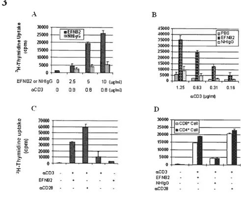
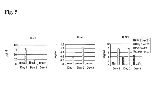
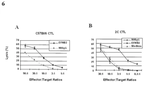
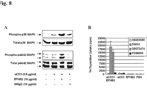
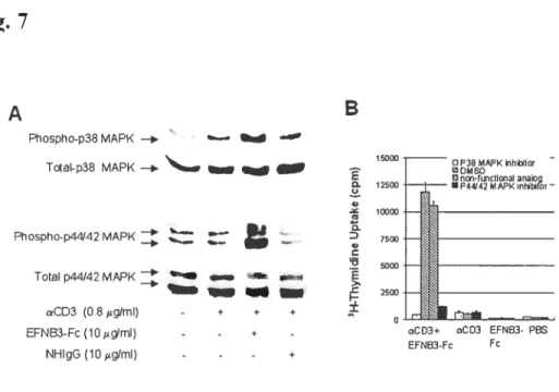
Fig 1. EfNB2 expression in the thymus and spleen according to in situ hybridization 72 Fig 2. FÏow cytometiy analysis oJEFNB2 and EfNB2R expression on immune ceÏÏs 73 Fig 3.EFNB2R crossÏinking enhances the T-ecu response to TC’R stimulation 76 Fig 4. Activationmarkerexpression oJEfNB2-Fc-costimuÏated T ceÏÏs 77 Fig 5.Solid phase EFNB2-Fc strongly augments IFN-y but not IL-2 or IL-4 production
byanti-’D3-stt,nulated Tcells 78
Fig 6. Effect ofsoÏid phase EFNB2-Fc on CTL deveÏopment 79 Fig 7. Rapid coÏocaiization ofEfNB2 receptors with TCR and raft caps afler anti-CD3
crosslinking 80
Fig 8. Activation ofp38 MAPK and p1-F-12 MAFK in T ceÏÏs by solid phase EfNB2-fc...81
Article 2:
Fig 1. In situ hybridization offFNB3 10$
Fig 2. FÏow cytometrv analysisofEfNB3 and EfNB3R expression on leukocvtes 109 Fig 3. $oÏid phase EFNB3-Fc enhances T-celÏ prohferation upon TR stimulation 111 Fig 4. Activation marker expression ofEfNB3-Fc-costimuiated T cells 113 Fig 5.EfN33-fc enhances T-cell effectorfiinction 114 Fig 6. Rapid co-locatization ofEfNB3R with TCR and rafi caps afler
anti-C’D3-crossÏinkzng 115
C
Fig 7. Actïvation ofp38 MÀFK andp44/42 MAFKin T ceÏÏs by soÏid phase
EFNB3-Fc 116
Article 3:
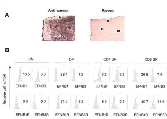
Fig 1. EFNBJ
and
EFNB]R expressionin
thethymus
144Fig 2.Soluble EFNB]-Fc skews thymocyte subpopulations
in
fTOC 145 Fig 3.SoÏubÏe EFNBJ influences protiferationand
apoptosis ofthymocytes in fTOC . ..147fig 4.Effect ofsoÏid phase and soÏubÏe EFNBJ-Fc on thymocyte survivaÏ in vitro 149
Fig 5. Rapid co-ÏocaÏization oJEFNB]R with TCR
and rafi
caps afier anti-CD3-and
CD4- crossÏinkzng 150
Fig 6.FhosphoiyÏation ofLA T and cbÏ
in
thymocytes after EFNB]R crossÏinking 151C
FACULTE DES ETUDES SUPERIEURES UNIVERSITÉ DE MONTRÉAL GUANG ‘JLIST 0F TABLES
Introduction:
Table 1. Expression ofEph and EFN members 4
Table 2. The expression ofEph and EFN members in murine orhurnan immune system 9
LIST 0F ABBREVIATIONS
AP-1 Transcription factor activator protein 1
APC Antigen-presenting cells
CD Cluster of differentiation
CTL Cytotoxic T lymphocyte
CTLA4 Cytotoxic T lymphocyte antigen-4
DP CD4 CD$ double positive
ERK Extracellular signal-regulated protein kinase. FADD Fas-associated death domain
FAK focal adhesion kinase
FasL Fas ligand
FTOC Fetal thymus organ culture
GEF Guanine exchange factor
GM-CSF Granulocyte-macrophage colony-stimulating factor GPI Glycosylphosphatidylinositol
Grb Growth factor receptor bound protein HVEM Herpes virusentry mediator
ICOS Inducible costimulatory molecule
IFN Interferon
1g Immunoglobulin
IL Interleukin
ITAMs Immunoregulatory tyrosine activation motifs
JNK Jun N-terminal stress kinase Jun N-terminal stress kinase
KO Knockout
LAT Linker for activation in T celis
LIGHT Hornologous to Lyrnphotoxins, Inducible expression, competes with H$V Glycoprotein D for HVEM, a receptor expressed on
T-lymphocytes
MAPK Mitogen-activated protein kinase MHC Major histocompatibility complex MIRR Multichain immune recognition receptor
MLR Mixed lymphocyte reaction NF-AT Nuclear factor of activated T celis NF-KB Transcription factor nuclear factor-KB
PAK p21 -activated kinase
PD-1 Phosphodiesterase-1
PUK1 Phosphoinositide-dependent kinase I PI3K Phosphoinositide 3-kinase
PMA Phorbol 1 ,2-rnyristate 1 ,3-acetate RT-PCR Reverse transcriptase PCR RTKs Receptor tyrosine kinases
SH2 Src-homology 2
SLAM Signaling Lymphocytic Activation Molecule
TCR T ceil receptor
Thl/2 T helper ce!! 1 and 2
TN CD3 CD4 CD8 triple negative
TNF Tumor necrosis factor
TRADD TNFR-associated death domain protein TRAIL TNF-re!ated apoptosis-inducing ligand TRANCE TNF related activation-induced cytokine
ACKNOWLEDGEMENTS
First, I would like to show my great thanks to Dr. Jiangping Wu, flot only for his support and guidance during my graduate studies, but also for his help in my living here at the
Université de Montréal.
Thanks also go to the staff of the Laboratory of Transplantation Immunology, Research Center, CHUM, for ail their help during the research for my dissertation.
DEDICATION
T would like to dedicatc this to my parents, Leihai Yu and Shaojie Peng, and to my daughter for their sufferings, and their constant encouragement and supports.
I would also like to dedicate this to my relatives and ail of my friends here and in China, without them I really don’t know how hard my time would be for
FACULTÉ DES ÉTUDES SUPÉRIEURES UNIVERSITÉ DE MONTRÉAL GUANO YU
I. INTRODUCTION
INTRODUCTION
In multiceflular organisms, celis can interact with each other to achieve their biological functions. The ccli surface receptors and their celi surface iigands are pivotai structures for such cell-to-ceÏl communication. Receptor tyrosine kinases (RTK5) are a large group of celi surface receptors that possess intrinsic cytoplasmic tyrosine kinase activity. Functionally, they play critical roles in celi proliferation, differentiation, migration and metabolism. There are several RTK families, such as the insulin, Eph, and the receptors for many growth factors, including the epidermal growth factor, platelet-derived growth factor, fibroblast growth factor and vascular endothelial growth factor. Ephs are the largest family of RTK, with a total of 15 members belonging to two subclasses (EphAs and EphBs subfamilies). They are membrane-bound glycoproteins and are activated by the binding of their cognate ligands, ephrins (EfNs). EfNs consist of 9 members oftwo subclasses, EfNAs and EfNBs. They are also membrane-bound. Both Ephs and EfNs can transduce the extracellular signai to the cytoplasm, either by phosphorylating tyrosine residues on the receptors themselves (autophosphorylation) andlor on downstrearn signaling proteins, playing their roles in
regulating various cellular activities.
In comparison to the extensive studies donc inthe central newous system, far fewer efforts
have been made in understanding the function of Ephs and EfNs in the immune system. Our work aims to enhance our knowledge on the function of EFNs and Ephs in immune regulation.
1.1. Eph receptors and EFNs Jigands
The term Eph originates from the name of a ce!! une, rythropoietin-producing human hepatocellular carcinoma, from which the first member of the farnily was identified (1). The !igands of Ephs, ephrins, may derive from two origins. One is the Greek word ejoros (or ephoros), meaning overseer or contro!ler; the other is the abbreviation ofph fami!y receptor interacting proteins (2).
Since the first Eph member, EphAl, was cloned (1), new members ofthis fami!y and their ligands were rapidly discovered. Many different names have been given to a same member, which is inconvenient for reference. In September 1996, a uniform nomenclature was recommended at the “Molecular Biology of Axon Guidanc&’ workshop, and since then the new name of these !argest RTK members have been wide!y adopted. A summarized nomenclature has been posted at the following website: http://cbweb.med.harvard.edu/eph nomenclature. To date, 9 members of EphAs (EphAl-9) and 6 members ofEphBs (EphBl 6) in the Eph family, and 6 members ofEfNAs (EFNA1-6) and 3 EFNBs (EfNB!-3) in the EFN fami!y have been identified (3, 4). Such a classification (A and B subfami!ies) is based on sequence simitarity and partner binding affinities. The members within each subclass share different degrees of homology. for examp!e, the extracellular regions of EfNB 1 show
50% arnino acid identity with EfNB2, and 42% identity with EFNB3 (5). Detai!ed sequence homology dendrograms of Eph receptors and EFN !igands can be viewed at the website:
http//cbweb. med. harvard. edtt/eph-noinenctature.
1.1.1. Expression ofEFNBs and Eph receptors
The expression of Eph and EfN protein has been shown in various tissues of zebrafish, xenopus, chicken ernbryos mice, rats and human (4). Physiologically, they are widely expressed during development, being most abundant in the nervous system and, to a lesser extent, in vascular endothelium and some specialized epithelia. They are also detected in some tumor ceils or tumor-related tissues. The expression of Ephs and EfNs in various organs, including immune ceils, is surnmarized in Table 1.
Table 1. Expression of Eph and EfN members
Name Expression in organs or tissues
EphAl In the embryonic or aduit mouse: the liver, lung, kidney, thymus, and placenta (6, 7).
In aduit rat: the liver, lung, kidncy, testis (8), and thymus (9).
In human: high in the bladder, and small intestine; moderate in the lung, liver,
colon, kidney, thymus, prostate and testis; low in the brain, spleen, and utems (10).
EphA2 In the embryonic mouse: at the early stage, neurogenic crest cells of the facial, acoustic, and distal regions of limb bud mesenchyme; at later stage, the cartilaginous model of the skeleton, tooth prirnordia, infundibular component of the pituitary, various fetal tissue epithelia (11, 12, 13); high levels in the developing lung, kidney, intestine, and salivary gland (14). In adult mouse: the lung (11) and thymus (7).
In the aduit rat: high level in the lung, skin, small intestine, and ovary; low in kidney, brain, spleen, submaxillary gland (15), and thymus (9).
In human: high level in the bladder, lung and kidney; moderate in the liver, spleen, small intestine, uterus and thymus; low in the brain, prostate and testis (10).
EphA3 In the embryonic mouse: the forebrain, midbrain, hindbrain, neural tube, branchial arches (lst & 2nd), sornites, and limb buds (16).
In the adult mouse and rat: the brain (17, 18, 19) and thymus or thymocyte (7,
9). In human: high level in the utems, bladder and prostate; moderate in the
brain, lung and testis; low in the thymus, liver, spleen, small intestine and kidney (10).
EphA4 In the ernbryonic mouse: multiple sites of the developing central nervous system, the striatum, ventricular zone of the cerebral cortex, hippocampus, thalamus, cerebellum, facial nucleus, and anterior hom ofthe spinaL cord. In the adult mouse: the hippocampus, cerebral cortex, striatum, thalamus, granule layer of the cerebellum (14), fetal and adult B-lineage cells (20), and thymus (7).
In rat: the adult brain (17) and thymus (9).
In human: high level in the brain and testis; moderate in kidney; low in the thymus, heart, lung, spleen, pancreas, small intestine, muscle, placenta, ________
bladder, utems, prostate and bone marrow (10, 21).
EphA5 In the embiyonic mouse: the telencephalon, olfactory bulb, and retina. In fetal and aduit mice: high expression at the hippocampus (22).
In the aduit rat: the hippocampus, ascending central cholinergic nuclei and
_________
monoaminergic nuciei (23, 24).
EphA6 In the neonatal mouse: the fleurons of the cochlear/spiral ganglion in the inner ear and whole brain (25). In the adult mouse: the thyrnocyte (7). In the embryonic rat: ail over the body and head. In the postnatal and adult rat: only in the brain (23).
In human: high in the testis; moderate in the brain and colon; low in the kidney, thymus, heart, lung, pancreas, small intestine, bladder, utems, ________
prostate and bone marrow (10).
EphA7 In the embryonic and postnatal mouse: predominantly in brain at embryo stage, also in various organs outside the nervous system during development: kidney, iung, limb buds, and to a lesser degree in the heart, liver, gut, and genital tubercle (26, 27). In the adult mouse: the brain, testes and, at lesser intensity, spleen (26), and thymus (7).
In rat: strong expression from fetus to adulthood, especially in the brain, and may also inthe retina and heart (28, 27, 29).
In human: the brain, heart, kidney, and lung (20); high in the kidney, bladder, brain and testis; moderate in the spleen, small intestine, uterus and prostate; Iow in the lung, thymus and bone marrow (10).
EphA$ In the adult rat: the brain (30) and thymus (7).
In human: high in the testis and moderate in the brain (10).
EphBl In the embryonic mouse: the brain, cerebellum, and spinal cord during development; In the adult mouse: the hippocampus and cerebellum (14). In the embryonic rat: high levels in the cerebellum, brainstem, diencephalon, and caudate nucleus (31).
In human: high in the brain and testis; moderate in the colon and kidney; low in the thymus, heart, tung, pancreas, small intestine, spleen, bladder, uterus and prostate (10).
EphB2 In the fetal mouse: the neurocctoderm (32), ventral midbrain, ventroposterior forcbrain, ventral neural tube, the developing dorsal root ganglia, hindbrain, retina, posterior haif ofthe foregut pocket, mesoderm around the heart (13), mesenchyme and heart (33).
In the fetal rat: high in the brain cortex, modest in ah fieÏds of the hippocampus, and in a subset of Purkinje ceils of the cerebellum, weakly in Schwann cells (17).
In human: the lung, thyroid, placenta and muscle (21, 30); high in the colon and small intestine; low in the brain, thymus, heart, lung, spleen, pancreas, kidney, bladder, uterus, testis, prostate (10, 21).
EphB3 In the embryonic mouse: cerebehlum, Purkinje cells, hippocampus (17), head mesenchyme, developing somites, foregut, first and second branchial arches, lateral rnesoderm of the forelimb bud (13), venous endothelial cells, and some arteries (33). In the adult mouse: the brain, liver, lung, kidney, intestine, testes, placenta, muscle, and heart (34).
In human: high in the coton, brain and lung; moderate in the uterus, prostate, small intestine, kidney and bladder; low in the thymus, spleen, testis and bone marrow (10).
EphB4 In the ernbryonic mouse: the epithehial lining of the brain and ventricles, thymus, lungs, heart, spleen, the entire glomeruli of kidney (34), and ah major veins (33). In the adult mouse: brain, heart, lung, kidney, placenta, testes and muscle (35)
In human: the fetal heart, lung, liver, and kidney, lesser signal in brain; aduit placenta and pancreas (36); high in the lung; moderate in the spleen, liver, testis, uterus, kidney, bladder, colon (10); low in the skeletal muscle, heart (36), prostate, small intestine, thymus, and bone marrow (10).
EphB5 No documented data in rodent and human tissue. In the chicken: the thymus (37).
EphB6 In the aduit mouse: high levels in the thymus and brain (3$).
In human: high levels in the thymus, moderate in the brain and testis, low in the lung, spleen, uterus, prostate, smahl intestine, kidney, bladder, colon and bone marrow (10).
EFNA1 In the fetal mouse: the lung, the epithelium of the gut, the ossifying bones of the face, salivary gland, central nervous system and low level in the connective tissue of septae in the thymus (39); the primitive streak, lateral mesoderm, tailbud, and at lower levels inthe blood vessels (40); In the aduit: the thymocyte (7).
In the embryonic rat: the epithelial ceils and the endothelial cells in lung, kidney, intestine, skin, skeletal system, teeth, heart (endocardium), and dorsal root ganglia neurons (41). In the postnatal rat: high levels in the lung, low levels scattered throughout the thymus (39).
In human: high in the lung, colon and liver; moderate in the bladder, kidney,
prostate and uterus; low in the brain, thymus, spleen, srnall intestine and testis (10).
EFNA2 In the embryonic mouse: high levels in the midbrain and lower levels in the dorsal anterior hindbrain; outside the nervous system, in the lst and 2nd branchial arches, somites, and limb buds (16, 40).
In postnatal mouse: the hippocampus in a lateral to medial gradient, and an increasing gradient from dorsal to ventral septum (22); the thymocytes (7). In human: high in the colon; moderate in the bladder, small intestine and liver; low in the brain, kidney and testis (10).
EFNA3 In embryonic mouse and rat: almost exclusively in the central nervous system besides the skin (42). In the aduit mouse: thymocyte (7).
In human: high in the brain; moderate in the colon; low in the thymus, lung,
liver, bladder, kidney, prostate, utems, small intestine and testis (10).
EFNA4 In the early embryonic mouse: the ectoderm of branchial arches, the forelimb bud, the anterior spinal cord, and throughout each sornite the limb over the proximal Iirnb and hand/foot plate (40); In the aduit: thymocytes (7).
In the embiyo rat: the forebrain, olfactory bulb, hippocampus and cortex; In the adult: low level in the brain (31).
In the human fetus: the heart, lung, and kidney offetus; In the aduit: high in
the colon; moderate in the lung, kidney, bladder, small intestine and prostate; low in the thymus, utems, liver, spleen, testis and boue rnarrow (10).
EfNA5 In mouse: the brain, heart, intestine, kidney, and at lower levels in the lung, liver (43) and thymocyte (7).
In human: the heart and placenta (44); high inbrain and kidney; moderate in the lung, spleen, bladder, colon and testis; low in the thymus, liver, prostate, utems and small intestine (10).
EfNB1 In the embryonic mouse and rat: high level in brain, sciatic nerve (42) and kidney; moderate in the heart, lung, skeletal muscle and thymus; low in the liver (42, 45).
In the aduit human: high in the bladder, lung and colon; moderate in the kidney, spleen, small intestine, uterus and testis; low in the brain, liver, prostate and thymus (10).
EFNB2 In the fetal mouse: the hindbrain, branchial arches, in two intense regions of new somite formation, and a weaker signal in the forebrain (46).
In the fetal hurnan: the heart, lung, kidney and brain.
In the adult human: high level in lung, kidney (47, 48) and colon (10); moderate in the brain, kidney, small intestine, testis and bladder; low in the thymus, spleen, liver, prostate and uterus (10).
EfNB3 In the mouse embryo: high level in the brain, lower levels in the heart, lung, and kidney.
In the aduit mouse: high levels in various sites ofthe central nervous system, and also in the heart (49).
In the aduit hurnan: high in the brain; moderate in the utems; low in the thymus, liver, kidney, small intestine, testis, bladder, prostate, lung and colon (10).
The expression of some Eph and EFN members in immunological compartments bas been reported during the past years. Hek (EphA3) protein was detected in a pre-B ceil acute lymphoblastic leukemia cdl une (LK63) in early 1992 (50), and later on, in chicken embiyo thymus (37). Following that, several other Eph and EFN members were found in mice, rat and hurnan lymphoid cells or immune organs (6, 7, 9, 14, 15, 20, 26, 38, 42, 45, 47,48, 51, 52, 53). However, the majority of these informations were derived from the detection at the mRNA level, and sorne contradictory resuits were reported. For example, EFNA1, A3 and AS were undetectable in thymocytes in early studies (54), but were later found to be expressed in these cells (7). Such a discrepancy may 5e due to the sensitivity ofthe different
assays employed. In the last three years, more and more Eph members and their ligands have been detected in the immune system (7, 9, 10). A general expression profile ofEph and EFN members in major immunological compartments is summarized in Table 2.
Table 2. The expression of Eph and EfN members in murine or human immune system
EphI EfN Spleen thymus Reference
EphAl + ++ 6, 9, 10 EphA2 +/++ +/++ 15, 9, 10 EphA3 + + 7,9,10 EphA4 + + 21,9, 10 EphA5 EphA6 + 10 EphA7 ++ + 26, 10 EphA$ + 7 EphA9 EFNA1 + + 39, 10 EFNA2 + 10 EfNA3 + + 7 EfNA4 + + 7 EFNA5 ++ + 7,10 EFNA6 EphBl + 10 EphB2 + + 30,21, 10 EphB3 + + 10 EphB4 + ++ 34, 36, 10 EphB5 EphB6 + +++ 38, 10 EfNB1 +/++ + 42,55, 10 EFNB2 +/++ + 47,48, 10 EFNB3 + + 10
There are variations in the distribution and expression levels in different species, tissues and developing stages. Pattems shown are for embryo or aduit ofmice, rat or human. The+++,
++, + represent strong, moderate, weak expression.
1.1.2. The structure of and interaction between EFNs and Eph receptors
The Eph receptors are type I transmembrane proteins (56) (Fig. 1 A). The extracellular sequence lias an N-terminal ligand-binding domain, a cysteine-rich domain and two fibronectin type III domains. In the cytoplasmic sequence, alrnost ail the Eph members have a juxtarnembrane segment, a highly conserved tyrosine kinase domain (57, 58), a sterile u motif (SAM), and a C-terminal PDZ-binding motif The cysteine-rich region close to the ligand-binding domain is believed to be involved in receptor—receptor interactions (59). The biological function ofthe two fibronectin repeats is not clear so far, but they possibly interact with other proteins (60). The SAM domain may have a role in receptor dimerization (61) or oligomerization (62), and it may also interact with adapter proteins (4).
Ail EFNs share a basic structural component, i.e., a unique conserved extracellular receptor-binding domain at the N-terminus. In the EFNB subclass, an additional highly conserved cytoplasmic domain and a C-terminal PDZ-binding motif are also found, and these are believed to be involved in protein-protein interactions that mediate membrane localization and reverse signaling (63). The EfNA subctass members have no transmembrane region, and they are linked to the celi by a glycosylphosphatidylinositol (GPI) linker (Fig. 1 B).
A B Cytoplasm PDZ op1asnc tau GPI—
,
Receptorbinding EphrinA EpluinBf ig.1. The structure ofEph and EFN
A. The Structure of Eph. B. The Structure of ephrinA and ephrinB. Abbreviations: GPI,
glycosylphosphatidylinositol; SAM, sterile-Œ-motif; PDZ, PSD95/Dlg/ZO 1 -binding motif.
The consequence of EFN and Eph interaction is the initiation of the signaling events in the
receptor bearing celis (forward signaling), or signaling into the ligand bearing ceils (reverse
signaling). It is also possible that the forward and reverse signaling, occur at the same time
(bidirectional signaling).
The Eph-EFN-mediated signaling depends on the association of Eph receptors and Iigands.
When Eph and EFN meet, there is initial conformational change
inboth the receptors and
ligands to achieve a better fit for the receptor-ligand interaction. Receptors and ligand
oligomerization, i.e., trimers composed oftwo receptors and one ligand, or tetrarner oftwo
Ligandbindingdornain
Cysteineiich domain
Juxtamembrane domain
Kinase dom ain
SAM
PDZ
Eph
receptors and two ligands, is basically needed to trigger forward and /or reverse signaling (64).
The interaction between Eph and EFN is prorniscuous, and recent evidences underline this characterisfic. In general, as illustrated in Fig.2, the EphAs interact with EFNAs, and the EphEs interact with EFNBs. However, sorne exceptions exist. For example, in addition to binding to ail ofthe five EFNA subclass members, EphA4 can also bind to EFNB2 (49, 65). Most recently, EphB2 and EFNA5 were shown to be able to bind one another with high affinity (66). This further complicates the existing profile of receptor-ligand interaction between different Eph andEFNmembers.
fig.2. Interactions between Ephs and EFNs
Solid unes represent the proven interactions between the receptors and ligands. Dashed unes indicate possible interactions. (Data corne from: 65, 5, 54, 67, 68, 7, 69, 70, 66)
qii.in
EFN1 EFHA
In addition to the prorniscuity, another characteristic ofthe Eph ligand-receptor interaction is that ligands and receptors need to be membrane-bound to be effective in triggering forward or reverse signaling. The soluble forms are not only inactive, but can be antagonistic to the membrane-bound ligands or receptors (42, 44, 71). Such a characteristic constitutes the basis for the requirement of direct contact between Eph and EfN-bearing ceils to initiate the biological function of Eph and /or EFN.
1.1.3. Function ofEFN and Eph receptors
Eph and EfN have been implicated in the regulation of many critical events during development, and to some extent in aduit life.
So far, the best-defined biological function of Eph and EfNs is the mutual ‘repulsive” or “attractive” effects on ceils expressing Eph and EFN, such effects resulting in structural modulation in tissue morphology, such as axon guidance (72), vascular development (73), and celi migration (74).
The roles that Eph and EfN may play in adulthood are related to angiogenesis (75) carcinogenesis (76
),
turnor metastasis (77), and T ceil regulation (78, 53, 9, 7, 79, 80).1.1.3.1 Eph/EFN in the development ofthe nervous system
One of the most important functions of Eph-EFN interaction is their involvernent in axon guidance or pathfinding. Both Ephs and EFNs are expressed on axons and ceils of neural
origin and their expression may follow a specific space and time pattem. During developrnent or structural reformation ofthe nervous system, the Eph and EFN bearing ceils receive signais from each other and thus rnodify their growth pattem. Although the effect of Eph!EfN interaction may also be pro-adhesive (81), the majority of evidence showed that such a signal is repulsive. for example, EphA3 expression follows gradient in the tectum and retina. When EfNA2, a ligand of EphA3, is overexpressed in the compiementary region of Eph expression, the axons tum to an abnormai direction (82, $3, $4, 85). Such a repulsive function ïs also supported by observations from mice with mutations of some Eph members (86, 87, 8$).
The importance of Eph receptors and EFNs in the aduit has drawn attention in recent years. Significant expression of some Eph and EFN members lias been found in the aduit nervous system (10), and a role for EphB in the reguiation of neurai stem ce!! migration has been described (4). How important Eph-EfN interaction is in the regulation of the aduit nervous system needs to be further eiucidated.
1.1.3.2 Eph/EFN in angiogenesis
In addition to their ftinction in the nervous system, Eph receptors and EfN Iigands are also involved in other morphogenic events, such as angiogenesis. Several Eph and EFN members play a critical role in normal vascular formation. for example, EfNA1 and EphA2 are highly angiogenic ($9), although the effect is not solely caused by the Eph-EfN interaction, and may depend on the contribution from other moiecules (90). Some EphB and EfNB subc!ass
members, sucli as EphB4 and EFNB2, also participate in the development of the cardiovascular system (33, 91, 73, 92). The pivotai roles of Eph and EFN members in angiogenesis have been dernonstrated both in vitro and in vivo, and this topic is reviewed in detail by Brantley-Sieders et al. (93). In accordance witli the observation ofthe formation of topographie projection maps in the neiwous system (81), Eph/EFN interaction can generate both positive and negative bioiogical effects on angiogenic celis, to promote neovascularization (74), regulate arteriovenous differentiation and controi vascular horneostasis (94).
1.1.3.3 Eph/EFN in tumor tissue
Mutation andlor over-expression of receptor tyrosine kinases have been shown to be associated with turnorigenesis a decade ago (95). The Eph famiÏy lias been known to be related to cancers since the first member, EphAl, was discovered (1). Severai Eph family receptors are found in turnor tissues or turnor-derived celi unes. For exampie, EphAl is over-expressed in various tumors, including breast, iung, iiver, colon, and prostate carcinomas (1, 8, 96); some other EphA and B members, including EphA2, A3, A4, 82, 33 and 34, are also over-expressed in tumor ceil unes (50, 97, 9$, 35, 99, 100).
The Eph expression levels seem to be related to the rnalignancy of tumors. Some Eph members are detected in the undifferentiated and invasive mammary tumors, but flot in well-differentiated and non-metastatic tumors (35). However, a comprehensive investigation on cells derived from normal tissues, and non-invasive and invasive tumors, respectively,
shows diverse EphIEfN expression profiles. Upregulation of some Eph and EFN members are positively related to tumorigenesis andlor invasiveness, while some other members are downregulated (101).
Although over-expression of EphAl in mouse NIH 3T3 fibroblast ceils induces colony formation in soft agar and tumor formation in nude mice (102), few data are available on the causal effect of Eph/EFN on tumorigenesis. Recent data support a model in which Eph-EFN interaction prornotes tumor growth by stimulating angiogenesis (103).
Thus, although a growing body of evidence supports the involvement of EfNs and Eph molecuies in tumor angiogenesis, the data are flot sufficient to prove a causal effect of Eph and EFN in inducing cell transformation. On the other hand, Eph and EfN are quite possibly involved in the metastatic phase of cancers and the angiogenesis of tumor tissues.
1.1.3.4 EphIEFN in the immune system
The expression of some of the Eph kinases and their ligands on immune cells has been documented (Table 1 and 2). However, the function of this largest RTK family in immune responses was basically unknown until we started our project in 2001. We have reported that a kinase defective Eph family member, EphB6, is capable oftransducing signais into T cells (104). Activation ofEphB6 with solid phase anti-EphB6 mAb results in Jurkat cell apoptosis. Later on, we found that EphB6 can augment normal human T-cell response to Ag stimulation (53). However, probably due to overlapping functions of Eph kinases, no abnormal
phenotype in terms of thymocyte structure and subpopulation composition is found in EphB6’ mice (105). It is to be noted that no detailed study on the functional defects in the EphB6 immune system was conducted in that study. In the same year, Munoz et al. reported that a few soluble recombinant EphAs and EFNAs interfere with T-cell development in thymie organ culture (9), and Sharfe et al. described that several EFNs, notably EFNA1, inhibit T-cell chemotaxis (7$). Recently, Roifman’s group (80) showed that EphB6 can influence T cell immune response by downregulating anti-CD3-induced IL-2 secretion and CD25 expression in a ligand-dependent manner. When murine thymocytes are stimulated with EFNB 1, TCR-mediated apoptosis is inhibited, suggesting that EphB6 may be involved in thymocyte development during positive or negative selection.
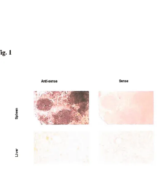
In addition to the resuits mentioned above, in situ hybridization revealed strong EfNB Ï, B2 and 33 mRNA signals in the cortex ofthe thymus and white pulp in the spleen (106, 107, 10$, 109). Taken together, the data raise the possibiiity that EFNBs might play pivotai roles in regulating T-cell development and function.
1.1.4. Signaling by EFN and Epli receptors
Although activation of Eph receptors may follow some general rules estabiished for other RTKs, the signal transduction by the Eph receptors and EFN ligands has a number of unique features. The EFN ligands need to be membrane-bound to be effective, and misregulated tyrosine kinase activities of many Eph members are flot oncogenic in contrast to other
receptor tyrosine kinases. It is also unusual that the Eph-EFN signaling is bi-directional, i.e., both receptors and ligands can transmit signais into ceils.
1.1.4.1. Forward signaling by Eph receptors
The exact process ofEph-EfN interaction is flot well understood for each receptor and ligand member. The crystal structure analysis on some Eph/EfN members revealed some dues in recent years (61, 110, 111, 112). Before ligand binding, the kinase domain in Eph sticks to the juxtamembrane segment, on an autoinhibited conformation. Upon ligand binding, juxtamembrane tyrosines of Eph can be phosphorylated; this releases the autoinhibition and free phosphotyrosine sites for SF12 domains-bearing signaling proteins (11 1). In addition to such a de-autoinhibition rnechanism, some phosphoiylation-independent interactions between EphB and substrate proteins also exist (113).
Many SH2-domain-bearing adapter proteins are involved in the Ephsignaling. for example, Grb2, Grb7 and GrblO bind to EphBl (114, 115); Abi and Arg bind to EphB2 (113). Through their SF12 binding domains, the adapter proteins, such as Shc and Nck, can interact with Eph receptors and influence the activation of the JNK or ERK pathway (116, 117). The recmitrnent of Shc may resuit in tyrosine phosphorylation, which provides binding sites for the SH2 domains on other adapter proteins. Nck can interact with downstream effectors, such
as Pak3 and WASP (72, 118). Notably, AbI and Arg may interact with Eph through their C
terminal tails other than the SF12 domains (113).
Some small GTPases of the Rho family (Rho, Rac and Cdc42) are involved in Eph receptor
signaling (78, 119, 120). Examples corne from both Eph A and B subfamilies, although there are sorne differences between these two groups. Stimulation of EphA receptors can activate Rho GTPases (119), and downregulate the activation ofRac (120) and Cdc42 (7$). On the other hand, EphB can influence the intersectin-Cdc42-WASP-actin pathway (121), or induce transiocation ofRho-GEF kalirin to the site where other target signaling molecules exist, and further activate downstream targets, such as small GTP binding protein Rad (ras-related C3 botulinum toxin substrate 1, rho family) (122).
Ligation of EphB receptors with EFNB Iigands can also activate Rapi (123, 124, 125), a R Ras-like protein that positively modulates integrin-rnediated adhesion (126, 127). Such activation may be mediated by some other adaptor proteins (128), such as Crk (123). The Src non-receptor kinases may also be related to Eph signaling (129). Some Src proteins can be activated afler Eph ligation, and the Src family kinase fyn is important to Eph signaling in modification of ccli adhesion (130, 131).
Recently evidences have shown that Eph/EfN5 signaling interferes with some growth factors, e.g., vascular endothelial growth factor (VEGF) (90). The effect of EphA and EfNA on angiogenesis is achieved with the involvement of VEGF, and phosphoinositide 3-kinase (PI3K) and Rad GTPase arc aiso related to such an activity (132). EphA ligation may inhibit proliferation ofprostatic epithelial ceils and endothelial cells through downregulation ofthe Ras/MAPK cascade (133, 134), which is activated by some growth factors, e.g., epidermal growth factor (EGf) and VEGf. However, such signaling events are not universai. for
instance, EfNA1 inhibits proliferation in prostatic epithelial celis and endothelial ceils, but
flot fibroblasts (133).
Our laboratory is the first one who discovered that the Cbl is invotvcd in Eph signaling in T celis (104, 135, 136, 137). EphB6 cross-linking by Ah in Jurkat celis causes Cbl dephosphorylation (104). CbI phosphoiylation is observed in both Jurkat ceils and peripheral blood T celis stimulated by EFNA1 (79); such EphA activation-induced Cbl phosphoiylation may employ adapter protein Crk-L and Crk-II, with an involvement of Src family kinases. However, stimulation of EphB receptors with EfNB Ï does not induce Cbl phosphorylation in Jurkat cells (79). Recently, Roifman’s group demonstrated that EFNA1 stimulation of Jurkat celis could cause down-regulation and degradation of endogenous EphA3 receptors. EphA receptor expression is down-regulated in ceils overexpressing of wild-type Chi, suggesting that CbI rnight be a negative regulator of Eph receptors ($0). In addition, they found that TCR-mediated activation of the JNK pathway is selectively inhibitcd upon stimulating overexpressed EphB6 with EFNB 1. According to their speculation, srnall GTPase Rad is possibly involved in this event.
So far, although the repertoire ofrnolecules involved in Eph fonvard signaling is expanding,
the available information is insufficient to develop a complete architecture of these Epli signaling pathways.
1.1.4.2. Reverse signaling by EFN ligands
Although the existence of EFN reverse signaling has been obsewed for almost ten years (138), and the reverse signaling is one of the critical feature of Eph-EfN interaction (139, 140, 92), the mechanisrn of reverse signaling stili Yacks details.
Several evidences suggest that EFNB-induced reverse signaling is mediated by proteins bearing SH2 domains (141, 142, 143), and that Src family kinases (SFK5) is responsible for EFNB phosphorylation upon Eph receptor engagement (144, 145). In the EFNB-expressing ceils, ligation with EphB causes rapid recmitment of SFKs, and induces transient SfK activation, which is possibly mediated by phosphorylation of the tyrosine residues and the engagement with PDZ domain proteins (145). Other PDZ-bearing proteins, such as RGS3, and SH2-bearing molecules, e.g., Grb4, can also induce the reverse signaling upon EFNBs receptor ligation (142, 146, 63). RGS3 can further regulate downstream G protein signaling. Grb4 can link EfNBs to a number of signaling molecules, such as the Cbl-associated protein, the Abl-interacting protein-1, dynamin, PAK1, and axin (63). Activation ofEFNB1 increases fak activity, redistributes the fak-binding protein paxillin, and leads to disassembling of focal adhesions (63, 123).
EFNA can also induce reverse signaling. Some evidences demonstrate that EFNA can recruit the Src family kinase Fyn to lipid rafts upon Eph ligation. As a consequence, tyrosine phosphorylation of a 120 kD lipid raft protein and activation of MAP kinases are observed
(147, 148); however, the mechanism is unclear.
1.2. T-ceIl development in the thymus
The thymus is the cradie of T celis, and is a complex prirnary iymphoid organ composed of stromal ceils and immigrant hemopoietic celis. Anatomically, the thymus can be divided into three regions: the subcapsuie region, the cortex, and the medulla (9). Hemopoietic stem celis enter the thymus and go through the early stage of developrnent within the subcapsule and cortex where they iose the abiiity of differentiating to non-T ccli lineages, and are graduaily comrnitted to the T-celi lineage (149).
At the eariiest stage in the murine thymus, the lymphoid precursors are characterized as CD4’°CD8CD25CD44c-Kit (150). Between 12.5 and 14.5 of embryonic days (E12.5’-E14.5), lymphoid precursors differentiate to the T-cell lineage; the thymic epitheliai ceils are flot abie to sufficiently support T-ceii developrnent at the stage earlier than E 12.5 (151). Sorne celi surface markers, such as CD25, CD44, CD117 (c-kit) and CD127 are considered to be indicators of T-ce!! lineage commitment (152, 153), and the Notch signaling is believed to play a pivotai role to the commitment (154). At this stage, CD3 CD4 CD8 are negative in the T ce!! precursors; so these celis are often named TN celis. These TN can be
further classified into TN1 (CD44CD25CD117CD127), TN2
(CD44CD25CD117CD127), and TN3 (CD4410CD25+CD117I0CD127E0). The TN2 ceils upregulate the expression ofpre-TCR (pT) gene and begin to rearrange the TCR gene (152,
153). Then, along with further TCR gene rearrangement, the TN3 ceils definitely commit to the T-cell lineage. These celis wifl tum to be CD4+CD8+CD3b0t, also called double positive (DP) celis (155).
The DP ceils then undergo extensive positive and negative setections, and differentiate into mature CD4 or CD8 single positive T ceils that express higb leveis of CD3-associtaed TCR, and are ready to move out of the thymus as functionai and self tolerant mature T celis.
1.2.1. Positive selection
Positive selection refers to a process that choose the developing DP celis capable of recognizing self-MHC (maj or histocornpatibility complex) molecules for further differentiation and survival (156, 157).
The positive selection requires the contact between TCR on the DP thymocytes and the MHC-peptide complex provided by other celis (158, 156, 159); the majority of MHC for efficient contact is suppiied by thymic stromal celis at the subcapsuie and cortex region (160). Both the concentration and specificity of antigenic peptides prcsented by MHC are important in determining the strength ofthe TCR signaling, and thus the fate ofthe DP cells (158, 161, 162, 163, 164, 165). Obviously, the molecules that can regutate TCR signaling, such as CD8O (166), Cbl (167) and LAT (168), may influence the outcome. Notably, it seems that the cortical epithelium expresses some unique moiecules that may provide additional signais required during the positive selection (156, 163).
The thyrnocytes that pass through the positive selection will further develop into CD4CD8 or CD4 CD8 single-positive (SP) thyrnocytes. Those who recognize class I MHC develop into CD8 cytotoxic T lymphocytes (CTLs), and the others who interact with class II MHC commit to helper CD4 cells (169, 170, 171).
1.2.2. Negative selection
Negative selection is a process to delete the maturing D? celis capable of recognizing self MHC-peptide complex with high affinity. This is a critical mechanism to keep the mature T ceils from being reactive to self-Ag.
Negative selection of thymocytes takes place either in the cortex at the cortex-medulla junction or within the thymic medulla (172, 173); it occurs probably not earlier than the CD4I0wCD8I0w TCRII1ÉCD69±stage (174). Dendritic cells (DC) play an important role for this
process (175). Some accessory molecules, which can influence TCR signaling, such as CD4O/CD4OL and CD28, also play important roles in negative selection (174, 176). However, their involvement appears flot just as simple as co-stimulation or modulation of TCR signaling. For example, Fas (174) and LIGHT (177) are able to affect negative selection, but FasL does not have obvious effect (174), suggesting the existence of a more complex mechanism than that ernployed in the mature T cells.
1.3 Co-stimulation in T-celI activation
The recognition of antigens by specific lymphocyte receptors constitutes a fundamental irnniune reaction, and T celis are a critical component for an effective immune response against a wide range of pathogens. Mounting an appropriate T-ce!! response depends on de!icate regu!ation of lymphocyte activation. To become fu!!y activated, two independent signais are required. The first is an antigen-specific signal that is de!ivered through theTCR. The second one is termed co-stimu!ation that works independent!y of TCR. Accumulating knowledge indicates that co-stimulation plays a crucial role in T-cel! activation (178), proliferation (179), differentiation to effector (180) or rnemory status (181), anergy (182), and apoptosis (183).
The co-stimulation mode! (or two-signal theory) of T-ce!! activation was first proposed by Lafferty and colleagues (184). Many celi surface mo!ecules and cytokines, such as 37(185), CD44 (186) and 1L2 (187
)
can transmit signaIs to enhance T-cell activation. Accumu!ated evidence indicates that bidirectional communication between T celis and accessory cel!s exists, and that this kind of interaction can evoke mutua! stimulation (188). Now it becomes clear that interactions between responder lymphocytes and “accessoiy” celis represent a critical event in the T-ceII activation process; such interaction is usually called co-stimulation. However, there is stiil no general agreement on what exactly defines “co-stirnulation”. The use of “negative co-stimulation” for inhibitory signaling events further confuses the terrninology. for the convenience of the discussion here, a co-stimu!atoiy molecule is defined as an initiator of a positive signal by increasing TCR avidity (for example, adhesionmoiecuies) or enhancing the recruitrnent of tyrosine kinases to the TCR compiex (for example, the co-receptor CD4) (188). Those inducing negative signais are flot called co stimulatory molecules, but are nevertheiess discussed.
1.3.1 Co-stimulatory molecules
The first ce!! surface molecule shownto function as a co-stimuiatory molecule is CD2$ (185). Since its identification, the number of candidate co-stimulatory molecules has significantly increased (189, 190).
Most mo!ecules that induce co-stimu!ation can be divided into two classes based on sequence homology.
The first c!ass is composed of members of the 1g superfami!y, including CD2$ family members. These molecules share several structural and functional features. Ail of the CD2$-!ike receptors are type I transmembrane g!ycoproteins (191). The CD28 fami!y comprises CD2$, CTLA4, ICOS (H4, AILIM) and PD-1; CD28 appears to be the major co stimuiatoiy molecule for the activation ofnaive T ce!!s. The ICOS mainiy works on activated or effector T celis. The othertworeceptors, CTLA-4 and PD- 1, a!though they share structura! hornoiogy with CD28 and ICOS, appear to be both inhibitoiy in nature. The ligands of the CD2$ fami!y members be!ong to the B7 fami!y, and these ligands are also members of the 1g superfami!y.
The second class of co-stimulatoiy molecules comprises members of the TNf receptor (TNFR) family, including CD4O, the major B ce!! co-stimu!atory mo!ecu!e, as well as OX-40, 4-1BB, CD27, CD3O, and HVEM (herpes-virus entry mediator). The ligands for these receptors are membrane-bound members of the TNF family, e.g., 0X40 ligand (OX4OL), 4-ÏBBL, CD7O, CD3OL and LIGHT, respectively. The co-stimu!atory TNFR-family members are mainly expressed on T celis, and their ligands, on antigen-presenting cel!s (192, 193).
1.3.1.1. The major co-stimulatory molecule CD2$
CD28 is constimtively expressed on the surface of 80% ofhuman T ce!!s (a!! CD4 ce!Is and about 50% ofCD8 ceils) and on virtually !00% ofmurine T ce!ls (185, !94). Studies in vitro and in vivo have shown that CD2S is the primary co-stimulatory molecule for naive T celis, and CD4 ce!!s are more dependent than CD8 ce!ls on CD28 co-stimu!ation in vivo (!95, 196).
The major function of CD28 signaling is to act in concert with TCR stimulation. CD28 !igation can enhance the transcription and stabi!ity of IL-2 mRNA, partially through cyclosporine insensitive signa!s (197). In recent years, accumulated data reveai more broad and proxirnal intersection of CD28 signais with those mediated by TCR (198, 199, 200). Ligation ofCD28 on naive T ce!!s by its ligand B7-1 or B7-2 on APC drasticatly enhances
the TCR signaling that activates protein tyrosine kinases (PTK5). The activated PTKs in tum phosphorylate critical scaffold proteins, such as the linker for activation of T celis (LAT) and the SH2 domain-bearing leukocyte-specific phosphoprotein of 76 kD (SLP-76) (201). Such events are critical for the further recruitment and activation of other key signaling molecules, such as phospholipase Cy-1 (PLCyI), growth factor receptor-bound protein 2 (Grb-2), son of sevenless (SOS), and protein kinase C theta (PKC8) (202, 203, 204, 205, 206, 207). Through these molecules, the CD28 signaling triggers downstream pathways leading to the activation of the nuclear factor of activated T ceils (Nf-AT), activator protein-1 (AP-1), and nuclear factor kappa B (NF-KB) transcription factors (203, 208, 209, 210).
Activation of PI3K is one ofthe controversial issues in CD28 down stream signaling. PI3K is an intracellular signaling enzyme that regulates a wide range of cellular functions (211). In T-celI activation, PI3K may tyrosine phosphorylation—dependently associate with the cytoplasmic tau of CD28, phosphoiylate membrane phospholipids phosphatidylinositol 4,5-bisphosphate (PIP2), and generate phosphatidylinositol-3 ,4,5-triphosphate (PIP3) that can activate a serial ofdownstream signaling targets. Recently, there are evidences either for and against an essential role of PI3K in CD2$ signaling (212). By introducing mutant CD28 constructs, which cannot bind PI3K, into CD2$-deficient mice, two groups of researchers obtained strikingly different resuits. One group (205) reported that CD28 Y189f transgenic T ceils severely reduce the ability of proliferation and IL-2 secretion at the early stage of anti-CD3 and anti-CD2S Ab stimulation, although the later responses are intact. On the other
hand, the IL-2 production is totally lost when the celis are stimulated with PMA and anti CD28 at any time point. In vivo test further demonstrated that CD28 Y1$9F transgenic spleen ceils do flot induce acute graft-vs-host reaction. Another group (213) showed that the CD28 mutant (Y 170 in mouse) is more susceptible to radiation-induced death, but does flot significantly compromise the ability of IL-2 production, celi proliferation and B ce!! help. The contradiction might be caused by different mutation points; this suggests that the performance of CD28 might be binding-site dependent. However, in another separate study, it was observed that T ce!!s from mice lacking the p85 G subunit of PI3K appear to function
normally (214). As the p85 u subunit is believed to be required for CD28 binding to PI3K, this observation suggests that PI3K is flot required for CD2$ signaling. Thus, the role of PI3K in the CD28 co-stimulation pathway remains undetcrmined.
Vav is another important signaling protein that regu!ates AP-1 activation, and plays an important ro!e in the TCRJCD28-induced stimulation (204). Acting as a guanine-nuc!eotide exchange factor for Rac and Cdc42, Vav a!!ows these molecu!es to switch from an inactive GDP-bound state to an active GTP-bound state (215). Signa!s from either CD3 or CD28 can independently phosphory!ate Vav and activate it, with CD28 being more potent and inducing more sustained phosphorylation. Vav may provide a link between CD28 and Rac/Cdc42— controlled events, possib!y through an adapter protein complex composed of SLP-76, LAT, and Grb2 (216). Vav may receive some CD28 signaIs via PI3K (217), and activate Rac-l, a ras-like guanosine triphosphatase (GTPase), and induce actin cytoskeletal rearrangement.
This cytoskeletal change is critical for activation-induced clustering ofTCR and aggregation ofÏipid microdomains (rafts) that harbour a variety of signaling molecules (199, 200).
In addition to PI3K and Vav, Akt has received attention recently. Akt, also called protein kinase B orPKB, is a serine/threonine kinase that is recruited to the plasma membrane when
its pleckstrin hornology domain binds to the phospholipid products of PI3K. In a PI3K-dependent way, Akt can be phosphoiylated by cross-linking CD28 inPI3K-dependent of TCR ligation (218, 219). The activation of Akt is achieved afler its phosphorylation by phosphoinositide-dependent kinase 1 (PDK1) and other kinases at the membrane (220). The activated Akt further phosphoiylates downstream targets. Notably, persistent activation of Akt can stimulate IL-2 production in mature CD28-deficient T cells (219), even though Akt has no effect on proliferation of CD28-deficient ceils. It seerns that Akt requires cooperative signaling from other molecules to mediate CD28 signaling. However, what molecules work downstream ofAkt to induce the CD2$-independent IL-2 production is so far unknown.
Cdc42 is another signaling molecule that is involved in the CD28-mediated signaling pathway. It plays a role in cytoskeletal rearrangement through the action of the Wiscott Aldrich syndrome protein (WASP) and augments IL-2 synthesis via MAPK activation (215).
1.3.1.2 Other co-stimulatory molecules related to T-celt activation and differentiation Although CD28 is a key co-stirnulatory molecule, it does flot account for ail co-stimulatory
functions in T celis. The existence of CD28-independent pathways has been demonstrated by convincing evidences derived from CD28 nul! mice (195), and from APCs lacking B7.2 (220, 221). Accumulating evidence suggests that other co-stimulatory molecules can compensate for the absence of CD28 signaling; ICOS is such an alternative that lias attracted mucli attention recently. The structure of ICOS is closely related to that of CD28, but there is no detectable binding ofICOS to B7.1 or 37.2. The ligand for ICOS, named ICOSL, is a novel B7 family member (190, 222). On naive T celis, ICOS is either absent or expressed at veiy low levets, while it is upregulated upon stimulation (223). Co-stimulation by ICOS contributes to the production of effector cytokines IfN-y, TNF- u , IL-4, IL-5, and IL-10, but much less for IL-2.
Multiple molecules on T ceils, such as CD2 (220), CD5 (221), CD9 (222) and CD44 (186), have the ability to costimulate T celis. In the presence ofimmobilized submitogenic doses of anti-CD3, mAbs against these co-stimulatory molecules aIl induce activation ofnaive T cells, and the proliferation rates determined 2 days after these co-stimulations are comparable to that induced by anti-CD28. Remarkably, co-stimulation by these molecules induces limited amounts of IL-2. Regarding the mechanism of such discrepancy, recent reports suggest that CD28 delivers a signa! different from that derived from the others (227). There are several examples to support this notion:
1). Analysis ofthe CD28RE (interleukin-2 CD28 response element) ofthe IL-2 promoter shows specific transcription factor recruitrnent at the CD28RE elernent upon induction by
B7-1/SEE (227).
2). Although CD58 (LFA-3, ligand of CD2) co-stimulation is able to induce T-ce!! proliferation as weIl as IFN-y and IL-4 production at similar levels as in celis induced by B7- 1, IL-2 promoter activity and production of IL-2 are only seen after B7- 1 co-stimulation. 3). Co-stimulation with either B7-1 or CD58 is both able to further enhance ERK-2 activity and strongly activate the p38 MAPK pathway, but only 37-1 co-stimulation induces high levels ofJNK-1 activity (227).
Human B7-H3 has been recently identified as a new co-stimulatory member of the B7 family. It is expressed on GM-CSf-stimulated monocytes and IFN-y—activated dendritic ceils, as welI as on CD3 T ceils activated with PMA and ionomycin (22$). 37-H3—Ig fusion protein can co-stimulate proliferation of CD4 and CD8 T celis through another receptor than CD28 and ICOS; the nature ofthis receptor is currently undetermined.
1.3.1.3 The TNF family
Several members ofthe tumor—necrosis factor receptor superfamily can also deliver positive co-stimulatory signais both during early and late stages after TCR ligation with antigen. f ive co-stimulation pairs of the TNFR-TNF family members have been weii recognized as positive reguiators of T ceil responses, i.e., 0X40-OX4OL, 4-1BB-4-1BBL, CD3O-CD3OL, CD27-CD7O and HVEM-LIGHT (193). Moreover, some members ofthe TNf ligand family,
such as FasL, CD4OL, TRAIL, TRANCE and LIGHT, can reversely transduce positive co stimuiatory signais into T cells (229, 230, 231, 232, 233, 234).
Expression of 0X40 (CD134) is restricted to activated T ceils in humansand rodents (235, 236), whiie CD134L are detected on severai kinds of human and murine celis, such as activated B ceils (237), dendritic ceils (238), and vascuiar endotheliai ceils (239). It was proposed that 0X40 signais act in a temporal manner after CD28 stimulation, and aliow effector T celis to survive and continue proiiferating in iater responses (240, 241).
4-1BB (CD137, ILA) is primariiy expressed on activatedCD4 and CD8 T ceiis (241, 242), as weli as on activated NK ceiis (243), B ceiis, macrophages, dendritic ceiis and eosinophiis (244). 4—1BBL is expressed on mature DC, andon activated B ceils and macrophages; its expression can be regulated by LPS, 1g or CD4O (244, 245, 246, 247). Stimulation of4—iBB induces higher ieveis of CD8 Tccii proiiferation than for CD4, and appears to be critical for CD8 celi survivai (248, 249).
CD3O is expressed on activated B or T ceiis. Expression of CD3O on T celis is dependent upon the presence of CD28 co-stirnulatoiy signais or exogenous IL-4 during primary T ccii activation (250, 251). CD3OL is expressedon T and B celis, macrophages, and a variety of hematopoietic celis and tumors (252, 253). On T ceiis, CD3O is primariiy expressed on
activated CD8 T celis, while CD3OL is on activated CD4 T cells (254). Both CD30 and
CD3OL can affect ce!! activation and ce!! death (251, 255).
CD27 is constitutive!y expressed on T ceils and this expression is significantly upregulated by reagents that cross-link TCR or CD3 (256, 257). It functions in T-cell activation, T-cet! development, and T-ce!!—dependent antibody production by B ce!ls (258, 259). CD27 is irreversibly lost from a subset of long-term repeatedly stimu!ated T celis, which are likely memoiy celis (256). The expression of CD27L (CD7O) is found on medul!ary thymic epithelium and can be rapidly induced on both T and B celis after activation. The specific interaction of CD27 with its !igand CD7O supports clonai expansion of both antigen stimu!ated CD4 and CD8 T ce!!s, and enhances the generation of cyto!ytic T celis (260).
As a member ofthe TNF !igand fami!y, LIGHT is constitutive!y expressed on “immature’t dendritic ce!is and expressed on resting as we!! as activated T ce!!s (234). It binds to three distinct members of the TNf receptor family, i.e., HVEM, LTI3R and DcR3 (261, 262). LIGHT provides costimu!ation by triggering HVEM on T ceils; such co-stimulation produces cytokines, including IfN-y and GM-C$F, that are pivotai for Thi type (1-helper
ce!! type 1) immune responses, whi!e IL-4 production is s!ight!y increased (261, 263, 264). On the other hand, LIGHT itself can reccive co-stimulatory signais and deliver “reverse
signaling” to the LIGHT-bearing T ce!!s (234, 265).
1.3.2 Inhibïtory molecules
Inhibitory molecules play important roles in the regulation of TCR stimulation. It is the harmony of co-stimuiatory and inhibitory signais that modulate the eventual magnitude and quality of immune responses.
The best-studied inhibitory receptor is CTLA-4 (cytotoxic T lymphocyte antigen-4, also called CD152), a member ofthe CD2$ farnily. CTLA-4 binds to 37.1 and B7.2 with higher affinity then CD2$. Resting T celis express iittie surface CTLA-4, whose expression increases upon T-ceil activation. CTLA-4 inhibits T-cell proliferation and cytokine production induced by stimulation with anti-CD3 and anti-CD28 or other co-stirnulatory molecules (197, 266, 267). Recent data suggest that CTLA-4 regulates T-ceii signaling by attenuating TCR accumulation and rctention within the immunological synapse (26$).
Another inhibitory member of the CD2$ family is PD-1. As with the other CD2$ family members, its ligands, PD-Ll and PD-L2 are related to the B7 proteins (210, 269). PD-l is expressed on activated T celis (270); engagement of PD-1 leads to inhibition of 1-ceil proliferation and cytokine production in response to anti-CD3 and anti-CD28 antibody stimulation (210, 269).
In sumrnary, there is enormous complexity in the ceil-celi interactions regulating T-cell
activation. Co-stimulatory receptors can bind to more than one ligand, and vice versa (190, 222); they may work in an synergistic (223) or antagonistic (197) way. Extensive studies lead to continued discoveries of new members of co-stimulatory and inhibitory molecules. The current known signaling molecules provide us with dues for further understanding of the molecular mechanism ofT-cell responses.
1.4. The role of lipid raft in signosome and T-cell signaling
1CR and B celi receptors (3CR) bind to Ag in a highly discriminative and sensitive fashion.
These receptors are complexes cornposed of extracellular ligand binding dornains and intracytoplasmic tails that are responsible for signal transduction. On the cytoplasmic tails of these molecules, there are conserved signaling components termed immunoregulatory tyrosine activation motifs (ITAMs). These components do flot have intrinsic kinase activity, but the tyrosines within the ITAMs become phosphorylated upon TCR or 3CR crosslinking, due to association of TCRJBCR with cytoplasmic RTKs. It is becoming clear that TCR/BCR and their downstream signaling molecules are spacially organized on the ceil membrane, and Iipid rafis is essential in such organization (271, 272, 273, 274, 275, 276, 277, 278, 279). Lipid rafts are conserved structures that exist in a variety of ccli types. They are relatively ordered membrane domains, and constitute of lipid-based platforms floating in the ccli membrane. Their components sphingolipid and cholesterol are usually used as markers for their detection.
In resting mature T cells, TCR is excluded from iipid rafis that contain severai key
components of the TCR signaling pathway, including the Src-family kinase, Lck, and LAT (170, 280, 281, 282). Upon TCR ligation, the CD3 complex moves into rafts where the Ç chain of TCR is phosphorylated. A number of T-ce!! signaling molecules, inc!uding the adapter protein SLP-76 and kinase PKC, are recruited to !ipid rafts following TCR activation (283, 284). The phosphorilated TCR also associates a portion of CD4 or CD8 in !ipid rafis where they interact with Lck through specific protein-protein interactions (280, 281). Notably, the proteins described above may only represent a subset of the raft-associated proteins, because over 70 different raft-associated proteins have been identified in resting T ceils by using recent protein identification techniques.
Lipid rafts appear necessary for T ce!! signaling. Structural disruption ofrafts interferes with the ear!iest steps of T-ce!! activation (282). Mice deficient in acid sphingomyelinase, which is needed for the formation of !ipid rafis, show defective T-cell signaling (285). In addition, raft-associated Lck is more catalytica!Iy active than Lck staying outside the rafis (286).
Whether CD28 associates with rafis is controversia!. Mounting evidences indicate that CD28 may enhance TCR sing!ing by augmenting !ipid raft aggregation (287, 199), with the recruitment and activation of signa!ing components, such as PI3K (!98, 287) and Vav (204, 206, 288), into rafis. However, a recent study on primary human T ce!!s demonstrated that CD3/CD2$ co-stimulation cannot induce lipid rafts polarization inCD8ce!ls (289, 290).
The composition of lipid rafts appears different in different ccli populations. Compared with CD4, CD$ T ceils express higlier levels of a microdomain component, glycosphingolipid GM 1, and have more Thy- Ï (a ceil surface protein) associated with lipid rafts. In addition, during the aging process, GM1 levels in CD8 T celis increase (291). Such differences may be the molecular basis ofthe functional difference between CD4 and CD8 ceils.
The raft also shows difference between naïve and effector T celis. GMÏ resides in intracellular membranes in the resting celis. The effector celis have very high levels of plasma membrane-associated GM1 (292).
Lipid rafts are aiso structurally unique in different stages ofthymocyte deveiopment and may piay an essential role in the positive selection of immature T cells in the thymus. The flrst rearranged chains of TCR are expressed on the surface of pre-T celis, and signais through such TCR are important for ccii survival. In contrast to mature TCR that is exciuded from rafts, a large number of pre-TCRs reside in lipid rafts (293, 294). The persistent presence of pre-TCR in iipid rafts during the positive selection stage might promote prc-T ccli survival. At the stage of negative seiection, pre-TCR5 of immature T ceils stay out of the rafts, and pre-TCR tigation cannot induce stable association betwccn pre-TCR and rafts (290, 295); this further suggests that iipid raft may piay a criticai role in regulating thymocyte development.
1.5. The objectives ofthis study