Biochem. J. (1984) 223, 271-274 271 Printed in Great Britain
The active site of the P99
f-lactamase
from Enterobacter cloacae
Bernard JORIS,* Jean DUSART,* Jean-Marie FRERE,*II Jozef VAN
BEEUMENJ
Ezard L.EMANUEL,:
Sigthor PETURSSON,$ Jean GAGNON§ and Stephen G.WALEYt
*Universite
de Liege, Service de Microbiologie, Institut deChimie, B6, B-4000 SartTilman,
Liege, Belgium,t Rijksuniversiteit-Gent,
LaboratoriumvoorMicrobiologie
enmicrobieleGenetica,
K.L.Ledeganckstraat,
35, B-9000 Gent, Belgium,I
Sir WilliamDunn School of Pathology, University of Oxford, South Parks Road,OxfordOX] 3RE, U.K., and§Departmentof Biochemistry, Universityof Oxford, South Parks Road, Oxford OX] 3QU, U.K.
(Received 12June 1984/Accepted24July 1984)
Labelling the
f,-lactamase
ofEnterobacter cloacae P99 with a poor substrate or a mechanism-based inactivator points to an active-site serine residue in a sequence closely resembling that of the ampC ,B-lactamase. These results establish the P99 enzyme as a class-C ,B-lactamase, and the concurrence of the two approaches helps to confirm the reliability of determining active-site sequences with the aid of mechanism-based inactivators.,B-Lactamases are clinically important enzymes notable for theirefficiencyanddiversity. That this diversity may not be extreme as it appears is suggestedby the division of
P-lactamases
into three classes (Ambler, 1980; Jaurin & Grundstrom, 1981). It is interesting that although members of bothclassesAand C are'serine enzymes' (Knott-Hunziker et al., 1979, 1980, 1982a,b; Cohen & Pratt, 1980; Fisher et al., 1980, 1981) their structuresdiffer so much thattheyareregardedasevolutionarily distinct (Jaurin & Grundstrom, 1981). Many Gram-negative bacteria produce chromosomally encoded
f,-lactamases,
and the recent increase in infections due to Enterobacter cloacae(Neu, 1983)has focused attentiononthef,-lactamase(called P99)thatcertain strainsproduce abundantly, and which has been crystallized (Charlieretal.,1983). The active-site residues of ,B-lactamases have been identified with
fl-lactams
behavingeitherasmechanism-based inhibitorsorsubstrates that turn over slowly. In the work reported here, both methods have been used. Materials and methods
Enterobacter cloacae,strainP99,wasgrown, and the
fi-lactamase
purified, as describedby
Ross(1975) and Cartwright & Waley
(1984).
Chymo-Abbreviations used:dansyl, 5-dimethylaminonaptha-lene-l-sulphonyl; h.p.l.c., high-pressure liquid chroma-tography; SP-Sephadex,.sulphopropyl-Sephadex.
H Towhomcorrespondence and requests forreprints should besent.
trypsin and tosylphenylalanylchloromethane ('TPCK')-treated trypsin were from Millipore (Freehold, NJ, U.S.A.).
6fl-Iodopenicillanate
was kindly given by Dr. Kemp (Pfizer Research, Sandwich, Kent, U.K.) and [3H]cloxacillin (sp. radioactivity 4.1pCi/pmol)
was the sample pre-pared previously (Knott-Hunziker et al., 1982a).Automatic sequencing was performed with an Applied Biosystem gas-phase sequenator for the peptides labelled with inhibitor (Hewick et al., 1981),or wasdoneasdescribed byCampbelletal. (1981) for the peptides labelled with substrate. Manualsequencingwascarriedoutbythe dansyl-Edman procedure (Bruton & Hartley, 1970). Results
Preparationoflabelled enzyme
The enzyme (4.5mg, 120nmol) in 0.5ml of 50mM-phosphate, pH7, was labelled with in-hibitor by treatment with
6f,-iodopenicillanate
(1.5mol/mol of enzyme). The appearance of a characteristic u.v. absorbance at 325nm (£ 12000w1 cm7l) was observed. This maximum shifted to 315 nm on denaturation with urea.
Labellingwith cloxacillinwaspossiblebecause the kcat.for thehydrolysisofthis substratewasvery low (about0.01min-I atpH7and30° C). The enzyme
(0.5mM) was incubated with [3H]cloxacillin in 50mM-sodium phosphate
(pH7)/0.5M-NaCl,
for 1 minat0° C;the reactionwasstopped byadding 1 vol. of acetic acid. Gel filtration wasthencarried out onSephadexG-25 in 30%(v/v)
acetic acid to Vol. 223B. Joris and others isolate labelledenzyme. The extent oflabellingwas
0.8-0.9mol of3H/mol of enzyme. Isolationofpeptides labelled with inhibitor
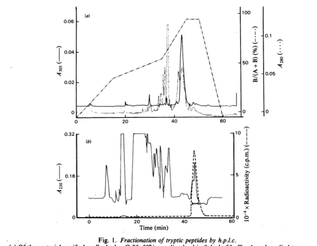
The solution of enzyme labelled with inhibitor was made 2M with respect to urea,0.5mg of trypsin was added and, after 60min at 37° C, a second portion of 0.5mg of trypsin was added and incubation continued for 60min. The digest was then fractionated on a column (140cm x 1cm) of Sephadex G-25. Fractions eluted with water, having absorbance at both 305 and 254nm (determined with fixed-wavelength detectors), were pooled (average Kd 0.38, where Kd is the distribution coefficient of the peptide). H.p.l.c. gave one major peak detected by A305 (Fig. la). The corresponding fractions (eluted between 42 and 45min) were further purified by a second run
under similar conditions, yielding peptide TI, which was 22 residues long and had N-terminal alanine (Table 1). The sequence, determined on 2nmol with the gas-phase sequenator, is given in Fig. 2; the penultimate, and antepenultimate residues (Ile, Ser) were not identified with cer-tainty. Hence 8nmol of Ti were digested with chymotrypsin and the peptide with absorbanceat 305nm (TICI) purified by h.p.l.c. The N-terminalresidue wasglutamic acid (Table 1), and the sequence, determined manually, showed that TICI comprisedresidues 16-20 (Fig. 2), and that the serine residuelabelled by inhibitor was
serine-19.
Isolation ofpeptides labelled with substrate The labelled enzyme (20mg) was digested with 0.4mg of trypsin in 1% NH4HCO3/2M-urea for 2h
10I I_ cd ._o~ -Time(min)
Fig. 1. Fractionationoftrypticpeptides byh.p.l.c.
(a)Qfthematerial purifiedonSephadex G-25,50%wasdissolved in 0.4ml of buffer A and applied to a reverse-phaseNucleosil-7C18Macherey-Nagel column(0mmx250mm). Buffer A was;50mM-NH4HCO3in water and bufferBa 2:3(v/v)mixture of bufferA andacetonitrile. The shape of the gradient[expressedas100xB/(A +B)]is shownontheFigureandthe-flow rate was2ml*min'1.LabelledpeptidesweredetectedbytheirA305.(b) PeptideT2,
labelledwith [3H]cloxacillin and previously fractionated on Sephadex G-25 (Kd=0.6) and SP-Sephadex, was
appliedto areverse-phase Lichrosorb RP (5pm) column(4.6mmx250mm). Buffer A was20mM-triethylamine acetate,pH4.5,inwaterand buffer Ba2:1: 2(byvol.)mixtureofacetonitrile, propanoland buffer A. Thegradient
was linear over60min and theflow rate Iml'min-'. Labelledpeptidesweredetectedby their radioactivity. 1984 272 ,-I x 1-1 -n 0 In 'lt. 1-1 0 In C4 '11:.
The active site of P99
P-lactamase
Pseudomonas aeruginosa Val-Thr-Pro-Glu-Thr-Leu-Phe-Glu-Ile-Gly-Ser-Val-Ser-Lys 62 70 80 ampC Ala-Asp-Ile-Ala-Lys-Lys-Gln-Pro-Val-Thr-Gln-Gln-Thr-Leu-Phe-Glu-Leu-Gly-Ser-Val-Ser-Lys-Thr 5 10 15 * 20 P99 Ala-Asp-Ile-Ala-Ala-Asn-Lys-Pro-Val-Thr-Pro-Gln-Thr-Leu-Phe-Glu-Leu-Gly-Ser-Ile-Ser-Lys-Thr Ti T2 --Z TlClC P1 P2Fig. 2. Active-site sequences of class-C/-lactamases
The sequencethatisdeduced for the 23-residue fragment of the P99
P-lactamase
is shown; the asterisk marks the serine residue that is labelled by substrate andinhibitor.Peptides obtained by the action of trypsin and pepsin are denoted by 'T' and 'P', respectively; further digestion of tryptic peptides Ti and T2 with chymotrypsin or thermolysin gave peptidesTlCl and T2H1 respectively. The sequence 1-19 of Ti was determined with the gas-phase sequenator. All the other peptides were sequenced except the last one. Above the continuous sequence are shown the sequences of ampC ,B-lactamase (middle line) (Jaurin & Grundstr6m, 1981) and Pseudomonas aeruginosa ,B-lactamase (top line) (Knott-Hunziker et al., 1982a). The numbering of the ampC ,B-lactamase is derived from the complete sequence of the gene, including the signal peptide.Table 1. Compositionoflabelledpeptides
The Tablegives residues/moleculefrom amino acidanalysis;the values inparentheses refertoresiduesnotfoundby
theEdman sequence analysis.The integral values in columns(3), (5), (7), (11)and (13) are from the sequence analysis, and in column (9) arecalculated for residues 17-19. The electrophoretic mobility (m) at pH6.5 was calculated relativetoAsp= -1(Offord, 1977).Columns(1)and(6)refertopeptide labelled with
6f,-iodopenicillan-ate, and columns(2), (4), (8), (10)and(12)refertoradioactivepeptideslabelled with[3H]cloxacillin. N-Terminal residueswereidentifiedbythedansylprocedure (Bruton&Hartley, 1970). T, P,TICI and T2Hl areexplained in thelegend toFig. 2.
Composition (residues/molecule) A Peptide ... T1 Column ... (1) 2.1 2.0 2.0 2.0 1.9 1.4 2.8 1.2 1.8 2.0 0.9 1.8 3H* ... 0.9 m ... -0.3
N-Terminus ... Ala Ala
No. of
residues ... 22
*Mol of labelled substrate bound/mol of peptide.
T2 TICI T2H1
(2) (3)
(4) (5)
(6)
(7)
1.9 2 (0.4) (0.3) 1.5 2 1.6 2 1.4 2 1.3 1 2.1 2 0.4 1 1.2 1 1.9 2 1.7 1 1.5 1 1.0 1 3.1 3 (1.3) 1.3 1 1.9 2 1.0 1 0.5 1 2.0 2 1.7 1 1.1 1 0.9 1 2.0 2 0.9 1 0.8 -0.27 Glu(8)
(9)
0.9 1(0.4).
1.1 I 0.9 -0.44 Glu Leu 7 5 P1 (10) (1 1) 1.0 1 1.0 2.2 2 2.0 0.9 1, 1.2 1 1.2 1 0.9 1 1.0 1 1.0 1 1.0 1 0.8 1 1.0 -0.31 Phe.9
3at 37° C and the digest fractionated on a column
(150cmxO.9cm) of Sephadex G-25 (superfine
grade)in 0.1M-acetic acidat4° C.Theradioactive fraction (Kd 0.34) contained peptide TI, and a
secdnd radioactive fraction (Kd 0.6) contained
peptide T2 (Table 1); further tryptic hydrolysis convertedpeptideTI intopeptideT2.The labelled
enzymewasalsodigested with 1% pepsin in 1
mM-HCl/3M-guanidinium chloride for60minat37° C. Thetrypticorpeptic digests,afterfractionationon
Vol. 223 Amino acid Asp Thr Ser Glu Pro Gly Ala Val Ile Leu Phe Lys P2 (12) (13) 1 2 1.0 1 1.1 1 0.8 1 1.0 -0.15 Leu 7 273
r-274 B. Joris and others Sephadex G-25, were further fractionated on
SP-Sephadex 50 and by h.p.l.c.; the peptic digest yielded two peptides, P1 and P2 (Table 1). The h.p.l.c. fractionation of the Sephadex fractions containing peptide T2 is shown on Fig. l(b).
The amino acid sequence of peptide T2 showed that it comprised residues 16-22 (Fig. 2). The sequences of peptides P1 and P2 showed that they comprised residues 15-23 and 17-23 respectively. Finally, peptide T2 was digested with thermolysin and the digestfractionatedby h.p.l.c. The radioac-tive tripeptide (T2H1) (Table 1) contained the labelled serine in the sequence Leu-Gly-Ser and comprised residues 17-19 (Fig. 2). Since serine is the only amino acid in this tripeptide with a reactivesidechain,itisserine-19that is labelled by substrate.
Discussion
The results on the P99
P-lactamase
in Fig. 2 establish that the sameserine residue islabelledby6fi-iodopenicillanate,
a 'branched-pathway' f3-lactamaseinactivator, and by cloxacillin, an 'inhibi-tory substrate' (Cartwright & Waley, 1983). The sequence ofthe 18 residues before, and the four residues after, serine-19 is firmly based on the structuresof sixpeptides. The sequence ofpeptides containing theactive-site serine residues of the ,B-lactamasesofPseudomonas aeruginosa and Escheri-chia coli K 12 (ampC gene) has been previously established (Knott-Hunziker et al., 1982a). The corresponding sequence obtained in the present work is closely similar to that of the ampC ,B-lactamase: 18 out of 23 residues are identical. Similarly, 11 out of 14residuesarethe same in the P99andPseudamonasaeruginosaP-lactamases.
In fact, among the 14 residues corresponding to positions 70-83 in the ampC,-lactamase, ten are identicalin these threefi-lactamases
(Fig. 2). Our results clearly establish the E. cloacae P99 /3-lactamase as amember of class C, and the serine residue labelled as the counterpart of serine-80 in the ampC ,B-lactamase (the position of peptide Ti in the sequence still requires determination).It was observed with variouspenicillin-sensitive enzymes that the homology was much more pronounced in theimmediate surroundings of the penicillin-binding serine residue (Frere & Joris, 1984). Although these homologies were not as strong as thoseobserved in the present study, one may wonderwhether, in the case of the class C /3-lactamases, the homology extends further away
from the active serine residue. This problem requires further investigation.
The isolation ofanacyl-enzyme from cloxacillin and the P99
P-lactamase
suggeststhat this covalent intermediate is important in catalysis. Kinetic studies are necessary to decide whether this intermediate is onthe main reaction pathway.The support of the Medical Research Council(U.K.) and of the National Fund for Scientific Research (Belgium), the Fonds de la Recherche Scientifique Medicale, Belgium (contract n° 3. 4507.83) and of the BelgianState(Actionconcertee n° 79/84-I1) is gratefully acknowledged.WethapkMr. N. Gascoyne forcarrying out the amino acidanalysesandGlaxo Group Research Ltd., Pfizer Researchand BeechamPharmaceuticals for gifts of various samples. B.J. and J.D. arerespectively 'Charge deRecherches'and 'ChercheurQualifie'of the NationalFund forScientific Research.
References
Ambler, R. P.(1980)Philos. Trans.R.Soc.London Ser. B 289, 321-331
Bruton,C. J. & Hartley, B. S.(1970)J.Mol. Biol. 52, 165-178
Campbell, D. G., Gagnon, J., Reid, K. B. M. & Williams, A. F. (1981)Biochem. J. 195, 15-30 Cartwright,S. J. &Waley,S. G.(1983)Med. Res. Rev. 3,
341-382
Cartwright,S. J. &Waley, S.G.(1984) Biochem.J.221, 505-511
Charlier,P.,Dideberg,O., Frere, J. M., Moews, P. C. & Knox, J. R.(1983)J. Mol.Biol. 171, 237-238 Cohen,S. A. & Pratt, R. F.(1980) Biochemistry 19,
3996-4003
Fisher, J., Belasco, J. G., Khosla, S. &Knowles, J. R. (1980) Biochemistry19, 2895-2901
Fisher, J.,Charnas, R. L., Bradley, S M. &Knowles,
J. R. (1981)Biochemistry 20, 2726-2731
Frere, J. M. & Joris, B.(1984)CRC Crit. Rev.in the press Hewick, R. M., Hunkapiller, M. W., Hood, L. E. & Dreyer, W. J. (1981)J. Biol. Chem. 256, 7990-7997 Jaurin,B. &Grundstrom,T.(1981)Proc.Nat!.Acad. Sci.
U.S.A. 78,4897-4901
Knott-Hunziker, V., Orlek, B. S., Sammes, P. G. & Waley, S. G. (1979) Biochem. J. 177, 365-367 Knott-Hunziker, V., Orlek, B. S., Sammes, P. G. &
Waley, S. G. (1980) Biochem. J. 187, 797-802 Knott-Hunziker, V., Petursson, S., Jayatilake, G. S.,
Waley, S. G., Jaurin, B. & Grundstrom, T. (1982a) Biochem. J. 201,621-627
Knott-Hunziker, V., Petursson, S., Waley,S.G., Jaurin, B. &Grundstrom,T.(1982b)Biochem. J.207, 315-322 Neu, H. C. (1983) Infection 11, Suppi. 2,74-80 Offord, R. E. (1977) Methods Enzymol. 47, 51-69 Ross, G. W. (1975)MethodsEnzymol. 43, 678-687