Electrophysiological
impact
of
multiple
concussions in asymptomatic athletes: a re-analysis
based on alpha activity during a visual-spatial
attention task
Samuel Guay, Louis De Beaumont, Brandi Lee
Drisdelle, Jean-Marc Lina, Pierre Jolicoeur
PII:
S0028-3932(17)30444-X
DOI:
https://doi.org/10.1016/j.neuropsychologia.2017.11.022
Reference:
NSY6578
To appear in:
Neuropsychologia
Received date: 10 September 2017
Revised date:
31 October 2017
Accepted date: 16 November 2017
Cite this article as: Samuel Guay, Louis De Beaumont, Brandi Lee Drisdelle,
Jean-Marc Lina and Pierre Jolicoeur, Electrophysiological impact of multiple
concussions in asymptomatic athletes: a re-analysis based on alpha activity during
a
visual-spatial
attention
task,
Neuropsychologia,
https://doi.org/10.1016/j.neuropsychologia.2017.11.022
This is a PDF file of an unedited manuscript that has been accepted for
publication. As a service to our customers we are providing this early version of
the manuscript. The manuscript will undergo copyediting, typesetting, and
review of the resulting galley proof before it is published in its final citable form.
Please note that during the production process errors may be discovered which
could affect the content, and all legal disclaimers that apply to the journal pertain.
Electrophysiological impact of multiple concussions in asymptomatic athletes: a re-analysis based on alpha activity during a visual-spatial attention task
Samuel Guaya,b, Louis De Beaumontb,c, Brandi Lee Drisdelled,e, Jean-Marc Linab,f, Pierre Jolicoeurd,e,g1 a
Department of Psychology, Université du Québec à Trois-Rivières, Trois-Rivières, QC, Canada bCentre de recherche de l’Hôpital du Sacré-Coeur de Montréal, Montreal QC, Canada
c
Department of Surgery, Université de Montréal, Montreal, QC, Canada d
Department of Psychology, Université de Montréal, Montreal, QC, Canada e
Centre de recherche en neuropsychologie et cognition (CERNEC), Université de Montréal, Montreal, QC, Canada
f
Montréal Polytechnique, Montreal, QC, Canada
gCentre de recherche de l’Institut universitaire de gériatrie de Montreal, Montreal, QC, Canada pierre.jolicoeur@umontreal.ca
Abstract
Most EEG studies used event-related potentials to assess long-term and cumulative effects of sport-related concussions on brain activity. Time-frequency methods provide another approach that allows the detection of subtle shifts in types and patterns of brain oscillations. We sought to discover whether event-related alpha activity would be significantly affected in asymptomatic multi-concussed athletes. We measured the amplitude of alpha activity (8-12 Hz) from the EEG recorded during a visual-spatial attention task to compare event-related alpha perturbations in 13 multi-concussed athletes and 14 age-equivalent, non-concussed teammates. Relative to non-concussed athletes, multi-concussed athletes showed significantly less event-related perturbations time-locked to stimulus presentation. Alpha activity alterations were closely related to the number of concussions sustained. Event-related alpha activity differed in asymptomatic multi-concussed athletes when compared to controls. Our study suggests that low-level neurophysiological underpinnings of the deployment of visual-spatial attention are affected in multi-concussed athletes even though their last concussion occurred on average 30 months prior to testing.
Key Words
Concussion; Long-term effects; Cumulative effects; EEG; Alpha; Event-related spectral perturbations
Introduction 1
Over the last decade, numerous studies have attempted to characterize the long-term and cumulative effects of sports-related concussions (SRC). While the vast majority of concussed athletes tend to
1 Département de Psychologie, Université de Montréal, C.P. 6128, succursale Centre-ville, Montréal, QC, Canada, H3C 3J7.
clinically recover within three to four weeks after an SRC (Henry, Elbin, Collins, Marchetti, & Kontos, 2016; McCrory, et al., 2017; Nelson, et al., 2016; Prichep, McCrea, Barr, Powell, & Chabot, 2013), electroencephalographic (EEG) measures can detect subclinical neurophysiological abnormalities in concussed athletes that persist long beyond the acute phase (Henry, Tremblay, & De Beaumont, 2016).
For instance, event-related potential (ERP) studies revealed significant and persistent N1 (Gosselin, Theriault, Leclerc, Montplaisir, & Lassonde, 2006), N2 (Ledwidge & Molfese, 2016), P2 (Gosselin, et al., 2006), P3 (De Beaumont, Brisson, Lassonde, & Jolicoeur, 2007; Gaetz, Goodman, & Weinberg, 2000; Gosselin, et al., 2006; Ledwidge & Molfese, 2016; Theriault, De Beaumont, Gosselin, Filipinni, & Lassonde, 2009) waveform components alterations when concussed athletes performed cognitive tasks relying on attentional processes in the acute phase as well as several years post-accident relative to controls. ERPs are computed from the averaged EEG signal time-locked to an event of interest, often the presentation of a stimulus. With appropriate stimuli and task conditions, the resulting waveforms can reveal components reflecting specific cognitive processes. Repeated concussions were found to significantly accentuate anomalies of ERP markers of attention and working memory (De Beaumont, et al., 2007; Gaetz, et al., 2000), supporting the notion that concussions are associated with long-term and cumulative effects on brain functions (Guskiewicz, et al., 2003; Henry, Tremblay, et al., 2016; Pearce, et al., 2014).
In parallel, other studies investigated resting oscillatory activity using time-frequency methods to closely track the recovery of neurophysiological function after concussion (Arciniegas, 2011; Haneef, Levin, Frost, & Mizrahi, 2013; Kutcher, et al., 2013). Time-frequency analyses characterize EEG in terms of power (squared amplitude) and phase of oscillations using a variety of analytic techniques that enable researchers to detect changes in the types and patterns of electrical oscillations as a function of a stimulus, task, psychological state, or neurological condition (Tallon-Baudry & Bertrand, 1999). A growing body of research seeks to relate oscillatory activity (e.g., averaged amplitude or power) with
underlying cognitive processes involved in information processing and increasingly with intra and inter-region neuronal communication (Bastos, et al., 2015; Frey, Ruhnau, & Weisz, 2015). The EEG spectrum is often described in terms of frequency bands, such as delta (0.5–4 Hz), theta (4–7 Hz), alpha (8–12 Hz), beta 1 (13–20 Hz), beta 2 (20–30 Hz), and gamma (> 30 Hz), although the precise boundaries between bands and often their names vary from author to author (Babiloni, et al., 2010; Pfurtscheller & Lopes da Silva, 1999). The impact of brain pathology on the alpha band has received much attention mostly due to the presumed association of alpha oscillations with a number of brain processes (Klimesch, 1999). The most documented changes found in brain activity after mild traumatic brain injury (mTBI) or SRC are a decrease in alpha power coupled with a concomitant increase in theta-alpha frequency ratio (Haneef, et al., 2013; Korn, Golan, Melamed, Pascual-Marqui, & Friedman, 2005; Kutcher, et al., 2013; Nuwer, Hovda, Schrader, & Vespa, 2005). Interestingly, this pattern is also found in the aging population as well as in patients presenting with a variety of neurological disorders (Chiang, Rennie, Robinson, van Albada, & Kerr, 2011; Klimesch, 1999; van Albada, Kerr, Chiang, Rennie, & Robinson, 2010). In the concussion literature, it was proposed that the suppression of alpha amplitude in posterior cortical regions could potentially be a sensitive tool in order to detect lingering cerebral dysfunctions (Rapp, et al., 2015; Slobounov, Sebastianelli, & Hallett, 2012; Thatcher, Walker, Gerson, & Geisler, 1989). Follow-up studies on acute SRC showed return-to-baseline of oscillatory activity within 45 days of the injury (Barr, Prichep, Chabot, Powell, & McCrea, 2012; McCrea, Prichep, Powell, Chabot, & Barr, 2010). In contrast, athletes who had suffered a second SRC exhibited a slower rate of recovery on alpha oscillation amplitudes at 7, 14, and 21 days post-injury (Slobounov, Cao, & Sebastianelli, 2009). Furthermore, one study observed a persistent reduction of resting alpha power in multi-concussion athletes tested at 12 months post-injury (Gosselin, et al., 2009). However, no clear EEG or time-frequency features seem specific to mTBI or SRC, especially beyond the acute post-injury phase (Nuwer, et al., 2005; Popescu, Hughes, Popescu, Riedy, & DeGraba, 2016).
Nonetheless, it is important to study alpha activity in concussion because of its implication in visual and spatial attention (Foxe & Snyder, 2011; van Diepen, Miller, Mazaheri, & Geng, 2016), which are affected in asymptomatic concussed athletes (Theriault, De Beaumont, Tremblay, Lassonde, & Jolicoeur, 2011).
Similar to ERPs, researchers can compute event-related spectral perturbation (ERSP) analyses that are time-locked to stimulus presentation. These analyses measure the average oscillatory amplitude induced by the presentation of stimuli relative to a prestimulus period (Makeig, 1993), enabling studies of spectral dynamics (i.e., how oscillatory amplitude changes over time). Spectral amplitude variations (increase/decrease) are thought to reflect changes in the activity of large assemblies of neurons in response to a stimulus or to other events of interest. Event-related synchronization (ERS) occurs when the amplitude of a given frequency increases with stimulus presentation, while event-related desynchronization (ERD) reflects a reduction of amplitude in response to a stimulus (Pfurtscheller & Lopes da Silva, 1999). According to some researchers, a large increase in alpha amplitude reflects a state of inhibition or cortical deactivation, whereas a decrease in power reflects a state of comparatively high neuronal excitability (Klimesch, 2012; Pfurtscheller, 2003). A decrease in alpha power is characteristically observed in the occipital regions when subjects process visual inputs or when they respond to internal events like mental activation or cognitive effort (Pfurtscheller & Lopes da Silva, 1999). Alpha power is also known to decrease as a function of attentional demands and generally spreads over the entire scalp, argued to reflect the gradual release of inhibition associated with the activation of attentional networks during information processing (Klimesch, Sauseng, & Hanslmayr, 2007).
Only a few ERSP studies have been conducted with clinical populations. Available results in patients presenting attention-deficit hyperactivity disorder (Gomarus, Wijers, Minderaa, & Althaus, 2009; Missonnier, et al., 2013); mild cognitive impairment (Caravaglios, Muscoso, Di Maria, & Costanzo,
2015; Deiber, et al., 2015); Alzheimer’s disease (Hogan, Swanwick, Kaiser, Rowan, & Lawlor, 2003), Parkinson’s disease (Heida, Poppe, de Vos, van Putten, & van Vugt, 2014), and schizophrenia (Haenschel, et al., 2009) show that ERSP analyses can sometimes reveal alterations in brain activity correlated with task performance and various neurological conditions. These clinical populations typically exhibit a pattern of attenuated ERD and larger ERS in alpha amplitude relative to healthy control groups during cognitive tasks. Given the known detrimental effects of concussion on resting alpha band activity, this study sought to determine whether persistent changes of event-related spectral perturbations within the alpha band (ERSP-α) occur while performing an attention task in concussed athletes.
A small number of studies that performed ERSP-α analyses in conjunction with ERP waveform components yielded enlightening results. Among them, a study showed associations between ERSP-α and the P3 component amplitude (Peng, Hu, Zhang, & Hu, 2012). An increase in P3 amplitude, which is thought to reflect attentional resource allocation and memory updating (Polich, 2007), is usually associated with a decrease in alpha amplitude (Ergenoglu, et al., 2004; Peng, et al., 2012; Sergeant, Geuze, & van Winsum, 1987; Yordanova, Kolev, & Polich, 2001). Interestingly, the P3 component shows chronic amplitude suppression among multi-concussion athletes (Broglio, Pontifex, O'Connor, & Hillman, 2009; De Beaumont, et al., 2007; Gaetz, et al., 2000; Gosselin, et al., 2006; Theriault, et al., 2009). Thus, studying the long-term effects of SRC on the alpha activity pattern during a cognitive task could reveal an important feature of the underlying neurophysiological underpinnings of persistent, subclinical anomalies after concussion.
The goal of the present study was to investigate the long-term effects of multiple SRC on alpha oscillatory activity modulations. Based on the SRC and oscillatory literature, we hypothesized that alpha desynchronization would be significantly reduced in multi-concussed athletes relative to control
athletes when performing a visual-spatial attention task. We also hypothesized that attenuation of event-related alpha desynchronization would be increase with the number of previous concussions.
Methods 2
In the present study, we used electrophysiological recordings from a previous EEG experiment that revealed P3 amplitude alterations in concussed athletes (De Beaumont, et al., 2007). This experiment was designed to assess possible differences in brain function between control and multi-concussed athletes in the context of a particular perceptual-cognitive task. The EEG recordings herein were analyzed on different EEG measures, thus not interfering or replicating previously published work. Only electrophysiological recordings from multi-concussed and non-concussed athletes were used for the purpose of this study. Given we are now looking at a different subset of the signal space, we occasionally see artefacts that were not apparent in the original work. Because of EEG signal-quality issues, one subject was removed from the control group and two from the concussion group of the original sample. Refer to the aforementioned published article for a more detailed description of the methods.
Participants 2.1
The sample analyzed in the present study consists of 27 active athletes recruited from a Canadian university football team. Athletes who took part in this study were those who were not rejected after having been screened for the following exclusion criteria: A history of alcohol and/or substance abuse, psychiatric illness, learning disability, neurological history (seizure, central nervous system neoplasm, or brain tumour), a history of TBI unrelated to contact sports or any daily medications. The study was vetted by the local ethics committee and all participants provided written informed consent prior to testing. Subjects received a financial compensation of $30 Canadian for their participation.
The total sample was divided into two experimental groups based on prior concussion history established in an experiment designed to elicit the N2pc ERP component (De Beaumont, et al., 2007). The control group consisted of 14 athletes who neither presented with a history of SRC nor a neurological trauma (e.g., motor vehicle accident) at the time of testing. Group classification was performed by a sports physician according to AAN clinical criteria on concussion at the time of testing (American Academy of Neurology, 1997). The second group consisted of 13 asymptomatic athletes who had reported two or more SRC sustained more than 9 months prior to testing. The number of concussions ranged from 2 to 6 (M = 3.08, SD = 1.26) and the time elapsed since the latest concussion ranged from 9 to 81 months (M = 31.61, SD = 21.90). Detailed information about concussion specifications (number, approximate date, accident description, nature and duration of on-field post-concussion severity) was obtained via a standardized post-concussion history form. Demographic and concussion information are summarized in Table 1. Both groups were considered equivalent in terms of age (t(25) = 1.00, p = .33), level of education (t(25) = 1.37, p = .18), and post-concussion symptoms reported on the Post-Concussion Symptom Scale (t(25) = 1.5, p = .16). Groups reported only few concussion-associated symptoms and were asymptomatic at the time of testing based on validated clinical threshold for post-concussion syndrome.
Table 1Demographic and concussion information
Control Multi-concussed (≥ 2)
Sample size (N) 14 13
Age 22.5 ± 2.53 23.5 ± 2.67
Education 17.0 ± 1.68 17.9 ± 1.73
Number of concussions - 3.08 ± 1.26
Time since last concussion (months) - 31.6 ± 21.90
LOC - 0.65 ± 0.92
PTA - 2.0 ± 1.84
PCS 8.9 ± 7.11 13.5 ± 9.26
Demographic, concussion severity markers and post-concussion symptoms information collected from the original N2pc experiment sample (De Beaumont et al., 2007). Values are expressed as means ± standard
deviation across experimental groups. Abbreviations: LOC = loss of consciousness; PTA = post-traumatic amnesia; PCS = post-concussion symptoms.
Visual spatial attention task. 2.2
Each participant performed one practice block of 48 trials followed by five experimental blocks of 96 trials. Following the presentation of a 500-ms fixation point, four colored squares were presented on each trial for 100 ms, then five seconds were given to the participants to provide their answer. Participants were instructed to maintain fixation on a point at the center of the screen throughout the trial and to indicate if the location of a gap in the target square (designated by color) was on the left, right, or upper side by pressing the ‘V’ key or pressing the ‘N’ key when the opening was on the bottom side, for half of the subjects, and the other response key pairing for the other half. In all trials, the odd-colored square (i.e., the possible target) had 0.25 probability of being presented in any of the four locations (near-left, near-right, far-left, far-right positions). The gap was presented with a 0.25 probability on each side of the target box (left, right, up, down). However, because there were only two possible responses (bottom vs. not-bottom), the probability of one of the response “bottom” was 0.25, whereas the probability of the response “not-bottom” was 0.75. This created a frequency difference across the responses, despite an equal probability of each possible gap location. Target stimuli were either red amongst green distractors or green amongst red distractors (counterbalanced across subjects). To control for low-level sensory responses, the colors were equiluminant and set according to measurements from a Minolta CS100 chroma meter.
Given the goal of the present study to examine alpha activity, some task conditions were irrelevant, considering that maximal alpha was recorded from centro-occipital electrode, a site primarily responsible for visual perception. Consequently, we examined alpha activity on every stimulus onset, collapsing many attention conditions together (e.g., no-go trials, gap opening position, frequency, and
color manipulations). Hence, the alpha activity was assessed based on the onset of visual stimuli, regardless of conditions.
Electrophysiological recordings 2.3
The electrophysiological recordings used in this study were taken from a previous experiment focusing on the cumulative and long-term effects of concussions. The N2pc experiment was initially conducted to elicit ERP waveforms related to visual spatial attention, but as it involved presentation of visual stimuli, we were able to compute ERSP analyses on background EEG activity within the alpha band.
The EEG was recorded from 64 active Ag/AgCl electrodes using a Biosemi Active Two System mounted on an elastic cap. Electrodes were referenced to averaged mastoids and positioned according to the extended International 10–10 system. Eye movements (e.g., saccades and blinks) were monitored by additional electrodes: two electrodes placed on external canthi to record horizontal electrooculogram (HEOG) and two placed on infra/supraorbital regions to record the vertical electrooculogram (VEOG). The EEG was digitized at 256 Hz and processed offline. The EOG channels were high-pass filtered at 0.1 Hz and low-pass filtered at 10 Hz. Independent component analyses (ICA) were conducted on all participants to remove eye blinks using methods described in Drisdelle, Aubin, and Jolicoeur (2017). Following the ICA, the VEOG difference wave was checked for any remaining artifactual activity by removing segments with a difference greater than 80 µV over a 150 ms interval, and the HEOG difference wave was screened for differences greater than 35 µV over a 300 ms. On average, 0.03% of the trials exceeded the VEOG threshold for the control group and 0.4% for the concussion group. For the HEOG test, 1.4% of the trials were excluded for the control group and 2.5% for the concussion group.
Estimation of alpha oscillation amplitude. 2.4
In order to estimate the amplitude of oscillations in the alpha band, we used an approach similar to Monto, Vanhatalo, Holmes, and Palva (2007). We took the absolute value of the Hilbert transform of the EEG signals high-pass filtered at 8 Hz, and low-pass filtered at 12 Hz, using infinite impulse response Butterworth, order 6, digital filters. This operation was performed on the continuous data, thus yielding a continuous estimate of the amplitude of oscillations at about 10 Hz. These amplitude values were then segmented from -200 ms to +1000 ms relative to stimulus onset. These segments were screened for artefacts defined as deflections greater than ±11 µV anywhere in the segment. ±11 µV was determined to reject large artefacts since the estimated instantaneous amplitudes of signal bandpassed from 8 to 12 Hz fluctuates in a smaller range than the original EEG (which is the sum of oscillations in a much broader range of frequencies). Therefore, we needed to use a smaller value than is used for broadband EEG (e.g., ±100 µV), which otherwise would have allowed artefacts in the alpha band to contaminate downstream analyses. With a cutoff at ±11 µV, we rejected between 5% and 6% of the trials. Channels that exceeded this amplitude criterion were interpolated from nearby channels if less than 8 channels were artefacted in a given segment; if 8 or more channels contained an artefact, the segment was rejected from further analysis. The screened segments were averaged for each subject from 350 to 550 ms post-stimulus and used for later statistical analyses. Then, a baseline correction was done by subtracting the mean amplitude of the activity recorded during the 200-ms prestimulus period from the entire segment. The results yielded values that were either positive, meaning an increase in alpha amplitude relative to the baseline, or negative, meaning a decrease in alpha amplitude. Because maximum alpha activity occurred over occipital areas, analyses were performed at the electrode site Oz. A decrease of alpha amplitude over occipital areas is thought to reflect cognitive demand and anticipatory attention or preparation to a visual-spatial task, whereas a decrease over central regions has
been associated with motor planning (Basar, Basar-Eroglu, Karakas, & Schurmann, 2001; Maclean & Arnell, 2011; Pfurtscheller & Lopes da Silva, 1999).
Given that alpha activity peaks at a specific frequency within the alpha band for each subject and that this inter-individual variability was found to affect study results (Haegens, Cousijn, Wallis, Harrison, & Nobre, 2014), we examined oscillatory amplitude within a broader range (i.e., 8 to 14 Hz) with a different technique. We performed the more detailed analysis by means of a set of Morse analytic wavelets (Lilly, 2017; Lilly & Olhede, 2010; Olhede & Walden, 2002). The center frequency of the wavelet ranged from 8 to 14 Hz, in 0.5 Hz steps (i.e., 8.0, 8.5, ... 13.5, 14.0 Hz). This finer exploration of the oscillation amplitude in the broad alpha range allowed us to estimate, for each subject, the 'best' frequency at which the amplitude decreased the most following stimulus onset. Using the 'best' ERSP-α frequency, we were able to estimate a baseline-corrected ERSP-α amplitude at each subject’s preferential alpha frequency from 350 ms to 550 ms post-stimulus.
Statistical analyses 2.5
Demographic and concussion information were subjected to groups t tests. Then, between-factor analyses of variances (ANOVA) were computed on the amplitude of the ERSP-α at electrode Oz for both techniques. The between-factor (i.e., group; concussed vs. control) ANOVAs were performed without within-factor because alpha activity was assessed regardless of conditions, as mentioned in section 2.2. We then conducted Pearson correlational analyses between ERSP-α amplitudes and clinical variables (number of concussions, time since the last concussion, post-concussion symptoms), and the amplitude of the P3 component from our previous N2pc experiment (De Beaumont, et al., 2007) since this ERP component seems closely associated with alpha rhythm in healthy controls (Peng, et al., 2012; Yordanova, et al., 2001). Two-tailed p-values less than .05 were considered statistically significant.
Results 3
Behavioral results 3.1
Participants accuracy and mean response time for correctly identifying the target were calculated and compared. Accuracy rate was higher than 90% and equivalent in both groups (F < 1). Similarly, the two groups did not differ in terms of mean response time either for the frequent (F(1, 25) = 0.73; p = .40) or infrequent response conditions (F(1, 25) = 1.26; p = .27).
Electrophysiological results: Event-related alpha activity 3.2
Using the two techniques described in section 2.4, we calculated the mean amplitude of the ERSP-α recorded at Oz from 350–550 ms post-stimulus. Here, we present our comparisons between both groups for the ERSP-α that were baseline-corrected from the prestimulus alpha activity from -200 to 0 ms.
3.2.1 ERSP-α using Hilbert transform method
ERSP-α evoked by target stimuli were significantly reduced in multi-concussed athletes (M = -0.38,
SD = 0.50) relative to non-concussed athletes (M = -1.00, SD = 0.64), when performing a visual-spatial
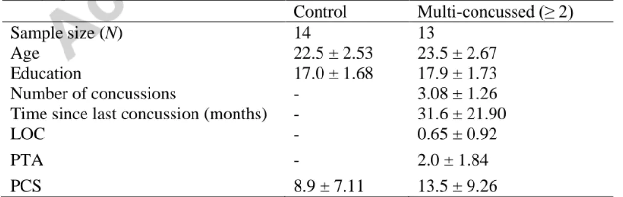
attention task, F(1, 25) = 8.03; p = .009, η2 = 0.2433, indicating a smaller post-stimulus alpha amplitude (in µV) change relative to the prestimulus period. Figure 1A depicts the baseline-corrected dynamics of the grand average alpha amplitude recorded at Oz for both groups. Figure 1B also shows the scalp topographies of the mean alpha amplitude (in the interval from 350–550 ms) after baseline correction in both groups.
One may wonder if the smaller ERSP-α reduction in multi-concussed athletes could be a consequence of lower alpha amplitude immediately before stimulus onset. Simply put, if alpha activity is reduced at baseline, there is simply less alpha to desynchronize after stimulus presentation. In order to investigate
this possibility, we expressed the amplitude of the ERSP-α as a percentage of the baseline alpha amplitude. The average reduction in alpha amplitude was 33.7% for non-concussed athletes and 14.5% for multi-concussed athletes, F(1, 25) = 11.34, p = .0025, η2 = 0.3120, thus reaffirming that the ERSP-α was disproportionately smaller in multi-concussed athletes relative to non-concussed athletes.
Figure 1: A) Baseline-corrected dynamics of the grand average alpha amplitude recorded at Oz (in the interval from 350–
550 ms) for both groups (unconcussed athletes group: blue line; Multiple concussion group: green dashed line). B) Scalp topographies of the mean alpha amplitude after baseline correction in both group from 350 to 500 ms poststimulus.
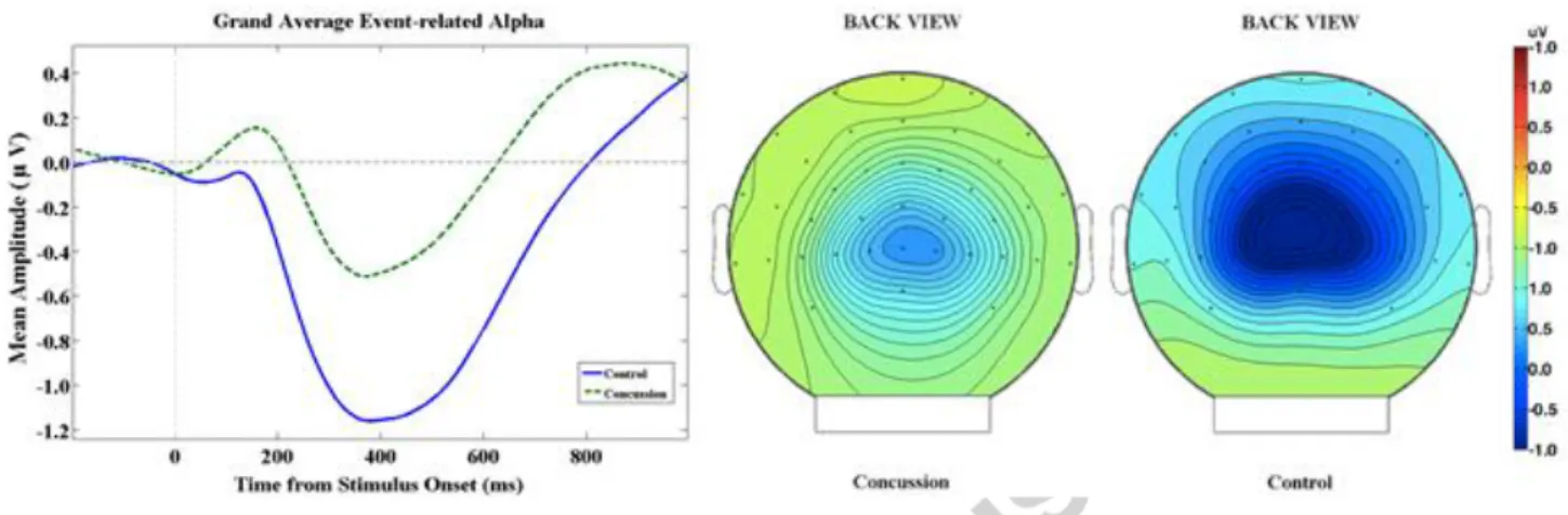
We then conducted two-tailed Pearson correlational analyses between the ERSP-α amplitude and clinical variables of interest. Figure 2 illustrates the relationship between significant correlations. Whereas ERSP-α amplitude did not correlate with either the time elapsed since last concussion (r(11) = 0.19, p = .53) or post-concussion symptoms at the time of testing (r(11) = 0.34, p = .26), ERSP-α amplitude correlated with the number of concussions (r(11) = 0.58, p = .037), indicating that athletes who had sustained more concussions showed smaller ERSP-α (i.e., a smaller change in amplitude relative to baseline). In addition, we verified whether the P3 amplitude, which differed in concussed athletes relative to controls, was associated with the ERSP-α amplitude in the present study. We found a significant negative correlation between these two variables in multiconcussed athletes (r(11) =
-0.56, p = 0., p = .04), suggesting that multi-concussed athletes with smaller ERSP-α were those with smaller P3 amplitudes.
Figure 2: A) Scatter plot illustrating the significant positive correlation between the number of concussions and the
averaged event-related spectral perturbations within the alpha band (ERSP-α) found at Oz between 350-550 ms after stimulus onset. B) Scatter plot illustrating the significant negative correlation between the ERSP-α and the P300 amplitudes in multi-concussed athletes.
3.2.2 ERSP-α using Morse analytic wavelets method
To investigate the possibility that individuals’ preferential alpha rhythm could influence our results, we applied a different analytic technique (described in section 2.4) to a broader alpha band ranging from 8 to 14 Hz. We first tested whether the 'best' alpha frequency in each group was different and computed the ERSP-α at each subject’s alpha frequency for both groups.
We were able to determine that the 'best' ERSP-α frequency did not differ significantly between controls (M = 10.96 Hz, SD = 1.35) and concussed athletes (M = 11.08 Hz, SD = 1.82), F(1, 25) = 0.03, p = .86). When we estimated the amplitude of the ERSP-α at each subject's 'best' frequency, the control group exhibited a significantly greater ERSP than the concussed group, F(1, 25) = 6.19, p = .02, thus reproducing our original result for difference in the mean amplitude from 350 to 550 ms.
Discussion 4
We studied the long-term and cumulative effects of concussion on event-related alpha activity during an attentional task in 27 varsity athletes. We observed significant differences between multi-concussed athletes relative to age-matched controls where concussed athletes showed significantly reduced ERSP-α that associated strongly with the number of sustained concussions and with the amplitude of the P3 waveform component. This pattern of results is consistent with previous reports on the cumulative deleterious effects of sports concussions on electrophysiological markers of cognitive processes (De Beaumont, Beauchemin, Beaulieu, & Jolicoeur, 2013; De Beaumont, et al., 2007; Gaetz, et al., 2000). This finding suggests a potential mechanistic linkage between the chronic pathophysiology of recurrent concussions and alterations of ERSP-α.
A major finding of the present study resides in that multi-concussed athletes exhibit significantly reduced ERSP-α in the 350-550 ms post-stimulus period. Using a multimodal oddball paradigm, a study by Peng, Hu, Mao, and Babiloni (2015) supported the inhibition-timing model introduced by Klimesch et al. (2007). This model suggests that the post-target stimulus decrease of alpha activity reflects the underlying neurophysiological mechanism of attentional deployment set off when the task-relevant cortical areas are released from previous inhibition. Large event-related changes of alpha activity are typically reflective of cognitive efficiency (Klimesch, 1999) and have been linked to processes related to the active processing of task-relevant stimuli (Peng, et al., 2015). Given that greater alpha amplitude at rest as well as larger ERSP-α are both associated with good cognitive and memory performance (Klimesch, 1999; Limbach & Corballis, 2016) and that the latter are also known to play an active role in the mechanisms of attention and consciousness (Palva & Palva, 2007; van Diepen, et al., 2016), one could have expected to observe cognitive performance decrements in this sample of multi-concussed athletes. In this study, however, behavioral performance on the visual-spatial attention task did not differ between groups despite significant ERSP-α alterations following in asymptomatic multi-concussed athletes tested on average 30 months after their last concussion. It is to
note that the experimental paradigm was originally designed to investigate the effects of concussion on electrophysiological markers of visual-spatial attention via the N2pc waveform component. The abnormal ERSP-α following concussion coupled with the absence of between-groups behavioral performance differences provides additional support for the notion that EEG measures can reveal changes in brain activity that would otherwise go undetected based on behavioral performance measures. Future studies using neurocognitive tasks known to be more sensitive to concussion could be useful to uncover functional links between alpha oscillations, concussion, and cognitive function (Dean & Sterr, 2013; Gosselin, et al., 2012).
Moreover, our results corroborate the typical pattern of maximal alpha band activity during a visual task localized near the occipital midline (electrode Oz), as evidenced in numerous EEG/MEG studies (Ciulla, Takeda, & Endo, 1999; Makeig, et al., 2002; Peng, et al., 2015; Pfurtscheller, Neuper, & Mohl, 1994) and fMRI-EEG studies (Brookes, et al., 2005; Moosmann, et al., 2003). Consistent with scalp topography findings from this study, lateral occipital and central occipital regions were identified as dominant components of posterior alpha oscillations in a human visual selective attention task (Makeig, et al., 2002). A recent study analyzing sources of resting- and task-related alpha generators concluded that both generators of alpha oscillations shared a common origin (Tenke, Kayser, Abraham, Alvarenga, & Bruder, 2015). Brain oscillations have been hypothesized to play a fundamental role in neuronal synchronization across functionally-connected brain regions. Such inter-regional communication appears to be critical in several cognitive processes (Engel, Gerloff, Hilgetag, & Nolte, 2013; Frey, et al., 2015; Klimesch, et al., 2007; Scholvinck, Leopold, Brookes, & Khader, 2013). In the visual brain areas, white matter pathways propagate alpha oscillations from primary visual cortex (V1) to higher-order visual areas, suggesting that V1 plays a central role in coordinating posterior alpha oscillations through white matter (WM) projections (Hindriks, et al., 2015). Significant associations between WM structure (assessed by Diffusion Tensor Franctional Anisotropy) and alpha rhythm have
been found in healthy individuals (Chu, et al., 2015; Jann, et al., 2012). More precisely, alpha oscillation propagation is thought to be dependent of structural as well as functional connectivity (Chu, et al., 2015; Nunez & Srinivasan, 2014; Nunez, Srinivasan, & Fields, 2015), such that the well-known effects of concussion on WM integrity could interfere with inter-regional communication. Interestingly, it has been suggested that cognitive alterations observed in TBI patients are partially due to damaged alpha generators (Dockree, et al., 2004; Dunkley, et al., 2015) and recent studies have reported evidence of diffuse and heterogeneous network connectivity alterations following concussion (Dimitriadis, Zouridakis, Rezaie, Babajani-Feremi, & Papanicolaou, 2015; Dunkley, et al., 2015). Importantly, long-term white matter anomalies induced by concussions have been hypothesized to disrupt pre-concussion neural networks through axonal shearing (Henry, Tremblay, et al., 2016; Tremblay, et al., 2013). Altered networks are characterized by weak local connections and strong long-range connections, an inverse pattern relative to the control group (Dimitriadis, et al., 2015). According to a model proposed by Molfese (2015), neural networks reorganize after concussion by developing new local and distal networks to compensate for reductions of efficiency caused by brain damage. Ledwidge and Molfese (2016) provided evidence that concussed athletes generate atypical brain patterns by recruiting additional neuronal resources relative to controls in such a way that allows concussed athletes to achieve similar performance levels (McAllister, et al., 2001; McDonald, Saykin, & McAllister, 2012; Ozen, Itier, Preston, & Fernandes, 2013). When applied to current study findings, the recruitment of additional brain resources that may have taken place over several months post-concussion could explain normal behavioral performance levels in multi-concussed athletes. Recently, it was found that reduced alpha activity in TBI patients could be reversed to normal levels after an intensive neuropsychological rehabilitation program (Castellanos, et al., 2011). Future magnetoencephalography studies could be useful to validate longitudinally the evolution of ERSP-α through the activation of neural populations that now form modified networks.
A reduction of event-related alpha activity analogous to what was found in the present study has also been observed in patients with ADHD (Missonnier, et al., 2013; ter Huurne, et al., 2013). Abnormal modulations of alpha oscillations in ADHD were thought to stem from malfunctions of neural assemblies as well as functional connectivity alterations within the failing attentional network (ter Huurne, et al., 2013). This interpretation is partially supported by functional imaging studies that revealed abnormal activation within occipital as well as cortico-thalamic networks during attention and working memory tasks as well as white matter anomalies (Chen, et al., 2016; Cortese & Castellanos, 2012; Paloyelis, Mehta, Kuntsi, & Asherson, 2007; Wu, et al., 2017). Interestingly, neuropsychological symptoms of ADHD closely resemble those observed after concussion (White, Harris, & Gibson, 2014). It remains to be tested whether the persistent reduction of ERSP-α found herein could be linked to recent findings demonstrating persistent functional connectivity as well as white matter integrity loss following concussion (Han, Chapman, & Krawczyk, 2016; Tremblay, et al., 2013; Tremblay, et al., 2014).
Although time elapsed since last concussion or post-concussion symptoms at the time of testing did not correlate in this report, studies that have examined cognitive function in relation with event-related electrophysiology (e.g., (De Beaumont, et al., 2011; 2009; 2013; Ozen, et al., 2013) have found clinically-relevant decline as opposed to progressive recovery. This is an important reason for believing that electrophysiological anomalies in young concussed athletes may reveal key information on the neurobiological substrates of the chronic installation of brain function damage after concussion. Accordingly, ERSP-α amplitude correlated with the number of concussions, providing further support for the sensitivity of the latter electrophysiological marker to the chronic detrimental effects of concussion.
Previous studies showed that the deployment of visual-spatial attention influences posterior alpha power through top-down processes (Romei, Gross, & Thut, 2010; Thut, Nietzel, Brandt, &
Pascual-Leone, 2006). Consistent with previous reports (Ergenoglu, et al., 2004; Peng, et al., 2012; Sergeant, et al., 1987; Yordanova, et al., 2001), the ERSP-α in the present study correlated negatively with the amplitude of the elicited P3 component. (Peng, et al., 2012) found that ERSP-α in the occipital lobe predicted the amplitude of the P3. This association was argued to reflect the influence of early attention and consciousness (indexed by ERSP-α) on subsequent cortical/information processing (indexed by P3). Multi-concussed athletes with smaller ERSP-α were those with smaller P3 amplitudes, further suggesting a mechanistic linkage between these two electrophysiological markers. Knowing the association between ERSP-α and the P3 component, it would stand to reason that alpha generators and WM integrity, which are both altered in concussed athletes, could contribute to the persistent post-concussion physiological anomalies, which seems to worsen through aging.
Limitations and future directions 5
Potential limitations of our study are the study design and the relatively small sample size. Given the retrospective design, our study only demonstrates differences in the dynamics of alpha activity between multi-concussed athletes relative non-concussed athletes from the same football team, but no causal relationship can be implied. A prospective design would explicitly demonstrate if changes observed herein are due to concussion. Also, a larger-scale study would be helpful to extend the current study findings and increase sensitivity to possible more subtle effects, including possible correlations with behavioral performance. Moreover, we focused on alpha oscillations but further studies examining other frequencies would be useful. In the same vein, oscillations found at anterior sites are thought to play an important role in the early deployment of cognitive processes, namely top-down processes (Basar, et al., 2001; Polich, 2007). Studies investigating other frequency bands would likely help to provide a more complete picture of the long-term effects of SRC on brain oscillations. Event-related theta oscillations in particular should be studied in conjunction with alpha oscillations, as it has
previously been suggested that both are inversely related to cognitive performance and attentional processes (Klimesch, 1999; Polich, 2007) and that they are both affected after concussions (Haneef, et al., 2013; Korn, et al., 2005; Kutcher, et al., 2013). Nonetheless, we preferred to examine a very strong natural oscillation known to reflect active stimulus processing and visual attention, which seems particularly relevant for the perceptual and cognitive functions of athletes who engage in fast visually-guided action.
Pertinently, non-invasive brain stimulation techniques, such as repetitive transcranial magnetic stimulation and transcranial alternating current stimulation, have proved effective in modulating alpha activity via an intervention tailored to each subject’s own alpha frequency (Feher & Morishima, 2016; Helfrich, Knepper, et al., 2014; Helfrich, Schneider, et al., 2014; Schutter & Wischnewski, 2016; Thut, et al., 2011; Veniero, Vossen, Gross, & Thut, 2015; Vossen, Gross, & Thut, 2015; Vosskuhl, Huster, & Herrmann, 2015). Thus, future studies should use these techniques to modulate altered alpha activity in concussed athletes.
Conclusions 6
While the majority of concussion studies investigated resting-state alpha activity, we explored the long-term effects of concussions on alpha activity while athletes performed a visual attentional task. Our findings show that alpha activity differences in asymptomatic multi-concussed subjects tested more than nine months after their last concussion outlast the acute post-concussion phase. Furthermore, we found a strong negative correlation between the altered P3 ERP component, which has been frequently associated to cumulative and long-term effects of SRC and poststimulus alpha activity, raising the possibility of shared neurophysiological mechanisms. Finally, our study findings highlight the pertinence of documenting the potential interplay between altered event-related alpha activity, WM integrity loss and network connectivity changes after concussion on brain dysfunction.
Conflicts of interest: none
This research did not receive any specific grant from funding agencies in the public, commercial, or not-for-profit sectors.
References
American Academy of Neurology. (1997). Practice parameter: the management of concussion in sports (summary statement). Report of the Quality Standards Subcommittee. Neurology, 48, 581-585. Arciniegas, D. B. (2011). Clinical electrophysiologic assessments and mild traumatic brain injury:
state-of-the-science and implications for clinical practice. Int J Psychophysiol, 82, 41-52.
Babiloni, C., Marzano, N., Iacoboni, M., Infarinato, F., Aschieri, P., Buffo, P., Cibelli, G., Soricelli, A., Eusebi, F., & Del Percio, C. (2010). Resting state cortical rhythms in athletes: a high-resolution EEG study. Brain Res Bull, 81, 149-156.
Barr, W. B., Prichep, L. S., Chabot, R., Powell, M. R., & McCrea, M. (2012). Measuring brain electrical activity to track recovery from sport-related concussion. Brain Inj, 26, 58-66.
Basar, E., Basar-Eroglu, C., Karakas, S., & Schurmann, M. (2001). Gamma, alpha, delta, and theta oscillations govern cognitive processes. Int J Psychophysiol, 39, 241-248.
Bastos, A. M., Vezoli, J., Bosman, C. A., Schoffelen, J. M., Oostenveld, R., Dowdall, J. R., De Weerd, P., Kennedy, H., & Fries, P. (2015). Visual areas exert feedforward and feedback influences through distinct frequency channels. Neuron, 85, 390-401.
Broglio, S. P., Pontifex, M. B., O'Connor, P., & Hillman, C. H. (2009). The persistent effects of concussion on neuroelectric indices of attention. J Neurotrauma, 26, 1463-1470.
Brookes, M. J., Gibson, A. M., Hall, S. D., Furlong, P. L., Barnes, G. R., Hillebrand, A., Singh, K. D., Holliday, I. E., Francis, S. T., & Morris, P. G. (2005). GLM-beamformer method demonstrates stationary field, alpha ERD and gamma ERS co-localisation with fMRI BOLD response in visual cortex. Neuroimage, 26, 302-308.
Caravaglios, G., Muscoso, E. G., Di Maria, G., & Costanzo, E. (2015). Patients with mild cognitive impairment have an abnormal upper-alpha event-related desynchronization/synchronization (ERD/ERS) during a task of temporal attention. Journal of Neural Transmission, 122, 441-453. Castellanos, N. P., Leyva, I., Buldu, J. M., Bajo, R., Paul, N., Cuesta, P., Ordonez, V. E., Pascua, C. L.,
Boccaletti, S., Maestu, F., & del-Pozo, F. (2011). Principles of recovery from traumatic brain injury: reorganization of functional networks. Neuroimage, 55, 1189-1199.
Chen, L., Hu, X., Ouyang, L., He, N., Liao, Y., Liu, Q., Zhou, M., Wu, M., Huang, X., & Gong, Q. (2016). A systematic review and meta-analysis of tract-based spatial statistics studies regarding attention-deficit/hyperactivity disorder. Neurosci Biobehav Rev, 68, 838-847.
Chiang, A. K., Rennie, C. J., Robinson, P. A., van Albada, S. J., & Kerr, C. C. (2011). Age trends and sex differences of alpha rhythms including split alpha peaks. Clin Neurophysiol, 122, 1505-1517. Chu, C. J., Tanaka, N., Diaz, J., Edlow, B. L., Wu, O., Hamalainen, M., Stufflebeam, S., Cash, S. S., &
Kramer, M. A. (2015). EEG functional connectivity is partially predicted by underlying white matter connectivity. Neuroimage, 108, 23-33.
Ciulla, C., Takeda, T., & Endo, H. (1999). MEG characterization of spontaneous alpha rhythm in the human brain. Brain Topogr, 11, 211-222.
Cortese, S., & Castellanos, F. X. (2012). Neuroimaging of attention-deficit/hyperactivity disorder: current neuroscience-informed perspectives for clinicians. Curr Psychiatry Rep, 14, 568-578. De Beaumont, L., Beauchemin, M., Beaulieu, C., & Jolicoeur, P. (2013). Long-term attenuated
electrophysiological response to errors following multiple sports concussions. J Clin Exp
Neuropsychol, 35, 596-607.
De Beaumont, L., Brisson, B., Lassonde, M., & Jolicoeur, P. (2007). Long-term electrophysiological changes in athletes with a history of multiple concussions. Brain Inj, 21, 631-644.
De Beaumont, L., Mongeon, D., Tremblay, S., Messier, J., Prince, F., Leclerc, S., Lassonde, M., & Theoret, H. (2011). Persistent motor system abnormalities in formerly concussed athletes. J Athl
Train, 46, 234-240.
De Beaumont, L., Theoret, H., Mongeon, D., Messier, J., Leclerc, S., Tremblay, S., Ellemberg, D., & Lassonde, M. (2009). Brain function decline in healthy retired athletes who sustained their last sports concussion in early adulthood. Brain, 132, 695-708.
De Beaumont, L., Tremblay, S., Henry, L. C., Poirier, J., Lassonde, M., & Theoret, H. (2013). Motor system alterations in retired former athletes: the role of aging and concussion history. BMC
Neurol, 13, 109.
Dean, P. J., & Sterr, A. (2013). Long-term effects of mild traumatic brain injury on cognitive performance. Front Hum Neurosci, 7, 30.
Deiber, M. P., Meziane, H. B., Hasler, R., Rodriguez, C., Toma, S., Ackermann, M., Herrmann, F., & Giannakopoulos, P. (2015). Attention and Working Memory-Related EEG Markers of Subtle Cognitive Deterioration in Healthy Elderly Individuals. J Alzheimers Dis, 47, 335-349.
Dimitriadis, S. I., Zouridakis, G., Rezaie, R., Babajani-Feremi, A., & Papanicolaou, A. C. (2015). Functional connectivity changes detected with magnetoencephalography after mild traumatic brain injury. Neuroimage Clin, 9, 519-531.
Dockree, P. M., Kelly, S. P., Roche, R. A., Hogan, M. J., Reilly, R. B., & Robertson, I. H. (2004). Behavioural and physiological impairments of sustained attention after traumatic brain injury.
Brain Res Cogn Brain Res, 20, 403-414.
Drisdelle, B. L., Aubin, S., & Jolicoeur, P. (2017). Dealing with ocular artifacts on lateralized ERPs in studies of visual-spatial attention and memory: ICA correction versus epoch rejection.
Dunkley, B. T., Da Costa, L., Bethune, A., Jetly, R., Pang, E. W., Taylor, M. J., & Doesburg, S. M. (2015). Low-frequency connectivity is associated with mild traumatic brain injury. Neuroimage
Clin, 7, 611-621.
Engel, A. K., Gerloff, C., Hilgetag, C. C., & Nolte, G. (2013). Intrinsic coupling modes: multiscale interactions in ongoing brain activity. Neuron, 80, 867-886.
Ergenoglu, T., Demiralp, T., Bayraktaroglu, Z., Ergen, M., Beydagi, H., & Uresin, Y. (2004). Alpha rhythm of the EEG modulates visual detection performance in humans. Brain Res Cogn Brain
Res, 20, 376-383.
Feher, K. D., & Morishima, Y. (2016). Concurrent Electroencephalography Recording During Transcranial Alternating Current Stimulation (tACS). J Vis Exp.
Foxe, J. J., & Snyder, A. C. (2011). The Role of Alpha-Band Brain Oscillations as a Sensory Suppression Mechanism during Selective Attention. Frontiers in Psychology, 2, 154.
Frey, J. N., Ruhnau, P., & Weisz, N. (2015). Not so different after all: The same oscillatory processes support different types of attention. Brain Res, 1626, 183-197.
Gaetz, M., Goodman, D., & Weinberg, H. (2000). Electrophysiological evidence for the cumulative effects of concussion. Brain Inj, 14, 1077-1088.
Gomarus, H. K., Wijers, A. A., Minderaa, R. B., & Althaus, M. (2009). Do children with ADHD and/or PDD-NOS differ in reactivity of alpha/theta ERD/ERS to manipulations of cognitive load and stimulus relevance? Clin Neurophysiol, 120, 73-79.
Gosselin, N., Bottari, C., Chen, J.-K., Christina Huntgeburth, S. C., De Beaumont, L., Petrides, M., Cheung, B., & Ptito, A. (2012). Evaluating the cognitive consequences of mild traumatic brain injury and concussion by using electrophysiology. Neurosurgical Focus, 33, E7.
Gosselin, N., Lassonde, M., Petit, D., Leclerc, S., Mongrain, V., Collie, A., & Montplaisir, J. (2009). Sleep following sport-related concussions. Sleep Med, 10, 35-46.
Gosselin, N., Theriault, M., Leclerc, S., Montplaisir, J., & Lassonde, M. (2006). Neurophysiological anomalies in symptomatic and asymptomatic concussed athletes. Neurosurgery, 58, 1151-1161. Guskiewicz, K. M., McCrea, M., Marshall, S. W., Cantu, R. C., Randolph, C., Barr, W., Onate, J. A., &
Kelly, J. P. (2003). Cumulative effects associated with recurrent concussion in collegiate football players: the NCAA Concussion Study. JAMA, 290, 2549-2555.
Haegens, S., Cousijn, H., Wallis, G., Harrison, P. J., & Nobre, A. C. (2014). Inter- and intra-individual variability in alpha peak frequency. Neuroimage, 92, 46-55.
Haenschel, C., Bittner, R. A., Waltz, J., Haertling, F., Wibral, M., Singer, W., Linden, D. E., & Rodriguez, E. (2009). Cortical oscillatory activity is critical for working memory as revealed by deficits in early-onset schizophrenia. J Neurosci, 29, 9481-9489.
Han, K., Chapman, S. B., & Krawczyk, D. C. (2016). Disrupted Intrinsic Connectivity among Default, Dorsal Attention, and Frontoparietal Control Networks in Individuals with Chronic Traumatic Brain Injury. J Int Neuropsychol Soc, 22, 263-279.
Haneef, Z., Levin, H. S., Frost, J. D., Jr., & Mizrahi, E. M. (2013). Electroencephalography and quantitative electroencephalography in mild traumatic brain injury. J Neurotrauma, 30, 653-656. Heida, T., Poppe, N. R., de Vos, C. C., van Putten, M. J., & van Vugt, J. P. (2014). Event-related
mu-rhythm desynchronization during movement observation is impaired in Parkinson's disease. Clin
Neurophysiol, 125, 1819-1825.
Helfrich, R. F., Knepper, H., Nolte, G., Struber, D., Rach, S., Herrmann, C. S., Schneider, T. R., & Engel, A. K. (2014). Selective modulation of interhemispheric functional connectivity by HD-tACS shapes perception. PLoS Biol, 12, e1002031.
Helfrich, R. F., Schneider, T. R., Rach, S., Trautmann-Lengsfeld, S. A., Engel, A. K., & Herrmann, C. S. (2014). Entrainment of brain oscillations by transcranial alternating current stimulation.
Current Biology, 24, 333-339.
Henry, L. C., Elbin, R. J., Collins, M. W., Marchetti, G., & Kontos, A. P. (2016). Examining Recovery Trajectories After Sport-Related Concussion With a Multimodal Clinical Assessment Approach.
Neurosurgery, 78, 232-241.
Henry, L. C., Tremblay, S., & De Beaumont, L. (2016). Long-Term Effects of Sports Concussions: Bridging the Neurocognitive Repercussions of the Injury with the Newest Neuroimaging Data.
Neuroscientist.
Hindriks, R., Woolrich, M., Luckhoo, H., Joensson, M., Mohseni, H., Kringelbach, M. L., & Deco, G. (2015). Role of white-matter pathways in coordinating alpha oscillations in resting visual cortex.
Neuroimage, 106, 328-339.
Hogan, M. J., Swanwick, G. R., Kaiser, J., Rowan, M., & Lawlor, B. (2003). Memory-related EEG power and coherence reductions in mild Alzheimer's disease. Int J Psychophysiol, 49, 147-163. Jann, K., Federspiel, A., Giezendanner, S., Andreotti, J., Kottlow, M., Dierks, T., & Koenig, T. (2012).
Linking brain connectivity across different time scales with electroencephalogram, functional magnetic resonance imaging, and diffusion tensor imaging. Brain Connect, 2, 11-20.
Klimesch, W. (1999). EEG alpha and theta oscillations reflect cognitive and memory performance: a review and analysis. Brain Res Brain Res Rev, 29, 169-195.
Klimesch, W. (2012). Alpha-band oscillations, attention, and controlled access to stored information.
Trends Cogn Sci, 16, 606-617.
Klimesch, W., Sauseng, P., & Hanslmayr, S. (2007). EEG alpha oscillations: the inhibition-timing hypothesis. Brain Research Reviews, 53, 63-88.
Korn, A., Golan, H., Melamed, I., Pascual-Marqui, R., & Friedman, A. (2005). Focal cortical dysfunction and blood-brain barrier disruption in patients with Postconcussion syndrome. J Clin
Kutcher, J. S., McCrory, P., Davis, G., Ptito, A., Meeuwisse, W. H., & Broglio, S. P. (2013). What evidence exists for new strategies or technologies in the diagnosis of sports concussion and assessment of recovery? Br J Sports Med, 47, 299-303.
Ledwidge, P. S., & Molfese, D. L. (2016). Long-Term Effects of Concussion on Electrophysiological Indices of Attention in Varsity College Athletes: An Event-Related Potential and Standardized Low-Resolution Brain Electromagnetic Tomography Approach. J Neurotrauma, Ahead of print. Lilly, J. M. (2017). Element analysis: a wavelet-based method for analysing time-localized events in
noisy time series. Proc Math Phys Eng Sci, 473, 20160776.
Lilly, J. M., & Olhede, S. C. (2010). On the Analytic Wavelet Transform. IEEE Transactions on
Information Theory, 56, 4135-4156.
Limbach, K., & Corballis, P. M. (2016). Alpha-power modulation reflects the balancing of task requirements in a selective attention task. Psychophysiology.
Maclean, M. H., & Arnell, K. M. (2011). Greater attentional blink magnitude is associated with higher levels of anticipatory attention as measured by alpha event-related desynchronization (ERD).
Brain Res, 1387, 99-107.
Makeig, S. (1993). Auditory event-related dynamics of the EEG spectrum and effects of exposure to tones. Electroencephalogr Clin Neurophysiol, 86, 283-293.
Makeig, S., Westerfield, M., Jung, T. P., Enghoff, S., Townsend, J., Courchesne, E., & Sejnowski, T. J. (2002). Dynamic brain sources of visual evoked responses. Science, 295, 690-694.
McAllister, T. W., Sparling, M. B., Flashman, L. A., Guerin, S. J., Mamourian, A. C., & Saykin, A. J. (2001). Differential working memory load effects after mild traumatic brain injury. Neuroimage,
14, 1004-1012.
McCrea, M., Prichep, L., Powell, M. R., Chabot, R., & Barr, W. B. (2010). Acute effects and recovery after sport-related concussion: a neurocognitive and quantitative brain electrical activity study. J
Head Trauma Rehabil, 25, 283-292.
McCrory, P., Meeuwisse, W., Dvorak, J., Aubry, M., Bailes, J., Broglio, S., Cantu, R. C., Cassidy, D., Echemendia, R. J., Castellani, R. J., Davis, G. A., Ellenbogen, R., Emery, C., Engebretsen, L., Feddermann-Demont, N., Giza, C. C., Guskiewicz, K. M., Herring, S., Iverson, G. L., Johnston, K. M., Kissick, J., Kutcher, J., Leddy, J. J., Maddocks, D., Makdissi, M., Manley, G. T., McCrea, M., Meehan, W. P., Nagahiro, S., Patricios, J., Putukian, M., Schneider, K. J., Sills, A., Tator, C. H., Turner, M., & Vos, P. E. (2017). Consensus statement on concussion in sport-the 5th international conference on concussion in sport held in Berlin, October 2016. Br J Sports Med. McDonald, B. C., Saykin, A. J., & McAllister, T. W. (2012). Functional MRI of mild traumatic brain
injury (mTBI): progress and perspectives from the first decade of studies. Brain Imaging Behav,
6, 193-207.
Missonnier, P., Hasler, R., Perroud, N., Herrmann, F. R., Millet, P., Richiardi, J., Malafosse, A., Giannakopoulos, P., & Baud, P. (2013). EEG anomalies in adult ADHD subjects performing a working memory task. Neuroscience, 241, 135-146.
Molfese, D. L. (2015). The need for theory to guide concussion research. Dev Neuropsychol, 40, 1-6. Monto, S., Vanhatalo, S., Holmes, M. D., & Palva, J. M. (2007). Epileptogenic neocortical networks
are revealed by abnormal temporal dynamics in seizure-free subdural EEG. Cereb Cortex, 17, 1386-1393.
Moosmann, M., Ritter, P., Krastel, I., Brink, A., Thees, S., Blankenburg, F., Taskin, B., Obrig, H., & Villringer, A. (2003). Correlates of alpha rhythm in functional magnetic resonance imaging and near infrared spectroscopy. Neuroimage, 20, 145-158.
Nelson, L. D., Guskiewicz, K. M., Barr, W. B., Hammeke, T. A., Randolph, C., Ahn, K. W., Wang, Y., & McCrea, M. A. (2016). Age Differences in Recovery After Sport-Related Concussion: A Comparison of High School and Collegiate Athletes. Journal of Athletic Training, 51, 142-152. Nunez, P. L., & Srinivasan, R. (2014). Neocortical dynamics due to axon propagation delays in
cortico-cortical fibers: EEG traveling and standing waves with implications for top-down influences on local networks and white matter disease. Brain Res, 1542, 138-166.
Nunez, P. L., Srinivasan, R., & Fields, R. D. (2015). EEG functional connectivity, axon delays and white matter disease. Clinical Neurophysiology, 126, 110-120.
Nuwer, M. R., Hovda, D. A., Schrader, L. M., & Vespa, P. M. (2005). Routine and quantitative EEG in mild traumatic brain injury. Clin Neurophysiol, 116, 2001-2025.
Olhede, S. C., & Walden, A. T. (2002). Generalized Morse wavelets. IEEE Transactions on Signal
Processing, 50, 2661-2670.
Ozen, L. J., Itier, R. J., Preston, F. F., & Fernandes, M. A. (2013). Long-term working memory deficits after concussion: electrophysiological evidence. Brain Inj, 27, 1244-1255.
Paloyelis, Y., Mehta, M. A., Kuntsi, J., & Asherson, P. (2007). Functional MRI in ADHD: a systematic literature review. Expert Rev Neurother, 7, 1337-1356.
Palva, S., & Palva, J. M. (2007). New vistas for alpha-frequency band oscillations. Trends Neurosci,
30, 150-158.
Pearce, A. J., Hoy, K., Rogers, M. A., Corp, D. T., Maller, J. J., Drury, H. G., & Fitzgerald, P. B. (2014). The long-term effects of sports concussion on retired Australian football players: a study using transcranial magnetic stimulation. J Neurotrauma, 31, 1139-1145.
Peng, W., Hu, L., Zhang, Z., & Hu, Y. (2012). Causality in the Association between P300 and Alpha Event-Related Desynchronization. PLoS One, 7, e34163.
Peng, W., Hu, Y., Mao, Y., & Babiloni, C. (2015). Widespread cortical alpha-ERD accompanying visual oddball target stimuli is frequency but non-modality specific. Behav Brain Res, 295, 71-77. Pfurtscheller, G. (2003). Induced oscillations in the alpha band: functional meaning. Epilepsia, 44, 2-8. Pfurtscheller, G., & Lopes da Silva, F. H. (1999). Event-related EEG/MEG synchronization and
Pfurtscheller, G., Neuper, C., & Mohl, W. (1994). Event-related desynchronization (ERD) during visual processing. Int J Psychophysiol, 16, 147-153.
Polich, J. (2007). Updating P300: an integrative theory of P3a and P3b. Clin Neurophysiol, 118, 2128-2148.
Popescu, M., Hughes, J. D., Popescu, E. A., Riedy, G., & DeGraba, T. J. (2016). Reduced prefrontal MEG alpha-band power in mild traumatic brain injury with associated posttraumatic stress disorder symptoms. Clin Neurophysiol, 127, 3075-3085.
Prichep, L. S., McCrea, M., Barr, W., Powell, M., & Chabot, R. J. (2013). Time course of clinical and electrophysiological recovery after sport-related concussion. J Head Trauma Rehabil, 28, 266-273.
Rapp, P. E., Keyser, D. O., Albano, A., Hernandez, R., Gibson, D. B., Zambon, R. A., Hairston, W. D., Hughes, J. D., Krystal, A., & Nichols, A. S. (2015). Traumatic brain injury detection using electrophysiological methods. Front Hum Neurosci, 9, 11.
Romei, V., Gross, J., & Thut, G. (2010). On the role of prestimulus alpha rhythms over occipito-parietal areas in visual input regulation: correlation or causation? J Neurosci, 30, 8692-8697. Scholvinck, M. L., Leopold, D. A., Brookes, M. J., & Khader, P. H. (2013). The contribution of
electrophysiology to functional connectivity mapping. Neuroimage, 80, 297-306.
Schutter, D. J., & Wischnewski, M. (2016). A meta-analytic study of exogenous oscillatory electric potentials in neuroenhancement. Neuropsychologia, 86, 110-118.
Sergeant, J., Geuze, R., & van Winsum, W. (1987). Event-Related Desynchronization and P300.
Psychophysiology, 24, 272-277.
Slobounov, S., Cao, C., & Sebastianelli, W. (2009). Differential effect of first versus second concussive episodes on wavelet information quality of EEG. Clin Neurophysiol, 120, 862-867.
Slobounov, S., Sebastianelli, W., & Hallett, M. (2012). Residual brain dysfunction observed one year post-mild traumatic brain injury: combined EEG and balance study. Clin Neurophysiol, 123, 1755-1761.
Tallon-Baudry, C., & Bertrand, O. (1999). Oscillatory gamma activity in humans and its role in object representation. Trends Cogn Sci, 3, 151-162.
Tenke, C. E., Kayser, J., Abraham, K., Alvarenga, J. E., & Bruder, G. E. (2015). Posterior EEG alpha at rest and during task performance: Comparison of current source density and field potential measures. Int J Psychophysiol, 97, 299-309.
ter Huurne, N., Onnink, M., Kan, C., Franke, B., Buitelaar, J., & Jensen, O. (2013). Behavioral consequences of aberrant alpha lateralization in attention-deficit/hyperactivity disorder. Biol
Psychiatry, 74, 227-233.
Thatcher, R. W., Walker, R. A., Gerson, I., & Geisler, F. H. (1989). EEG discriminant analyses of mild head trauma. Electroencephalogr Clin Neurophysiol, 73, 94-106.
Theriault, M., De Beaumont, L., Gosselin, N., Filipinni, M., & Lassonde, M. (2009). Electrophysiological abnormalities in well functioning multiple concussed athletes. Brain Inj, 23, 899-906.
Theriault, M., De Beaumont, L., Tremblay, S., Lassonde, M., & Jolicoeur, P. (2011). Cumulative effects of concussions in athletes revealed by electrophysiological abnormalities on visual working memory. J Clin Exp Neuropsychol, 33, 30-41.
Thut, G., Nietzel, A., Brandt, S. A., & Pascual-Leone, A. (2006). Alpha-band electroencephalographic activity over occipital cortex indexes visuospatial attention bias and predicts visual target detection. J Neurosci, 26, 9494-9502.
Thut, G., Veniero, D., Romei, V., Miniussi, C., Schyns, P., & Gross, J. (2011). Rhythmic TMS causes local entrainment of natural oscillatory signatures. Current Biology, 21, 1176-1185.
Tremblay, S., De Beaumont, L., Henry, L. C., Boulanger, Y., Evans, A. C., Bourgouin, P., Poirier, J., Theoret, H., & Lassonde, M. (2013). Sports concussions and aging: a neuroimaging investigation. Cereb Cortex, 23, 1159-1166.
Tremblay, S., Henry, L. C., Bedetti, C., Larson-Dupuis, C., Gagnon, J. F., Evans, A. C., Theoret, H., Lassonde, M., & De Beaumont, L. (2014). Diffuse white matter tract abnormalities in clinically normal ageing retired athletes with a history of sports-related concussions. Brain, 137, 2997-3011.
van Albada, S. J., Kerr, C. C., Chiang, A. K., Rennie, C. J., & Robinson, P. A. (2010). Neurophysiological changes with age probed by inverse modeling of EEG spectra. Clin
Neurophysiol, 121, 21-38.
van Diepen, R. M., Miller, L. M., Mazaheri, A., & Geng, J. J. (2016). The Role of Alpha Activity in Spatial and Feature-Based Attention. eNeuro, 3.
Veniero, D., Vossen, A., Gross, J., & Thut, G. (2015). Lasting EEG/MEG Aftereffects of Rhythmic Transcranial Brain Stimulation: Level of Control Over Oscillatory Network Activity. Frontiers in
Cellular Neuroscience, 9, 477.
Vossen, A., Gross, J., & Thut, G. (2015). Alpha Power Increase After Transcranial Alternating Current Stimulation at Alpha Frequency (alpha-tACS) Reflects Plastic Changes Rather Than Entrainment. Brain Stimul, 8, 499-508.
Vosskuhl, J., Huster, R. J., & Herrmann, C. S. (2015). BOLD signal effects of transcranial alternating current stimulation (tACS) in the alpha range: A concurrent tACS-fMRI study. Neuroimage. White, R. D., Harris, G. D., & Gibson, M. E. (2014). Attention deficit hyperactivity disorder and
athletes. Sports Health, 6, 149-156.
Wu, Z. M., Bralten, J., Cao, Q. J., Hoogman, M., Zwiers, M. P., An, L., Sun, L., Yang, L., Zang, Y. F., Franke, B., & Wang, Y. F. (2017). White Matter Microstructural Alterations in Children with ADHD: Categorical and Dimensional Perspectives. Neuropsychopharmacology, 42, 572-580.
Yordanova, J., Kolev, V., & Polich, J. (2001). P300 and alpha event-related desynchronization (ERD).
Psychophysiology, 38, 143-152.
Highlights
Event-related alpha activity is attenuated in athletes with multiple concussions.
Event-related alpha perturbations significantly correlated with P300 amplitude suppression in multi-concussed athletes.