Point mutations
of
two
arginine residues
in the
Streptomyces
R61
DD-peptidase
Catherine BOURGUIGNON-BELLEFROID,* Bernard JORIS,* Jozef
VAN BEEUMEN,tJean-Marie GHUYSEN* and Jean-Marie
FRERE*T
*Laboratoire d'Enzymologie et Centre
d'Ing6nierie
des Proteines, Universite de Liege, Institut de Chimie, B6, B-4000 Sart-Tilman, Belgium, and tLaboratorium voor Microbiologie en Microbiele Genetica, Rijksuniversiteit-Gent, Ledeganckstraat 35, B-9000 Gent, BelgiumIncubation of the exocellular DD-carboxypeptidase/transpeptidase of Streptomyces R61 withphenylglyoxal resulted ina
time-dependent decreaseintheenzymeactivity. This inactivation wasdemonstratedtobe duetomodification of the Arg-99side chain. Inconsequence, therole of that residuewasinvestigated by site-directed mutagenesis. Mutation of Arg-99
into leucine appearedtobe highly detrimentaltoenzymestability,reflectingadetermining structural role for this residue.
The conserved Arg-103 residue was also substituted by using site-directed mutagenesis. The modification to a serine
residue yieldedastableenzyme,the catalyticpropertiesofwhichweresimilartothose of the wild-typeenzyme.Thus
Arg-103, although strictlyconserved orreplaced byalysine residue inmostof the active-site penicillin-recognizingproteins,
didnot appeartofulfil anyessential role in either theenzyme activity or structure.
INTRODUCTION
The exocellular DD-carboxypeptidase/transpeptidase of
Streptomyces R61 serves as a model for the membrane-bound
DD-peptidases involved in bacterial cell-wall biosynthesis (Frere
& Joris, 1985). It is inactivated by /?-lactam compounds in a
reaction involving acylation of the Ser-62 residue (Frere et al., 1976; Joris etal., 1987).
Active-siteserineDD-peptidases and /-lactamases formalarge
family of penicillin-recognizing proteins, and studies of the
functional role of the active-site residues have untilrecentlybeen
restricted to the latter enzymes. Although somemutations that
influencepenicillin bindingorthephysiological activity of various
penicillin-binding proteins havebeen described (Dowson etal., 1989; Laibleetal., 1989; Branniganetal., 1990; Schusteretal., 1990; Lujan et al., 1991), no detailed analysis of the kinetic
properties of the mutants has been reported. In the R61
DD-peptidase,the role of thetwotryptophan residues, Trp-233 and
Trp-271, has been probed by chemical modification and
site-directed mutagenesis (Bourguignon-Bellefroid et al., 1992)and
that of His-298 by the latter technique (Hadonou etal., 1992), but the residues that assist the active Ser-62 residue incatalysis
and in the interaction with
fl-lactams
remain unknown. In thepresent study, wehave investigated theimportance ofarginine residues in this enzyme. Indeed, it has been generally
assumed thatapositively chargedgroupof theenzymeis involved incharge-pairingwith the C-terminalcarboxylateof the substrate
orthecarboxylateonC-3 ofpenicillins (orC-4 ofcephalosporins).
Such a positively charged group might be important for the
recognitionof substrates and inactivators bythe DD-peptidases
and the 8-lactamases (Varetto etal., 1987, 1991; Laws &Page, 1989).The first residue of the'HTG' motif(box VII;Jorisetal.,
1988) has been considered as a likely candidate for charge-pairing (Kelly et al., 1989), but the pH-dependence of both
hydrolysisof the substratesand inactivationby penicillinsseems
to indicate the importanceofagroup ofpKclose to orlarger
than 9(Varettoetal., 1987). Theonly phenomenon exhibitinga strong pH-dependence between 6 and 8 is thetranspeptidation
reaction (Frere etal., 1973). The lysine residue of the S*XXK
motif,where S* is the active-site serine,isstrictlyconserved in all
active-site-serine penicillin-recognizing proteins. Many results
obtained with
fl-lactamases
indicate that substitutions affecting thisresidue result inaveryimportant loss of activity (Madgwick&Waley, 1987; Tsukamoto et al., 1990), although it does not
seemtobe involved inaninteraction withthe penicillin
carboxy-late(Moewsetal., 1990; Herzberg, 1991). Chemical-modification experiments (Georgopapadakou et al., 1981) have been
inter-preted by assumingthat an arginine residue waspresent in the
active site of the Streptomyces R61 DD-peptidase. However, a
reaction withone ormorelysineresidues could alsoexplain these
results(Means&Feeney, 1964).The R61 DD-peptidase contains
14arginine and seven lysine residues (Duezetal., 1987). In the
present investigation we have examined the effect of
phenyl-glyoxalontheenzymeactivity and penicillin-binding properties.
An apparently essential arginine residue was identified and its
role further investigated by site-directed mutagenesis. Another
arginine residue, formerly characterized as being conserved in
most penicillin-recognizing enzymes, was also modified by the
lattermethod.
MATERIALS AND METHODS
Enzymes, chemicals and antiserum
Theenzymes used in the recombinant DNA techniques were
from Bethesda Research Laboratory (Gaithersburg, MD, U.S.A.), New England Biolabs (Beverly, MA, U.S.A.), Boerhinger (Mannheim, Germany) and Amersham
Inter-national, Amersham, Bucks., U.K. Thewild-type R61 enzyme was produced by the Streptomyces lividans TK24 strain
con-tainingplasmid pDMLI15 (Hadonouetal., 1992) andpurified
as described previously (Fossati et al., 1978). The class A
,-lactamaseofBacilluslicheniformis 749/Cwaspurified according
tothe method ofMatagne etal. (1990).
[y-[35S]Thio]dATP
(1350 Ci/mmol) was from NEN (Boston, MA, U.S.A.), and[7-14C]phenylglyoxal
(25 nCi/nmol) and['4C]benzylpenicillin (54 nCi/nmol)werefromAmersham
Inter-national; benzylpenicillin was from Rhone-Poulenc (Paris, France), ampicillinwasfromBristol Benelux(Brussels, Belgium),
carbenicillin was from Beecham Research Laboratories
(Brent-ford, Middx., U.K.), cefuroxime was from Glaxo Group Re-search (Greenford, Middx., U.K.) and cefoxitin was from Merck, Sharp and Dohme(Rahway, NJ,U.S.A.). All the unlabelled /3-lactams were kindlygiven by the respective companies.Nitrocefin wasfrom Oxoid (Basingstoke, Hampshire, U.K.). Phenylglyoxal
and cyclohexane-1,2-dione were from Janssen Chimica (Beerse, Belgium);thetripeptideAc2-L-Lys-D-Ala-D-Ala was from UCB Bioproducts(Braine-l'Alleud, Belgium). The poor substrate Ac-L-Ala-D-Glu-Gly was agiftfrom Dr. H. R. Perkins (University
of Liverpool, Liverpool, U.K.). The thioester substrate
(C6H.-C
NH-CH2-C
S-CH2<02-)was
synthesized inourlaboratory(Adametal., 1990). Rabbit anti-(R61 DD-peptidase) serumwas from GammaS. A. (Angleur, Belgium).
Modification procedure
Modification experiments with phenylglyoxal and
cyclohexane-1,2-dionewerecarriedoutin 10 mM-sodiumborate
buffer,pH 8,and 20mM-boricacid, pH 9,respectively. Samples
were withdrawn and diluted at least 40-fold, and the residual
enzymic activity wasdetermined. Labelling experiments
For the binding of [14C]phenylglyoxal, reaction mixtures
contained 1mM-['4C]phenylglyoxal (5nCi/nmol) and 38,ug of pure enzyme(1 nmol) in a total volume of 25 ,1 of 10 mM-sodium
borate,pH 8.Bindingwasdeterminedbymeasuring the amount ofradioactivity associatedwith theprotein after separation from theexcessreagentbyt.l.c. (solvent: butanol/water/acetic acid/
ethanol,10:4: 3: 3,byvol.). Paralleldetermination oftheresidual hydrolytic activity allowed the computation of the number of
inactivator molecules bound per mol of inactivated enzyme.
Another sample was incubated with 5 nmol ofbenzylpenicillin before thetreatment withphenylglyoxal.
["4C]Benzylpenicillin-binding
assays wereperformed at 37°Cfor 10min. Assay mixtures containing 38,g of pure enzyme 80%-inactivated by25nmol of
phenylglyoxal
werecentrifuged on Centricon-10 (molecular-mass cut-off 10kDa) to eliminate the excess of phenylglyoxal and incubated with 2nmol of[I4C]benzylpenicillin
(40/SM,
final concentration). The labelled proteinwasisolatedbyt.l.c. beforeliquid-scintillationcounting.Oligonucleotides
Oligodeoxynucleotideswereobtained from Eurogentec(Liege, Belgium). The crude oligonucleotides were purified by
electro-phoresison a20 %(w/v)polyacrylamide gel containing8M-urea
and desaltedby usingaspuncolumn
(Maniatis
etal., 1982).Theoligonucleotides
used to introduce the mutations were thefollowing: R99L, TGCTGCCCGACGAC CTGATC ACC
GTGCGTC; R103S, TCACCGTGAGTCAGGTGAT.
Strains, plasmidsandgrowthconditions
The Escherichia coli TG1 strain was used as a host for
bacteriophage M13,and theS. lividans TK24 strain
(Hopwood
etal.,1985)wasused forenzyme
expression
andproduction.
The Streptomycesplasmid pIJ702 (Katzetal., 1983)wasmodifiedin ordertoremove itsSphI and PstI sites. Plasmid pDML110, asprepared byDuezetal.(1987), wasusedas a sourceof the
DD-peptidasegene. The
expression
vector waspDML1
15(Hadonou
etal.,
1992).
Growthconditions for theexpression
of the mutantproteins were asdescribed in Jacobetal. (1990).
DNArecombinant techniques
The procedures used were those described in Maniatis et al. (1982) andHopwoodetal.(1985). Site-directed
mutagenesis
wasperformed using the
'Oligonucleotide-directed
in vitromuta-genesis' kit from Amersham. Nucleotide sequencingwascarried
outby thebacteriophageM13dideoxy chain-terminationmethod usingthe USBSequenase kit.
Mutagenesis, expression and purification
These were as described in Bourguignon-Bellefroid et al. (1992). The concentration of the pure enzymes ( > 90 %) was
determined by u.v.-absorbance measurementsby using amolar
absorption coefficient at 280 nm of 38000 M-l cm-'.
Instrumentation
Isoelectric focusing was performed on a Pharmacia Phast systemapparatusin the pH range 3-10.T.l.c.wasperformed on PolygramSil-Gplates (Macherey and Nagel, Diiren, Germany). The solvent was butanol/water/acetic acid/ethanol (10:4:3:3, by vol). High-voltage electrophoresiswascarried out on What-man 3MM paper for 45 min in pyridine/acetic acid/water (25:1:225, by vol), pH 6.5, in a Gilson model DW electro-phorator (60V/cm). Radioactive peptides were localized by liquid-scintillation counting. The
['4C]phenylglyoxal-labelled
Arg-99-containing peptide was purified on a
C,8
reverse-phase PEP-RPC HR 5/5 chromatography column connected to an f.p.l.c. system (Pharmacia, Uppsala, Sweden). Solvent A was0.1 % (v/v) trifluoroacetic acid and solvent B was 70% (v/v)
acetonitrile in 0.1 % (v/v) trifluoroacetic acid. Elution was
performed with a linear gradient (27-40% of solvent B) over 11ml at aflow rate of 0.5 ml/min. The peptide was sequenced with the help ofan Applied Biosystem gas-phase Sequenator (Hewick et al., 1981). Amino acid analysis was performed with a Pico-Taganalyser (Waters, Millipore).
Fractionation of the Streptomyces cells
Three fractions were separated: the 'lysozyme-releasable'
fraction, the membranes and the cytoplasm; fractionation was
performed asdescribed byLeyh-Bouille et al. (1977). Determination of kinetic parameters
Absorbance measurements were performed with a Uvikon 860 spectrophotometer coupled to a microcomputer via an RS232 interface. Hydrolysis of the tripeptide
Ac2-L-Lys-D-Ala-D-Ala
wasmonitored by estimating the amount of released D-alanine
withthehelpof theD-aminoacid oxidaseprocedure(Frere et al., 1976). The
kcat./Km
values were determined by using first-orderanalyses of the time-courses at [S]<Km and on the basis of
Hanesplots.Hydrolysisandaminolysis ofthethioestersubstrate
were monitored spectrophotometrically at 250nm by using an
absorptioncoefficientvariationvalueof2200 M-1
cm-'.
Separate values forkcat
and Km were computed from complete timecourses as described by De Meesteretal. (1987).
Inactivation experiments were performed by mixing enzyme, reporter thioester substrate and inactivator and analysing the time courseof thioester
hydrolysis
as described in De Meesteretal.(1987). The
deacylation
rate constant(k3)
wasdetermined asdescribedinFrereetal.(1974):
aftercomplete
inactivationof the enzyme and elimination ofthe excess of free,3-lactam by
addition of a small amount of the Enterobacter cloacae P99
/,-lactamase,
recoveryof the enzymeactivity
wasmonitored with the tripeptideAc2-L-Lys-D-Ala-D-Ala.
All incubations were performed at 37°C in 10mM-sodium phosphatebuffer, pH7.
RESULTS
Inactivation byphenylglyoxal
Incubation of the
DD-peptidase
withphenylglyoxal
resulted inprogressive lossofthe enzyme
hydrolytic
activity
asassayed
on02
4-0
0
0 20 40
Time(min)
Fig.1. Inactivation of the R61enzyme byphenylgly
1,2-dione
Residual hydrolyticactivity after incubationof
with(a) phenylglyoxalat the concentrations in 10mM-sodium borate buffer,pH 8and (b)cyclo
the concentrations indicated at 30°C in 20 m! Activities are expressed as apercentage of tha
control. -0 0 c) 0 4-~0 -*5 .t IL 7 7.5 8 pH
Fig.2. Effect ofpHon inactivationbyphenylglyoxa
0 0 50 .D 50 \ 3 0 20 40 60 80 Time(min)
Fig. 3. Protection of the R61 enzyme against phenylglyoxal inactivation by theAc-L-Ala-D-Glu-Gly peptide
All incubations were carried out at 37 'C. The R61 enzyme was
preincubated with the 20mM-peptide for 15min before 0.5
mM-phenylglyoxal was added. Curve 1, R61 + peptide; curve2, R61 +
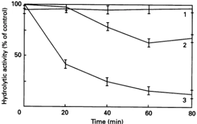
peptide+phenylglyoxal; curve 3, R61 +phenylglyoxal. Activities areexpressedasinFig. 1.
2 by a 17 h incubation at 37'C. Inactivationrate increased with increasing pH, control activity remaining virtually unchanged in
60 80 the pH range examined
(7.0-9.0)
(Fig. 2, curve a).Whenthe enzyme was inactivated by a 10-fold stoichiometric
oxaland cyclohexane- excess of benzylpenicillin before phenylglyoxal treatment, a complete recovery of the enzyme activity was observed after the enzyme
(25,UM)
addition of the ,-lactamase and dilution. The decay of the idicated at 37 'C in inactive acyl-enzyme occurred with a half-life similar to that,hexane- 1,2-dione at recorded in the absence of phenylglyoxal treatment. In conditions
m-boric acid, pH
9u
similar to those of Fig.1(a),
the activity loss caused by 2mM-~tofanunmodified phenylglyoxal decreased from 80 to
400%
in 60 min when theexperimentwasperformedinthe presence of 20 mM-Ac-L-Ala-D-Glu-Gly, a very poor substrate for which a
Ki
value of about 15mm was measured by competition with the usual substrate Ac2-L-Lys-D-Ala-D-Ala (Fig. 3). Protection against inactivationwasthus afforded if benzylpenicillin or the poor substrate Ac-L-Ala-D-Glu-Gly occupied the enzyme active site, suggesting the presenceofareactivearginine or lysine residue in the active site.
['4C]Benzylpenicillin
binding was also inhibited by phenylglyoxal. After a 10min incubation with [14C]benzylpenicillin, the >\b phenylglyoxal-treated 80%-inactivated enzyme was found tobind only
54%
of the labelled benzylpenicillin bound by thea ; same amount of intact enzyme. This showed that the rate of
binding of penicillin to the modified enzyme was drastically
decreased, since, with the intact enzyme, the same level of 8.5 labelling should be reached in about 1 s.
*.5
9 Similar inactivation time courses (Fig. lb), pH-dependence(Fig. 2, curveb) andprotection by benzylpenicillin (results not
dandcyclohexane-1,2- shown) were also obtained withcyclohexane-1,2-dione.
dione
Residualhydrolytic activity after a 60 min incubation ofthe R61 enzyme(25
#M)
with(curve a)0.5mM-phenylglyoxal at 37°C and(curve b) 2mM-cyclohexane-1,2-dione at 30°C at the pH values
indicated. ActivitiesareexpressedasinFig.1.Thefollowingbuffers
were used: pH7.0 and 7.5, 50mM-sodium phosphate; pH 8.0,
10mM-sodium borate; pH8.5, 10mM-Hepes+25-,uMEDTA;
pH9.0,20mM-boric acid.
thesynthetic tripeptideAc2-L-Lys-D-Ala-D-Ala. The time-course of inactivationbyfour concentrations ofphenylglyoxalis shown inFig. l(a).Inactivationwas not reversed by overnight dialysis (4 °C) against10mM-potassiumphosphate buffer, pH 7,followed
Stoichiometry of inactivation oftheDD-peptidase by
phenylglyoxal
Incubation of 1 nmol of enzyme with 25 nmol of
[14C]phenylglyoxalfor 20 min
(residual hydrolytic activity
about 20%)
followedbyt.l.c.resulted inincorporationofradioactivity
into the protein peak corresponding to 3.4+0.34nmol of['4C]phenylglyoxal/nmol
of inactivated enzyme. Ifthemodifi-cationreactionwasperformedwhen theactive sitewas
protected
by
benzylpenicillin,
only
2.5+0.5 nmol of[14C[phenylglyoxal
wasincorporatedper nmolof enzyme. This indicated that several enzymeresidues weremodifiedand confirmed that the presence of the
penicilloyl
residue in the active siteapparently
prevented
the modification ofoneof them.Table 1. Separation oftheI14Clphenylglyoxal-labeiledpeptidesbyhigh-voltage electrophoresisatpH 6.5
Mobilitiesarerelativetolysine= +1.00(+ and - indicatemigration towards cathode and anode respectively). The radioactivity found in the various peptides essentially accounted forthat present inthe initial sample beforeproteolytic digestion (about 100 %). Abbreviation: PenG, benzylpenicillin.
R61+[14C]phenylglyoxal R61 +PenG +['4C]phenylglyoxal Mobility Peptide at pH 6.5 (c.p.m.) (%) (c.p.m.) (%) 2 3 4 S 6 7 +1.00 +0.77 +2.51 +0.11 +0.00 -0.26 -0.80 2100+70 900+40 3500+80 36800+270 27000 +230 4500+100 5300+ 100 80100+900 2.6+0.08 1.1+0.05 4.4+0.10 45.9+0.30 33.7 +0.30 5.6+0.10 6.6+0.10 100+ 1.00 1300+ 50 400+30 2000+60 14700+170 17400+ 190 5500±100 5000+100 46300+700 2.8 +0.10 0.9+0.10 4.3+0.10 31.7+0.40 37.6+0.40 11.9+0.20 10.8 +0.20 100+ 1.50
Identification of the amino acid residueinvolved inthe inactivation reaction
The enzyme was inactivated to 80% by [14C]phenylglyoxal
and the complex thusformedwasfiltered onSephadex G-25 in 50
mM-NH4HCO3
and freeze-dried. The labelled enzyme was digested with pepsin[2%(w/w) with respect to the DD-peptidase] in 5% (v/v) formic acid at 37 °Cfor4h andthen with trypsin [2 % (w/w)] in 25mM-NH4HCO3at37 °C for4h.The digest was then submitted toelectrophoresis atpH6.5,giving seven radio-active peaks (Table 1). The radioactivity of peak 4 decreased when the enzyme was protected by benzylpenicillin beforereaction with phenylglyoxal suggesting that it contained the active-site' peptide. Peptide 4 was eluted from the
electro-phoretogram with 1 % (v/v) formic acid and purified by three successive identical
chromatographies
on thereverse-phase
column. Its amino acid sequence was determined and corresponded to that of the Thr-89-Arg-99 segment of the
DD-peptidase
(Thr89-Tyr-Leu-Pro-Gly-Leu-Leu-Pro-Asp-Asp-Arg99). Inparallel to this, a similar analysis of Pronase digests [Pronase(10%,w/w)in 10
mM-phosphate
buffer, pH 7,
for2hat 37°C] ofthe modifiedand subsequently heat-denatured DD-peptidase was performed. The 'active-site' labelled protein fragment isolated under those conditions contained only an
arginine residue, as revealed by amino acid analysis. These results indicated that Arg-99 was the residue involved in the
inactivation reaction by phenylglyoxal. Its role, together with thatof theconserved Arg-103
residue,
wasfurtherinvestigated
bysite-directedmutagenesis:Arg-99wasreplaced byleucine and Arg-103 by serine.
Production of the mutantDD-peptidases
The leucine and serine residues were chosen to replace the
arginine residues in order to eliminate the
positive
charge
associated with thearginine sidechain.After mutagenesis and sequencing, the
PstI-SphI fragments
(0.5
kb) coding
for the N-terminal part of the mutantDD-peptidases were cloned into the
previously
described Strepto-mycesplasmid (Hadonouetal.,1992)
toreplace
theequivalent
wild-typefragment.Aftertransformationofthe S.lividansTK24
protoplasts, the recombinant
plasmids
pDML22
(R103S)
and pDML32(R99L)werepurified,
and the DNAfragments coding
for theDD-peptidases werereleased
by
restriction,
recloned intobacteriophage
M13 andresequenced
in theregion
ofthemu-tation. Thisprocedure was
designed
toavoidthe occurrenceofa
wild-type
genein theplasmid
preparations.
The presence of themutations inpDML22andpDML32wasthusconfirmed. Mutant DD-peptidases were then produced in 5-litre 7-day cultures and
purified as described by Bourguignon-Bellefroid et al. (1992).
The R103S enzyme preparation was more than 90% pure as
estimatedbySDS/PAGE.
R99Lmutant DD-peptidase
Noenzymic activity was detected in the culture supernatant. Attempts to detect an inactive protein were made by using
SDS/PAGE, with the wild-type enzyme as an Mr standard,
followedbyWesternblottingforimmunological localization. All these testsremained negative. Cells were thenfractionated and all fractions submitted to SDS/PAGE and Western blotting.
Only a very weak response was obtained in the 'Iysozyme-releasable', membrane and cytoplasmic fractions, corresponding
to aproductionlower than 0.05 mg of recombinant protein/I of
culture. Nohydrolytic activity could be detected with thegood
substrate Ac2-L-Lys-D-Ala-D-Ala, which showed that the immunological method detected a protein with less than 0.1 % activity of the wild-type. It thus appeared that the enzyme was eitherunderexpressed or that its rate offolding was significantly decreased, resulting in amuch faster proteolysis of the nascent
protein, and that its secretion was also impaired. This under-expression could not be attributed to the choice of aninfrequent codon for leucine, since CTG is the most often used in the Streptomyces R61 DD-peptidase gene (Duez et al., 1987). The modified enzyme could not beproduced andpurified.
R103Smutant DD-peptidase
The mutation R103Syielded an enzymethe stability of which at 37°C did not differsignificantly from that of the wild-type (inactivation ti> 1500 min for both enzymes).
Isoelectrofocusingrevealedthe presence of two bandsforboth wild-type and mutant enzyme preparations, which, however, behavedhomogeneouslyonSDS/PAGE. The pI valueswere4.2 and4.5 for thewild-type and 4.1 and 4.4 for the R103S enzymes, thusconfirming themoreacidiccharacter of themutant. For the wild-type enzyme, the two forms have been separated by ion-exchange chromatography and exhibit identical enzymic and
penicillin-binding properties (M. Jamin & J.-M.Frere, unpub-lished work). The structural basis for the presence oftwoactive protein variants ispresentlyunder
investigation.
It isinterestingtonotethat suchaphenomenon hadnotbeenpreviouslyobserved when the enzyme was producedby the R61 strain itself(Frere etal., 1973).
Table 2.Kineticparameters forthe interactionofthe wild-type and R103S enzymes with substrates andAOilactams
The kinetic parameters for the hydrolytic + transpeptidase activity weredetermined in the presence of 290 1sM-thioester substrate and 10mM-D-alanine. Unless otherwise stated S.D. values are within 2000.
Wild-type R103S
Hydrolytic activity(Ac2-L-Lys-D-Ala-D-Ala)
kcat/Km(M1s-1) 3900* 2250
Esteraseactivity (thioester)
kcat (s-1 5+0.3 5.3 +0.6
Km
(/M)
41 +4 49.5 +4kcat./Km
(M-1* S-1) 120000+19000 107000+20000Esterase +transpeptidaseactivity(thioester-D-Ala)
kcat.
(s-')
47+13 32.5+4.5Km
(aM)
308 +60 385+60kcat./Km
(M-1S-1) 153000+70000 84000+24000Interaction with/-lactamst
Benzylpenicillin k2/K 14000 14170 k3 1.4x10-4* 1.1x10-4 Carbenicillin k2/K 830* 600 k3 1.4x10-4* 1.3x105 Ampicillin k2/K 110* 100 k3 1.4x10-4* 0.9X10-4 Nitrocefin k2/K 4100 4140 k3 3x10-4* 2x10-4 Cefuroxime k2/K 1400 520 k3 4x10-6* 5.3x10-6 Cefoxitin k2/K 1500* 20100 k3 5x10-5* 8.3x10-6
* FromFrere & Joris (1985).
t k2/Kandk3valuesarein M-1 s-' and s-'respectively.
tripeptide and thioester substrates were not drastically affected (Table 2). Thetranspeptidation/hydrolysis ratio determined by
h.p.l.c. (Jamin etal., 1991)after incubation of the enzymes with 2901sM-thioestersubstrate and 20 mM-D-alanineasacceptorwas
thesame(8.5) forthewild-typeandR103Senzymes. TheR103S
enzyme lost 98% of its hydrolytic activity after a 60 min
treatmentwith 2mM-phenylglyoxal.Re-activation of the enzyme
wasobserved after elimination of theexcessreagentifthe active site was
protected
bybenzylpenicillin
before treatment withphenylglyoxal.
Thecharacteristicparametersof the interaction of themutant
with/3-lactamantibiotics also appeared tobe similartothose of the wild-type enzyme with the
striking exception
ofcefoxitin,
which inactivated the mutant 10-fold faster than thewild-type
enzyme (Table 2).DISCUSSION
Modification of theR61 DD-peptidase
by
phenylglyoxal
resul-ted in inactivation time courses similar to those obtainedby
Georgopapadakou
etal.(1981); however,
our results indicatedthat more than one enzyme residue was modified. Protection
against inactivation was afforded
by benzylpenicillin
and thepoorsubstrateAc-L-Ala-D-Glu-Gly,suggestingthepresenceofa
reactivearginine (or
lysine)
residueinthe activesite.Arg-99
wasidentified as the
protected
residue involved in the inactivationreaction and its role was further
investigated by
site-directedmutagenesis.
The R99L mutant protein was apparently underexpressed, segregated inside the cell and inactive. Such low-level expression of unstable mutant proteins has been reported in the case of the D131G mutant/3-lactamaseof StreptomycesalbusG,where the conserved 'SDN' motifwasmodified (Jacobetal., 1990),andin other instances (Schultz & Richards, 1986). Another totally inactive mutant R61 DD-peptidase (S62C) was expressed in normal amounts in the culture supernatant, and its folding appeared to be grossly correct (B. Joris & J.-M. Frere, un-published work). The R99L mutation thus appeared to be highly detrimental to enzyme stability, reflecting a determining struc-tural role for this residue. Yet, Arg-99 is not conserved among the active-site-serine penicillin-recognizing proteins, and /3-lactamases even appeared to exhibit a deletionencompassing this position (Joris et al., 1988). According to the available X-ray data, Arg-99 lies in the a-domain, outside the active site (Kelly etal., 1989), and is involved in hydrogen bonds with the chain bearing the conserved 'YSN' motif (J.Lamotte-Brasseur, per-sonal communication). The complete elimination of the side chain can be expected to induce some 'floppiness' of the 'YSN' loop.
Residue Arg- 103 (box IV; Joris et al., 1988) is strictly conserved or replaced by a lysine residue in most of the active-site-serine penicillin-recognizing proteins. Surprisingly, the modification to aserine residueyieldedastable enzyme, the catalytic properties of which were similar to those of the wild-type enzyme. Thus a positive chargeatthat level did not appear to be essential for the enzymeactivity or structure. A totally unexpected result was the significant increase in the sensitivity of the mutant to one particular 3-lactam, cefoxitin. Interpretation of this observation mustawaitthe refinement of the three-dimensional structure and the modelling of the interaction of the wild-type and mutant enzymeswith various inactivating ,-lactams.
Fromthese results, it appeared that the important residue in this regionof the R61 DD-peptidase primary structure was Arg-99instead of Arg- 103. In the analogous class A
/8-lactamases,
a conserved lysine residue is found 40 residues after the active serine residue (Ambler et al., 1991), forming box IV in the nomenclature of Joris et al. (1988). In the Streptomyces R61 DD-peptidase, Arg-99 and Arg-103 occur respectively 37 and 41 residues after the active-site serine residue. Our results suggest that Arg-99 is more likely to be an important residue in a structurally importantconserved box. Finally, the present study alsoshowed that protection afforded by inhibitorsorsubstrates against modification by chemical reagents is not indisputable evidence for the presence of the target amino acid in the active site.Thisworkwassupported,in part,byanAction Concertee with the BelgianGovernment(Convention 86/91-90),theFonds de la Recherche
Scientifique Medicale (Contractn° 3.4537.88),a Convention Tripartite
between the Region Wallone, SmithKline Beecham, U.K., and the
University ofLiege,and theBelgianNationalincentive programme on fundamental research in Life Sciences (Contract BIO 22). This text
presents research results of theBelgian programme on Interuniversity
Polesof AttractioninitiatedbytheBelgianState,Prime Minister'sOffice,
SciencePolicyProgramming.Thescientificresponsibility isassumedby
its authors. B. J. is Chercheur Qualifie of the F.N.R.S. (Brussels,
Belgium), C.B. B. performedalargepart ofthe work described hereas fellow of the I.R.S.I.A. (Brussels, Belgium).
REFERENCES
Adam, M., Dazmblon, C., Plaitin, B., Christiaens, L. & Frkre, J.-M.
(1990) Biochem.J. 270, 525-529
Ambler, R.P.,Coulson, A. F.W., Frere,J.-M., Ghuysen, J.-M., Joris, B.,Forsman,M., Levesque,R.C.,
Tiraby,
G. &Waley,
S. G.(1991)
Bourguignon-Bellefroid, C., Wilkin, J. M., Joris, B., Aplin, R.T., Houssier, C.,Prendergast,F.G.,VanBeeumen, J.,Ghuysen,J.-M. & Frere,J.-M. (1992) Biochem. J.282,361-367
Brannigan, J.A., Tirodimos, I.A., Zhang, Q.Y., Dowson, C. G. & Spratt, B.G.(1990)Mol. Microbiol. 4,913-919
DeMeester, F.,Joris,B.,Reckinger, G.,Bellefroid-Bourguignon, C.&
Frere, J.-M. (1987)Biochem. Pharmacol. 36, 2396-2403
Dowson, C.G.,Hutchison,A. &Spratt,B. G.(1989) Mol.Microbiol.3, 95-102
Duez,C.,Fraipont, C., Joris, B., Dusart, J.,Urdea,M.S.,Martial,J.A.,
Frere, J.-M.&Ghuysen,J.-M.(1987) Eur. J.Biochem. 162,509-518
Fossati, P., Saint-Ghislain, M., Sicard, P.J., Frere, J.-M., Dusart, J., Klein, D.&Ghuysen,J.-M.(1978) Biotechnol. Bioeng. 20, 577-587 Frere, J.-M. &Joris,B.(1985) CRC Crit.Rev. Microbiol. 11,299-396 Frere,J.-M.,Ghuysen,J. M.,Perkins,H. R.&Nieto,M.(1973) Biochem.
J. 135,483-492
Frere,J.-M.,Leyh-Bouille, M., Ghuysen,J. M.&Perkins, H. R.(1974)
Eur.J. Biochem. 50, 203-214
Frere, J.-M., Duez, C., Ghuysen, J.-M. & Vandekerkhove, J. (1976) FEBSLett.70,257-260
Georgopapadakou, N.H., Liu, F.Y., Ryono, D.E., Neubeck, R. & Ondetti,M.A. (1981) Eur. J.Biochem.115, 53-57
Hadonou,A. M.,Jamin,M.,Adam, M.,Joris, B., Dusart, J., Ghuysen,
J.-M.& Frere, J.-M.(1992) Biochem.J. 282, 495-500 Herzberg, 0.(1991)J.Mol. Biol.217, 701-719
Hewick,R.M.,Hunkapiller,M.W.,Hood,L. E. &Dreyer,W. J.(1981)
J. Biol. Chem. 256, 7990-7997
Hopwood,D.A.,Bibb,M.J.,Chater,K.F.,Kieser,T.,Brutton, C.J.,
Kieser,H.M.,Lydiate,D. J.,Smith,C. P.,Ward,J. M.&Schrempf, H. (1985) Genetic Manipulation of Streptomyces -A Laboratory Manual,John InnesFoundation, Norwich
Jacob, F.,Joris,B.,Lepage,S., Dusart,J.&Frere, J.-M.(1990) Biochem.
J.271, 399-406
Jamin, M., Adam, M., Damblon, C., Christiaens, L. & Frere, J.-M.
(1991) Biochem.J.280, 499-506
Joris, B., Jacques, Ph., Frere, J.-M., Ghuysen,J.-M.&VanBeeumen, J.
(1987) Eur. J.Biochem. 162, 519-524
Joris, B.,Ghuysen, J.-M., Dive, G., Renard, A., Dideberg,O.,Charlier, P., Frere, J.-M., Kelly, J. A., Boyington,J.C.,Moews,P. C. &Knox,
J. R. (1988) Biochem. J. 250, 313-324
Katz, E., Thompson, C. J. & Hopwood, D. A. (1983) J. Gen. Microbiol. 129, 2703-2714
Kelly, J. A., Knox, J. R. & Zhao, H. (1989) J. Mol. Biol.209,281-295
Laible, G., Hakenbeck, R., Sicard, M.A., Joris, B. &Ghuysen,J.-M.
(1989) Mol. Microbiol. 3, 1337-1348
Laws,A. P. & Page, M. I.(1989) J.Chem.Soc.PerkinTrans. 21577-1581
Leyh-Bouille, M., Dusart, J., Nguyen-Disteche, M., Ghuysen, J.-M.,
Reynolds,P. E.&Perkins, H.R. (1977) Eur. J.Biochem.81, 19-28
Lujan,R.,Zhang,Q.Y., SaezNieto,J.A.,Jones, D. M. &Spratt, B. G.
(1991) Antimicrob. Ag. Chemother.35,300-304
Madgwick,P. J. &Waley, S. G. (1987) Biochem.J. 248,657-662
Maniatis,T., Fritsch,E. F.&Sambrook,J.(1982)Molecular Cloning:
A Laboratory Manual, Cold Spring Harbor Laboratory Press,
Cold SpringHarbor, NY
Matagne,A.,Misselyn-Bauduin, A.M.,Joris, B., Erpicum, T.,Granier, B.& Frere, J.-M. (1990) Biochem.J.265, 131-146
Means,G. E.& Feeney,R.E.(1964) Chemical Modification of Proteins,
p. 197,Holden-Day, San Francisco
Moews, P.C., Knox,J. R., Dideberg, O., Charlier, P. & Frere, J.-M.
(1990) Proteins7, 156-171
Schultz, S. C. &Richards,J. H.(1986)Proc. Natl.Acad. Sci.U.S.A.83, 1588-1592
Schuster, C., Dobrinski, B. &Hakenbeck, R.(1990) J. Bacteriol. 172,
6499-6505
Tsukamoto, K., Tachibana, K., Yamazaki, N., Ishii, Y., Ujiie, K.,
Nishida,N. &Sawai,T. (1990) Eur. J. Biochem. 188, 15-22 Varetto, L., Frere, J.-M. & Ghuysen, J.-M. (1987) FEBS Lett. 225,
218-222
Varetto,L.,DeMeester, F., Monnaie, D.,Marchand-Brynaert,J., Dive, G.&Frere,J.-M.(1991)Biochem.J. 278,801-807
Received 22July 1991/13 September 1991; accepted4October 1991