HAL Id: hal-02404499
https://hal.archives-ouvertes.fr/hal-02404499
Submitted on 11 Dec 2019HAL is a multi-disciplinary open access archive for the deposit and dissemination of sci-entific research documents, whether they are pub-lished or not. The documents may come from teaching and research institutions in France or abroad, or from public or private research centers.
L’archive ouverte pluridisciplinaire HAL, est destinée au dépôt et à la diffusion de documents scientifiques de niveau recherche, publiés ou non, émanant des établissements d’enseignement et de recherche français ou étrangers, des laboratoires publics ou privés.
Oxytocin effects on eye movements in schizophrenia
Lilla Porffy, Victoria Bell, Antoine Coutrot, Rebekah Wigton, Teresa
d’Oliveira, Isabelle Mareschal, Sukhwinder Shergill
To cite this version:
Lilla Porffy, Victoria Bell, Antoine Coutrot, Rebekah Wigton, Teresa d’Oliveira, et al.. Oxytocin effects on eye movements in schizophrenia. Schizophrenia Research, Elsevier, 2019. �hal-02404499�
In the eye of the beholder? Oxytocin effects on eye movements in schizophrenia
Lilla A. Porffy1*, Victoria Bell1*, Antoine Coutrot4, Rebekah Wigton2,3, Isabelle Mareschal5,
Sukhwinder S. Shergill 1
* Authors contributed equally and should be acknowledged as joint first authors
1. Department of Psychosis Studies, Institute of Psychiatry, Psychology & Neuroscience, King's College London, London, UK
2. Department of Neurology, Harvard Medical School, Boston, MA, US
3. Department of Neurology, Beth Israel Deaconess Medical Center, Boston, MA, US
4. Laboratoire des Sciences du Numérique de Nantes, CNRS, Université de Nantes, Nantes, France 5. Biological and Experimental Psychology, School of Biological and Chemical Sciences, Queen Mary University, London, UK
Corresponding author: Lilla Porffy
Department of Psychosis Studies
Institute of Psychiatry, Psychology & Neuroscience 16 De Crespigny Park, London, SE5 8AF, UK
Email: [email protected] Phone: (+44) 7738 244217
Keywords
Abstract
Background: Individuals with schizophrenia have difficulty in extracting salient information from faces. Eye-tracking studies have reported that these individuals demonstrate reduced exploratory viewing behaviour (i.e. reduced number of fixations and shorter scan paths) compared to healthy controls. Oxytocin has previously been demonstrated to exert pro-social effects and modulate eye gaze during face exploration. In this study, we tested whether oxytocin has an effect on visual attention in patients with schizophrenia. Methods: Nineteen male participants with schizophrenia received intranasal oxytocin 40UI or placebo in a double-blind, placebo-controlled, crossover fashion during two visits separated by seven days. They engaged in a free-viewing eye-tracking task, exploring images of Caucasian men displaying angry, happy, and neutral emotional expressions; and control images of animate and inanimate stimuli. Eye-tracking parameters included: total number of fixations, mean duration of fixations, dispersion, and saccade amplitudes. Results: We found a main effect of treatment, whereby oxytocin increased the total number of fixations, dispersion, and saccade amplitudes, while decreasing the duration of fixations compared to placebo. This effect, however, was non-specific to facial stimuli. When restricting the analysis to facial images only, we found the same effect. In addition, oxytocin increased fixation rates in the eye and nasion regions. Discussion: This is the first study to explore the effects of oxytocin on eye gaze in schizophrenia. Oxytocin had enhanced exploratory viewing behaviour in response to both facial and inanimate control stimuli. We suggest that the acute administration of intranasal oxytocin may have the potential to enhance visual attention in schizophrenia.
1. Background
Deficits in social-cognitive functioning are a major problem in schizophrenia, impacting significantly on socio-occupational functioning (Fett et al., 2011), and current treatments demonstrate limited efficacy (Kurtz & Richardson, 2012). These deficits have an effect on a wide range of social behaviours, such as attribution (Brüne, Abdel-Hamid, Sonntag, Lehmkämper, & Langdon, 2009; Fett & Maat, 2013), empathy (Shamay-Tsoory, Shur, Harari, & Levkovitz, 2007; Sparks, McDonald, Lino, O’Donnell, & Green, 2010), identification of emotional expressions (Huang et al., 2011; Kohler et al., 2003), and maintenance of appropriate eye contact (Choi et al., 2010; Davison, Frith, Harrison-Read, & Johnstone, 1996). The visual attention and scanning of human faces can be seen as an elementary step before social cues are made available to the higher order cognitive functions, in order to establish emotional expression, and facilitate appropriate social interaction. The neuropeptide oxytocin has previously been demonstrated to exert pro-social effects in schizophrenia (Averbeck, Bobin, Evans, & Shergill, 2012). Here we seek to evaluate the actions of oxytocin on the elementary processing of eye-tracking parameters.
Face perception is a fundamental process in social communication (Haxby, Hoffman, & Gobbini, 2002). Key information is extracted primarily from socially meaningful or salient features of the face, such as the eyes, nose and mouth, to assess another individual’s expressions. For example, fixating on the lip area when one smiles, enables the recognition of the facial expression as happy. However, individuals with schizophrenia tend to focus less on these areas when compared to healthy controls (Delerue, Laprévote, Verfaillie, & Boucart, 2010; Loughland, Williams, & Gordon, 2002). Moreover, they tend to exhibit reduced scanning behaviour characterised by fewer and longer fixations and travel shorter distances between fixation points (i.e. reduced dispersion) (Asgharpour, Tehrani-Doost, Ahmadi, & Moshki, 2015; Gaebel, Ulrich, & Frick, 1987; Kurachi et al., 1994; Manor et al., 1999; Phillips & David, 1998). These deficits have been observed both in the acute phase of the illness, and in remission (Streit, Wölwer, & Gaebel, 1997).
Abnormal scanning behaviour is likely to contribute to the impaired emotion perception observed in schizophrenia. While reduced attention to salient facial features may not dramatically affect the identification of less ambiguous emotions such happiness, it can have a significant impact on the recognition of negative expressions. For example, it has been reported that the accuracy with which schizophrenia patients identify happy emotions does not differ from healthy controls, but they are more likely to attribute fearful, angry, or sad expressions to another emotion (Bediou et al., 2005; Lee, Lee, Kweon, Lee, & Lee, 2010). More crucially, increased accuracy of emotion recognition in patients
is associated with longer scan paths and distance between fixations (i.e. higher dispersion), and fixation on more salient features (Green, Williams, & Davidson, 2003; Loughland et al., 2002). Emotion recognition is key to the cognitive model of schizophrenia that proposes that hallucinations and delusions occur when anomalous experiences are misattributed in a way that has extreme and threatening personal meaning, which leads to increased levels of distress (Garety, Kuipers, Fowler, Freeman, & Bebbington, 2001). In addition, the beliefs an individual hold can be ‘self-fulfilling’ – that is, once they are in place, the person will selectively attend to information that reinforces them, and rarely gathers information to challenge and expose them as biased and inaccurate, serving to perpetuate this cycle of threat perception which, in turn, can exacerbate psychotic symptoms. Visual attention and processing of faces are not typically improved using anti-psychotics (Penn et al., 2009) despite this being an important determinant of poor functional outcome (Fett et al., 2011). Psychosocial interventions have shown limited promise and benefits have been narrow in their scope, mainly improving isolated functions such as performance on theory of mind tasks (Kurtz & Richardson, 2012). Despite animal research demonstrating positive social improvements with oxytocin (Kim et al., 2007), relatively few studies have examined the effect of its administration on social-cognitive processes in humans. Oxytocin is considered to have a role in facilitating social interactions by ameliorating social bias and response to emotional faces. Several studies have shown that oxytocin enhances eye gaze to facial stimuli in healthy participants (Domes, Steiner, Porges, & Heinrichs, 2013) and can increase the number of fixations towards salient features of the face, compared to participants receiving placebo (Guastella, Mitchell & Dadds, 2008). Such research has led it to be considered as a possible intervention for use in disorders with severe social deficits such as schizophrenia (Evans, Shergill, & Averbeck, 2010; Guastella et al., 2013).
Experimental studies of the acute administration of oxytocin demonstrate facilitation of pro-social behaviours such as attenuation of aversion to angry faces (Bartz, Zaki, Bolger, & Ochsner, 2011; Evans et al., 2010), as well as an improved ability to accurately identify facial emotions in patients with schizophrenia (Averbeck et al., 2012). This raises the question of which level is oxytocin operating on. Does it impact non-specifically at an early visual level on socially relevant stimuli during the scanning of faces, or is it more specifically attuned to displays of emotional salience while observing emotional faces? Thus, we explored the acute effects of a single dose of intranasally administered oxytocin on visual attention and processing of human faces. We measured standard eye-tracking parameters - including the total number and duration of fixations, dispersion and saccade amplitudes to provide an objective proxy as to how oxytocin modulates visual attention to human faces. We hypothesised that
patients would exhibit an increased exploration of the facial stimuli following oxytocin administration compared to placebo; indexed by 1) an increased number of total fixations, and 2) fixations of shorter duration, 3) increased dispersion, and increased 4) saccade amplitudes. We also performed an exploratory examination of the specificity of oxytocin for facial and emotional facial stimuli, using control and neutral face stimuli; and conducted region of interest analysis to establish fixation rates on salient facial features.
2. Methods
2.1. Participants
Nineteen right-handed male subjects with a diagnosis of schizophrenia (n=16) or schizoaffective disorder (n=3) were recruited from outpatient services in South London. Patients were diagnosed according to International Statistical Classification of Diseases and Related Health Problems (ICD-10; World Health Organisation, 1993) criteria. All participants were screened and excluded if they had a neurological impairment or traumatic head injury; history of cardiovascular disease, suicidal ideation; and history of alcohol and/or substance dependence. We included male participants only due to differential neuropsychological effects of oxytocin on women compared to men (Wigton et al., 2015). Participants were prescribed various antipsychotic medications including: olanzapine (n=9), risperidone (n=2), clopixol (n=1), clozapine (n=3), haloperidol (n=1), and fluphenazine decanoate (n=1). Demographic and clinical information are presented in Table 1. Ethics approval was obtained from the Camberwell and St Giles Research Ethics Committee, London, UK. All participants gave informed written consent and were compensated for their time and travel.
TABLE 1
2.2. Oxytocin administration
Drug administration was carried out using a double-blind, placebo-controlled, cross-over design. Participants attended two testing sessions separated by 7 days, during which they either received 40IU oxytocin or placebo (saline) in a counterbalanced fashion. The administration protocol was devised following the guidance by Guastella et al. (2013). Participants were asked to self-administer using a nasal spray while being supervised by the researcher. Both oxytocin 40IU and placebo were delivered through eight puffs, four per nostril, with 45 second intervals. 40IU oxytocin was chosen based on previous clinical studies reporting positive effects on symptoms e.g. (Feifel et al., 2010; Modabbernia et al., 2013). Studies evaluating the effects of oxytocin on social cognition also reported positive findings with 40IU e.g. (Shin et al., 2015; Woolley et al., 2014).
2.3. Eye-tracking
Eye movements were recorded with a sampling rate of 500 Hz, using EyeLink 1000 (SR Research Ltd, Ottawa, Ontario, Canada). The eye-tracker was controlled by the Psychophysics Toolbox Version 3 (PTB-3) software for Matlab. A standard 9-point calibration was carried out before task initiation. Monocular sampling method was used tracking pupil of the dominant eye. The task took 3 minutes to complete and was carried out without head support.
2.4 Free-viewing Task
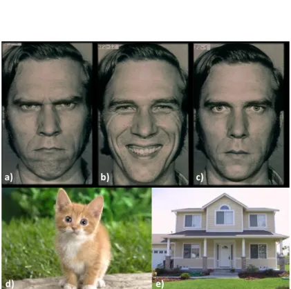
Six coloured images of two Caucasian men displaying angry (x2), happy (x2), and neutral (x2) facial expressions were presented to participants in addition to 4 inanimate control images including a bus, boat, house, mountain; and 2 animate control images including a cat and a puppy (see Figure 1). Facial stimuli were selected from the Ekman series (Ekman & Friesen, 1971), based on a previous study looking at oxytocin effects on social decision making (Evans et al., 2010). Stimuli were displayed on a 17-inch LCD monitor (height: 38.4 cm, width: 37.6 cm) with a resolution of 1280 x 1024 pixels and refresh rate of 60 Hz at the viewing distance of 76 cm, 1 pixel subtended 0.0278 degree. The 12 images were presented 5 times each, in a random order, amounting to 60 trials. This was a free-viewing visual attention task, participants did not need to respond. Images were displayed in colour in the middle of the screen for 3 seconds; the inter-stimulus interval was 0 seconds. Faces were resized to 27.8° x 25.4°, while control images were resized to 26.2° x 28.1° of visual angle.
FIGURE 1
2.5 Procedure
The eye-tracking task was performed approximately 2-hours after oxytocin administration (during this time participants underwent functional magnetic resonance imaging [fMRI] (these findings are in prep). Participants were shown the equipment and verbally instructed that they would be viewing a series of images during which they should look at the screen without turning away, while staying as still as possible. Instructions were purposely kept brief to ensure that participants were blind to the purpose of the study. Prior to the task, eye dominance of patients was assessed by having them pick a spot on the wall and then first point at it with both eyes open and then with their left eyes closed. If their finger jumped from the selected location, they were considered to be left eye dominant (1/19 participants). Once comfortable, participants were seated 76 cm away from the display screen to complete the task.
2.6. Data analysis
In order to assess exploratory scanning behaviour in response to facial stimuli, four eye movement parameters were extracted including: 1) total number of fixations, 2) duration of fixations, 3) dispersion, and 4) saccade amplitude. A fixation was defined, by EyeLink default, as a time period during which the pupil is visible, and there are no saccades. Duration of fixations was measured in milliseconds (ms). Any fixation longer than 5000 (ms) were removed from the analysis. Dispersion represents the average distance between fixation points and is measured in degrees of visual angle (°). A saccade is defined as a movement of the eye when its displacement is >0.10°, its velocity is >302/s and its acceleration is >8,000°/s.
We also defined Regions of Interest (ROIs) for the facial stimuli: eyes, nasion, nose, and mouth; see Figure 1. Since the face stimuli had the same size and were aligned on their nasion, we used the exact same ROIs for every stimulus. ROI fixation rates were defined as the number of sample eye position landing in a given ROI divided by the total number of sample eye positions. Hence, for a given trial, the sum of all ROI fixation rates equals to one.
FIGURE 2
Prior to analysis, data were checked for missing values, normality and outliers. Eighty-three or 3.6% of values were missing for dispersion, 11.5% (n=261) for saccade amplitude, and 12.9% (n=293) for duration of fixations. Normality was explored using visual plots and the Kolmogorov-Smirnov test. All eye-tracking variables were somewhat positively skewed, and the Kolmogorov-Smirnov test was statistically significant for all variables. No extreme outliers were found; all data points were within 2.5 SDs.
Exploratory viewing behaviour under oxytocin versus placebo were assessed using linear mixed models (LMMs). In the first model, participant IDs were included as random effect; and treatment (placebo / oxytocin), stimuli type (face / animate control / inanimate control), repetition of stimuli (1-2-3-4-5), and counterbalancing (placebo visit-1 / placebo visit-2) were included as fixed effects. We employed a full covariance matrix using Cholesky parameterization, and Maximum Likelihood method of estimation. Dependent variables (DVs) included the total number of fixations, duration of fixations, dispersion, and saccade amplitudes. The equation of the model was described as follows: dependent_variable ~ 1+ treatment + stimulus_type + repetition + counterbalancing + (1 | sub_id).
We also evaluated the effects of repetition on treatment. In this model, participant IDs were included as random effect; stimuli type (face / animate control / inanimate control), repetition of stimuli (1-2-3-4-5), and counterbalancing (placebo visit-1 / placebo visit-2) were included as fixed effects. DVs were defined as the difference between the DVs (number of fixations, duration of fixations, dispersion, and saccade amplitudes) in the oxytocin and placebo conditions. Again, we employed a full covariance matrix using Cholesky parameterization, and Maximum Likelihood method of estimation. The equation of the model was described as follows: (DV_oxytocin – DV_placebo) ~ 1+ stimulus_type + repetition + counterbalancing + (1 | sub_id).
In the third model, we focused on facial stimuli. Participant IDs were included as random effect; and treatment (placebo / oxytocin), emotion (neutral / angry / happy), repetition of stimuli (1-2-3-4-5), and counterbalancing (placebo visit-1 / placebo visit-2) were included as fixed effects. DVs were the same as in the first model. We employed a full covariance matrix using Cholesky parameterization, and Maximum Likelihood method of estimation. The equation of the model was described as follows: dependent_variable ~ 1+ treatment + emotion + repetition + counterbalancing + (1 | sub_id).
Finally, we looked at fixation rates at specific ROIs including the eyes, nose, nasion, mouth, and rest of the face (Figure 1). Again, we employed a full covariance matrix using Cholesky parameterization, and Maximum Likelihood method of estimation. The equation of the model was described as follows: ROI ~ 1+ treatment + emotion + repetition + counterbalancing + (1 | sub_id).
3. Results
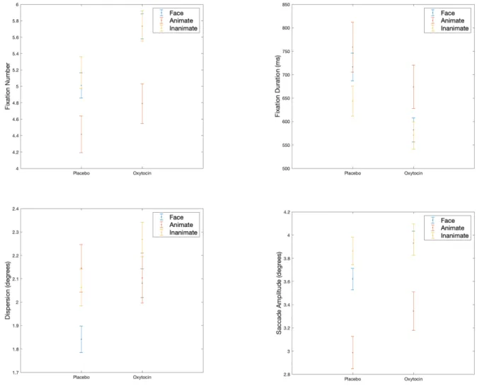
3.1. Animate Control vs. Inanimate Control vs. Facial Stimuli
There is a main effect of treatment on all eye-tracking parameters. The mean number of fixations is 4.9 (95%CI = 4.6-5.1) under placebo versus 5.4 (95%CI = 5.2-5.7) under oxytocin; F1, 2272 = 25.50, p <
.001. The mean duration of fixations reduces to 607.9 ms (95%CI = 564.1-651.7) under oxytocin compared to 705.5 ms under placebo (95%CI = 661.0-750.0); F1, 1979 = 16.45, p < .001. Dispersion
increases under oxytocin (mean = 2.15°, 95%CI = 2.05-2.24) compared to placebo (mean = 2.01°, 95%CI = 1.92-2.11); F1, 2187 = 11.81, p < .001. Similarly, under oxytocin, saccade amplitudes increase
(mean = 3.74°, 95%CI = 3.58-3.90) compared to placebo (mean = 3.48°, 95%CI = 3.32-3.64); F1, 2011 =
9.04, p = .002.
There is also a main effect of stimuli type. The total number of fixations is the highest in response to inanimate control images (mean = 5.45, 95%CI = 5.20-5.70), followed by faces (mean = 5.37, 95%CI =
5.17-5.79), and animate control images (mean = 4.60, 95%CI = 4.25-4.96); F2, 2272 = 8.24, p > .001.
Duration of fixations is the longest in response to animate control images (mean = 712.97 ms, 95%CI = 643.97-781.96), followed by faces (mean = 650.33 ms, 95%CI = 610.58-690.08), and inanimate control images (mean = 650.33 ms, 95%CI = 610.58-690.08); F2, 1979 = 3.05, p = .048. Dispersion is the
highest when viewing inanimate control stimuli (mean = 2.17°, 95%CI = 1.98-2.27), followed by animate control images (mean = 2.13°, 95%CI = 2.06-2.27), and then faces (mean = 1.96°, 95%CI = 1.89-2.05); F2, 2187 = 5.09, p = .006. Finally, saccade amplitudes were the largest in response to
inanimate control stimuli (mean = 3.90°, 95%CI = 3.73-4.08), followed by faces (mean = 3.77°, 95%CI = 3.63-3.92), and animate control images (mean = 3.16°, 95%CI = 2.91-3.40); ); F2, 2011 = 12.25, p < .001.
Results are displayed in Figure 2. FIGURE 3
3.2. Effects of repetition on treatment
There is a main effect of repetition on the total number of fixations (F4,2271 = 20.00, p < .001), and
dispersion (F4,2188 = 8.38, p < .001). On these eye-tracking parameters, treatment effect decreases as
the number of repetitions (R) increases. Results are displayed in Table 2 for number of fixations, and in Table 3 for dispersion. There is no effect of repetition on duration of fixations (F4,1978 = 1.22, p =
0.30), and saccade amplitudes (F4,2010 = 0.56, p = 0.69).
TABLE 2 TABLE 3
There is no interaction between treatment and stimuli type or treatment and stimuli repetition on any DVs. Finally, there is no effect of counterbalancing on any DVs.
3.3. Angry vs. Happy vs. Neutral Faces
There is a main effect of treatment on all eye-tracking parameters when viewing different expressions. Oxytocin increases the total number of fixations by 0.72 (95%CI = 0.37-1.06), F2, 1132 = 17.05, p < 0.001;
reduces the duration of fixations by 136.63 ms (95%CI = 63.88-209.37), F2, 994 = 13.58, p < 0.001;
increases dispersion by 0.28° (95%CI = 0.14-0.41), F2, 1078 = 16.06, p < 0.001; and finally, increases
On dispersion, there is also a main effect of valance. Mean dispersion is 2.11° (95%CI = 1.97-2.26) in response to happy faces, 1.89° (95%CI = 1.75-2.03) in response to neutral faces, and 1.88° (95%CI = 1.73-2.02) in response to angry faces; F2, 1077 = 4.14, p = .002 There is no effect of valance on any other
DV. Furthermore, there is no interaction between treatment and valance. Finally, there is no effect of counterbalancing on any DVs. Results are displayed in Figure 3.
FIGURE 4
3.4. Regions of Interest
Both treatment (F1,1078 = 7.99, p = .005) and valence (F2,1078 = 3.35, p = .035) have a main effect on
fixation rates in the eye region, with oxytocin decreasing fixation rates by 7%. Furthermore, there is main effect of treatment in the nasion region (F1,1078 = 5.37, p = .021), where oxytocin increases
fixations rates by 22%. Finally, there is both a main effect of treatment (F1,1078 = 4.22, p = .040) and
main effect of valence (F2,1078 = 3.34, p = .036) on the rest of the face, outside the main regions of
interest, where oxytocin increased fixation rates by 11%. There is no main effect of treatment or valence in the nose or mouth regions. There is no main effect of repetition nor counterbalancing on the fixation rates in any ROIs, or interaction effect.Results are displayed in Figure 4.
FIGURE 5 4. Discussion
To our knowledge, this is the first study to investigate eye-tracking parameters following oxytocin administration in individuals with schizophrenia. We report that oxytocin modulates all eye-tracking parameters measured by increasing the total number of fixations, average distance between fixation points (i.e. dispersion), and speed of eye movements (i.e. saccade amplitudes), while decreasing the duration of fixations. Contrary to our hypothesis, inanimate control images are found to be the most visually engaging followed by facial and animate control stimuli. Furthermore, for the total number of fixations and dispersion, the effect of treatment decreases as the number of repetition increases. We report the same trend when separately evaluating exploratory viewing behaviour in response to different emotional expressions (angry, happy, neutral), whereby oxytocin increases the total number of fixations, dispersion, and saccade amplitudes, and decreases the duration of fixations. Finally, ROI analyses revels that oxytocin increases fixation rates specifically in the eye region, nasion, and the rest of the face, while having no effect in the nose or mouth regions.
Our findings add to the growing literature showing that oxytocin can enhance exploratory viewing behaviour in humans. Previous research found that oxytocin modulates visual attention in healthy volunteers (Domes, Steiner, et al., 2013; Guastella et al., 2008), and in clinical populations with autistic spectrum disorder (ASD) (Kanat et al., 2017). However, in the present study, the effects of oxytocin seem to be non-specific to emotional expressions. It is possible that non-facial stimuli attract more attention than facial stimuli as humans are fine-tuned to rapidly scan and recognise facial features. Evidence shows that while there are a number of similarities between facial and object recognition, there are also important differences; for example, faces are examined in a more holistic manner compared to objects (Bruce & Humphreys, 1994). This may explain why participants found inanimate control images the most engaging.
Given that we did not find significant processing differences between facial and inanimate control stimuli, rather than increasing salience to emotional faces, our results indicate that oxytocin may have an effect on early-stage visual processing by either directly interacting with the visual cortex (Beard, Singh, Grundschober, Gee, & Tate, 2018; Grinevich & Stoop, 2018), or through modulation via other brain regions such as the amygdala (Adolphs & Spezio, 2006). Evidence suggests that the amygdala activates at the early stages of visual processing when viewing emotional faces (Morris et al., 1998). However, amygdala activation may also be observed in relation to self-relevant stimuli that include, but are not restricted to, social stimuli (Zalla & Sperduti, 2013). Therefore, it is plausible that oxytocin may have an impact on amygdala activity regardless of the stimuli type.
In the present study, oxytocin increased exploratory viewing behaviour in response to all emotional expressions in a similar fashion. This is contrary to previous research in healthy volunteers, which found that oxytocin increased eye gaze towards happy faces relative to neutral faces, but decreased eye gaze towards angry faces relative to neutral faces (Domes, Steiner, et al., 2013). In addition, it has been previously reported that oxytocin enhanced covert attention to happy faces but not angry faces (Domes, Sibold, et al., 2013). In the present sample, dispersion was significantly higher when viewing happy faces compared to neutral and angry faces, which may be an indication of enhanced visual attention. However, this does not provide a conclusive evidence that oxytocin differently modulated eye gaze in response to angry, happy and neutral expressions. Moreover, increased dispersion may be explained by the stimuli presented. Happy faces displayed to participants depict both men with a pronounced wide smile, potentially requiring increased dispersion for thorough image exploration.
We also report that oxytocin increases fixation rates in the eye region, nasion, and the rest of the face. Increased attention to the eyes under oxytocin is well-documented in the literature. In marmosets, oxytocin enhances attention specifically in this region (Kotani et al., 2017). Similar findings have been reported in healthy human volunteers e.g. (Guastella et al., 2008); and in individuals with ASD, where oxytocin facilitated increased eye-contact during a real-time, naturalistic social interaction (Auyeung et al., 2015). This, together with the propensity for enhanced general exploration under oxytocin, may have important clinical implications for individuals with schizophrenia. As discussed above, more thorough scanning can lead to more accurate affect recognition in this population (Green et al., 2003; Loughland et al., 2002); which, in turn, may translate into improved social-cognitive functioning. Indeed, studies have found a strong correlation between affect recognition and social functioning in various domains including social presentation, communication skills, and occupational functioning (Hooker & Park, 2002). Furthermore, deficits in affect recognition seem to be prevalent and persistent both in first-episode and chronic schizophrenia, and may partially mediate the relationship between cognitive and social functioning (Addington, Saeedi, & Addington, 2006). Taken together, this suggests that oxytocin may have the potential to improve some of the social-cognitive deficits observed in schizophrenia.
There are a number of limitations to this study to consider. First, our sample size was small and contained only males; therefore, results must be interpreted with caution. The addition of a healthy comparison and female patient groups would allow the investigation of the specificity of oxytocin to schizophrenia and document the response patterns in female adults. Second, we administered a single dose of oxytocin, plasma levels were not recorded, and assessments took place 2-hours after administration. Therefore, although the doses administered were in line with standard clinical doses, it is not clear if the dosage used would have elicited adequate levels in all subjects. While oxytocin levels in the plasma tend to peak at 30 minutes and then steadily decline until it is eliminated in approximately 90-120 minutes (Striepens et al., 2013), there is evidence that a significant proportion of men still have elevated oxytocin plasma levels 150 minutes following intranasal administration (Gossen et al., 2012). In addition, oxytocin becomes elevated in the cerebrospinal fluid (CSF) 75 minutes after administration and remains elevated at 120 minutes (Freeman et al., 2016; Striepens et al., 2013). Therefore, we have assumed that oxytocin was still effective in influencing visual behaviour during the eye-tracking task. Future studies should investigate the effects of intranasal oxytocin at peak plasma levels in larger samples and include female participants to evaluate gender effects. Thirdly, all patients were self-reported to be compliant with medication at admission to the study; except one patient who did not take any antipsychotic medication at the time, which could have
moderated the effects of oxytocin (Bradley & Woolley, 2017). Future studies may wish to objectively evaluate medication compliance by assessing antipsychotic plasma levels.
Overall, our findings support the notion that acute oxytocin administration may improve exploratory viewing behaviour compared to no treatment. Standard antipsychotic treatment produces little or no improvement on facial affect recognition tasks (Penn et al., 2009); therefore, it is crucial to consider other potential therapeutics such as oxytocin in future studies as a potential candidate for treatment of social-cognitive dysfunction in schizophrenia.
Acknowledgments
We would like to thank Dr Teresa D’Oliveira, Dr Dan W. Joyce, and Dr M. Berk Mirza for their help, comments, and suggestions.
LAP is supported by the UK Medical Research Council (MR/N013700/1) and King’s College London member of the MRC Doctoral Training Partnership in Biomedical Sciences.
SSS is supported by a European Research Council Consolidator Award (Grant Number 311686) and the National Institute for Health Research (NIHR) Mental Health Biomedical Research Centre at South London and Maudsley NHS Foundation Trust and King’s College London.
Funding Source
This study represents independent research funded by the National Institute for Health Research (NIHR) Biomedical Research Centre at South London and Maudsley NHS Foundation Trust and King’s College London. The views expressed are those of the author(s) and not necessarily those of the NHS, the NIHR or the Department of Health and Social Care. The funders had no role in the design, analysis, write-up or decision to submit for publication.
Conflict of Interest
References
Addington, J., Saeedi, H., & Addington, D. (2006). Facial affect recognition: A mediator between cognitive and social functioning in psychosis? Schizophrenia Research, 85(1–3), 142–150. https://doi.org/10.1016/j.schres.2006.03.028
Adolphs, R., & Spezio, M. (2006). Role of the amygdala in processing visual social stimuli. In S. Anders, G. Ende, M. Junghofer, J. Kissler, & D. B. T.-P. in B. R. Wildgruber (Eds.), Understanding
Emotions (Vol. 156, pp. 363–378). Elsevier.
https://doi.org/https://doi.org/10.1016/S0079-6123(06)56020-0
Asgharpour, M., Tehrani-Doost, M., Ahmadi, M., & Moshki, H. (2015). Visual attention to emotional face in schizophrenia: An eye tracking study. Iranian Journal of Psychiatry, 10(1), 13–
18. https://www.ncbi.nlm.nih.gov/pmc/articles/PMC4434423/
Auyeung, B., Lombardo, M. V., Heinrichs, M., Chakrabarti, B., Sule, A., Deakin, J. B., … Baron-Cohen, S. (2015). Oxytocin increases eye contact during a real-time, naturalistic social interaction in males with and without autism. Translational Psychiatry, 5(2), e507-6.
https://doi.org/10.1038/tp.2014.146
Averbeck, B. B., Bobin, T., Evans, S., & Shergill, S. S. (2012). Emotion recognition and oxytocin in patients with schizophrenia. Psychological Medicine, 42(02), 259–266.
https://doi.org/10.1017/S0033291711001413
Bartz, J. A., Zaki, J., Bolger, N., & Ochsner, K. N. (2011). Social effects of oxytocin in humans: Context and person matter. Trends in Cognitive Sciences, 15(7), 301–309.
https://doi.org/10.1016/j.tics.2011.05.002
Beard, R., Singh, N., Grundschober, C., Gee, A. D., & Tate, E. W. (2018). High-yielding (18)F
radiosynthesis of a novel oxytocin receptor tracer, a probe for nose-to-brain oxytocin uptake in vivo. Chemical Communications (Cambridge, England), 54(58), 8120–8123.
https://doi.org/10.1039/c8cc01400k
Bediou, B., Franck, N., Saoud, M., Baudouin, J. Y., Tiberghien, G., Daléry, J., & D’Amato, T. (2005). Effects of emotion and identity on facial affect processing in schizophrenia. Psychiatry
Research, 133(2–3), 149–157. https://doi.org/10.1016/j.psychres.2004.08.008
Bradley, E. R., & Woolley, J. D. (2017). Oxytocin effects in schizophrenia: reconciling mixed findings and moving forward. Neuroscience & Biobehavioral Reviews, 80(December 2016), 36–56. https://doi.org/10.1016/j.neubiorev.2017.05.007
Bruce, V., & Humphreys, G. W. (1994). Recognizing Objects and Faces. Visual Cognition, 1(2–3), 141– 180. https://doi.org/10.1080/13506289408402299
cognition with social interaction: Non-verbal expressivity, social competence and “mentalising” in patients with schizophrenia spectrum disorders. Behavioral and Brain Functions, 5, 1–10. https://doi.org/10.1186/1744-9081-5-6
Choi, S. H., Ku, J., Han, K., Kim, E., Kim, S. I., Park, J., & Kim, J. J. (2010). Deficits in eye gaze during negative social interactions in patients with schizophrenia. Journal of Nervous and Mental
Disease, 198(11), 829–835. https://doi.org/10.1097/NMD.0b013e3181f97c0d
Davison, P. S., Frith, C. D., Harrison-Read, P. E., & Johnstone, E. C. (1996). Facial and other non-verbal communicative behaviour in chronic schizophrenia. Psychological Medicine, 26(4), 707–713. https://doi.org/https://doi.org/10.1017/s0033291700037727
Delerue, C., Laprévote, V., Verfaillie, K., & Boucart, M. (2010). Gaze control during face exploration in schizophrenia. Neuroscience Letters, 482(3), 245–249.
https://doi.org/10.1016/j.neulet.2010.07.048
Domes, G., Sibold, M., Schulze, L., Lischke, A., Herpertz, S. C., & Heinrichs, M. (2013). Intranasal oxytocin increases covert attention to positive social cues. Psychological Medicine, 43(8), 1747–1753. https://doi.org/10.1017/S0033291712002565
Domes, G., Steiner, A., Porges, S. W., & Heinrichs, M. (2013). Oxytocin differentially modulates eye gaze to naturalistic social signals of happiness and anger. Psychoneuroendocrinology, 38(7), 1198–1202. https://doi.org/10.1016/j.psyneuen.2012.10.002
Ekman, P., & Friesen, W. V. (1971). Constants across cultures in the face and emotion. Journal of
Personality and Social Psychology, 17(2), 124–129. https://doi.org/10.1037/h0030377
Evans, S., Shergill, S. S., & Averbeck, B. B. (2010). Oxytocin decreases aversion to angry faces in an associative learning task. Neuropsychopharmacology, 35(13), 2502–2509.
https://doi.org/10.1038/npp.2010.110
Feifel, D., MacDonald, K., Nguyen, A., Cobb, P., Warlan, H., Galangue, B., … Hadley, A. (2010). Adjunctive intranasal oxytocin reduces symptoms in schizophrenia patients. Biological
Psychiatry, 68(7), 678–680. https://doi.org/10.1016/j.biopsych.2010.04.039
Fett, A. K. J., & Maat, A. (2013). Social cognitive impairments and psychotic symptoms: What is the nature of their association? Schizophrenia Bulletin, 39(1), 77–85.
https://doi.org/10.1093/schbul/sbr058
Fett, A. K. J., Viechtbauer, W., Dominguez, M. de G., Penn, D. L., van Os, J., & Krabbendam, L. (2011). The relationship between neurocognition and social cognition with functional outcomes in schizophrenia: A meta-analysis. Neuroscience and Biobehavioral Reviews, 35(3), 573–588. https://doi.org/10.1016/j.neubiorev.2010.07.001
(2016). Plasma and CSF oxytocin levels after intranasal and intravenous oxytocin in awake macaques. Psychoneuroendocrinology, 66, 185–194.
https://doi.org/10.1016/j.psyneuen.2016.01.014
Gaebel, W., Ulrich, G., & Frick, K. (1987). Visuomotor performance of schizophrenic patients and normal controls in a picture viewing task. Biological Psychiatry, 22(10), 1227–1237.
https://doi.org/10.1016/0006-3223(87)90030-8
Garety, P. A., Kuipers, E., Fowler, D. G., Freeman, D., & Bebbington, P. (2001). A cognitive model of the positive symptoms of psychosis. Psychological Medicine, 31(2), 189–195.
https://doi.org/10.1017/S0033291701003312
Gossen, A., Hahn, A., Westphal, L., Prinz, S., Schultz, R. T., Gründer, G., & Spreckelmeyer, K. N. (2012). Oxytocin plasma concentrations after single intranasal oxytocin administration - A study in healthy men. Neuropeptides, 46(5), 211–215. https://doi.org/10.1016/j.npep.2012.07.001 Green, M. J., Williams, L. M., & Davidson, D. (2003). Visual scanpaths to threat-related faces in
deluded schizophrenia. Psychiatry Research, 119(3), 271–285. https://doi.org/10.1016/S0165-1781(03)00129-X
Grinevich, V., & Stoop, R. (2018). Interplay between Oxytocin and Sensory Systems in the Orchestration of Socio-Emotional Behaviors. Neuron, 99(5), 887–904.
https://doi.org/10.1016/j.neuron.2018.07.016
Guastella, A. J., Hickie, I. B., McGuinness, M. M., Otis, M., Woods, E. A., Disinger, H. M., … Banati, R. B. (2013). Recommendations for the standardisation of oxytocin nasal administration and guidelines for its reporting in human research. Psychoneuroendocrinology, 38(5), 612–625. https://doi.org/10.1016/j.psyneuen.2012.11.019
Guastella, A. J., Mitchell, P. B., & Dadds, M. R. (2008). Oxytocin Increases Gaze to the Eye Region of Human Faces. Biological Psychiatry, 63(1), 3–5. https://doi.org/10.1016/j.biopsych.2007.06.026 Haxby, J. V, Hoffman, E. A., & Gobbini, M. I. (2002). Human neural systems for face recognition and
social communication. Biol Psychiatry, 51(1), 59–67. https://doi.org/10.1016/S0006-3223(01)01330-0
Hooker, C., & Park, S. (2002). Emotion processing and its relationship to social functioning in schizophrenia patients. Psychiatry Research, 112(1), 41–50. https://doi.org/10.1016/S0165-1781(02)00177-4
Huang, J., Chan, R. C. K., Gollan, J. K., Liu, W., Ma, Z., Li, Z., & Gong, Q. (2011). Perceptual bias of patients with schizophrenia in morphed facial expression. Psychiatry Research, 185(1–2), 60– 65. https://doi.org/10.1016/j.psychres.2010.05.017
of oxytocin on the attentional preference for faces in autism. Translational Psychiatry, 7, e1097. Retrieved from https://doi.org/10.1038/tp.2017.67
Kim, K., Kim, J.-J., Kim, J., Park, D.-E., Jang, H. J., Ku, J., … Kim, S. I. (2007). Characteristics of social perception assessed in schizophrenia using virtual reality. Cyberpsychology & Behavior : The
Impact of the Internet, Multimedia and Virtual Reality on Behavior and Society, 10(2), 215–219.
https://doi.org/10.1089/cpb.2006.9966
Kohler, C. G., Turner, T. H., Bilker, W. B., Brensinger, C. M., Siegel, S. J., Kanes, S. J., … Gur, R. C. (2003). Facial emotion recognition in schizophrenia: Intensity effects and error pattern.
American Journal of Psychiatry, 160(10), 1768–1774.
https://doi.org/10.1176/appi.ajp.160.10.1768
Kotani, M., Shimono, K., Yoneyama, T., Nakako, T., Matsumoto, K., Ogi, Y., … Ikeda, K. (2017). An eye tracking system for monitoring face scanning patterns reveals the enhancing effect of oxytocin on eye contact in common marmosets. Psychoneuroendocrinology.
https://doi.org/10.1016/j.psyneuen.2017.05.009
Kurachi, M., Matsui, M., Kiba, K., Suzuki, M., Tsunoda, M., & Yamaguchi, N. (1994). Limited visual search on the WAIS picture completion test in patients with schizophrenia. Schizophrenia
Research, 12(1), 75–80. https://doi.org/10.1016/0920-9964(94)90086-8
Kurtz, M. M., & Richardson, C. L. (2012). Social cognitive training for schizophrenia: A meta-analytic investigation of controlled research. Schizophrenia Bulletin, 38(5), 1092–1104.
https://doi.org/10.1093/schbul/sbr036
Lee, S. J., Lee, H. K., Kweon, Y. S., Lee, C. T., & Lee, K. U. (2010). Deficits in facial emotion recognition in schizophrenia: A replication study with Korean subjects. Psychiatry Investigation, 7(4), 291– 297. https://doi.org/10.4306/pi.2010.7.4.291
Loughland, C. M., Williams, L. M., & Gordon, E. (2002). Visual scanpaths to positive and negative facial emotions in an outpatient schizophrenia sample. Schizophrenia Research, 55(1–2), 159– 170. https://doi.org/10.1016/S0920-9964(01)00186-4
Manor, B. R., Gordon, E., Williams, L. M., Rennie, C. J., Bahramali, H., Latimer, C. R., … Meares, R. A. (1999). Eye movements reflect impaired face processing in patients with schizophrenia.
Biological Psychiatry, 46(7), 963–969. https://doi.org/10.1016/S0006-3223(99)00038-4
Modabbernia, A., Rezaei, F., Salehi, B., Jafarinia, M., Ashrafi, M., Tabrizi, M., … Akhondzadeh, S. (2013). Intranasal oxytocin as an adjunct to risperidone in patients with schizophrenia: An 8-week, randomized, double-blind, placebo-controlled study. CNS Drugs, 27(1), 57–65. https://doi.org/10.1007/s40263-012-0022-1
neuromodulatory role for the human amygdala in processing emotional facial expressions.
Brain, 121 ( Pt 1, 47–57. https://doi.org/10.1093/brain/121.1.47
Penn, D. L., Keefe, R. S. E., Davis, S. M., Meyer, P. S., Perkins, D. O., Losardo, D., & Lieberman, J. A. (2009). The effects of antipsychotic medications on emotion perception in patients with chronic schizophrenia in the CATIE trial. Schizophrenia Research, 115(1), 17–23.
https://doi.org/10.1016/j.schres.2009.08.016
Phillips, M. L., & David, A. S. (1998). Abnormal visual scan paths: A psychophysiological marker of delusions in schizophrenia. Schizophrenia Research, 29(3), 235–245.
https://doi.org/10.1016/S0920-9964(97)00097-2
Shamay-Tsoory, S. G., Shur, S., Harari, H., & Levkovitz, Y. (2007). Neurocognitive basis of impaired empathy in schizophrenia. Neuropsychology, 21(4), 431–438. https://doi.org/10.1037/0894-4105.21.4.431
Shin, N. Y., Park, H. Y., Jung, W. H., Park, J. W., Yun, J. Y., Jang, J. H., … Kwon, J. S. (2015). Effects of oxytocin on neural response to facial expressions in patients with schizophrenia.
Neuropsychopharmacology, 40(8), 1919–1927. https://doi.org/10.1038/npp.2015.41
Sparks, A., McDonald, S., Lino, B., O’Donnell, M., & Green, M. J. (2010). Social cognition, empathy and functional outcome in schizophrenia. Schizophrenia Research, 122(1–3), 172–178. https://doi.org/10.1016/j.schres.2010.06.011
Streit, M., Wölwer, W., & Gaebel, W. (1997). Facial-affect recognition and visual scanning behaviour in the course of schizophrenia. Schizophr.Res., 24, 311–317. https://doi.org/10.1016/S0920-9964(96)00126-0
Striepens, N., Kendrick, K. M., Hanking, V., Landgraf, R., Wüllner, U., Maier, W., & Hurlemann, R. (2013). Elevated cerebrospinal fluid and blood concentrations of oxytocin following its intranasal administration in humans. Scientific Reports, 3, 1–5.
https://doi.org/10.1038/srep03440
WHO. (1993). The ICD-10 classification of mental and behavioural disorders: Diagnostic criteria for research. Geneva: World Health Organization.
Wigton, R., Radua, J., Allen, P., Averbeck, B., Meyer-Lindenberg, A., McGuire, P., … Fusar-Poli, P. (2015). Neurophysiological effects of acute oxytocin administration: systematic review and meta-analysis of placebo-controlled imaging studies. Journal of Psychiatry & Neuroscience,
40(1), E1–E22. https://doi.org/10.1503/jpn.130289
Woolley, J. D., Chuang, B., Lam, O., Lai, W., O’Donovan, A., Rankin, K. P., … Vinogradov, S. (2014). Oxytocin administration enhances controlled social cognition in patients with schizophrenia.
Zalla, T., & Sperduti, M. (2013). The amygdala and the relevance detection theory of autism: an evolutionary perspective. Frontiers in Human Neuroscience, 7, 894.
Tables and Figures
Table 1. Sample demographics and clinical characteristics.
Mean SD Range
Age 38.4 7.3 28-50
Education (years) 13.2 2.9 9-18 WASI score 99.4 10.7 84-125 NS-SEC classification 2.8 1.8 1-5 Age at illness onset 24.9 6.4 16-37 Illness duration (years) 13.5 6.7 2-28 Antipsychotic dose (mg) a 436.9 240.7 0-1000 PANSS Positive symptoms 14.3 4.2 8-22 Negative symptoms 18.3 5.1 11-28 General symptoms 29.6 5.4 21-40 Total score 62.2 12.7 42-87
Notes. WASI, Wechsler Abbreviated Scale of Intelligence; NS-SEC, National Statistics
Socio-economic Classification; PANSS, Positive and Negative Syndrome Scale; a Chlorpromazine equivalent
a) b) c)
d) e)
Figure 1. Examples of stimuli presented: a) Angry face; b) Happy
Figure 2. Regions of interest.
Table 2. The effects of stimuli repetition (R) on the total number of fixations.
Mean Difference 95%CI F-statistics p-value
R1 vs R2 1.09 0.36-1.82 F4, 2271= 0.01 p = .91
R1 vs R3 1.55 0.82-2.28 F4, 2271= 5.34 p = .02
R1 vs R4 1.21 0.48-1.94 F4, 2271= 11.70 p < .001
R1 vs R5 1.23 0.50-1.96 F4, 2271= 5.60 p = .02
Table 3. The effects of stimuli repetition (R) on dispersion.
Mean Difference 95%CI F-statistics p-value
R1 vs R2 0.36 0.06-0.66 F4, 2188= 0.01 p = .40
R1 vs R3 0.47 0.17-0.77 F4, 2188= 5.34 p = .47
R1 vs R4 0.33 0.03-0.63 F4, 2188= 11.70 p = .006
R1 vs R5 0.32 0.02-0.62 F4, 2188= 5.60 P = .001
Figure 5. Fixation rates under oxytocin versus placebo at regions of interest including the eyes, nasion,