Université de Montréal
Mechanism of Inducible Costimulator
(ICOS)-mediated calcium signaling
Par Julien Leconte
Département de microbiologie, immunologie et infectiologie Université de Montréal
Faculté de médecine
Mémoire présenté à la Faculté de médecine en vue de l’obtention du grade de maitrise ès science en microbiologie et immunologie
Décembre, 2013
RÉSUMÉ
Le Costimulateur Inductible (ICOS) est un récepteur exprimé à la surface des cellules T CD4 auxiliaires et T CD8 cytotoxiques. Il fut démontré à l’aide de modèles murins de transplantation de moelle osseuse que ICOS joue un rôle important dans l’induction de la maladie du greffon contre l’hôte aigüe (GVHD). ICOS potentialise deux signaux médiés par le récepteur de cellules T (TCR) : l’activation de la phosphoinositide 3-kinase (PI3K) ainsi que la mobilisation interne de calcium. En conditions in vitro, dans les cellules CD4 et CD8, ICOS réussi à potentialiser le flux de calcium médié par le TCR indépendamment de PI3K. La voie de signalisation de ICOS impliquée dans la GVHD demeure inconnue. Cependant, en utilisant une lignée de souris ‘knock-in’ nommée ICOS-Y181F, dans laquelle le cellules T ont sélectivement perdu la capacité d’activer PI3K par l’entremise d’ICOS, nous avons démontré que les cellules T peuvent utiliser un mécanisme ICOS indépendant de PI3K afin d’induire la GVHD.
La mobilisation interne du Ca2+ mène à l’activation de NFAT, un facteur de transcription clé régulant des gènes comme IFN-γ, qui exprime une des cytokines clés impliquées dans la GVHD. Nous émettons comme hypothèse que la capacité pathogénique intacte des cellules T ICOS-Y181F à induire la GVHD, repose sur la signalisation du Ca2+ indépendante de PI3K. Le but de mon projet est d’identifier les résidus responsables de cette signalisation de Ca2+ médiée par ICOS ainsi que le mécanisme par lequel ce récepteur fonctionne. À l’aide de la mutagénèse dirigée, j’ai généré des mutants d’ICOS et j’ai analysé par cytométrie en flux leur capacité à activer le flux de Ca2+. J’ai ainsi identifié un groupe de lysine sur la queue cytoplasmique d’ICOS situé à proximité de la membrane comme étant essentiel à la fonction de potentialisation du flux de Ca2+. Je fournis également des preuves de l’implication de la kinase Lck, membre de la famille de kinases Src, dans la voie de signalisation de ICOS médiant la potentialisation du flux de Ca2+. Ainsi, ICOS s’associe à Lck et mène à une augmentation de l’activation de PLCγ1, la protéine effectrice clé causant la sortie de Ca2+ de la réserve intracellulaire.
En conclusion, notre étude permet de comprendre davantage une des voies de signalisation d’ICOS. L’influx de Ca2+ dans les cellules T implique la voie ICOS-Lck-PLCγ1. Une compréhension plus approfondie de cette voie de signalisation pourrait s’avérer bénéfique afin d’élaborer de nouvelles stratégies menant à la prévention de maladies reliées à ICOS, comme la GVHD.
ABSTRACT
The Inducible Costimulator (ICOS) is a receptor expressed on activated CD4 helper and CD8 cytotoxic T cells.It was previously shown that ICOS plays an important role in inducing acute graft versus host disease (GVHD) in murine models of allogeneic bone marrow transplantation (BMT). ICOS potentiates TCR-mediated phosphoinositide 3-kinase (PI3K) activation and intracellular calcium mobilization. In both CD4+ and CD8+ T cells, ICOS can potentiate TCR-mediated calcium flux in a PI3K-independent manner in vitro. However, the ICOS signal transduction pathway involved in GVHD remains unknown. Using a knock-in straknock-in of mice (termed ICOS-Y181F) in which T cells have selectively lost the ability to activate PI3K, we have recently shown that T cells can utilize PI3K-independent ICOS signaling pathways to induce GVHD.
The mobilization of intracellular Ca2+ leads to the activation of NFAT, a key transcription factor regulating genes such as IFN-γ, one of the key T cell cytokines involved in GVHD. Therefore, we hypothesize that the intact pathogenic capacity of ICOS-Y181F T cells to induce GVHD relies on ICOS-dependent, PI3K-independent calcium signaling. My goal is to identify the residue(s) responsible for this ICOS-mediated Ca2+ signaling and find the mechanism by which the receptor achieves its function. Through site-directed mutagenesis and flow cytometric analysis of calcium fluxing capacities of mutant ICOS proteins, I identified a membrane proximal cluster of lysine residues that is essential in inducing ICOS-mediated Ca2+ signaling. I also provide evidence for the involvement of the Src family kinase Lck in ICOS-mediated Ca2+signaling. ICOS associates with Lck molecules, leading to the activation of PLCγ1, the key effector protein causing the release of Ca2+ from the intracellular pool.
Taken together, our study is beginning to unravel a complexity in ICOS signaling, and implicates the ICOS-Lck-PLCγ1 axis in T cell calcium signaling and potentially the induction of GVHD. Further understanding of this pathway could prove beneficial in designing new strategies to prevent ICOS-related diseases such as GVHD.
TABLE OF CONTENT
Résumé III
Abstract IV
Table of content V
List of figures VIII
List of tables IX
List of abbreviations X
Acknowledgements XVII
Chapter 1: Introduction 1
1.1 Signal transduction pathways in T cells 2
1.1.1 General overview 2 1.1.2 Src family kinases 4 1.1.3 Lck 5 1.1.4 Molecules downstream of Lck 6 1.1.5 Fyn 7 1.1.6 Ras/Erk 7 1.1.7 Lipid Rafts 8 1.1.8 Costimulatory receptors 9
1.1.9 CD28 and its ligands B7-1/B7-2 9
1.2 The Inducible Costimulator 12
1.2.1 General description 12
1.2.2 ICOS structure 12
1.2.3 ICOS signaling mechanism and functions 13
1.2.4 Regulation of expression 14
1.2.5 B7-H2 (ICOSL or CD275) 15
1.2.6 Disease relevance 16
1.3 Calcium signaling pathway in T cells 17
1.3.2 PLCγ1 18
1.3.3 CRAC Channels 19
Chapter 2: Materials and Methods 21
Mice 22
Antibodies and cytokines 22
Reagents 23
In vitro CD4 T cells isolation, activation and restimulation 23
Jurkat cells 24
Ca2+ flux 24
Immunoprecipitation and immunoblot analysis 25 Plasmid construction and cell transfection 26 Sucrose density gradient and lipid raft isolation 26
Chapter 3: Results 28
Four membrane proximal residues in the cytoplasmic tail of ICOS is
sufficient for Ca2+ flux 29
The triple lysine motif is necessary for Ca2+ fluxing capacity
of ICOS 32
Interruption of the triple lysine stretch affects Ca2+ flux
initiation 34
ICOS induces intracellular Ca2+ release from the intracellular
pool 36
ICOS-mediated Ca2+ flux depends on Src family kinases 37 Following stimulation, ICOS associates with Lck and ZAP70 and
increases activation of PLCγ1 40
ICOS does not relocalize into lipid rafts upon stimulation 41
Discussion 45
Summary 53
References 54
Appendix 66
LIST OF FIGURES
Chapter 1: IntroductionFigure 1.1 Simplified overview of TCR signaling leading to activation of
the Ca2+ signaling pathway 11
Figure 1.2 Overview of Ca2+ signaling pathways in T cells 20
Chapter 3: Results
Figure 3.1 Mapping the minimal segment of ICOS cytoplasmic tail
sufficient for Ca2+ flux 31
Figure 3.2 Membrane proximal motif KKKY is crucial for ICOS-mediated
Ca2+ flux 33
Figure 3.3 Single mutations in the membrane proximal triple lysine cluster of ICOS lead to delayed Ca2+ initiation 35 Figure 3.4 ICOS is involved in the release of intracellular Ca2+ 37 Figure 3.5 ICOS mediates its Ca2+ flux function through Src family
kinases 39
Figure 3.6 ICOS associates with Lck and ZAP-70 and increases PLCγ1
activation 41
Figure 3.7 ICOS doesn’t relocalize into lipid rafts upon stimulation 43
Appendix
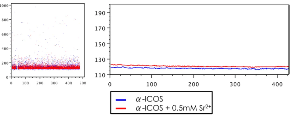
Figure S1 Addition of Sr2+ is not sufficient to allow autonomous ICOS- mediated Ca2+ flux in primary CD4+ T cells 68 Figure S2 Inhibition of HSP90 by Tanespimycin treatment leads to
delayed ICOS-mediated Ca2+ flux initiation 68 Figure S3 ICOS and CD28 act synergistically to further potentiate TCR-
LIST OF TABLES
AppendixLIST OF ABBREVIATIONS
3’-UTR Three Prime Untranslated Region
Ab Antibody
A/Ala Alanine
Akt Serine/Threonine-specific kinase
APC Antigen Presenting Cell
ATCC American Type Culture Collection
B7-1 B-lymphocyte activation antigen 7-1
B7-2 B-lymphocyte activation antigen 7-2
B7-H2 B7 homolog 2
Bcl-xl B-Cell Lymphoma Extra Large
BMT Bone Marrow Transplant
Brij-58 Polyethylene Glycol Hexadecyl Ether
BTLA B- and T-Lymphocyte Attenuator
Ca2+ Calcium
Ca2+ATPase Calcium Adenylpyrophosphatatse
CaCl2 Calcium Chloride
CamK2 Calcium/Calmodulin-dependent Kinase 2
CD3 Cluster of Differentiation 3 CD4 Cluster of Differentiation 4 CD8 Cluster of Differentiation 8 CD28 Cluster of Differentiation 28 CD45 Cluster of Differentiation 45 CD80 Cluster of Differentiation 80 CD86 Cluster of Differentiation 86
CRAC Calcium Release Activated Channel
Csk C-terminal Src Kinase
CTLA-4 Cytotoxic T-Lymphocyte Antigen 4
CVID Common Variable Immunodeficiency
cyTOF Mass Cytometry Instrument
DAG Diacyl-Glycerol
DAPP1 Dual Adapter for Phosphotyrosine and
3-Phosphoinositide
DNA Deoxyribonucleic Acid
dNTPs DeoxyriboNucleotide tri-phosphates
dsDNA Double Strand DNA
dSMAC Distal SMAC
EDTA Ethylene-Diamine-Tetraacetic Acid
EGTA Ethylene-Glycol-Tetraacetic Acid
ER Endoplasmic Reticulum
Erk Extracellular signal-Regulated Kinase
F/Phe Phenylalanine
FBS Fetal Bovine Serum
Fyn Proto-oncogene tyrosine-protein kinase
GADS GRB2-related Adapter Protein 2
GATA-3 Trans-acting T-cell-specific transcription
factor 3
GC Germinal Center
Grb2 Growth factor Receptor-Bound protein 2
GTP Guanosine Triphosphate
GVHD Graft Versus Host Disease
HEPES 4-(2-hydroxyethyl)-1-piperazine
ethanesulfonic acid
Hrp HorseRadish Peroxydase
ICOS Inducible Costimulator
Ig Immunoglobulin
IgG Immunoglobulin G
IgSF Immunoglobulin Super Familly
IgV Immunoglobulin V domain
IκB Inhibitor of Kappa light polypeptide gene
enhancer in B-Cells
IκK IκB Kinase
ITAMs Immunoreceptor Tyrosine-based Activation
Motifs
IL-2 Interleukine 2
Itk IL-2-inducible T-cell Kinase
K+ Potassium
K/Lys Lysine
kDA Kylo Dalton
Lat Linker of Activated T cells
Lck Lymphocyte-specific protein tyrosine
Kinase
LFA-1 Lymphocyte Function-associated Antigen
1
M/Met Methionine
mAB Monoclonal Antibody
MCD Methyl-β-cyclodextrin
Mek Mitogen-activated Protein Kinase Kinase
MgCl2 Magnesium Chloride
MgSO4 Magnesium Sulfate
MHC Major Histocompatibility Ceomplex
N/Asn Asparagine
NaCl Sodium Chloride
Na4P2O7 Sodium Pyrophosphate Tetrabasic
Na3VO4 Sodium Orthovanadate
NFAT Nuclear Factor of Activated T cells
NFATc2 NFAT cytoplasmic 2
NFκB Nuclear Factor
Kappa-light-chain-enhancer of activated B cells
NP-40 Nonidet P-40
ORAI1 Calcium release-activated calcium
channel protein 1
P/Pro Proline
p-X Phosphorylated protein X
PAK1 Serine/Threonine-protein Kinase 1
PCR Polymerase Chain Reaction
PD-1 Programmed cell Death Protein 1
PE Phycoerythrin
PH (domain) Pleckstrin Homology domain
PI3K Phosphatidylinositide 3-Kinase
PIP2 Phosphatidylinositol 4,5-bisphosphate
PIP3 Phosphatidylinositol 3,4,5-triphosphate
PKCθ Protein Kinase C Theta
PLCγ1 Phospholipase C gamma 1
PMSF Phenylmethanesulfonylfluoride
pSMAC Peripheral SMAC
PTEN Phosphatase and Tensin Homolog
PTPN22 Protein tyrosine phosphatase, non-receptor
type 22
R/Arg Arginine
Ras Rat Sarcoma protein
RasGRP1 Ras Guanyl-Releasing Protein 1
Raf Proto-Oncogene Serine/Threonine-protein
Kinase
RPMI Roswell Park Memorial Institute Medium
SCID Severe Combined Immunodeficiency
SDS-PAGE Sodium Dodecyl Sulphate Polyacrylamide
Gel Electrophoresis
SFK Src Family Kinase
SH2 Src Homology domain 2
SLAM Signaling Lymphocytic Activation Molecule
SLE Systemic Lupus Erythematosus
SLP76 SH2 Domain Containing Leukocyte Protein
of 76 kDA
SMAC Supramolecular Activation Cluster
Sos1 Son Of Sevenless Homolog 1
Sos2 Son Of Sevenless Homolog 2
STAT6 Signal Transducer and Activator of
Transcription 6
STIM1 Stromal Interaction Molecule 1
Syk Spleen tyrosine kinase
TCR T cell receptor
TGF-β Transforming Growth Factor Beta
Th0 T helper cell 0
Th1 T helper cell 1
Th2 T helper cell 2
Th17 T helper cell 17
TNF Tumour Necrosis Factor
TNFSF TNF Super Family
ZAP-70 Zeta-chain-Associated-Protein kinase of 70 kDa
AKNOWLEDGMENTS
First and foremost special thanks to Dr. Woong-Kyung Suh for accepting me in his lab two years ago and giving me the chance to progress in my academic path. You have shown a lot of patience training me and never let any details of my training unnoticed. You have given me the tools to become a better scientist. You were a great mentor and I will always carry with me the knowledge that I acquired in your lab. I am forever grateful. Thanks for everything.
Mat and Joanne, you accepted me as one of your peer, you showed me the way. Thanks for the good memories, the daily chats about life. Mat, I enjoyed our scientific discussions but also those on music, movies and literature; essential in keeping us sane throughout the bumps and bruises! Good luck in your future career! I wish you a long life alongside Marissa! Joanne, special thanks for the laughs and the hilarious life analyses! Ha-ha! Good luck in your future, professionally and personally; may you fulfill your dreams. Thanks to Olivier, our summer student, for your help and I wish you success in your academic progress. Good luck to Sahar, the newest Suh lab member! I wish you a good time in this lab, you will learn a lot from Kyung, enjoy it while it lasts!
I would like to thank some IRCM staff and members of other labs for their wonderful and crucial help throughout my Master’s degree: Éric Massicotte, Julie Lord, Odile Neyret, Agnès Dumont, Mylène Cawthorn, Viviane Beaulieu, Jeannine Amyot as well as all the investigators and research assistants that helped me along the way. Thanks to Dr. Yu for the fruitful collaboration. Also special thanks to Dominique Davidson; for your guidance and precious advice.
Thanks to all the students/friends at the IRCM. You made my stay at the institute a pleasant ride.
I couldn’t thank enough my fellow Primers… our wonderful IRCM Ultimate Frisbee team. Jen, Alex, Vas, Nou, Gab, Sarah, Aurèle, Jin, Val, Steve (both!), Max, Monika, Joe, Mel and Ludi: it was fun coaching you guys. I’ve had a blast teaching you the rudiments of the sport. However, what I will remember the most are the great friendships I’ve made while playing on this team. Time spent outside the field was even more enjoyable and I will cherish the fond memories for a long a time!
Thanks to my family. Merci pour tout, votre support fut essentiel tout au long de mes études et de ma vie. Vous m’avez permis de devenir la personne que je suis aujourd’hui. Merci à mes parents, Francine et Yves, ma sœur Elyse, je vous aime! Durant ma maitrise j’ai eu l’immense honneur de devenir le parrain de ma filleule Miyu. Je t’aime petite princesse, sache qui tu me réchauffes le cœur chaque fois que je te vois! Un jour tu deviendras une femme incroyable et j’espère y contribuer en te transmettant le même amour que ma famille m’a donné durant mon parcours. Merci à mon cher cousin, Marco. Thanks buddy pour le support, les bons moments présents et futurs.
Lastly, thanks to Jennifer, my dearest. Merci du fond du cœur pour ton soutien, ta présence, ta compréhension et ton amour. Ta rencontre fut le plus beau moment de ma vie. J’ai extrêmement hâte de parcourir le monde à tes côtés et de vivre des expériences enrichissantes et inoubliables. Love, always.
You have all contributed to my training in one-way or another, and I couldn’t have done it without you. Thank you all.
Chapter 1
INTRODUCTION
INTRODUCTION
1.1 Signal transduction pathways in T cells
1.1.1 General Overview
The immune system’s main role in the body is to rid it of invading pathogens by mounting a protective immunity. It also serves to prevent harm to the host by inducing tolerance to self-tissues. T cells stand as key effectors in adaptive immune responses and calibration of their activities is a founding concept of the immune response1. T cells also play a large part in autoimmune diseases. To acquire specific effector functions, T cells must have their T cell receptor (TCR) engaged. This process requires the participation of multiple signals coming from the T cell environment. Understanding how the various T cell signals integrate to lead to appropriate functional outcome and dissecting the basis of T cell activation and regulation, can eventually help us to control infections and autoimmunity.
T cells only recognize foreign antigens that are displayed on the surface of the host’s own cells. These antigens are derived from components of pathogens that are processed inside the cells; e.g. viruses, intracellular bacteria or pathogen products in the extracellular environment internalized by endocytosis. T cell activation can be broken down in four different stages: cell adhesion, antigen-specific activation, costimulation, and cytokine production or cytotoxicity (signaling). Adhesion between T cells and antigen presenting cells (APC) requires molecules such as integrins and selectins and is essential in coupling the cells together before ensuing cellular activation takes place. APCs acquire antigens through internalization (by receptor-mediated
endocytosis or phagocytosis) or infection and process the antigen-specific peptides. Next, they display the antigenic peptides on their surface in association with major histocompatibility complexes (MHCs) class I or class II molecules. These peptide-MHC molecules are to be recognized by T lymphocytes through their TCRs. The interaction between the MHC and TCR are not sufficient for full T cell responses, as TCR signal alone does not lead to sustained expansion and differentiation of T cells. To bolster the subsequent pathways, intercellular costimulatory signals are required. Costimulation promotes downstream signaling leading to proliferation, differentiation and survival of T cells. Cells involved in costimulatory signals need to communicate this way to determine if such course of actions goes through or terminates. Alongside perpetuating these T cell functions, costimulation can also mediate proinflammatory signals or generate immune tolerance in the periphery to avoid unwanted tissue damage due to staggeringly active proinflammatory effector signals2.
The T cell program of proliferation and differentiation following MHC peptide presentation by APCs is controlled by signaling events that mainly take place in immunological synapse. The immunological synapse consists of an assembly of supramolecular activation clusters (SMACs) forming a ring-like structure3. The central SMAC (cSMAC) is composed of the TCR and its associated kinases. It represents the pivotal location where TCR signaling events take place. The peripheral SMAC (pSMAC) is composed of integrins (like lymphocyte function-associated antigen 1(LFA-1)4) stabilizing the interaction between the T cell and the APC and increasing the T-cell sensitivity to the antigen5. And lastly, the distal SMAC (dSMAC) comprises the molecules excluded from the central signaling center, e.g. phosphatases like CD456.
The first signaling molecules activated lead to a branched network of signaling cascades surrounding the TCR. A signaling balance exists in order to prevent activation of naïve T cells by self-antigens. The signal the cell receives must reach an established threshold in order for the cell to be activated. Such a strong signal can normally only occur from an interaction with an APC presenting a foreign antigen. However, autoimmune diseases ensue when a self-reactive T cells are erroneously activated.
In the recent years, knowledge of the molecules involved in early TCR signaling has improved greatly, due in part to advances in super-resolution microscopy (reviewed7). Also several new approach combine different techniques together to create powerful tools allowing better understanding of the events of TCR signaling. Coupling of mass spectrometry and cytometry created mass cytometry (cyTOF)8 and a combination of imaging and flow cytometry saw the conception of Imaging flow cytometry9. These novel techniques now allow us to address molecular interactions in a dynamic manner. In the following sections, I will depict the events happening after the TCR is engaged and what key molecules are involved in that process (A schematic representation of TCR signaling events relevant to my project is in Figure 1.1, p.11).
1.1.2 Src family kinases
Src family kinases (SFKs) are essential in providing the signals required for the survival of naïve T cells10. Regulation of SFK functions is central to the TCR signaling events. Since the TCR itself has no inherent enzymatic activity, it largely depends on the SFKs to trigger signaling after initial TCR engagement. SFKs are positively regulated by phosphorylation
of an activating tyrosine residue located in their catalytic domain. The activating tyrosine stabilizes an open conformation and promotes the full kinase activity of the molecule. The negative regulation happens by phosphorylation of an opposing tyrosine located in the carboxy-terminal domain. This negative phosphorylation is accomplished by the SRC kinase Csk11. This leads to a closed conformation of the kinase and an inhibition of its kinase activity12-14. Activation of SFKs is achieved by autophosphorylation and dephosphorylation is mediated by several phosphatases such as CD45 and PTPN2215, 16.
1.1.3 Lck
Following TCR engagement, the SFK member lymphocyte-specific protein tyrosine kinase (Lck) is the first molecule that becomes activated. Lck binds to the cytoplasmic tail of the TCR co-receptors CD4 and CD817. The co-receptors target the delivery of Lck into close proximity with its target: the TCR CD3 ζ-chains and their immunoreceptor tyrosine-based activation motifs (ITAMs)18. In naïve T cells, Lck is constitutively active in order to stabilize basal levels of CD3 ζ-chains phosphorylation19.
Dynamic segregation of the signaling molecules is apparent as some transmembrane phosphatase like CD45 are forced out of the immunological synapse, enabling optimal downstream activation. The exclusion of CD45 could be either due to its larger size or the binding energy of the TCR-peptide-MHC interaction forcing it out20, 21. Abundance of Lck at the immunological synapse dictates the phosphorylation of subsequent targets: ITAMs, CD3 chains, and the ζ-chain associated protein kinase of 70 kDa (ZAP-70).
Recent evidence suggests that clustering in the early events of TCR activation is regulated by the conformational states of Lck22. However, the level of phosphorylation of Lck is relatively unchanged before and after T cell stimulation. Lck molecules are ≈40 % constitutively active23. This suggests that the local concentration of Lck rather then their phosphorylation state is more likely to direct TCR triggering.
1.1.4 Molecules downstream of Lck
Once Lck phosphorylates the Syk-family kinase Zap-70, the latter undergoes conformational changes as well24. This leads to the ensuing phosphorylation of its target molecule: the key adaptor molecule linker of activated T cells (LAT). Another molecule that is phosphorylated by ZAP-70 that associates with LAT is SLP7625. SLP76 colocalizes and connects with PLCγ1 and ITK.
The main role of LAT is to colocalize signaling molecules; to form a signalosome. LAT gets phosphorylated on several residues. These phosphorylated residues act as docking sites for kinases and other adaptors. For example, Lat recruits PLCγ1 through its tyrosine 136. LAT assures propagation of the TCR-mediated signals in a tightly regulated manner. Indeed, LAT-deficiency in Jurkat cells leads to impediment of TCR signals26. Also, partial loss-of-function mutation of LAT (Tyr136Phe) accounts for LAT-signaling pathology as severe as a total loss of LAT27. The multiple phosphorylated sites of LAT eventually lead to the recruitment of downstream molecules and adaptors, which then branch to several different signaling pathways. One of the consequences of these signaling
pathways are the nuclear mobilization of key transcription factors crucial for gene expression responsible for T cell differentiation and function.
1.1.5 Fyn
Fyn is another SFK crucial for TCR signaling 28. The vast majority of Fyn is not constitutively associated with other proteins. Biochemical and genetic evidence have shown a partial redundancy between Lck and Fyn29. Lck is has now been shown to induce Fyn activation30. Some studies have tried to show that in cell lines deficient for Lck, Fyn could replace the activity of Lck, but T cell activation was not sustainable31, 32. Fyn may in fact be rather more important for T cell anergy33, 34 and may indicate that Fyn does not necessarily act downstream of Lck in a given signaling pathway.
1.1.6 Ras/Erk
The complex formed by PLCγ1 and SLP-76 is one that regulates Ras activation35. Ras is a small G protein central to numerous physiological conditions. In T cells, the Ras guanine exchange factor (RasGEF) RasGRP1 is phosphorylated on its tyrosine 184 by diacylglycerol (DAG), a secondary messenger generated by PLCγ136. The RasGEFs Sos1 and Sos2 are constitutively associated with the adaptor Grb2. They are recruited to the membrane where they have basal RasGEF activity via Grb2/LAT interactions37. Sos proteins contain an allosteric Ras–GTP binding site. When that site is engaged, the RasGEF activity is greatly enhanced38. Ras-GTP binding to Sos allows the engagement of a positive feedback loop
between the two proteins39, 40. When Ras becomes activated, it induces multiple downstream pathways, including the Raf/MEK/ERK kinase cascade driving both T cell development and their effector functions41, 42. The Raf/MEK/Erk pathway can also be activated in a LAT-independent manner. It forms a complex with DAPP1, PLCγ1 and PAK143.
1.1.7 Lipid Rafts
Plasma membranes of several cell types contain microdomains enriched with cholesterol and sphingomyelin, called lipid rafts44. Sphingolipids allow ordered assembly beyond regular phospholipid bilayer. These rafts can be seen as organized platforms that mobilize in the plane of the plasma membrane. Their main function is to segregate molecules from one another45. Lipid rafts were initially discovered by their insolubility in non-ionic detergents as opposed to other areas of the plasma membrane46, 47. This facilitates their isolation from the rest of the membrane. In T cells, some molecules involved in TCR signaling are found in lipid rafts and disrupting these rafts can abrogate TCR signaling. Lipidation of Lck in its membrane anchoring N-terminal motif relocates the protein into lipid rafts48. Approximately 25-50% of total Lck molecules co-purifies with detergent insoluble fractions during lipid raft isolation49. Fyn is another molecule shown to be active in lipid rafts50. Some proteins such as ZAP-70 and PLCγ151, 52 are only transiently recruited into the lipid rafts. For the past two decades, numerous studies have been reported on lipid rafts without achieving a full comprehension. Not all studies are in accordance with the existence of the lipid rafts as some have questioned the approach to identify them53, 54. It’s still up for debate regarding their true existence as well as their role in TCR signaling55.
1.1.8 Costimulatory receptors
T cell co-signaling receptors (a broader concept accommodating positive and negative aspects of costimulation) positively or negatively modulate signaling pathways induced by TCR triggering. Co-signals consist of co-stimulatory and co-inhibitory signals56, 57. The crosstalk between co-signaling receptors and their respective ligand takes place in the T-APC contact area including the immunological synapse, where they influence the T cells for activation or inhibition58. Co-signaling receptors are broadly divided into those belonging to the immunoglobulin (Ig) superfamily (IgSF), the tumor necrosis factor (TNF) superfamily (TNFSF), and signaling lymphocytic activation molecule (SLAM) family. This classification is based on their phenotypic and signaling features. The CD28 and CD80 (B7-1)/CD86 (B7-2) costimulatory pathways represent prototypes of the Ig family of co-signaling receptors and ligands. The extended CD28 family includes CD28, CTLA-4, PD-1, ICOS, and BTLA.
1.1.9 CD28 and its ligands B7-1/B7-2
CD28 has been characterized as a homodimeric costimulatory receptor for the TCR complex and is responsible for providing signals required for T cell activation. During the very early stages of T-cell activation, CD28 is expressed on T cells and is ligated by B7-1/B7-2, which are constitutively expressed on dendritic cells (DCs) and inducible in other APCs such as B cells and monocytes59-62. CD28 stimulation has been shown to increase IL-2 production, promote survival of activated T cells and prevent T cell anergy 63. The cytoplasmic tail of CD28 contains three signaling motif. The first motif contains an YMNM sequence. It serves as a binding site for the SH2-containg proteins (p85, GADS and Grb2). The
second motif contains a PRRP sequence. It interacts with the SH3 domain of Itk. The third motif contains a PYAP sequence. It associates with the SH3 domain of Grb2, GADS, Lck, and filamin-A. The association of Grb2 to the motifs YMNM and PYAP is crucial for the recruitment of PKCθ and RASGRP to the immunological synapse and its ensuing activation64, 65. The B7-1/B7-2/CD28 pathway decreases the threshold for T cell activation. Subsequently, this results in T cell proliferation, the upregulation of anti-apoptotic proteins like Bcl-xL, and the increase of IL-2 production66-68.
In contrast, B7-1 and B7-2 also deliver a co-inhibitory signal to activated T cells through CTLA-4, the CD28 antagonist. The cytoplasmic tail of CTLA-4 contains the immunoreceptor tyrosine inhibitory motif (ITIM) recruiting SHP-1 and SHP-269, 70. CTLA-4 possesses a higher affinity to B7 ligands and competes out CD28 for their binding leading to signal-independent T cell suppression by sequestering the ligands from the APC surface71. Contrary to the CD28 pathway, the B7/CTLA-4 pathway increases the threshold to reduce T cell activation and ultimately terminate it. B7-1 and B7-2 are considered to deliver bidirectional signaling critical for the downregulation of T cell response and induction of T cell tolerance.
Once CD28 is engaged in TCR-mediated signaling, it is thought to bind to phosphatidylinositde-3 kinase (PI3K). CD28 brings PI3K to the membrane where it will generate phosphatidylinositol (3,4,5)-triphosphate (PIP3) and anchor proteins containing pleckstrin homology (PH) domains. The catalytic PI3K helps to convert phosphatidylinositol (4,5)-bisphosphate (PIP2) into PIP3. The excess of PIP3 is removed by phosphatase and tensin homolog (PTEN). PI3K is required to activate Akt, the regulator of many downstream targets. PI3K binding alone doesn’t account for all the CD28 effects. Indeed, recent data from knock-in mouse models show that it is
the PYAP motif that plays most important role in CD28-mediated costimulation whereas the contribution of YMNM motif is minimal72. Nuclear factors of activated T cells (NFAT), as well as Nuclear Factor KappaB (NFκB), play a crucial role in the regulation of gene transcription, a function triggered by CD28 stimulation, independently of PI3K.
Figure 1.1 Simplified overview of TCR signaling leading to activation of the Ca2+ signaling
pathway. Once an MHC molecule presents an antigenic peptide and engages the TCR,
signaling pathways are activated leading to effector functions of the cell. One of the downstream signaling events that are activated is the nuclear relocalization of NFAT that induces the transcription of several genes, e.g. IL-2 and IFN-Υ. During the recognition of antigens by TCR complexes Lck is delivered nearby through association with coreceptors (CD4/CD8) and phosphorylates its target molecules: ITAM motifs on CD3 ζ-chains. Phosphorylated ITAMS recruit ZAP-70 kinases that activate LAT/GADS/SLP-76 signalosomes. Phosphorylated LAT recruits PLCγ1. Activated PLCγ1 generates secondary messengers DAG and IP3; IP3 in turn leads to the activation of calcineurin and eventually the nuclear relocalization of NFAT. It has been unclear how ICOS achieves potentiation of TCR-mediated Ca2+ flux and if the activation of PI3K is required in regulation of the
downstream Ca2+ signaling pathway.
ICOS TCR Lck CD4 L A T ! PLCγ1 ZAP 70 PIP2 DAG IP3 [Ca2+] Calcineurin Calmodulin NF-AT PLCγ1 L A T NF-AT GSK-3 PDK1 PI3K AKT
Lipid Raft Lipid Raft
Fyn
?
?
APC
GADS
1.2 The Inducible Costimulator (ICOS)
1.2.1 General Description
It has almost been 15 years since the Inducible Costimulator (ICOS) was discovered73-75. ICOS is a costimulatory receptor of the CD28 family of costimulatory receptors and was found to be selectively ‘induced’ on activated T cells, hence its given name. It was later discovered that it was expressed on T cells in the germinal centers76, 77. ICOS also displayed a structural similarity with the prototypical costimulatory receptor CD28. However, ICOS binds to its ligand B7-H2, but not other members of B7 family proteins78. Costimulation of ICOS enhances mainly cytokine production but promotes proliferation minimally. Both mice and humans, ICOS-deficiency leads to reduced antibody production due to impaired germinal center reaction. We and others have shown that this is due to its crucial role in the development of Tfh cells79.
1.2.2 ICOS Structure
Structurally, ICOS is similar to CD28. Both are type I transmembrane glycoproteins. The amino acid sequence of ICOS is divided in three distinctive sections: a signal peptide, a single IgV-like domain, a 23 amino-acid transmembrane region and a 35 amino-amino-acid cytoplasmic tail80. The whole ICOS sequence is composed of numerous amino acid clusters that are conserved through evolution between species. The two conserved cysteine residues at position 42 and 109 within the Ig domain are supposed to stabilize the Ig fold by forming disulfide bonds. On the surface, ICOS assembles as a homodimeric receptor. A conserved cysteine residue at position 136 is predicted to help forming the disulfide bridge between the homodimeric chains. In mice, ICOS has an apparent relative
molecular mass of 47-57 kDa75, 81.
In mice and humans, the amino-acid sequence of ICOS shares closely to 40% of sequence similarity with CD2878, 80; a significant number as 26% amino-acid identity is found between CD28 and CTLA-482. The human and mouse amino-acid sequences of ICOS share approximately 70% identity, which is comparable to identity between human and mouse CD28 (69%)78. The similarities of sequences between species imply that the mechanism of action for ICOS in both mice and humans should be conserved, indicating that studies in mouse models are likely reflecting what would happen in humans.
1.2.3 ICOS signaling mechanisms and functions
ICOS is known to potentiate two TCR-mediated signaling pathways: the PI3K pathway83-85 and the Ca2+ pathway85, 86. While ICOS binds PI3K directly, the mechanism of Ca2+ mobilization by ICOS remains unclear. At first, it was assumed that ICOS-mediated Ca2+ potentiation was an indirect result of PI3K activity, as PI3K induces Ca2+ mobilization in T cells87, but our lab showed that this function was occurring in a PI3K-independent manner79.
Similar to CD28’s YMNM motif, the cytoplasmic tail of ICOS contains a YMFM motif. Once the tyrosine in the motif is phosphorylated, it can recruit the SH2 domain- containing PI3K regulatory subunits p85α and p50α, but not Grb283, 88. Compared to CD28, ICOS has a much more potent capacity to activate PI3K79, 85. This may be due to a competition for binding between PI3K and Grb2 for the p-Tyr motif that happens in CD28 cytoplasmic tail does not occurring ICOS. ICOS augments T cell effector
rather through enhancement of Th1 and Th2 cytokine production89-91. ICOS also regulates humoral immune responses by enabling germinal center T cells to achieve cognate interaction with B cells, providing a signal leading to a germinal center reaction and consequently, antibody maturation79, 92, 93. In preactivated CD4 T cells, ICOS is constitutively bound to PI3K and ICOS ligation further increases PI3K recruitment94. Co-ligation
of the TCR and ICOS gives rise to a maximal PI3K signaling. It was highlighted by imaging and biochemical studies that ICOS is in complex with TCR complexes, and ICOS may get recruited into the immunological synapses. This supports the view that ICOS probably functions in conjunction with the TCR75, 88, 95. Moreover, inactivation of the p110δ
isoform of PI3K also leads to impaired humoral immunity, reduced generation of Tfh cells, and impaired germinal center reaction 96, 97.
Therefore, ICOS-PI3K signaling axis play critical role to support generation of Tfh cells.
Although ICOS-mediated Ca2+ flux can take place independently of ICOS-mediated PI3K activation, the molecular mechanisms and its biological significance were not known at the beginning of this project (depicted in Figure 1.1, p.11).
1.2.4 Regulation of Expression
ICOS is expressed at low levels on naïve T cells and is significantly upregulated after TCR and CD28 costimulation78, 80. Fyn and Erk can regulate ICOS expression at the transcriptional level after T cell activation98. Fyn activates calcineurin, which in turn dephosphorylates NFATc2 and induces its nuclear translocation99. In the nucleus, NFATc2 and Erk bind independently to the Icos promoter and activate its
transcription.
ICOS is expressed on Tfh cells, Th1, Th2, Tregs, Th17 and unpolarized activated CD4 T cells (Th0)100. Recent studies have shown that ICOS is regulated differently depending on the T cell subsets. In Th1 cells, the master regulator of Th1 cells, T-bet, binds to the Icos promoter and synergizes with NFATc2 to upregulate ICOS transcription. In Th2 cells, NFATc2 also binds to the ICOS promoter, but it is GATA-3 that operates via an Icos 3’UTR elements101. This proves relevant as Th2 cells express higher levels of ICOS than do Th1 cells83.
1.2.5 B7-H2 (ICOSL or CD275)
B7-H2 is a co-stimulatory ligand that only binds ICOS on the T cell surface73, 78, 102. B7-H2 is detected on the surface of APCs including B cells, DCs, and macrophages and a subset of CD3 T cells, but as well on non-hematopoietic cells such as endothelial cells and some epithelial cells103, 104. B7-H2 mRNA is constitutively expressed in several non-hematopoietic tissues like the liver, kidney, testes and lung74, 105. Anatomically, B7-H2 is expressed in areas where B cell are present like the lymph nodes and the spleen76. It was recently discovered that human H2, but not mouse B7-H2, also binds to CD28 and CTLA-4. Thus, ICOS, CD28 and CTLA-4 may compete for a similar binding site on human B7-H2106. The questions regarding the physiologic role of the B7-H2 interaction with CD28 and CTLA-4 in vivo remain unresolved.
1.2.6 Disease relevance
ICOS defect in humans has been reported to cause common variable immunodeficiency (CVID), a disease characterized by a severe reduction in class-switched antibodies, a failure to mount specific antibody responses to vaccination or natural infection and hypogammaglobulinemia and recurrent bacterial infections93, 107, 108. Consistently, mice with ICOS or ICOSL deficiencies also have severe defects in Tfh cell generation and GC reactions92, 109-113. On the contrary, mice with increased surface expression levels of ICOS have increased Tfh cell numbers and are prone to autoimmunity114, 115. ICOS was found highly expressed on activated CD4+ T cells in patients with autoimmune conditions such as rheumatoid arthritis (RA), systemic lupus erythematosus (SLE), and inflammatory bowel disease116-118. ICOS-deficient mice were noted to have reduced total IgG and anti-dsDNA production119. ICOS-L blockade of a mouse cardiac allograft model was shown to enhance cardiac graft survival where CD28 costimulation was absent and while CD8 T cells, CTLA-4, and the STAT-6 pathway were functionally active120.
Recent studies published in collaboration with our lab showed that ICOS plays an important role in inducing acute graft-versus-host disease (GVHD) in murine models of allogeneic bone marrow transplant (BMT)121. Furthermore, using the ICOS-Y181F mouse strain, we showed that this contribution was mediated by PI3K-independent mechanisms122. However, the mechanism by which ICOS triggers the onset of the disease remains unknown. GVHD is a major complication following allogenic hematopoietic stem cell transplantation, in which the grafted cells recognizes allogeneic antigens on host tissue cells leading to subsequent inflammation and tissue damage. Standard treatments are 1-2 mg/day of prednisone with continued administration of calcineurin inhibitor for
steroid sparing. However, the general prognosis remains weak123. Several players are hinted as being potentially involved in the acute manifestation of the disease, with TGF-β124, T regulatory cells (Tregs)125 and Th1/Th2/Th17 cytokine126-128 having potential roles. It becomes apparent that further research on the pathophysiology of GVHD may facilitate the establishment of novel strategies leading to prevention and cure of this disease. With the promising results observed with ICOS122 and the favourable effects of blocking Ca2+ signaling in standard treatments, understanding in depth how ICOS-mediated Ca2+ signaling functions might prove beneficial in preventing GVHD.
1.3 Calcium signaling pathway in T cells
1.3.1 General overview
A rapid increase in intracellular Ca2+ concentration is crucial for T cell activation and modulation of TCR signal intensity129, 130. A remarkable variety of Ca2+ signals in T cells, ranging from infrequent spikes to sustained oscillations and plateaus, derived from the interactions of multiple Ca2+ sources and sinks in the cell. By depleting the stores prolonged Ca2+ influx is triggered through calcium release activated calcium channels (CRAC) in the plasma membrane. The range and dynamics of Ca2+ signals are shaped by the action of several intertwining mechanisms. These different events include potassium (K+) channels and membrane potentials, mitochondria that buffer Ca2+ preventing the activation of CRAC channels and plasma membrane Ca2+-ATPase (PMCA).
activated, resulting in the phosphorylation of, amongst other CD3 modules, the ζ chain of the TCR complex. This leads to the recruitment and activation of ZAP70, and then LAT. Tyrosine- phosphorylated LAT then recruits several SH2-containing proteins, including PLC-γ1.
1.3.2 PLCγ1
Upon activation, PLCγ1 interacts with the phosphorylated tyrosine 136 of LAT. Stabilization of this interaction by the adaptors GADS (that bind LAT p–Y175 and p-Y195) and SLP-76 allows phosphorylation of PLCγ1 on residues critical for its activation37. Once activated, PLCγ1
cleaves phosphatidylinositol 4,5-bisphosphate (PIP2) to generate two distinct secondary messengers: inositol 1,4,5-triphosphate (IP3) and diacylglycerol (DAG).
DAG activates RasGRP1 by either directly binding its C1 domain of RasGRP1 or indirectly by activating novel protein kinase C (PKC) isoforms (notably PKCθ). RasGRP1 is phosphorylated at the tyrosine 184 and its RasGEF activity is increased. PKCθ along with calmodulin and calcium/calmodulin-dependent protein kinase-2 (CalmK2) regulates the phosphorylation state of the inhibitor of kappa light polypeptide gene enhancer in B-Cells (IkB) kinase (IκK) complex through direct and indirect interactions. Activated IκK induces phosphorylation and degradation of IkB, and nuclear factor-kappaB (NF-κB) can be released from IkB and translocate into the nucleus.
The other second messenger IP3 travels through the cytoplasm and induces the release of intracellular Ca2+ by binding to the IP3
(ER). Ca2+ activates the phosphatase calcineurin, which dephosphorylates the transcription factor nuclear factor of activated T cells (NFAT) in the cytoplasm. This induces the translocation of NFAT to the nucleus and the transcription of a large array of genes such as IL-2 and IFN-γ. The relationship between PLCγ1 and ICOS, as well as the role of ICOS-mediated Ca2+ flux potentiation has yet to be fully examined (depicted in
Figure 1.2 along with a summary of TCR-mediated Ca2+ signaling, p.20).
CRAC Channels
In T lymphocytes, CRAC channels constitute the only pathway for Ca2+ entry following TCR engagement. Their function is essentially to drive
the program of gene expression that underlies T-cell activation by antigen. Prolonged Ca2+ entry through CRAC channels is essential to
activate transcription factors such as NFAT initiating many of the changes in gene expression131. Abrogated signaling through CRAC channels results
in a lethal severe combined immunodeficiency (SCID) syndrome in human patients, characterized by defective T-cell activation and proliferation132, 133. Studies have established that CRAC channels are the
primary Ca2+-influx pathway that is activated upon TCR engagement in T
cells and their essential role in T-cell function and human health133-135.
Since the discovery of CRAC channels no mechanism had been proposed to explain how their function is regulated. In the last few years, the first molecular components of this pathway have been identified. The first one is the ER Ca2+ sensor, the stromal interaction molecule 1 (STIM1)136, 137. The second one is Orai1, a pore-forming subunit of the CRAC
channel138, 139. Recent work shows that CRAC channels are activated in a
the plasma membrane140, 141. They align to face opposite to Orai1
molecules. These studies reveal an abundance of sites where Ca2+ signaling might be controlled to modulate the activity of T cells
during the immune response. To balance the concentration of Ca2+ inside
the cells, PMCAs provide the dominant mechanism though which clearance of the ions occurs142, 143. At the beginning of this project, there
was no information as to where ICOS fits in these general Ca2+ signaling
pathways in T cells.
Figure 1.2: Overview of Ca2+ signaling pathways in T cells. IP3 releases Ca2+ from the ER.
Depletion of the intracellular Ca2+ stores activates plasma membrane CRAC channels.
Ca2+ depletion is detected by Ca2+ sensor STIM1 on the ER membrane. It then associates
with the CRAC channels (purple dashed line). Ca2+ entering the cell activates the
K+/Ca2+ channels and upregulates PMCA activity. Calcineurin is activated and it leads to
dephosphorylation of NFAT allowing its translocation into the nucleus. Mitochondria take up Ca2+ near the CRAC channels to prevent their auto-inactivation by negative
feedback-loop, redistributing it elsewhere in the cell. The route by which ICOS achieves its function in mediating Ca2+-flux needs to be investigated (green dashed lines).
Chapter 2
MATERIALS AND METHODS
Mice
C57BL/6 mice (WT) were purchased from Jackson Laboratory (Bar Harbor, ME, USA). ICOS-KO (Icos-/-) or ICOS-YF knock-in (Icosyf/yf) mouse strains were previously described79, 111. FYN-KO (Fyn-/-) mice were provided by Dr.
A. Veillette (IRCM, Montreal, QC, Canada) and were previously described34, 145. All the mice were in C57BL/6 background (minimum N10) and were housed in the IRCM Animal Facility under specific pathogen-free conditions. Animal experiments were performed according to animal use protocols approved by the IRCM Animal Care Committee.
Antibodies and cytokines
For T cell stimulation, the following functional grade purified Armenian hamster anti-mouse antibodies were used: antibodies against ICOS (mAb C398.4A), CD3 (145.2C11) and CD28 (37.51)(eBioscience). For flow cytometry, the following Armenian hamster anti-mouse antibodies were used: PE-conjugated anti-ICOS (mAb 15F9), biotinylated anti-ICOS (mAb C398.4A), biotinylated anti-CD3ε (mAb 145.2C11), biotinylated anti-CD28 (mAb 37.51)(eBioscience). Goat anti-Armenian hamster IgG (Jackson Immunoresearch) or avidin (Calbiochem) were used to crosslink primary antibodies. For immunoblots, the following antibodies were used: goat anti-mouse ICOS (Santa Cruz, sc-5748), rabbit anti-mouse PLCγ1 (Santa Cruz, sc-81), mouse anti-human/mouse GAPDH (Santa Cruz, sc-32233), mouse anti-human/mouse pTyr (4G10; Millipore), rabbit anti-mouse pY505-Lck (Cell Signaling, 2751P), rabbit anti-mouse pY783-PLCγ1 (Cell Signaling,
2821S). HRP-conjugated goat anti-rabbit or sheep anti-mouse secondary antibodies were purchased from BioRad. Rabbit antisera against mouse Lck, Fyn or ZAP-70 were kind gifts from Dr. A. Veillette (IRCM, Montreal, QC, Canada). Recombinant IL-2 was purchased from Peprotech.
Reagents
The PLCγ1 inhibitor U73122 and its non-specific analog U73343 were from Calbiochem. Hexadimethrine bromide (polybrene), sucrose, calcium chloride, magnesium chloride, EGTA, Brij58, methylcyclodextrin and n-Dodecyl β-D-maltoside were purchased from Sigma. Nonidet p-40 was purchased from Calbiochem.
In vitro CD4 T cells isolation, activation and restimulation
CD4 T cells were isolated from splenocytes and superficial lymph nodes (popliteal, axillary, inguinal and submandibular) using Mouse CD4 T cell Enrichment KitTM (EasySep) according to the manufacturer’s instructions. T cells were cultured in RPMI1640 medium supplemented with 10 % FBS, 300 mg/ml glutamine, 1 Unit/ml penicillin, 1 µg/ml streptomycin, 55 mM β-mercaptoethanol and 10 mM HEPES. Purified CD4 T cells were activated by culturing with plate-bound anti-CD3 (3 µg/mL) and soluble anti-CD28 (2 µg/mL) for 2 days and were subsequently expanded in media containing 100 U/mL IL-2 (Peprotech) for 3 days. For restimulation experiments, CD4 T cell blasts were harvested and incubated for 1 min at room temperature with primary antibodies: anti-CD3 (1 µg/mL) alone or anti-CD3 (1 µg/mL) plus anti-ICOS (2 µg/mL). Immediately after addition of
anti-hamster IgG (20 µg/mL) for crosslinking, the cells were transferred to a water bath at 37 °C and incubated for 1–5 min depending on experimental settings.
Jurkat cells
Jurkat, JCam1 (Lck-deficient Jurkat), P116 (ZAP70-deficienct Jurkat), and their reconstituted counterpart cell lines were generous gifts from Dr. A. Weiss (UCSF, San-Francisco, CA, USA). Jgamma1 (PLCγ1-deficient Jurkat) and its reconstituted counterpart cell lines were purchased from ATCC, All Jurkat-derived cell lines were cultured according to ATCC guidelines in RPMI1640 medium supplemented with 10 % FBS, 300 mg/ml glutamine, 1 Unit/ml penicillin, 1 µg/ml streptomycin, 55 mM β-mercaptoethanol and 10 mM HEPES. For reconstituted cell lines P116_WT and Jgamma1_WT geneticin (G418) (Sigma) was added (2 mg/ml) to maintain the ectopic expression of the reconstituted genes. The Jurkat-Eco (Jurkat derivative expressing ecotropic receptor) cell line was obtained from Dr. Linda Penn (OCI, Toronto, Canada).
Ca
2+flux
For Indo-1 loading, CD4+ T blasts (1 X 10⁷ cells/ml) were incubated for 30 min with Indo-1 AM (Life Technologies) in Ca2+ buffer (HBSS buffer supplemented with 0.1 % BSA, 1 mM CaCl2 and 1 mM MgCl2). After washing, cells were stained with anti-ICOS-PE (15F9). For stimulation, Indo-1 loaded ICOS stained cells (1 X 10⁶ cells in 50 μl) were incubated for 1 min at room temperature with biotinylated antibodies: 0.2 μg/ml of anti-CD3
+/- 2 μg/ml anti-ICOS. When needed, EGTA was added to the media to chelate Ca2+ ions (at a concentration of 2.5 mM). After diluting in 500 μl of total Ca2+ buffer, cells were run in LSR II flow cytometer (BD). After recording baseline for 30 sec, avidin (28 µg/ml) was added and the mobilization of intracellular Ca2+ was monitored by measuring FL4/FL5 ratio. Equal loading of Indo-1 was confirmed by releasing intracellular Ca2+ by ionomycin (Sigma-Aldrich, 1 µg/ml). The same procedure was performed for Jurkat cells, except that the stimulation was achieved with biotinylated anti-ICOS antibody without anti-TCR antibody.
Immunoprecipitation and immunoblot analysis
Restimulation was stopped by adding ice-cold Ca2+ buffer with 10 % FBS, 1 mM Na3VO4, 1 mM EDTA pH 8.0. Cells were lysed in NP-40 lysis buffer (1 % NP-40 in 10 mM Tris pH 7.5, 5 mM Na4P2O7, 100 µM Na3VO4, 5 mM NaF, 150 mM NaCl, 1 mM PMSF and protease inhibitor cocktail (Sigma)) or digitonin lysis buffer (the same recipe with 1 % Digitonin instead of NP-40) for 20 min on ice. Cell debris was removed by centrifugation at 16,000 x g for 10 min at 4 °C and the cleared lysates were collected. For immunoprecipitation, the cleared lysates were incubated for 1 hour on ice after addition of indicated antibodies (2 µg/ml). Immune complexes were recovered with protein A beads (Pierce) (1 hour on rocker at 4 C) and then washed in NP-40 lysis buffer (two times in 1 ml). Cleared lysates or immunoprecipitates were boiled in SDS-PAGE sample buffer. The samples were run on 8 % (for PLCγ1) or 12 % (all other proteins) SDS-PAGE gels and transferred to Amersham Hyperfilm ECL nitrocellulose membranes (GE Healthcare). Blocking was performed in either 5 % fat-free skim milk powder (Lck, ZAP-70, Fyn and ICOS) or 5 % BSA (PLCγ1, pY783-PLCγ1, pY394-Lck, pY505-Lck,
GAPDH), both in TBST. Detection was achieved with Amersham ECL PlusTM Western Blotting Detection Reagents (GE Healthcare), membranes were revealed using ChemiDocTM MP Imaging System (BioRad) and protein bands were quantified using the Image LabTM software (BioRad).
Plasmid construction and cell transfection
Mutant ICOS constructs were generated by site-directed mutagenesis using the GeneArt Site-Directed Mutagenesis System (Invitrogen) according to the manufacturer’s instructions with modifications: Platinium Pfx® high capacity DNA polymerase (Life technology) was used instead of Accuprime Pfx® and 50mM MgSO4 and 10mM dNTPs were added to the PCR mixture for each mutagenesis reaction as normally these components are present in the Accuprime Pfx® reaction mix. . Methylation of the template DNA was accomplished in a 37°C water bath prior to mutagenesis PCR reaction. Primers are detailed in Appendix (Table 1,
p.67). Template DNA used was pBMN_ICOS_IRES_GFP plasmid. ICOS
mutant constructs were expressed in ICOS KO CD4 T cells or Jurkat-Eco (ICOS negative) cells through retroviral transduction146. In case of Jurkat and their derivatives, endofectin-mediated lentiviral transduction method was used according to the manufacturer’s instructions to express murine ICOS (Ex-Mm07236-Lv81; Genecopoeia).
Sucrose density gradient and lipid raft isolation
Stimulated cells were lysed in Brij 58 Lysis Buffer (1 % Brij 58, 25 mM Tris [pH 7.6], 150 mM NaCl, 5 mM EDTA, 1mM Na3VO4, protease/ phosphatase inhibitor mix) for 30 min on ice. Lysate was mixed in 1:1 ratio of 80% Sucrose (in Brij 58 lysis buffer) and poured in polyallomer centrifuge tubes (Beckman Coulter). Column was assembled with subsequent layers of 40 %, 30 % and 5 % sucrose/lysis buffer. Columns were spun at 39, 000 rpm for 20 hours with a SW 41 Ti rotor (Beckman Coulter). 1 ml fractions were collected and 1 % maltoside was added to each tube to solubilize proteins. A portion of the fractions were then boiled with sample buffer and analyzed by immunoblot.
Chapter 3
RESULTS
RESULTS
Four membrane proximal residues in the cytoplasmic tail of
ICOS are sufficient for Ca
2+flux
I sought to determine the residues in the cytoplasmic tail of ICOS responsible for the potentiation of TCR-mediated Ca2+ release in a PI3K-independent way. To this end, I generated a series of ICOS mutants through site-directed mutagenesis on ICOS-Y181F backbone cDNA in a retroviral vector (pBMN-IRES-GFP). The murine ICOS cytoplasmic tail has several evolutionarily conserved regions as shown in Figure 3.1A (p.31). In order to delineate the minimal segment of the ICOS tail required for Ca2+ flux, I first made truncation mutants by introducing stop codons right after Ala185 or Tyr170 (Figure 3.1B, p.31). Once these mutants were generated, I expressed them in either Jurkat-Eco cells, which are known to be negative for the expression of ICOS88, 147, or in activated primary CD4 T cells isolated from ICOS-KO mice. I then tested the ability of these ICOS mutants to induce Ca2+ flux. Importantly, I found that ICOS was able to induce Ca2+ flux upon ligation without co-ligation with TCR in Jurkat-Eco cells. This is in sharp contrast with CD4 T cells in which ICOS can only potentiate TCR-mediated Ca2+ flux but cannot function by itself (Figure 3.1C; first panel, p.31). This “autonomous” nature of ICOS-mediated Ca2+ flux in Jurkat-Eco cells facilitated biochemical and genetic analyses that can be limited or impossible in primary T cells. Through this approach, I found that the membrane distal clusters beyond Ala-185 were dispensable for ICOS-autonomous Ca2+ flux in Jurkat cells as well as potentiation of TCR-mediated Ca2+ flux in primary CD4 T cells (Figure 3.1C; third panel, p.31). Remarkably, when most of the cytoplasmic tail was truncated except the four membrane proximal residues, ICOS still maintained its Ca2+ fluxing
capacities in both cell types (Figure 3.1C; fourth panel, p.31). Therefore, I conclude that the membrane proximal cluster KKKY in the cytoplasmic tail of ICOS is sufficient to induce Ca2+ flux.
31
Figure 3.1: Mapping the minimal segment of ICOS cytoplasmic tail sufficient for Ca2+ flux.
(A) Schematic representation of the transmembrane and cytoplasmic sequence of ICOS
in multiple species. Evolutionarily conserved regions are highlighted in black. (B) Schematic representation of ICOS mutants (C) Autonomous calcium fluxing capacities (Jurkat-Eco) or potentiation of TCR-mediated calcium flux by ICOS mutants. Data shown are representative of two independent experiments.
A
Pig : Cow : Dog : Human : Mouse : Rat : 160 * 180 * 200 VVYIIGCVLTCWLTKKKYRPSVHDPNSEYMFMAAVNTTKKAGPTDVTRNLVRSGTRA TVCVFGCVLMYWLTKKKYPTSVHDPNSEYMFMAAVNTAKKPAPTDVTRNLELPGTQA VVYIFGCIFLCWLTKKKYRSSVHDPNSEYMFMAAVNTAKKPGLTGVTHNLELCGTQA VVCILGCILICWLTKKKYSSSVHDPNGEYMFMRAVNTAKKSRLTDVT L---VVLLFGCILIIWFSKKKYGSSVHDPNSEYMFMAAVNTNKKSRLAGVT S---AALLFGCIFIVWFAKKKYRSSVHDPNSEYMFMAAVNTNKKSRLAGMT : 209 : 209 : 208 : 199 : 200 : 200B
Y170% Y181% Cytoplasmic Transmembrane Cytoplasmic domain WT Y181F A185-Stop Y170-Stop Y181MFM KKKY170 F181MFM KKKY170 *% F181MFMA185 KKKY170 *% KKKY170C
Jurkat-Eco Cells Primary CD4+ T Cells-ICOS -CD3 -CD3 + -ICOS -ICOS Y170-Stop Time In d o -A M ra tio WT Y181F A185-Stop CF28_2_121126.jo Layout 11/23/13 7:01 PM Page 1 of 1 (FlowJo v9.4.4) Jurkat WT Icos alone from CF28-2 100 200 300 400
Time: Time (512.00 sec.) 200 300 400 FL4/FL5 CF29_121208.jo Layout 11/23/13 6:59 PM Page 1 of 1 (FlowJo v9.4.4) 0 100 200 300 400 150 200 250 300 350 400 Sample Yf_3.002 Yf_3+I.003 primary cells cd4 ICOS_Y181F 3 vs 3+I 0 100 200 300 400 500 140 190 240 290 340 390 Sample Kstop_3+I.009 Kstop_3.008 primary cells cd4 ICOS_KKKY_stop 3 vs 3+I from cf29 CF29_121208.jo Layout 11/23/13 6:59 PM Page 1 of 1 (FlowJo v9.4.4) 0 100 200 300 400 150 200 250 300 350 400 Sample Yf_3.002 Yf_3+I.003 primary cells cd4 ICOS_Y181F 3 vs 3+I 0 100 200 300 400 500 140 190 240 290 340 390 Sample Kstop_3+I.009 Kstop_3.008 primary cells cd4 ICOS_KKKY_stop 3 vs 3+I from cf29 CF20-2_120611.jo Layout-1 11/23/13 7:16 PM Page 1 of 1 (FlowJo v9.4.4) 0 100 200 300 210 260 310 360 fig1 primary 185_stop from CF20 CF28_121126.jo Layout 11/23/13 7:05 PM Page 1 of 1 (FlowJo v9.4.4) 0 100 200 300 400 100 150 200 250 300 jurkat icos alone KKKY_stop from cf28 CF21-2_120622.jo Layout-1 11/23/13 6:56 PM Page 1 of 1 (FlowJo v9.4.4) 50 100 150 200 250 180 230 280 330 380 fig1 jurkat stop_185 icos alone from cf21-2 CF28_121126.jo Layout-1 11/23/13 7:57 PM Page 1 of 1 (FlowJo v9.4.4) 50 100 150 200 250 300 150 200 250 fig1 Jurkat Y181F ICOS alone from cf28 CF28_121126.jo Layout 11/23/13 7:05 PM Page 1 of 1 (FlowJo v9.4.4) 0 100 200 300 400 100 150 200 250 300 jurkat icos alone KKKY_stop from cf28 CF28_121126.jo Layout 11/23/13 7:05 PM Page 1 of 1 (FlowJo v9.4.4) 0 100 200 300 400 100 150 200 250 300 jurkat icos alone KKKY_stop from cf28 CF29_121208.jo Layout 0 100 200 300 400 150 200 250 300 350 400 Sample Yf_3.002 Yf_3+I.003 primary cells cd4 ICOS_Y181F 3 vs 3+I 0 100 200 300 400 500 140 190 240 290 340 390 Sample Kstop_3+I.009 Kstop_3.008 primary cells cd4 ICOS_KKKY_stop 3 vs 3+I from cf29 CF29_121208.jo Layout 11/23/13 6:59 PM Page 1 of 1 (FlowJo v9.4.4) 0 100 200 300 400 150 200 250 300 350 400 Sample Yf_3.002 Yf_3+I.003 primary cells cd4 ICOS_Y181F 3 vs 3+I 0 100 200 300 400 500 140 190 240 290 340 390 Sample Kstop_3+I.009 Kstop_3.008 primary cells cd4 ICOS_KKKY_stop 3 vs 3+I from cf29 CF29_121208.jo Layout 11/23/13 6:59 PM Page 1 of 1 (FlowJo v9.4.4) 0 100 200 300 400 150 200 250 300 350 400 Sample Yf_3.002 Yf_3+I.003 primary cells cd4 ICOS_Y181F 3 vs 3+I 0 100 200 300 400 500 140 190 240 290 340 390 Sample Kstop_3+I.009 Kstop_3.008 primary cells cd4 ICOS_KKKY_stop 3 vs 3+I from cf29 CF29_121208.jo Layout 11/23/13 6:59 PM Page 1 of 1 (FlowJo v9.4.4) 0 100 200 300 400 150 200 250 300 350 400 Sample Yf_3.002 Yf_3+I.003 primary cells cd4 ICOS_Y181F 3 vs 3+I 0 100 200 300 400 500 140 190 240 290 340 390 Sample Kstop_3+I.009 Kstop_3.008 primary cells cd4 ICOS_KKKY_stop 3 vs 3+I from cf29
CF_4_auto scale.jo Layout-2
12/11/13 10:05 PM Page 1 of 1 (FlowJo v9.4.4) 0 50 100 150 200 250 200 250 300 primary cd4 t cells Icos_WT 3 vs 3+I
for FIG 1 thesis
from CF4 + Strontium
CF_4_auto scale.jo Layout-2
12/11/13 10:05 PM Page 1 of 1 (FlowJo v9.4.4) 0 50 100 150 200 250 200 250 300 primary cd4 t cells Icos_WT 3 vs 3+I
for FIG 1 thesis
from CF4 + Strontium