HAL Id: hal-02393969
https://hal.archives-ouvertes.fr/hal-02393969
Submitted on 4 Dec 2019HAL is a multi-disciplinary open access archive for the deposit and dissemination of sci-entific research documents, whether they are pub-lished or not. The documents may come from teaching and research institutions in France or abroad, or from public or private research centers.
L’archive ouverte pluridisciplinaire HAL, est destinée au dépôt et à la diffusion de documents scientifiques de niveau recherche, publiés ou non, émanant des établissements d’enseignement et de recherche français ou étrangers, des laboratoires publics ou privés.
A tutorial for the assessment of the stability of
organometallic complexes in biological media
Sarah Keller, Yih Ching Ong, Yan Lin, Kevin Cariou, Gilles Gasser
To cite this version:
Sarah Keller, Yih Ching Ong, Yan Lin, Kevin Cariou, Gilles Gasser. A tutorial for the assessment of the stability of organometallic complexes in biological media. Journal of Organometallic Chemistry, Elsevier, 2019, pp.121059. �10.1016/j.jorganchem.2019.121059�. �hal-02393969�
Special Issue for ISBOMC19
A Tutorial for the Assessment of the Stability of
Organometallic Complexes in Biological Media
Sarah Keller,a Yih Ching Ong,a Yan Lin,a Kevin Cariou,a,b,* and Gilles Gassera,*a Chimie ParisTech, PSL University, CNRS, Institute of Chemistry for Life and Health Sciences,
Laboratory for Inorganic Chemical Biology, F-75005 Paris, France. Email: [email protected]; [email protected]; www:www.gassergroup.com b Université Paris-Saclay, CNRS, Institut de Chimie des Substances Naturelles, UPR 2301,
91198, Gif-sur-Yvette, France.
ORCID-ID:
Sarah Keller: 0000-0003-2667-9157 Yih Ching Ong: 0000-0003-0411-1114 Kevin Cariou: 0000-0002-5854-9632 Gilles Gasser: 0000-0002-4244-5097
Keywords: Bioorganometallic Chemistry, Medicinal Inorganic Chemistry, Metal-based Drugs, Stability Studies, Drug Metabolism.
Abstract
The use of metal-based drugs is increasingly gaining importance in medicine and renowned conferences such as the International Symposium on Bioorganometallic Chemistry (ISBOMC) are excellent opportunities for scientists in the field to exchange ideas and present their research on this topic. We decided to dedicate one of our group’s presentations at ISBOMC'19 to the detailed experimental description of the stability studies on metallocene-containing compounds that we are currently developing. The stability of (organometallic) drug candidates in biological media such as plasma is indeed a crucial parameter in medicinal chemistry and drug design. The presentation was very well received by the audience, which prompted us to contribute to this special issue with an article in the form of a tutorial. More specifically, we are aiming to show how such experiments can be easily performed in standard chemistry laboratories, without the need of specific pharmacological or biological expertise. In this tutorial article, we describe the detailed protocols for studies with human plasma and human microsomes, which simulate the conditions in the blood and liver, respectively, so that medicinal chemists from organometallic backgrounds can take the plunge without fear.
1. Introduction
While the majority of approved drugs are purely organic,1 the immense potential of
metal-based/organometallic drugs is increasingly being recognized.2 , 3 , 4 , 5 , 6 , 7 , 8 , 9 Chemists with
inorganic and organometallic backgrounds conduct promising research in the area of medicinal chemistry and drug development,10 and high-level conferences such as the biannual
International Symposium on Bioorganometallic Chemistry (ISBOMC) allow for valuable exchanges about new developments and techniques in the field. While the properties of metallodrugs can differ from those of classic organic pharmaceuticals, the requirements for successful drug candidates, such as high stability or low toxicity stay essentially the same. Until the 1980s, in vivo testing was often performed at an early stage of the drug development process.11 Today a wide range of in vitro methods are commonly applied before moving on to
in vivo testing, therefore reducing cost and animal sacrifices while at the same time allowing
to screen more compounds. (Side note: there are, however, other factors such as sharpening of the criteria for drug approval that lead to increased costs of the drug development and approval process.)12 Commonly used tests are for example selectivity assays to identify the drug
target(s), or determination of IC50 values to measure the efficiency of a compound. While
parameters such as bioactivity and solubility are often routinely determined, this is rarely the case for the drug stability profile. These extremely crucial parameters of a potential drug candidate include blood plasma stability, microsomal stability, buffer stability and others. For example, rapid degradation in human plasma is usually a knock-out criterion for the pharmaceutical industry, with few exceptions such as prodrugs. Stability tests serve to pre-select and narrow down promising candidates for in vivo testing, to profile prodrugs and to identify hydrolytic products thus allowing for a superior and more systematic drug design.13
Metabolism & Elimination) profile of the drug candidate in the body. The hepatocyte stability is generally of high interest, as the majority of drugs are metabolized in the liver.
Pharmaceutical companies usually have high-end instruments and techniques for stability tests, often including automation for fast screenings of large libraries of compounds, as well as access to in vivo testing to directly analyze the blood and urine of the treated animals to follow the distribution of the administered drug and its metabolites in the body. On the contrary, the average (organometallic) chemistry laboratory, especially in academic institutions, is normally not specialized in the pharmacokinetic evaluation of the potential drugs that they synthesize. It appears that when it comes to organometallic drug candidates developed within academia, the efficiency of the drug is often the only criterion that is assessed. Indeed, most studies stop after basic activity tests, such as cytotoxicity assays, have been performed. It is still often common practice that the most promising compounds are being tested in vivo, usually by collaborating partners that are specialized in this, without carrying out the prerequisite stability evaluation. However, in vitro stability studies in blood plasma and using microsomes are less complicated and cheaper to perform than it might appear at the first glance, and chemists can gain valuable insights into the behavior of their compounds their respective metabolites, thus enabling them to respond faster to stability issues and design alternative structures. In addition, , the ready availability of these stability data sets can increase the interest of the compounds for biologist when establishing collaborations for the development of in vivo models. We would therefore like to share our protocols with the community of organometallic medicinal chemistry and encourage our colleagues to investigate their compounds more in detail under simulated biological conditions. The described methods are fast and straightforward, using standard equipment such as high performance liquid chromatography (HPLC) and the employed compounds and substances (plasma, microsomes) can easily be obtained commercially.
2. Description of the protocols
2.1 Stability studies in (human) plasma
2.1.1 Background
For intravenous, intramuscular or subcutaneous drug administration, the behavior of the compound in blood plasma is of high interest. Plasma is the yellow liquid part of the blood that holds the blood cells in suspension. Although it is mostly made of water, it also contains important components such as proteins, sugar, electrolytes and hormones, as well as oxygen and CO2. As mentioned in the introduction, the stability and metabolic behavior of a drug in
human plasma is of high importance. It is therefore an ideal experiment in order to narrow down on the most promising candidates of a series of potential drugs, so that only the most stable ones (or the ones surpassing a given stability threshold) will be selected for in vivo testing, thus rendering the drug development process more efficient and more economic. In addition, these studies can give insights into the transformation process of prodrugs into their active compounds. For instance , several types of derivatives of the antiparasitic drug albendazole can act as prodrugs and be transformed into albendazole under biological conditions.14 And as a very prominent example, platinum anticancer complexes are usually
given to the patient in form of prodrugs,10 which then undergo hydrolysis to the active
compounds in the body. Studying the stability of these drugs in plasma illuminates their behavior in the bloodstream after their intravenous administration, which facilitates devising strategies to mitigate deleterious side effects.15
Apart from the hydrolytic stability in human plasma, one should also keep in mind that metallodrugs, as for example ruthenium-based anti-tumor agents, can bind to plasma proteins such as serum transferrin and albumin.16,17 Depending on the target of the drug plasma protein
binding reports are important at later states of the drug development process, but will not be discussed further in the limited scope of this tutorial.
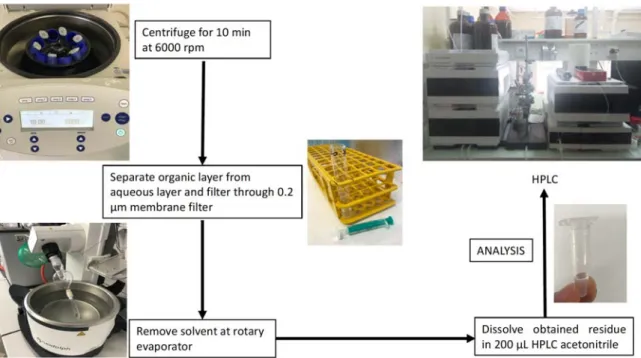
An example of a concrete experiment with human plasma that we performed is described in section 2.1.2 below and in Fig. 1 the respective steps are illustrated in a flow chart. The drug of interest is incubated in plasma at body temperature (37 °C) for a given time in the presence of an analytical reference (e.g. caffeine). The addition of the reference before the incubation period is possible because caffeine exhibits a high stability in human plasma.18 It is also
possible to utilize a 1:1 mixture of plasma with buffer (pH 7.4), which should not affect the hydrolytic rate of the compound. Besides saving biological resources, this has the additional benefit that the pH effect (increase of pH in plasma over long-time storage) does not influence the experiments.13
After the given time period the reaction is quenched with MeOH. In order to extract the drug, its (possible) metabolites and the analytical standard, CH2Cl2 is added. It is our solvent of
choice because it is easily separated from the aqueous phase, dissolves the drug and caffeine and can be rapidly removed in vacuo. Of course, other organic solvents can be suitable for this process, depending on the properties of the studied compound. For example, in the case of water soluble drug candidates, a miscible solvent such as MeOH can be employed. In such a case, the phase separation step can be skipped and the proteins and other solid parts from the plasma are only separated by centrifugation. The supernatant can then be directly injected into the HPLC (after filtration). We also strongly recommend testing the stability of the compound in the solvent in which the compound is dissolved before addition to the plasma (or the microsomes, respectively).Doing a test run on the HPLC of just the compound with the solvent is also useful to exclude eventual additional peaks that might stem from interactions between the solvent and the compound. For example in the case of ruthenium complexes, a non-coordinating solvent would usually be a better choice than acetonitrile.2.1.2 Methodology The following protocol was derived from a previously published procedure by Patra et al.19
human plasma was obtained from Bio-west and caffeine from TCI Chemicals. For each experiment, fresh stock solutions were prepared in DMSO and water, for the compound and caffeine respectively. To 950 μL of plasma, 25 μL of the solution containing the compound (5.0 mM) to be studied and 25 μL of the caffeine solution (5.0 mM) were added to reach a total volume of 1000 μL. The resulting solutions were incubated for 0 h, 1 h, 3 h, 6 h, 18 h and 24 h at 37 °C with continuous and gentle shaking (ca. 500 rpm) while protected from light. Afterwards, the plasma solution was quenched with 1 mL MeOH and 2 mL CH2Cl2 and the
mixture was shaken for 15 min at room temperature followed by centrifugation at 6000 rpm for 10 min. The organic layer was separated from the aqueous layer and the CH2Cl2 removed
under reduced pressure. The obtained residue was dissolved in 200 μL CH3CN. The solution
was filtered through a 0.2 μm membrane filter and analyzed using a 1260 Infinity HPLC System (Agilent Technology). A Pursuit XRs C18 (250 x 4.6 mm) reverse phase column was used with a flow rate of 1 mL/min.
Figure 1 Step-by-step guide for investigating the stability of compounds in human plasma.
2.1.3 Interpretation of the results
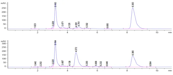
After extraction from the plasma, the complexes were analyzed via HPLC and the chromatogram before and after incubation were compared. With increasing incubation time, the peak size of the test compound decreases as decomposition products appear (Fig. 2.). The ratio of the analyzed compound with respect to the internal standard is then plotted against incubation time (Fig. 3) after having been normalized. At t = 0, when there should be no other compounds detectable yet, the ratio of the compound peak area to the internal standard peak area is set to one, respectively 100%. This has the advantage, that the analytical recovery rate of the compound, which can sometimes not be optimal in the case of metal compounds, does not affect the experiment as this factor should be constant for the different time points. Ideally, the decomposition products can be analyzed by LC-MS, however, even without this additional information one can clearly obtain valuable information about the stability profile of the compound. The time points of this experiment should be chosen wisely. While some compounds react within a few minutes in human plasma, other compounds such as for example
the ferrocenyl derivatives of the antiparasitic drug praziquantel are stable for several hours in human plasma under physiological conditions.19
Figure 2 Example of the UV traces at 265 nm of the HPLC analysis of a test compound treated
with human plasma at t = 0 h (top) and t = 24 h (bottom). Internal Standard at 3.18 min, test compound at 8.38 min. The peak at 4.57 min can be attributed to decomposition products.
Figure 3 Example of the normalized graph showing the ratio of test compound to caffeine
(internal standard) at different incubation times in human plasma.
2.2 Stability studies using microsomes
0 0.2 0.4 0.6 0.8 1 1.2 0 5 10 15 20 25 30 Ratio of tested compound vs. internal standard t / h
2.2.1 Background
In modern drug design and development, analysis and optimization of metabolic stability play an immensely important role, usually already at an early stage of the process.20 Metabolic
stability is defined as the susceptibility of a chemical compound to biotransformation. Once a drug is in the body, it is predominantely the liver that is responsible for its metabolization, transforming it into more easily excretable compounds. Hepatic enzymes such as for example the cytochromes P450 (CYP) can oxidize a broad range of compound classes. However, there are of course also other enzymes and mechanisms to be considered. In order to get a first and realistic idea of what happens to a drug in the functioning liver, it can be exposed in vitro to liver microsomes and the metabolic fate of the compound can be monitored over time. Essentially, microsomes consist of different sorts of membrane proteins (phase I proteins), such as CYP, UGT (Uridine 5'-diphospho-glucuronosyltransferase) or FMO (flavin monooxygenase). Whereas freshly extracted microsomes are often employed in (pre)clinical studies (for example obtained from cancer patients), microsome solutions can also be readily acquired from various suppliers. If stored correctly at low temperatures (−80 °C), the enzymatic activity of the microsomes can be maintained,21 and thus the same batch of microsomes can
easily be used for different experiments or compounds without the additional deviation between microsome batches.22 The microsomes can either come from a single donor, or, as it
is preferred because it gives a better median and less deviations between the experiments, from a pool of different donors, usually around 50. Of course, in addition to human microsomes, animal microsomes are also frequently used. It is important to mention that the microsomal activity can strongly differ between the species, depending on which exact (CYP) enzymes are involved in the metabolic pathways. Such differing behavior was for example observed for the organometallic drug ferroquine which was tested in mice, rats, dogs, monkeys and humans.23
From the comparison of the microsomal activity of different animal microsomes with human microsomes, the most suitable animal model can be chosen.22
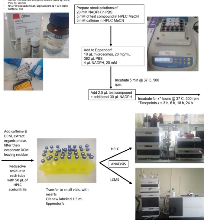
For the enzymatic activity the cofactor NADPH is required and if desired, the experiment can be performed without the addition of NADPH in order to get a negative (non NADPH-dependent) control. Other options for negative controls are to leave out the substrate or the microsomes from the incubation mixture. In order to recreate ideal conditions for the microsomes, they are first incubated in buffer (PBS) at body temperature (37 °C) with NADPH for the predetermined time. An internal standard (IS) can be included either at the start of the incubation (only if the IS proves to be stable to metabolism) or at the end during the quenching phase (if the IS is susceptible to microsomal metabolism). The substrate concentration should be low in order to assure that the drug-enzyme reaction follows first-order kinetics and the reaction rate can be assumed to be proportional to the drug concentration. It has also been shown that high substrate concentrations can lead to overestimation of the compound stability in microsomal studies, and should thus be avoided. Concerning the practical aspect of the sample preparation, either one batch per time point could be prepared, and aliquots withdrawn at the desired time, or several samples for each desired time point could be withdrawn from a single batch. In the example shown below (Fig. 4) the chosen time points are 3, 6, 18 and 24 hours because in this case the analyzed compound was very stable, however we advise to test relatively close time points at the beginning of the measurements when analyzing a new compound as metabolic processes can take place rather fast. The microsomes can tolerate the presence of small amounts of organic solvent (eg. 2.5 μL test compound solution in DMSO for a total volume of around 430 μL, less than 1 %), although the percentage of DMSO should be kept as low as possible because higher concentrations can give adulterated stability of the analyzed compound.24 The addition of larger volumes (3 mL of CH
2Cl2, CH3CN or CH3OH)
For more background information on metabolic studies, varied protocols and good practice we recommend these articles by J. R. Hill25 and by Jia and Liu.22 It is needless to say that if a larger
number of compounds are to be evaluated , specialized laboratories are much better equipped and able to perform these experiments faster and with more efficiency. For example, by application of parallel liquid chromatography/mass spectrometry (LC/MS) whole compound libraries can be screened with high throughput for their microsomal stability.26
2.2.2 Methodology
As an example, the following protocol we are currently using is derived from a previously published procedure by Hess et. al.27 The stability of the complexes was evaluated with
caffeine as internal standard, which was added during the quenching phase. The 1xPBS (phosphate buffered saline, pH 7.4) and pooled human microsomes were obtained from Gibco, NADPH tetrasodium salt from Sigma Aldrich and caffeine from TCI Chemicals. For each experiment, fresh stock solutions of the test compound were prepared in CH3CN and those of
caffeine were prepared in H2O. To 382 μL of PBS in an Eppendorf tube, 10 μL of microsomes
(20 mg/mL) and 4 μL of NADPH (20 mM) were added and the mixture was incubated for 5 minutes at 37 °C. Following this, 2.5 μL of the test compound (5 mM) and an additional 30 μL of NADPH (20 mM) was added and the resulting samples were incubated for the desired time points with continuous shaking at 500 rpm at 37 °C. After incubation, the samples were quenched with 3 mL of CH2Cl2 and 2.5 μL of caffeine was added. The mixture was then shaken
for 10 minutes on a plate shaker at room temperature. The organic layer was separated from the aqueous layer in which the proteins remain via centrifugation, and all volatiles of the organic phase were removed in vacuo. The resulting residue was redissolved in 50 μL of HPLC grade CH3CN and analyzed on a 1260 Infinity HPLC System (Agilent Technology). An
Agilent Pursuit XRs C18 (250 x 4.6 mm) reverse phase column was used with a flow rate of 1 mL/min.
Figure 4. Step-by-step guide for metabolic stability studies with microsomes.
2.2.3 Interpretation of the results
As described in section 2.1.3 for the human plasma studies, the integration of the standard and compound peak is done for the different time points and a normalized decay curve is plotted. To determine the in vitro half‐life (t1/2), the following process was derived from Tan et al.28
are expressed as a percentage of the peak area of the test compound at t = 0, which is taken as 100% (remaining parent %). The remaining parent % of the test compound were plotted against incubation time. Data points were the average of three measurements with standard deviation as error bars. The in vitro t1/2 can be calculated from the slope of the linear regression from the
half‐life equation below (1), where k is the slope of the line of the natural logarithm of the remaining parent % of the compound plotted against time.
t1/2 = 0.693/k (min), where k is the slope of the plot
Estimated in vitro clearance can be determined by the equations below:
CLint, invitro = V × 0.693/t1/2 (L/min/mg protein)
CLint, app = CLint (45 mg microsomal protein/g of liver)(45 g of liver/kg of body weight)
The shorter the half life time t1/2, the faster the clearance will be. The in vitro clearance
parameter CLint, invitro is correlated with in vivo clearance, which can be estimated with the
apparent intrinsic clearance parameter CLint, app, thus building a bridge between in vitro and in
vivo experiments.
3. Conclusion
In this article, we have given an overview of the protocols and benefits of relatively simple stability tests in blood plasma and microsomes with the aim of encouraging our colleagues in the field of metal-based drug development to conduct such experiments, which can be easily performed using standard chemical laboratory equipment. The benefits are more detailed insights into the drug behavior, resulting in a more efficient and intelligent drug design. A further advantage is the reduction of animal sacrifices for in vivo testing which, in addition to ethical factors, also makes the drug development process faster and more economic. Since these often unnecessary expenses are usually assumed by the biologist collaborators, we are
convinced that conducting these valuable studies upstream will induce more fruitful and robust collaborations towards the development of innovative organometallic drugs.
4. Acknowledgments
This work was financially supported by the Swiss National Science Foundation (Grant Sinergia CRSII5_173718 to G.G.) and has received support under the program «Investissements d’Avenir » launched by the French Government and implemented by the ANR with the reference ANR-10-IDEX-0001-02 PSL (G.G.). It was further supported by a Feodor Lynen Research Fellowship from the Alexander von Humboldt Foundation (S.K.), an Early Postdoc. Mobility Fellowship from the Swiss National Science Foundation (S.K., Grant P2 BSP2_181760) and the Chinese Scholarship Council (Y.L.).
5. References
1 B. R. Smith, C. M. Eastman, J. T. Njardarson, J. Med. Chem. 57 (2014) 9764−9773. 2 C. G. Hartinger, P. J. Dyson, Chem. Soc. Rev. 38 (2009) 391−401.
3 C. G. Hartinger, N. Metzler-Nolte, P. J. Dyson, Organometallics 31 (2012) 5677−5685. 4 N. P. E. Barry, P. J. Sadler, Chem. Commun. 49 (2013) 5106−5131.
5 G. Jaouen, A. Vessieres, S. Top, Chem. Soc. Rev. 44 (2015) 8802–8817. 6 D. Dive, C. Biot, 14 (2014) 1684–1692.
7 G. Gasser, N. Metzler-Nolte, Curr. Opin. Chem. Biol. 16 (2012), 84−91. 8 G. Gasser, I. Ott, N. Metzler-Nolte, J. Med. Chem. 54 (2011) 3−25. 9 Y. C. Ong, G. Gasser, Drug. Discov. Today Tech. (2019) accepted, DOI:
10.1016/j.ddtec.2019.06.001.
10 K. D. Mjos, C. Orvig, Chem. Rev. 114 (2014), 4540−4563.
11 J. G. Lombardino, J. A. Lowe, Nat. Rev. Drug Discovery 3 (2004) 853–862.
12 P. Baranczewski, A. Stañczak, K. Sundberg, R. Svensson, Å. Wallin, J. Jansson, P. Garberg, H. Postlind, Pharmacol. Rep. 58 (2006) 453–472.
13 L. Di, E. H. Kerns, Y. Hong, H. Chen, Int. J. Pharm. 297 (2005) 110–119.
14 F. Hernández-Luis, A. Hernández-Campos, L. Yépez-Mulia, R. Cedillo, R. Castillo, Bioorg. Med. Chem. Lett. 11 (2001) 1359−1362.
15 M. Sooriyaarachchi, A. Narendran, J. Gailer, Metallomics 3 (2011) 49–55.
16 C. S. Allardyce, A. Dorcier, C. Scolaro, P. J. Dyson, Appl. Organometal. Chem. 19 (2005), 1–10. 17 B. Demoro, R. F. M. de Almeida, F. Marques, C. P. Matos, L. Otero, J. Costa Pessoa, I. Santos,
A. Rodríguez, V. Moreno, J. Lorenzo, D. Gambinoa, A. I. Tomaz, Dalton Trans. 42 (2013) 7131–7146.
18 S. N. alvi, M. M Hammami, J. Chromatogr. Sci. 49 (2011) 292–296.
19 M. Patra, K. Ingram, V. Pierroz, S. Ferrari, B. Spingler, J. Keiser, G. Gasser, J. Med. Chem. 55 (2012) 8790−8798.
20 T. N. Thompson, Med. Res. Rev. 21 (2001) 412−449.
21 R. E. Pearce, C. J. McIntyre, A. Madan, U. Sanzgiri, A. J.Draper, P. L.Bullock, D. C.Cook, L. A. Burton, J. Latham, C. Nevins, A. Parkinson, Arch. Biochem. Biophys. 331 (1996) 145−169. 22 L. Jia, X. Liu, Curr. Drug Metab. 8 (2007) 822−829.
23 W. Daher, L. Pelinski, S. Klieber, F. Sadoun, V. Meunier, M. Bourrié , C. Biot, F. Guillou, G. Fabre, J. Brocard, L. Fraisse, J.-P. Maffrand, J. Khalife, D. Dive, Drug Metab. Dispos. 34 (2006) 667–682.
24 L. Di, E. H. Kerns, Y. HONG, T. A. Kleintop, O. J. McConnell, D. M. Huryn, J. Biomol. Screening 8 (2003) 453–462.
25 J. R. Hill, Current Protocols in Pharmacology (2003) 7.8.1−7.8.11.
26 R. Xu, C. Nemes, K. M. Jenkins, R. A. Rourick, D. B. Kassel, C. Z. C. Liu, J. Am. Soc. Mass. Spectrom. 13 (2002), 155−165.
27 M. Patra, K. Ingram, A. Leonidova, V. Pierroz,, S. Ferrari, M. N. Robertson, M. H. Todd, J. Keiser, G. Gasser, J. Med. Chem. (56) 2013, 9192−9198.