Article type:
Advanced Review
Article title: Bringing computational models of bone regeneration to the clinic
Authors:
Full name and affiliation; email address if corresponding author; any conflicts of interest First author
Aurélie Carlier
Biomechanics Section, KU Leuven, Celestijnenlaan 300 C, PB 2419, 3000 Leuven, Belgium
Prometheus, Division of Skeletal Tissue Engineering, KU Leuven, O&N 1, Herestraat 49, PB 813, 3000 Leuven, Belgium
Second author
Liesbet Geris
Biomechanics Section, KU Leuven, Celestijnenlaan 300 C, PB 2419, 3000 Leuven, Belgium
Prometheus, Division of Skeletal Tissue Engineering, KU Leuven, O&N 1, Herestraat 49, PB 813, 3000 Leuven, Belgium
Biomechanics Research Unit, University of Liege, Chemin des Chevreuils 1 – BAT 52/3, 4000 Liege 1, Belgium
Third author
Johan Lammens
Prometheus, Division of Skeletal Tissue Engineering, KU Leuven, O&N 1, Herestraat 49, PB 813, 3000 Leuven, Belgium
University Hospitals of KU Leuven, Department of Orthopaedics, KU Leuven, Weligerveld 1 - blok 1, 3212 Pellenberg, Belgium
Fourth author
Hans Van Oosterwyck (*) [email protected]
Biomechanics Section, KU Leuven, Celestijnenlaan 300 C, PB 2419, 3000 Leuven, Belgium
Prometheus, Division of Skeletal Tissue Engineering, KU Leuven, O&N 1, Herestraat 49, PB 813, 3000 Leuven, Belgium
Abstract
Although the field of bone regeneration has experienced great advancements the last decades, integrating all the relevant, patient-specific information into a personalized diagnosis and optimal treatment remains a challenging task due to the large number of variables that affect bone regeneration. Computational models have the potential to cope with this complexity and to improve the fundamental understanding of the bone regeneration processes as well as to predict and optimize the patient-specific treatment strategies. However, the current use of computational models in daily orthopedic practice is very limited or inexistent. We have identified three key hurdles that limit the translation of computational models of bone regeneration from bench to bed side. First, there exists a clear mismatch between the scope of the existing and the clinically required models. Second, most computational models are confronted with limited quantitative information of insufficient quality thereby hampering the determination of patient-specific parameter values. Third, current computational models are only corroborated with animal models whereas a thorough (retro- and prospective) assessment of the computational model will be crucial to convince the health care providers of the capabilities thereof. These challenges must be addressed so that computational models of bone regeneration can reach their true potential, resulting in the advancement of individualized care and reduction of the associated health care costs.
Bone is a truly remarkable and interesting tissue. Not only provides the human adult skeleton support and protection for various organs in the body, the collection of 206 bones also stores minerals, produces blood cells and allows movement. Moreover, unlike other adult biological tissues, bone is the only tissue that can heal without the production of scar tissue. The regeneration of bone tissue is a complex, well-orchestrated process of cell recruitment, proliferation and differentiation regulated by several biochemical and mechanical factors. Unfortunately, despite bone’s remarkable healing capacity and the continuing research efforts, 5 to 10 percent of the over 6 million fractures occurring annually in the USA develop into delayed or non-unions 1,2. In a more recent 5-year cross-sectional epidemiological study Mills et al. report a non-union incidence in the Scottish population of 22.45 in men and 15.65 in women per 100 000 population per annum 3. The complications in fracture healing often result in the need of reoperations or additional treatments, costing society large amounts of money. In 2005 the cost of the more than 2 million osteoporosis-related fractures occurring in the USA was estimated at 17 billion dollar and the annual costs are projected to rise by 50% by 2025 due to the aging population 4. Moreover, it is estimated that by 2020, traffic accidents (a major cause of fractures) will rank in the top three causes of disability 5,6. As such, more knowledge of the complex physiological process of bone healing is a prerequisite for the prevention and effective treatment of complex fractures.
This article will focus on the translation of computational models of bone regeneration towards clinical practice, including both models of the fundamental biological process of bone regeneration as well as of devices improving the bone healing outcome (e.g. fixation plates). First, the most recent modelling efforts will be summarized. Although many computational models of the fundamental biological process of bone regeneration exist, none have already entered the clinical arena to aid in the clinical decision process (e.g. prevention, diagnosis, treatment and monitoring) whereas patient-specific finite element (FE) models have been adopted for the evaluation of individualized implant solutions in orthopedic bone and joint reconstruction surgery. The key challenges associated with the translation from bench to bed side will be identified and thoroughly discussed. Finally, some opportunities and conclusive remarks will be formulated.
Computational modelling of bone regeneration
Although the field of bone regeneration has experienced great advancements the last decades 7, integrating all the relevant, patient-specific information into a personalized diagnosis and optimal treatment remains a challenging task due to the large number of variables that affect bone regeneration 7–10. However, (patient-specific) computational models have the potential to cope with this complexity and to improve the fundamental understanding of the bone regeneration processes as well as to predict and optimize the patient outcome. Given the extensive amount of work in this modelling field, we only highlight some recent advances and focus specifically on the clinical potential of the computational models. For further information on the bioregulatory and mechanoregulatory algorithms used in these computational models we refer the reader to some excellent reviews 11–14.
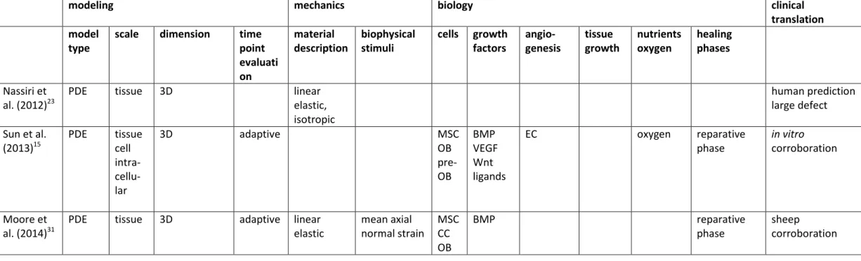
The most important computational models of bone regeneration, including their essential characteristics, are summarized in Table 1. Clearly, most models focus on the tissue scale, using differential equations to describe the mechanoregulatory or bioregulatory processes. Only Sun et al. 15
and Carlier et al. 16 use a multiscale approach to capture the bone regeneration dynamics. Note that the more recent modeling frameworks often include some bioregulatory aspects of the bone regeneration process. Depending on the application, mechanoregulatory models use a linear elastic or a poroelastic material description as well as various definitions of the mechanical stimulus. The majority of the bioregulatory models includes a description of cells, growth factors and angiogenesis. More recent modeling efforts also focus on the influence of oxygen on the bone regeneration processes. Tissue growth during callus formation is only accounted for in the models of Chen et al. 17, Simon et al. 18 and Gomez-Benito et al. 19. Interestingly, none of the models listed in Table 1 capture the inflammation phase and very little frameworks include the remodeling phase. The last column of Table 1 indicates the level of clinical translation. It appears that most models are only corroborated in small or large animal models and extrapolate their findings for human predictions. Nevertheless, various computational models of bone regeneration hold great potential to address particular clinical questions as detailed in the following paragraphs.
Impaired bone healing has been associated with a variety of factors including the mechanical and biological micro-environment. In order to ensure a correct anatomic alignment and to provide enough stability to allow (partial) loading while maintaining a certain interfragmentary movement for optimal secondary healing, orthopedic fixation is used (in combination with bone grafts or other bone substitutes if the biological micro-environment is also compromised). A variety of internal and external fixation devices are available that combine an adequate device stiffness with sufficient device strength and acceptable surgical technicalities: screws, plates, staples, wires, rods, Ilizarov fixator, etc. 20. These fixation designs can be critically evaluated and optimized with mechanoregulatory and FE models. Nasr et al. performed for example an idealized poroelastic finite element analysis to evaluate 19 different plate-screw combinations 21. They showed that a 4-screw symmetrical construct with a sufficient distance between the screws provides an optimal balance between stability (to allow weight bearing) and flexibility (to promote callus formation) 21. Similarly, Moazen et al. concluded that the bridging length made a more substantial difference to the stiffness and interfragmentary movement than varying the plate material, plate thickness or screw-plate fixation 22. Also Nassiri et al. have recently reported similar findings 23. The calculations of Boccaccio et al. indicated that the presence of percutaneous fixation devices significantly shortened the healing times of the fractured body of the L4 vertebra 24. Moreover, they also found that Cobalt-Chrome would be a better alloy than Ti6Al4V due to its greater stiffness 24.
The more challenging orthopedic cases do not only require adequate stabilization of the fracture but also biological support through e.g. distraction osteogenesis, bone grafting and/or the administration of growth factors. The influence of the pre-traction stresses, the distraction rate as
well as the fixator stiffness on the bone regeneration process during bone transport was computationally investigated by Reina-Romo et al. 25–27. Their mechanoregulatory model showed that the inclusion of pre-traction stresses, i.e. the stress level in the gap tissue before each distraction step, affects the evolution of the bone regeneration process and consequently the reaction forces 25. Moreover, in agreement with clinical findings, a distraction rate of 1 mm/day was found to stimulate osteogenesis optimally 28. They also reported that a stiff fixator promotes bone formation while flexible fixators will give rise to excessive motion and adverse bone healing 27. Mechanoregulatory and FE models can also be used to evaluate bone grafting methods, a technique commonly performed in clinical practice for the skeletal reconstruction of large bone defects. In order to improve the access of surrounding vascularity and increase the graft incorporation, bone allografts are sometimes longitudinally or transversely perforated. Santoni et al. have used a finite element analysis to evaluate the structural and mechanical integrity of these constructs and conclude that longitudinal perforation does not adversely affect the mechanical properties of the graft 29.
To date very little bioregulatory models of bone regeneration exist that can aid in the evaluation of bioactive molecules and tissue engineering strategies. Burke et al. have included biological cues such as oxygen tension in a mechanoregulatory model to study stem cell differentiation during fracture healing 30. A similar approach was taken by Moore et al. who include a mechanical regulation of BMP-2 production thus establishing a novel mechanobioregulatory framework 31. After comparing the in silico predictions with the observed in vivo outcome, Geris et al. have used a continuum-type model of fracture healing to simulate the injection of cultured MSCs 32. They conclude that eccentric injection resulted in unicortical bridging of an atrophic non-union. Moreover, a suitable time point for intervention was found to be three weeks post-osteotomy so that the blood supply to the fracture gap had already partially recovered 32. Similar conclusions were drawn by Carlier et al. who used a hybrid, multiscale bioregulatory model of fracture healing for an in depth investigation of the mechanisms of action underlying critical size defects 33. More specifically, the formation of a non-union was attributed to the severe hypoxia in the central callus area 33. Motivated by these results, the timing of administration of osteoprogenitor cells or growth factors was explored further. Carlier et al. conclude that the timing of administration is only critical for cell therapies since the local oxygen tension will determine the survival as well as proliferation and differentiation potential of the administered cells and consequently the extent of the bone formation process. The calculations suggest a minimal delay of 5 weeks (for a 5 mm segmental defect in mouse bone) in order to allow for a (partial) restoration of the blood supply that can nurture the administered cells 33. Sun et al. also propose a computational framework to study the effect of different growth factors on the bone regeneration process and tailor their respective release profiles by controlling the pore size of a tissue engineered scaffold 15.
Although these examples illustrate that various clinical questions can be adequately addressed by in
silico techniques, the current use of computational models in daily orthopedic practice is very limited
or inexistent. Indeed, there are several barriers to bring in silico models from the (computer) bench to the bed side which will be further elaborated in the next section.
Key challenges
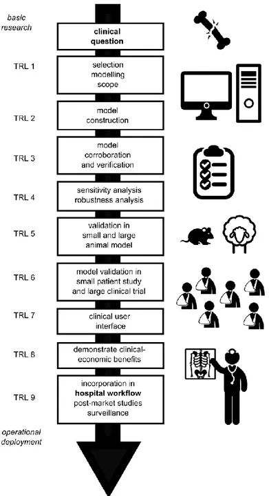
While computational models of bone regeneration hold great promise to advance individualized care and reduce the associated health care costs, several key challenges need to be addressed including the selection of scope of the computational model, the generation of data for model construction and validation as well as the creation of user-friendly interfaces tailored to the clinical purpose (Figure 1). In this section we will highlight some of the most important challenges and apply
them to the field of computational modelling of bone regeneration. We would like to refer the reader to the “Digital Patient Roadmap” for a broader and more exhaustive overview of the scientific and technological challenges associated with clinical computational models 34.
Selection of the appropriate modelling scope
Recent advances in fracture management, including better protocols, more strict patient follow-up and improvements in hardware as well as surgical techniques have contributed to a better prognosis, even in complex fractures 35. However, the treatment of for example atrophic non-unions, characterized by a severely hampered biological support for bone healing 7, or of osteoporotic fractures, characterized by limited fixation capabilities due to a poor bone quality 36, continues to represent a therapeutic challenge. Clearly, these two examples of complex orthopedic challenges are associated with an impaired host environment. Although clinical complications mostly occur in patients with preexisting risk factors including old age 37, cachexia and malnutrition 38, immune compromise 8, genetic disorders (e.g. type 1 neurofibromatosis 39), osteoporosis 40, anticoagulants 41, smoking 42 and anti-inflammatory agents 43, computational models of bone regeneration to date mostly consider an average (i.e. healthy, young) subject. Consequently, there is a mismatch of the existing computational models of bone regeneration and the clinically required models as the (diseased) host environment is not adequately captured. The first important challenge in bringing computational models to the bed side is the development of clinically relevant models of bone regeneration that consider the compromised, diseased state.
Some promising steps have already been taken in this direction. For example, Sabalic et al. used an osteoporotic finite element model to compare three fixation configurations for distal humerus fractures 44. They conclude that a Y-shaped plate is a potential alternative for the standard two-plate osteosynthesis method although further biomechanical studies are required 44. Ode et al. used whole-genome expression analysis to identify the key genes that are influenced by an interaction between the effects of mechanical fixation stability and age 45. The differentially expressed genes indicated an association with the following biological processes: extracellular space, cell migration and vascular development 45. By altering the parameter values of the functional forms corresponding to the biological processes identified by Ode et al. 45, the influence of patient age could be incorporated in existing computational models. In a next step, these improved models allow to simulate stratified patient populations (e.g. fracture healing in old versus young patients), taking a first step in the direction of patient-specific models of bone regeneration.
Generation of (patient-specific) data
Computational models massively rely on quantitative and qualitative data to (i) identify the theoretical backbone of the computational model and determine its parameter values, (ii) validate the in silico predictions and (iii) update the framework with patient-specific information. Nevertheless, most computational models are confronted with limited quantitative information of insufficient quality due to a variety of reasons.
A first question arises as to which aspects of the mechano- and bioregulatory models should be made patient-specific: the geometry, the parameter values, the boundary conditions, the initial conditions or a combination thereof? Sensitivity analysis methods, including Design of Experiments (DOE) can be a very valuable tool to determine the importance of the patient-specific aspects to the predicted model outcome 46. DOE (or experimental design) is a statistical tool that generates an array of combinations of different parameter values within a predefined parameter space. Next, the computational model is run with these parameter combinations and finally the results are statistically analyzed. This approach was taken by Isaksson et al. to determine the most important cellular characteristics of a mechanoregulatory model of bone healing. The parameters related to cartilage formation and degradation were found to significantly influence the bone healing outcome
as was confirmed by in vivo animal experiments in the literature 47. Another example, which is closer to clinical application, is given by Valente et al. who investigated the sensitivity of patient-specific model predictions (i.e joint angles, joint moments, muscle and joint contact forces) during walking to the uncertainties in the identification of body landmark positions, maximum muscle tension and musculotendon geometry 48. They concluded that the patient-specific models are not markedly sensitive to the parameter identification, depending on the intended application of the model 48. The second barrier is represented by the limited technologies available to measure the key patient-specific aspects. A lot of progress has been made concerning patient-patient-specific FE modeling of bones, as reviewed by Poelert et al. 49. Indeed, with current imaging modalities such as MRI and CT, the bone geometry can be relatively easily obtained. Moreover, power laws exist to correlate the bone density, obtained from the CT readout, to Young’s moduli thereby assigning the patient-specific material properties to every element. Difficulties are however associated with the determination of the in vivo loading conditions since there exists no simple method to non-invasively measure the muscle and joint reaction forces 49. Therefore, forces are currently indirectly determined from musculoskeletal models. In order to provide mechano- and bioregulatory models with more in vivo quantitative data on cellular properties (e.g. proliferation rate), the patient-specific (biological) host environment, the spatial and temporal distribution of cells, growth factors and tissues and the mechanical regulation thereof, novel tools will need to be developed since adequate technologies are currently inexistent. According to a recent review of Pountos et al., none of the existing biomarkers can be recommended for routine clinical use to assess the progression of fracture healing although some of the investigated biomarkers (e.g. ALP, TGF-β1, VEGF, BMP-2) might be indicative of distinctive processes occurring during fracture healing (e.g. proliferation, differentiation, matrix production) 50. Calori et al. have combined several risk factors (e.g. smoking, osteoporosis, gap size) into a Non-Union Scoring System (NUSS) which provides an index of severity (0-100 points) and classifies patients in four treatment groups 51. As such, appropriate biomarkers and NUSS-scores can potentially provide an important input for the parameter determination and validation of computational models. Another promising technique is intravital microscopy (IVM) which allows a dynamic, non-invasive and high resolution visualization of the region of interest 52,53. A third reason for the limited quantitative information can be found in the clinical adoption of the (novel) measurement technologies. Firstly, there exists a gap between the technologies available to monitor and quantify the (patho)physiological phenomena in a research setting with respect to daily clinical practice. In a research context a detailed 3D patient-specific anatomy can for example be derived from advanced 3D imaging methods like CT and MRI, whereas conventional 2D X-ray images are still the method of choice in daily orthopedic practice since they can be obtained through a fast and inexpensive procedure. In order to acquire the 3D geometries existing CT and MRI imaging modalities should become part of routine diagnosis and therapy planning in orthopedics whereby the additional imaging costs are predicted to be compensated by the large amount of costs savings that an improved in silico designed treatment presents (see further). Alternatively, novel tools can be developed to extract additional information from existing 2D X-ray images. The latter approach has been followed by Ehlke et al. who maximize the correspondence between a virtual X-ray projection derived from a 3D deformable tetrahedral mesh and the anatomy depicted in a clinical X-ray 54. By using a computational efficient projection algorithm, they are able to reconstruct 3D models of patient-specific bone shapes from a single or few X-ray images 54. Similar approaches can also be used to determine the bone density distribution, necessary to populate the (bio)mechanoregulatory models with patient-specific material properties.
Secondly, during clinical imaging or sensing examinations, often only limited information is acquired. As such, dedicated examination protocols should be established in collaboration with modelling partners so that the necessary information for modelling purposes is acquired. Bode et al. have for example designed a clinical study protocol during which structural and functional data at the
vascular level will be collected and the vascular access will be functionally evaluated during follow-up 55. By implementing a strict imaging protocol, they aim to maximize the amount of demographical data in the difficult and heterogeneous target population 55. In this way the adopted strategy will allow for the calibration and validation of the computational models developed within the ARCH project (patient specific image-based computational modeling for improvement of short-term and long-term outcome of vascular access in patients on hemodialysis therapy) 55.
Thirdly, the limited patient-specific information might be obtained from different systems, using different settings or different technologies 56. As such, protocols are needed that define how the data should be acquired and represented.
Finally, patient specific data might be missing due to e.g. the invasiveness of the method or local unavailability. For these cases, machine learning methods can be used to learn from the remaining data set and predict missing values 57. Alternatively, atlases, built from patient-specific data in large databases 58,59, can provide subject-specific data through mapping procedures.
Translation and clinical utilization of models
The clinical adoption of patient-specific computational models will be highly dependent on the confidence clinicians feel to use them as a tool for personalized diagnosis and treatment. One of the key challenges will be to convince the health care providers of the technical capabilities and limitations through a thorough assessment of the computational models, including validation (i.e. the model predictions match the experimental reality), verification (i.e. the claimed outputs can be achieved for specified inputs by someone other than the model developer) as well as a sensitivity analysis (i.e. identification of the most influential model parameters) and a robustness analysis (i.e. evaluation of the deviation of the reference state due to external perturbations) 34.
In a first step, computational models are generally corroborated with animal models (e.g. rat, sheep) (Table 1) since animal models allow for an extensive experimental characterization and quantification. Clearly, a key requirement for this strategy to be successful is that the animal models correctly capture the key disease mechanisms present in the human patient 60–62. If not, an animal-validated computational model will fail in a human patient although it was able to nicely predict the bone regeneration process in the respective animal model. Likewise, the underlying mechanisms captured in the computational models need to match those of the animal model and the human patient to increase the chances of a successful translation from animal-validated to patient-validated models. Moreover, if the disease mechanisms are conserved between animal and patient, an animal-validated mechanistic model can potentially assist in the clinical translation of an in silico designed therapy by recalibrating a number of parameter values (e.g. the geometry).
In a second step, the most promising computational models should be evaluated in a limited number of patient-specific study cases. Subsequently, a larger retrospective validation can be performed, although the existing retrospective data are often insufficient, improper for the modelling purposes or incomplete. As such, the challenge of thorough statistical (retrospective) validation and sensitivity analysis is intimately tied to the availability of patient-specific data, a key challenge that was discussed above. Alternatively or as a second stage, thorough prospective investigations can be performed using dedicated clinical trial protocols. A few good examples do exist such as the study by Trabelsi et al. (2011) who validated patient-specific finite element models of human cadaver femurs based on quantitative computer tomography in a double-blinded manner by biomechanical in vitro experiments performed in two different research institutes 63. A single leg loading configuration was used to determine the strains and local displacements on the bone surface as well as the axial stiffness 63. They demonstrated an excellent agreement between the in silico and in vitro results, highlighting the advanced stage of the computational model 63.
After a thorough assessment of the technical and clinical capabilities of the computational models, clear improvements in health outcome measures and economic benefits should be demonstrated.
Many techniques and tools are available within the field of health economics to aid in such an analysis (e.g. minimization analysis, effectiveness analysis, utility analysis and cost-benefit analysis) 64. A multilevel generic methodological framework to assess both the clinical and socio-economic impact of biomedical computational models in specific was developed by Thiel et al. 65
. When applying this framework to the predictive computational models for osteoporosis developed during the Osteoporotic Virtual Physiological Human Project (VPHOP), Thiel et al. conclude that the extra costs needed to implement the VPHOP framework are by far compensated by the large amount of costs savings that the improved fracture risk prognosis of VPHOP presents 66. Finally, following acceptance by the clinicians and other health care professionals, the computational models can be integrated in the hospital workflow. In order to facilitate this transition, attention needs to be paid to the usability of the models including a user-friendly interface and clinically relevant calculation times.
Opportunities
Computational models hold great promise to improve patient-specific treatment and reduce the associated health care costs. Moreover, as ethical and economic considerations increasingly challenge in vitro and in vivo methods, computational models will play a critical role in the replacement, reduction and refinement of animal testing. However, computational models need to take several steps before they can be adopted in clinical practice. Figure 1 schematically summarizes the roadmap of this clinical translation process, including the Technology Readiness Levels (TRL) which are typically used to assess the maturity of novel technologies. From Table 1 it can be noted that the usage of computational models as a research tool is growing steadily (small and large animal corroboration, TRL 5) whereas their use in clinical practice is very limited (TRL 9). A nice example of patient-specific implant solutions for orthopedic bone and joint reconstruction surgery is described by Delport et al.. This proven technology, marketed by Mobelife (Belgium) comprises three highly automated steps. First, the bony structures are presented with advanced 3D image processing techniques. Second, a patient-specific implant is designed, including a pre-operative planning that will be transferred into surgery using jig guiding technology (Materialise, Belgium). In a last step, the design is evaluated with a patient-specific finite element model that accounts for patient-specific bone quality and thickness as well as individualized muscle attachments and muscle and joint forces 67
.
Besides aiding in the identification of novel treatment strategies or surgical planning, in silico models have also been accepted by the US Food and Drug Administration (FDA) as substitutes to animal trials in the preclinical testing phase. Kovatchev et al. present for example a system for in silico testing of control algorithms linking continuous glucose modeling to insulin delivery in an artificial pancreas 68. Zhao et al. also review the use of PBPK models in regulatory decision making 69. They conclude that computational models can facilitate the decision making concerning the need for specific clinical pharmacological studies, specific study designs and the appropriate labeling language 69
.
Conclusion
Recent advances in fracture management, including better protocols, more strict patient follow-up and improvements in hardware as well as surgical techniques have contributed to a better prognosis, even in complex fractures 35. However, the treatment of for example atrophic non-unions, characterized by a severely hampered biological support for bone healing, or of osteoporotic fractures, characterized by limited fixation capabilities due to a poor bone quality, continues to represent a therapeutic challenge. In order to find clinically relevant solutions for these complex orthopedic cases a combined in vitro, in vivo and in silico research approach is imperative. Indeed,
current computational models of bone regeneration hold great promise to improve our fundamental understanding of impaired bone healing and design novel treatment strategies. Unfortunately, the translation of computational models from bench to bed side has been hampered by a number of barriers such as the mismatch between the open clinical questions and the current modelling efforts, the scarcity of patient-specific quantitative data and the lack of adequate model validation. Further research is required to overcome these challenges so that computational models of bone regeneration can reach their true potential, resulting in the advancement of individualized care and reduction of the associated health care costs.
Figure captions
Figure 1: Roadmap for the clinical translation of computational models of bone regeneration. On the left the well-known TRL (technology readiness levels) are indicated 70.
Acknowledgements
Aurélie Carlier is a post-doctoral research fellow of the Research Foundation Flanders (FWO-Vlaanderen). The authors gratefully acknowledge support from the BOF-KU Leuven GOA project 3M120209 and the European Research Council under the European Union's Seventh Framework Programme (FP7/2007-2013)/ERC Grant Agreement n° 279100 and 308223). The work is part of Prometheus, the Leuven Research and Development Division of Skeletal Tissue Engineering of the Katholieke Universiteit Leuven: www.kuleuven.be/Prometheus.
modeling mechanics biology clinical translation model
type
scale dimension time point evaluati on material description biophysical stimuli cells growth factors angio- genesis tissue growth nutrients oxygen healing phases Carter et al. (1998)71 PDE tissue 2D+ (axi-symmetric) single linear elastic principal tensile and hydrostatic stress reparative phase mouse corroboration Claes et al. (1999)72 PDE tissue 2D+ (axi-symmetric) single linear elastic and hyper- elastic principal strain and hydrostatic pressure reparative phase sheep corroboration Ament (2000)73 PDE, fuzzy logic tissue 2D+ (axi-symmetric) adaptive linear elastic SED fuzzy logic reparative, remodelling phase sheep corroboration Bailon-Plaza et al. (2003)74 PDE tissue 2D+ (axi-symmetric) adaptive linear elastic deviatoric strain and dilatational strain MSC CC OB CGGF OGGF reparative phase Gomez-Benito et al. (2005)19 PDE tissue 2D+ (axi-symmetric) adaptive linear elastic second invariant of the deviatoric strain tensor MSC CC FB OB volume growth reparative, remodelling phase sheep corroboration human prediction Shefelbine et al. (2005)75 PDE, fuzzy logic
tissue 3D adaptive linear
elastic octahedral shear strain, hydrostatic strain fuzzy logic reparative, remodelling phase Santoni et al. (2007)29
PDE tissue 3D single linear
elastic
sheep
modeling mechanics biology clinical translation model
type
scale dimension time point evaluati on material description biophysical stimuli cells growth factors angio- genesis tissue growth nutrients oxygen healing phases Andreykiv et al. (2008)76 PDE tissue 2D+ (axi-symmetric) adaptive poro- elastic shear strain and fluid flow
MSC FB CC OB reparative phase sheep corroboration Isaksson et al. (2008)77 PDE tissue 2D+ (axi-symmetric) adaptive poro- elastic shear strain and fluid flow
MSC FB CC OB reparative phase non-union prediction Geris et al. (2008)78 PDE tissue 2D+ (axi-symmetric) adaptive poro- elastic fluid flow MSC FB CC OB CGGF OGGF VEGF vascular matrix reparative phase rat corroboration non-union prediction Chen et al. (2009)17 Simon et al. (2011)18 PDE, fuzzy logic tissue 2D+ (axi-symmetric) adaptive linear elastic dilational , distortional strain vascular matrix volume growth nutrient reparative phase sheep corroboration non-union prediction Wehner et al. (2010)79 PDE, fuzzy logic
tissue 3D adaptive linear elastic volumetric, distortional strain vascular matrix, perfusion reparative phase human corroboration human prediction Reina-Romo et al. (2009-2011)25–28 PDE tissue 2D+ (axi-symmetric) adaptive poro-elastic principal strain and hydrostatic pressure MSC vasculariz ation reparative phase sheep corroboration distraction osteogenesis Byrne et al. (2011)80
PDE organ 3D adaptive biphasic
poroelastic
shear strain and fluid flow
MSC FB CC OB reparative and remodelling phase human corroboration human prediction
modeling mechanics biology clinical translation model
type
scale dimension time point evaluati on material description biophysical stimuli cells growth factors angio- genesis tissue growth nutrients oxygen healing phases Boccaccio et al. (2012)24 PDE, fuzzy logic tissue cell 3D adaptive biphasic poroelastic octahedral shear strain, hydrostatic strain reparative phase, remodeling phase human prediction Vetter et al. (2012)81 PDE tissue 2D+ (axi-symmetric) adaptive linear elastic principal, shear, volumetric, octahedral shear strain ‘biol ogic al pote ntial’ ‘biologic al potentia l’ ‘biological potential’ reparative phase sheep corroboration Burke et al. (2012)30 PDE tissue 2D+ (axi-symmetric)
adaptive biphasic deviatoric strain vascular matrix oxygen reparative, remodelling phase sheep corroboration Peiffer et al. (2011)16,16,8 2,82,82,83 PDE, ABM tissue cell intra- cellu-lar 2D adaptive MSC FB CC OB GF VEGF EC oxygen reparative phase rat corroboration non-union prediction Moazen et al. (2012)22
PDE tissue 3D single isotropic human
corroboration human prediction Nasr et al.
(2013)21
PDE tissue 3D adaptive isotropic, poroelastic octahedral shear strain, interstitial fluid velocity reparative phase human predictions large defect
Table 1 : Summary of computational models of bone tissue regeneration, indicating their major constituents. (PDE, partial differential equation; SED, strain energy density; GF, growth factor; MSC, mesenchymal stem cell; FB, fibroblast; CC, chondrocyte; OB, osteoblast; EC, endothelial cell; CGGF, chondrogenic growth factor; OGGF, osteogenic growth factor; VEGF, vascular endothelial growth factor; EC, endothelial cell; BMP, bone morphogenetic protein.)
modeling mechanics biology clinical
translation model
type
scale dimension time point evaluati on material description biophysical stimuli cells growth factors angio- genesis tissue growth nutrients oxygen healing phases Nassiri et al. (2012)23
PDE tissue 3D linear
elastic, isotropic human prediction large defect Sun et al. (2013)15 PDE tissue cell intra- cellu-lar 3D adaptive MSC OB pre-OB BMP VEGF Wnt ligands EC oxygen reparative phase in vitro corroboration Moore et al. (2014)31
PDE tissue 3D adaptive linear
elastic mean axial normal strain MSC CC OB BMP reparative phase sheep corroboration
Reference List
1. Einhorn TA The cell and molecular biology of fracture healing. Clinical Orthopaedics and
Related Research 1998, S7-S21.
2. Bhandari M, Jain AK Bone stimulators: Beyond the black box. Indian J Orthop 2009, 43: 109-110. 10.4103/0019-5413.50842 [doi].
3. Mills LA, Simpson AH The relative incidence of fracture non-union in the Scottish population (5.17 million): a 5-year epidemiological study. BMJ Open 2013, 3. bmjopen-2012-002276 [pii];10.1136/bmjopen-2012-bmjopen-2012-002276 [doi].
4. Burge R, Dawson-Hughes B, Solomon DH, Wong JB, King A, Tosteson A Incidence and economic burden of osteoporosis-related fractures in the United States, 2005-2025.
J Bone Miner Res 2007, 22: 465-475. 10.1359/jbmr.061113 [doi].
5. Fong K, Truong V, Foote CJ, Petrisor B, Williams D, Ristevski B, Sprague S, Bhandari M Predictors of nonunion and reoperation in patients with fractures of the tibia: an observational study. Bmc Musculoskeletal Disorders 2013, 14.
6. Dormans JP, Fisher RC, Pill SG Orthopaedics in the developing world: present and future concerns. J Am Acad Orthop Surg 2001, 9: 289-296.
7. Roberts TT, Rosenbaum AJ Bone grafts, bone substitutes and orthobiologics The bridge between basic science and clinical advancements in fracture healing. Organogenesis 2012, 8: 114-124.
8. Claes L, Recknagel S, Ignatius A Fracture healing under healthy and inflammatory conditions.
Nat Rev Rheumatol 2012, 8: 133-143. nrrheum.2012.1 [pii];10.1038/nrrheum.2012.1
[doi].
9. Marsh D Concepts of fracture union, delayed union, and nonunion. Clin Orthop Relat Res 1998, S22-S30.
10. Dimitriou R, Jones E, McGonagle D, Giannoudis PV Bone regeneration: current concepts and future directions. BMC Med 2011, 9: 66. 1741-7015-9-66 [pii];10.1186/1741-7015-9-66 [doi].
11. Isaksson H Recent advances in mechanobiological modeling of bone regeneration.
Mechanics Research Communications 2012, 42: 22-31.
12. Pivonka P, Dunstan CR Role of mathematical modeling in bone fracture healing. BoneKEy Rep 2012, 1. 10.1038/bonekey.2012.221.
13. Geris L, Gerisch A, Schugart RC Mathematical modeling in wound healing, bone regeneration and tissue engineering. Acta Biotheor 2010, 58: 355-367. 10.1007/s10441-010-9112-y [doi].
14. Geris L Regenerative orthopaedics: in vitro, in vivo and in silico. International Orthopaedics
15. Sun XQ, Kang YQ, Bao JG, Zhang YY, Yang YZ, Zhou XB Modeling vascularized bone regeneration within a porous biodegradable CaP scaffold loaded with growth factors. Biomaterials 2013, 34: 4971-4981.
16. Carlier A, Geris L, Gastel NV, Carmeliet G, Oosterwyck HV Oxygen as a critical determinant of bone fracture healing-A multiscale model. J Theor Biol 2014, 365C: 247-264. S0022-5193(14)00601-8 [pii];10.1016/j.jtbi.2014.10.012 [doi].
17. Chen G, Niemeyer F, Wehner T, Simon U, Schuetz MA, Pearcy MJ, Claes LE Simulation of the nutrient supply in fracture healing. J Biomech 2009, 42: 2575-2583.
S0021-9290(09)00391-1 [pii];10.1016/j.jbiomech.2009.07.010 [doi].
18. Simon U, Augat P, Utz M, Claes L A numerical model of the fracture healing process that describes tissue development and revascularisation. Comput Methods Biomech
Biomed Engin 2011, 14: 79-93. 929761607 [pii];10.1080/10255842.2010.499865
[doi].
19. Gomez-Benito MJ, Garcia-Aznar JM, Kuiper JH, Doblare M Influence of fracture gap size on the pattern of long bone healing: a computational study. J Theor Biol 2005, 235: 105-119. S0022-5193(05)00003-2 [pii];10.1016/j.jtbi.2004.12.023 [doi].
20. Taljanovic MS, Jones MD, Ruth JT, Benjamin JB, Sheppard JE, Hunter TB Fracture fixation.
Radiographics 2003, 23: 1569-1590. 10.1148/rg.236035159 [doi];23/6/1569 [pii].
21. Nasr, S., Hunt, S., and Duncan, N. Effect of screw position on bone tissue differentiation within a fixed femoral fracture. Journal of Biomedical Science and Engineering 2013, 6: 71-83. 10.4236/jbise.2013.612A009.
22. Moazen M, Jones AC, Leonidou A, Jin ZM, Wilcox RK, Tsiridis E Rigid versus flexible plate fixation for periprosthetic femoral fracture-Computer modelling of a clinical case.
Medical Engineering & Physics 2012, 34: 1041-1048.
23. Nassiri M, MacDonald B, O'Byrne JM Locking compression plate breakage and fracture non-union: a finite element study of three patient-specific cases. European Journal of
Orthopaedic Surgery and Traumatology 2012, 22: 275-281.
24. Boccaccio A, Kelly DJ, Pappalettere C A model of tissue differentiation and bone remodelling in fractured vertebrae treated with minimally invasive percutaneous fixation.
Medical & Biological Engineering & Computing 2012, 50: 947-959.
25. Reina-Romo E, Gomez-Benito MJ, Garcia-Aznar JM, Dominguez J, Doblare M Growth mixture model of distraction osteogenesis: effect of pre-traction stresses. Biomechanics and
Modeling in Mechanobiology 2010, 9: 103-115.
26. Reina-Romo E, Gomez-Benito MJ, Garcia-Aznar JM, Dominguez J, Doblare M Modeling distraction osteogenesis: analysis of the distraction rate. Biomechanics and
Modeling in Mechanobiology 2009, 8: 323-335.
27. Reina-Romo E, Gomez-Benito MJ, Dominguez J, Niemeyer F, Wehner T, Simon U, Claes LE Effect of the fixator stiffness on the young regenerate bone after bone transport: Computational approach. Journal of Biomechanics 2011, 44: 917-923.
28. Reina-Romo E, Gomez-Benito MJ, Garcia-Aznar JM, Dominguez J, Doblare M Modeling distraction osteogenesis: analysis of the distraction rate. Biomechanics and
Modeling in Mechanobiology 2009, 8: 323-335.
29. Santoni BG, Womack WJ, Wheeler DL, Puttlitz CA A mechanical and computational
investigation oil the effects of conduit orientation on the strength of massive bone allografts. Bone 2007, 41: 769-774.
30. Burke DP, Kelly DJ Substrate Stiffness and Oxygen as Regulators of Stem Cell Differentiation during Skeletal Tissue Regeneration: A Mechanobiological Model. Plos One 2012, 7: e40737. 10.1371/journal.pone.0040737 [doi];PONE-D-12-01592 [pii].
31. Moore SR, Saidel GM, Knothe U, Knothe Tate ML Mechanistic, mathematical model to predict the dynamics of tissue genesis in bone defects via mechanical feedback and mediation of biochemical factors. PLoS Comput Biol 2014, 10: e1003604.
10.1371/journal.pcbi.1003604 [doi];PCOMPBIOL-D-13-01236 [pii].
32. Geris L, Reed AA, Vander SJ, Simpson AH, Van OH Occurrence and treatment of bone atrophic non-unions investigated by an integrative approach. PLoS Comput Biol 2010, 6: e1000915. 10.1371/journal.pcbi.1000915 [doi].
33. Carlier A, van GN, Geris L, Carmeliet G, Van OH Size does matter: an integrative in vivo-in silico approach for the treatment of critical size bone defects. PLoS Comput Biol 2014, 10: e1003888. 10.1371/journal.pcbi.1003888 [doi];PCOMPBIOL-D-14-00735 [pii].
34. Díaz, V., Viceconti, M., Stroetmann, V., and Kalra, D. Roadmap for the Digital Patient 2013,Discipulus, FP7/2007-2013/n°288143, http://www.digital-patient.net. 35. Gomez-Barrena E, Rosset P, Lozano D, Stanovici J, Ermthaller C, Gerbhard F Bone fracture
healing: Cell therapy in delayed unions and nonunions. Bone 2014. S8756-3282(14)00296-8 [pii];10.1016/j.bone.2014.07.033 [doi].
36. Rothberg D, Lee M Internal Fixation of Osteoporotic Fractures. Curr Osteoporos Rep 2015, 13: 16-21.
37. Bak B, Andreassen TT The effect of aging on fracture healing in the rat. Calcif Tissue Int 1989, 45: 292-297.
38. Day SM, DeHeer DH Reversal of the detrimental effects of chronic protein malnutrition on long bone fracture healing. J Orthop Trauma 2001, 15: 47-53.
39. Schindeler A, Little DG Recent insights into bone development, homeostasis, and repair in type 1 neurofibromatosis (NFI). Bone 2008, 42: 616-622.
40. Nikolaou VS, Efstathopoulos N, Kontakis G, Kanakaris NK, Giannoudis PV The influence of osteoporosis in femoral fracture healing time. Injury 2009, 40: 663-668. S0020-1383(08)00495-6 [pii];10.1016/j.injury.2008.10.035 [doi].
41. Stinchfield FE, Sankaran B, Samilson R The Effect of Anticoagulant Therapy on Bone Repair.
42. Scolaro JA, Schenker ML, Yannascoli S, Baldwin K, Mehta S, Ahn J Cigarette Smoking Increases Complications Following Fracture. Journal of Bone and Joint
Surgery-American Volume 2014, 96A: 674-681.
43. Altman RD, Latta LL, Keer R, Renfree K, Hornicek FJ, Banovac K Effect of nonsteroidal antiinflammatory drugs on fracture healing: a laboratory study in rats. J Orthop
Trauma 1995, 9: 392-400.
44. Sabalic S, Kodvanj J, Pavic A Comparative study of three models of extra-articular distal humerus fracture osteosynthesis using the finite element method on an osteoporotic computational model. Injury 2013, 44 Suppl 3: S56-S61. S0020-1383(13)70200-6 [pii];10.1016/S0020-S0020-1383(13)70200-6 [doi].
45. Ode A, Duda GN, Geissler S, Pauly S, Ode JE, Perka C, Strube P Interaction of Age and Mechanical Stability on Bone Defect Healing: An Early Transcriptional Analysis of Fracture Hematoma in Rat. Plos One 2014, 9.
46. Geris, L. and Gomez-Cabrero, D. (2014) Uncertainty in biology: a computational modeling approach.
47. Isaksson H, van Donkelaar CC, Huiskes R, Yao J, Ito K Determining the most important cellular characteristics for fracture healing using design of experiments methods. J Theor Biol 2008, 255: 26-39. S0022-5193(08)00398-6 [pii];10.1016/j.jtbi.2008.07.037 [doi]. 48. Valente G, Pitto L, Testi D, Seth A, Delp SL, Stagni R, Viceconti M, Taddei F Are
Subject-Specific Musculoskeletal Models Robust to the Uncertainties in Parameter Identification? Plos One 2014, 9: e112625. doi:10.1371/journal.pone.0112625. 49. Poelert S, Valstar E, Weinans H, Zadpoor AA Patient-specific finite element modeling of
bones. Proceedings of the Institution of Mechanical Engineers Part H-Journal of
Engineering in Medicine 2013, 227: 464-478.
50. Pountos I, Georgouli T, Pneumaticos S, Giannoudis PV Fracture non-union: Can biomarkers predict outcome? Injury 2013, 44: 1725-1732. S0020-1383(13)00397-5
[pii];10.1016/j.injury.2013.09.009 [doi].
51. Calori GM, Colombo M, Mazza EL, Mazzola S, Malagoli E, Marelli N, Corradi A Validation of the Non-Union Scoring System in 300 long bone non-unions. Injury 2014, 45, Supplement 6: S93-S97.
52. Rucker M, Laschke MW, Junker D, Carvalho C, Schramm A, Mulhaupt R, Gellrich NC, Menger MD Angiogenic and inflammatory response to biodegradable scaffolds in dorsal skinfold chambers of mice. Biomaterials 2006, 27: 5027-5038.
53. Schumann P, Tavassol F, Lindhorst D, Stuehmer C, Bormann KH, Kampmann A, Mulhaupt R, Laschke MW, Menger MD, Gellrich NC, Rucker M Consequences of seeded cell type on vascularization of tissue engineering constructs in vivo. Microvascular Research 2009, 78: 180-190.
54. Ehlke M, Ramm H, Lamecker H, Hege HC, Zachow S Fast generation of virtual X-ray images for reconstruction of 3D anatomy. IEEE Trans Vis Comput Graph 2013, 19: 2673-2682. 10.1109/TVCG.2013.159 [doi].
55. Bode A, Caroli A, Huberts W, Planken N, Antiga L, Bosboom M, Remuzzi A, Tordoir J Clinical study protocol for the ARCH project - computational modeling for improvement of outcome after vascular access creation. J Vasc Access 2011, 12: 369-376. 60D2485C-CC0E-4CBF-B3B2-BA3B5417E2DD [pii];10.5301/JVA.2011.8382 [doi].
56. Lu YT, Engelke K, Puschel K, Morlock MM, Huber G Influence of 3D QCT scan protocol on the QCT-based finite element models of human vertebral cancellous bone. Medical
Engineering & Physics 2014, 36: 1069-1073.
57. Khan, A., Doucette, J. A., Cohen, R., and Lizotte, D. J. (12-12-2012) Integrating Machine Learning Into a Medical Decision Support System to Address the Problem of Missing Patient Data. Machine Learning and Applications (ICMLA), 2012 11th International Conference on 1: 454-457.
58. Parnell SE, Wall C, Weinberger E Interactive digital atlas of skeletal surveys for common skeletal dysplasias. Pediatric Radiology 2013, 43: 803-813.
59. Carballido-Gamio J, Folkesson J, Karampinos DC, Baum T, Link TM, Majumdar S, Krug R Generation of an Atlas of the Proximal Femur and Its Application to Trabecular Bone Analysis. Magnetic Resonance in Medicine 2011, 66: 1181-1191.
60. Garcia P, Histing T, Holstein JH, Klein M, Laschke MW, Matthys R, Ignatius A, Wildemann B, Lienau J, Peters A, Willie B, Duda G, Claes L, Pohlemann T, Menger MD Rodent animal models of delayed bone healing and non-union formation: a comprehensive review. Eur Cell Mater 2013, 26: 1-12. vol026a01 [pii].
61. Histing T, Garcia P, Holstein JH, Klein M, Matthys R, Nuetzi R, Steck R, Laschke MW, Wehner T, Bindl R, Recknagel S, Stuermer EK, Vollmar B, Wildemann B, Lienau J, Willie B, Peters A, Ignatius A, Pohlemann T, Claes L, Menger MD Small animal bone healing models: standards, tips, and pitfalls results of a consensus meeting. Bone 2011, 49: 591-599. S8756-3282(11)01085-4 [pii];10.1016/j.bone.2011.07.007 [doi].
62. Reifenrath J, Angrisani N, Lalk M, Besdo S Replacement, refinement, and reduction:
necessity of standardization and computational models for long bone fracture repair in animals. J Biomed Mater Res A 2014, 102: 2884-2900. 10.1002/jbm.a.34920 [doi]. 63. Trabelsi N, Yosibash Z, Wutte C, Augat P, Eberle S Patient-specific finite element analysis of
the human femur-A double-blinded biomechanical validation. Journal of
Biomechanics 2011, 44: 1666-1672.
64. Goeree R, Diaby V Introduction to health economics and decision-making: Is economics relevant for the frontline clinician? Best Pract Res Clin Gastroenterol 2013, 27: 831-844. S1521-6918(13)00179-0 [pii];10.1016/j.bpg.2013.08.016 [doi].
65. Thiel R, Stroetmann KA, Stroetmann VN, Viceconti M Designing a socio-economic
assessment method for integrative biomedical research: the Osteoporotic Virtual Physiological Human project. Stud Health Technol Inform 2009, 150: 876-880. 66. Thiel R, Viceconti M, Stroetmann K Assessing biocomputational modelling in transforming
clinical guidelines for osteoporosis management. Stud Health Technol Inform 2011, 169: 432-436.
67. Delport H, Mulier M, Gelaude F, Clijmans T Complex Acetabular Revision Using Computer-Aided Planning for Patient-Specific Implant and Guide. Journal of Bone & Joint
Surgery, British Volume 2012, 94-B: 40.
68. Kovatchev BP, Breton M, Man CD, Cobelli C In silico preclinical trials: a proof of concept in closed-loop control of type 1 diabetes. J Diabetes Sci Technol 2009, 3: 44-55. 69. Zhao P, Zhang L, Grillo JA, Liu Q, Bullock JM, Moon YJ, Song P, Brar SS, Madabushi R, Wu TC,
Booth BP, Rahman NA, Reynolds KS, Gil BE, Lesko LJ, Huang SM Applications of physiologically based pharmacokinetic (PBPK) modeling and simulation during regulatory review. Clin Pharmacol Ther 2011, 89: 259-267. clpt2010298 [pii];10.1038/clpt.2010.298 [doi].
70. by Freepik (http:, www.freepik.com) from (http:, and www.flaticon.com (2015) Title icons: broken bone / desktop computer / checklist on clipboard / medical-doctor-standing-beside-x-ray_48633 / health-insurance-symbol-of-a-man-with-broken-arm_48956 / Sheep front / Mouse pet.
71. Carter DR, Beaupre GS, Giori NJ, Helms JA Mechanobiology of skeletal regeneration. Clin
Orthop Relat Res 1998, S41-S55.
72. Claes LE, Heigele CA Magnitudes of local stress and strain along bony surfaces predict the course and type of fracture healing. J Biomech 1999, 32: 255-266.
S0021-9290(98)00153-5 [pii].
73. Ament C, Hofer EP A fuzzy logic model of fracture healing. J Biomech 2000, 33: 961-968. S002192900000049X [pii].
74. Bailon-Plaza A, van der Meulen MC Beneficial effects of moderate, early loading and adverse effects of delayed or excessive loading on bone healing. J Biomech 2003, 36: 1069-1077. S0021929003001179 [pii].
75. Shefelbine SJ, Augat P, Claes L, Simon U Trabecular bone fracture healing simulation with finite element analysis and fuzzy logic. J Biomech 2005, 38: 2440-2450. S0021-9290(04)00515-9 [pii];10.1016/j.jbiomech.2004.10.019 [doi].
76. Andreykiv A, van KF, Prendergast PJ Simulation of fracture healing incorporating mechanoregulation of tissue differentiation and dispersal/proliferation of cells.
Biomech Model Mechanobiol 2008, 7: 443-461. 10.1007/s10237-007-0108-8 [doi].
77. Isaksson H, van Donkelaar CC, Huiskes R, Ito K A mechano-regulatory bone-healing model incorporating cell-phenotype specific activity. J Theor Biol 2008, 252: 230-246. S0022-5193(08)00050-7 [pii];10.1016/j.jtbi.2008.01.030 [doi].
78. Geris L, Gerisch A, Sloten JV, Weiner R, Oosterwyck HV Angiogenesis in bone fracture healing: a bioregulatory model. J Theor Biol 2008, 251: 137-158. S0022-5193(07)00567-X [pii];10.1016/j.jtbi.2007.11.008 [doi].
79. Wehner T, Claes L, Niemeyer F, Nolte D, Simon U Influence of the fixation stability on the healing time--a numerical study of a patient-specific fracture healing process. Clin
Biomech (Bristol , Avon ) 2010, 25: 606-612. S0268-0033(10)00066-5
80. Byrne DP, Lacroix D, Prendergast PJ Simulation of fracture healing in the tibia:
mechanoregulation of cell activity using a lattice modeling approach. J Orthop Res 2011, 29: 1496-1503. 10.1002/jor.21362 [doi].
81. Vetter A, Witt F, Sander O, Duda GN, Weinkamer R The spatio-temporal arrangement of different tissues during bone healing as a result of simple mechanobiological rules.
Biomech Model Mechanobiol 2012, 11: 147-160. 10.1007/s10237-011-0299-x [doi].
82. Peiffer V, Gerisch A, Vandepitte D, Van Oosterwyck H, Geris L A hybrid bioregulatory model of angiogenesis during bone fracture healing. Biomech Model Mechanobiol 2011, 10: 383-395. 10.1007/s10237-010-0241-7 [doi].
83. Carlier A, Geris L, van Gastel N, Carmeliet G, Van Oosterwyck H Oxgen as a critical determinant of bone fracture healing - a multiscale model. J Theor Biol 2014.