ISSN 0079-6123
Copyright r 2009 Elsevier B.V. All rights reserved
CHAPTER 19
Another kind of ‘BOLD Response’: answering
multiple-choice questions via online decoded
single-trial brain signals
Bettina Sorger
1,2,3,�
, Brigitte Dahmen
3, Joel Reithler
1,2, Olivia Gosseries
3,
Audrey Maudoux
3, Steven Laureys
3and Rainer Goebel
1,21Department of Cognitive Neuroscience, Faculty of Psychology and Neuroscience,
Maastricht University, Maastricht, The Netherlands
2Maastricht Brain Imaging Centre (M-BIC), Maastricht, The Netherlands 3Coma Science Group, Cyclotron Research Centre, University of Lie`ge, Lie`ge, Belgium
Abstract: The term ‘locked-in’ syndrome (LIS) describes a medical condition in which persons concerned are severely paralyzed and at the same time fully conscious and awake. The resulting anarthria makes it impossible for these patients to naturally communicate, which results in diagnostic as well as serious practical and ethical problems. Therefore, developing alternative, muscle-independent communication means is of prime importance. Such communication means can be realized via brain–computer interfaces (BCIs) circumventing the muscular system by using brain signals associated with preserved cognitive, sensory, and emotional brain functions. Primarily, BCIs based on electrophysiological measures have been developed and applied with remarkable success. Recently, also blood flow–based neuroimaging methods, such as functional magnetic resonance imaging (fMRI) and functional near-infrared spectro-scopy (fNIRS), have been explored in this context.
After reviewing recent literature on the development of especially hemodynamically based BCIs, we introduce a highly reliable and easy-to-apply communication procedure that enables untrained participants to motor-independently and relatively effortlessly answer multiple-choice questions based on intentionally generated single-trial fMRI signals that can be decoded online. Our technique takes advantage of the participants’ capability to voluntarily influence certain spatio-temporal aspects of the blood oxygenation level–dependent (BOLD) signal: source location (by using different mental tasks), signal onset and offset. We show that healthy participants are capable of hemodynamically encoding at least four distinct information units on a single-trial level without extensive pretraining and with little effort. Moreover, real-time data analysis based on simple multi-filter correlations allows for automated answer decoding with a high accuracy (94.9%) demonstrating the robustness of the presented method. Following our ‘proof of concept’, the next step will involve clinical trials with LIS patients, undertaken in close collaboration with their relatives and caretakers in order to elaborate individually tailored communication protocols.
�Corresponding author.
Tel.: +31 (0) 43 388 2177; Fax: +31 (0) 43 388 4125; E-mail: [email protected]
As our procedure can be easily transferred to MRI-equipped clinical sites, it may constitute a simple and effective possibility for online detection of residual consciousness and for LIS patients to communicate basic thoughts and needs in case no other alternative communication means are available (yet) — especially in the acute phase of the LIS. Future research may focus on further increasing the efficiency and accuracy of fMRI-based BCIs by implementing sophisticated data analysis methods (e.g., multivariate and independent component analysis) and neurofeedback training techniques. Finally, the presented BCI approach could be transferred to portable fNIRS systems as only this would enable hemodynamically based communication in daily life situations.
Keywords: ‘locked-in’ syndrome; motor disability; communication; consciousness; neurorehabilitation; clinical neuroscience; brain–computer interface; real-time functional magnetic resonance imaging; online data analysis; mental imagery
Introduction
The ‘locked-in’ syndrome (LIS) constitutes a medical condition in which patients suffer from an almost complete motor de-efferentiation lead-ing to quadriparesis or quadriplegia and anarthria
(Plum and Posner, 1966). However, cognitive,
sensory, and emotional functions can be widely preserved (e.g., Schnakers et al., 2008). Conse-quently and most characteristically, LIS patients are unable to communicate naturally but are fully awake and conscious.
The acute LIS is most commonly caused by stroke, more precisely by small circumscribed bilateral ventro-pontine lesions in the brainstem. Other potential causes are various neurological diseases, such as infections (e.g., encephalitis), central pontine myelinolysis, or dysequilibrated states like hypo- or hyperglycemia (Gosseries et al., 2008; Leon-Carrion et al., 2002). A slowly developing LIS is found in the context of progressive motor neuron diseases, such as amyotrophic lateral sclerosis (ALS) (Birbaumer et al., 1999; Bruno et al., 2008).
The prevalence of the LIS is difficult to establish considering the probably high number of unregistered cases caused by the challenging diagnostics. However, the prevalence is estimated to lie around 0.7 or 0.8 per 100,000 inhabitants in Western countries. In 2008, the French Associa-tion for the ‘Locked-in’ Syndrome (ALIS) counted about 490 LIS patients in France since its foundation in 1997 (La lettre d’ALIS, August 2008). In Western countries, the yearly incidence
of ALS cases is about two persons out of 100,000
(Wijesekera and Leigh, 2009).
Three LIS subtypes have been defined (Bauer et al., 1979): (1) the so-called incomplete LIS, in which voluntary movements are still possible to a small extent, (2) the classical LIS, in which the whole body is immobile except for eye blinking and small vertical eye movements, and finally, (3) the complete (or total) LIS, in which patients are completely unable to voluntarily move any part of their body. The patient’s inability to communicate naturally poses serious problems, especially in terms of diagnostics and treatment as detailed below.
Diagnostic of the LIS
Misdiagnosis of the LIS as vegetative state or as minimally conscious state occurs frequently, espe-cially during the first months after brain injury onset (Leon-Carrion et al., 2002). Studies report percentages of up to 40% of erroneous diagnoses
(Majerus et al., 2005). Interestingly, in most cases
(55%) the first signs of consciousness in LIS patients are detected by family members and not by treating physicians (Leon-Carrion et al., 2002). Therefore, it remains a great challenge to reliably assess the patient’s residual state of consciousness and therewith to diagnose a LIS immediately following acute stroke or traumatic brain injury. Functional neuroimaging can provide information on the presence, degree, and location of residual brain function in patients with severe brain damage (Laureys et al., 2004) and may thus play
a key role in detecting consciousness. Recently, Owen and colleagues successfully used functional magnetic resonance imaging (fMRI) to assess residual brain activation associated with pre-served cognitive function. Through letting the patient — initially diagnosed with vegetative state — perform mental imagery tasks (e.g., imagining playing tennis), preserved conscious awareness could be demonstrated by revealing brain activation that unmistakably resulted from the patient’s cooperation (Owen et al., 2006). Patient care and treatment
In patients with incomplete or classical LIS, residual control over small (mostly eye) move-ments can enable social interactions (Laureys
et al., 2005). Of course, these remaining
capabil-ities are very limited but at least they allow for basic communication. However, the complete LIS prevents even this rudimentary form of commu-nication. A complete inability to communicate can result in serious psychological, practical, and ethical problems. For example, as communication is a basic human need, the unavoidable social isolation associated with the complete LIS can reduce the quality of life to an unacceptable degree and result in depression. Note that for the patient’s relatives and caretakers, the situation can constitute a tremendous burden as well. The inability to express thoughts, feelings, and desires also impedes individualized patient care and treatment, and can leave ethical issues unresolved. The development of alternative muscle-indepen-dent communication means constitutes a possibi-lity to cope with these two main problems and is thus of great importance. Such devices can be realized via brain–computer interfaces (BCIs). A BCI is a system that ‘translates’ an individual’s thoughts via brain signals into commands to control or communicate via computer or electro-mechanical hardware. It therewith establishes a direct connection between thoughts and the external world in the absence of motor output
(Kubler and Neumann, 2005).
In this article, we first review recent BCI research relevant for the development of alter-native communication devices under particular
consideration of techniques based on hemody-namic brain signals. Moreover, we present a novel method for answering multiple-choice questions that exploits intentionally generated single-trial blood oxygenation level–dependent (BOLD) responses and real-time fMRI. Finally, we indi-cate potential clinical applications for the diag-nosis and treatment of noncommunicative patients and promising paths for future research in the field of hemodynamically based BCI development.
Brain–computer interfaces for severely motor-disabled patients
BCI techniques rely on either electrophysiologic (neuroelectric) or hemodynamic brain signals.
Figure 1 provides an overview of currently
avail-able and potential techniques. Effective BCI-based communication or device control in severely motor-disabled patients has been demonstrated using:
1. electroencephalography (EEG) employing slow cortical potentials (Birbaumer et al., 1999; Karim et al., 2006; Kubler et al., 1999), brain oscillations (Pfurtscheller et al., 2000), or event-related potentials (Nijboer et al., 2008),
2. intracortical recordings (ICoR) using ensem-ble spiking activity (Hochberg et al., 2006), and
3. magnetoencephalography (MEG) through volitional modulation of micro-rhythm amplitudes (Buch et al., 2008).
Other functional brain imaging techniques, namely electrocorticography (ECoG) (Felton
et al., 2007; Leuthardt et al., 2006; Ramsey
et al., 2006; Scherer et al., 2003) and blood flow-based methods, such as fMRI (Lee et al., 2009b;
Weiskopf et al., 2003; Yoo et al., 2004) and
func-tional near-infrared spectroscopy (fNIRS) (Coyle et al., 2004; Naito et al., 2007; Sitaram et al.,
2007b), also show potential for communication
and device control in motor-disabled patients. In the following, we will focus our review on hemodynamically based BCI research [for elec-trophysiological BCI techniques see other reviews
Fig. 1. Classification of current and potential BCI techniques. The figure provides an overview of both already available and currently explored BCI techniques classified according to the specific nature of the signal they are based on. Moreover, relevant references of pioneering research are quoted. Since EEG-based communication devices have played a prominent role so far, the most commonly known speller interfaces and the particular brain measure they rely on are shown at the lowest branch. Remarks: References marked with ‘‘�’’ indicate clinical studies in motor-disabled patients. Non-invasive methods are underlined (green in the web version) and invasive methods are not underlined (red in the web version).
(Allison et al., 2007; Birbaumer and Cohen, 2007;
Birbaumer et al., 2008; Lebedev and Nicolelis,
2006; Schwartz et al., 2006)]. Hemodynamically
based brain imaging modalities (fMRI, fNIRS or invasive optical imaging) exploit the physiological fact that neural activity in a certain brain region results in an increased local blood flow and metabolic changes (hemodynamics). Therefore, these methods provide relatively indirect mea-sures of brain activation.
To our knowledge, invasive optical imaging has not been explored in the context of BCI research yet, but this might be promising given its high spatio-temporal resolution in the order of a few microns and milliseconds (Vanzetta, 2006). Most hemodynamic BCI studies have actually used fMRI. This imaging technique has developed at a breathtaking pace, especially during the last ten years. Recent advances, e.g., in computational power, data acquisition and analysis techniques, gave rise to a variety of potential real-time fMRI applications for research and clinical use
(Bagarinao et al., 2006).
fMRI-based BCI research
Besides research that has explicitly dealt with the development of fMRI-based BCI techniques for communication and control purposes (see below), there is another stream of BCI research — focusing on neurofeedback training effects and exploiting fMRI-based BCI as a tool for neuros-cientific research and treatment (deCharms, 2007,
2008; Sitaram et al., 2007a; Weiskopf et al., 2004b,
2007), e.g., to learn more about and enhance cognitive functioning in healthy humans (e.g.,
Rota et al., 2009; Scharnowski et al., 2004).
Moreover, fMRI-based neurofeedback training may help to understand and ultimately treat certain pathological conditions as recently shown by
deCharms et al. (2005): Chronic pain patients were
trained to control BOLD activation in the rostral anterior cingulate cortex — a region putatively involved in pain perception and regulation — and reported accordant decreases in the ongoing level of chronic pain. Further clinical applications are conceivable (see e.g., Birbaumer et al., 2006).
One major goal in BCI research focusing on the development of alternative communication and control means is to increase the number of different commands that can be generated by the BCI user (e.g., a LIS patient), measured by the applied brain imaging method, and ‘interpreted’ (decoded) by the BCI system as this would increase communication efficiency. Since human brain functions can be spatially localized and fMRI provides relatively high spatial resolution (i.e., the source location of the measured signal can be determined quite well), this method provides a great opportunity to increase the degrees of freedom in BCI applications: Separate commands can be encoded by employing different cognitive brain functions. Since different cognitive states evoke spatially different brain activation patterns and fMRI techniques can disentangle these, the original intention of the encoder can be derived.
This possibility was tested by Yoo et al. (2004)
in a pioneering study: They asked participants to perform four different mental tasks (‘right hand motor imagery’, ‘left hand motor imagery’, ‘mental calculation’, and ‘inner speech’) that evoke differential brain activation in four distinct brain locations and were interpreted as predeter-mined BCI commands (‘‘right,’’ ‘‘left,’’ ‘‘up,’’ and ‘‘down’’). This allowed the participants to navi-gate through a simple two-dimensional (2D) maze by solely using their thoughts. Each movement command (e.g., ‘‘up’’) was based on the average of three separate (e.g., ‘mental calculation’) trials and took 2 min and 15 s. Only recently, this research group demonstrated that it is also possible to control 2D movements of a robotic arm by using the same principles (Lee et al.,
2009b). A similar approach was followed by
another research group (Monti et al., 2008): Participants were asked autobiographical ques-tions that they answered with ‘‘yes’’ or ‘‘no’’ by generating two different mental states (‘motor imagery’ and ‘spatial navigation’). Based on multiple trials, the experimenters were able to infer the answers of 16 participants with 100% accuracy by the end of each 5 min-run.
Our research group has tested another approach, namely utilizing the fMRI signal
amplitude to encode discrete information units. By using real-time fMRI-based neurofeedback, participants were able to differentially adjust regional brain activation to three different target levels (‘‘low,’’ ‘‘middle,’’ and ‘‘high’’) within one fMRI session (Sorger et al., 2004). In a later study, extending the training to four fMRI sessions, a differentiation of four levels was possible when averaging across all sessions (Dahmen et al., 2008). Finally, we could show that participants can play an analog of the computer game ‘pong’ just by using their differentially generated brain signal level (Goebel et al., 2004, 2005). Note that this approach relied on single-trial responses for coding one command which is of course much more desirable for BCI applications. During the last years, our research group has further focused on the possibility to increase the degrees of freedom that can be coded by a single cognitive event. In a later section, we will propose a new fMRI-based BCI communication technique that works on the single-trial level.
fNIRS-based BCI research
fNIRS offers a noninvasive, safe, potentially portable, and relatively inexpensive possibility to indirectly measure brain activity (Irani et al., 2007; Villringer and Chance, 1997). Its suitability for BCI applications has been demonstrated by several studies using multi-channel systems (Luu
and Chau, 2009; Sitaram et al., 2007b). However,
the results of the study by Naito et al. (2007) using a single-channel fNIRS system are of particular importance — showing that for about 40% of the 17 tested patients in a complete LIS state, voluntary control via performing different mental tasks was possible (coding ‘‘yes’’ and ‘‘no’’ answers with 80% accuracy). Before that, no other kind of BCI had been successfully applied to this patient group.
Answering multiple-choice questions based on single-trial BOLD responses
Based on previous research reviewed above, we have developed a novel information encoding
technique that allows to further increase the number of distinct information units transmitted. Next to the advantageous high spatial resolution of fMRI, we exploited the fact that the signal-to-noise ratio in fMRI time courses is sufficiently high to reliably detect BOLD signal onsets and offsets on a single-trial level. This led us to the hypothesis that a systematic variation of temporal aspects of executing a mental task would result in differenti-able dynamic BOLD activation patterns, which might be exploited to encode distinctive BCI commands — even using only one mental task. To test our hypothesis, we performed a real-time
fMRI communication experiment in which parti-cipants motor-independently answered multiple-choice questions based on intentionally generated single-trial BOLD responses. Figure 2 shows the encoding parameters that we used for generating differential BOLD responses necessary to answer multiple-choice questions with four response options in a reasonable timeframe (1 min). The parameters were chosen in such a way that each of the expected BOLD responses differed with respect to at least two of three influenceable BOLD signal aspects (source location, onset, offset). Furthermore, given the sluggishness and
Fig. 2. Answer coding scheme. The figure displays the particular parameters (columns 2/3) assigned for coding the four different answer options (column 1) used by participant 7. Schematic single-trial time courses for coding the four different answer options are shown in order to illustrate their expected temporal differences (curves in column 4). Remarks: Gray-shaded areas (red-shaded in web version) in column 4 represent phases in which the participant does not perform any mental task; brighter areas (green-shaded in web version) respectively represent active encoding phases within the general answer encoding period (0–20 s).
therewith the temporal limits of the BOLD response (Menon and Kim, 1999), we expected that varying the temporal encoding parameters in steps of 5 s would still result in clearly distinguish-able fMRI responses (see Fig. 2).
Materials and methods
General procedure of the study
At first, a ‘localizer experiment’ was performed in order to determine brain regions (regions-of-interest; ROIs) that were differentially engaged in the performance of three mental tasks (‘motor imagery’, ‘mental calculation’, and ‘inner speech’) and showed clear and consistent task-related BOLD signal changes. Later, fMRI activation time courses derived from two of these three ROIs were used for deciphering the participants’ answers (see section Real-time data analysis).
Then, questions (that could be generally answered by all participants and of which the answer was unknown to the experimenters) and possible multiple-choice answers were visually presented to the volunteers (see Table 1 and Fig. 3).
Participants were asked to select a response option and encode the corresponding letter (A, B, C, or D) by performing a certain mental task in a specific time window (see Fig. 2). The encoding process was facilitated by a convenient dynamic display that fully guided the answer encoding. To encode a particular answer option, participants only had to visually attend to the corresponding letter and perform the designated mental task as long as the letter was highlighted in the display (see Fig. 4).
Immediately after answer encoding, an auto-mated decoding procedure deciphered the answer by analyzing the generated single-trial BOLD responses online (see below).
Table 1. Questions and multiple-choice answers provided to the participants (selection)
Question A B C D
Which color do you like most? Red Blue Green Black
Which animal do you like best? Cat Dog Bird Horse
Which fruit do you like most? Pear Apple Orange Banana
Do you have children? None A son A daughter More children
How did you get to work today? Walk Car Public transport (motor-) Bike What music style do you like? Punk Jazz Classical music Pop/rock
Which TV genre do you prefer? News Sport Movies Documentary
What do you prefer to drink? Tea Coffee Milk Soft drink
Fig. 3. Multiple-choice question and appropriate answer options. This figure demonstrates an example display presented to the participant immediately before scanning to initiate answer encoding. A multiple-choice question and four appropriate answer possibilities were visually presented.
Fig. 4. Answer encoding display with time-line. The dynamic display consists of a sequence of monochrome images. Participants are asked to attend to the letter corresponding with their selected answer and to perform the particular mental task assigned to the respective row (indicated in the first column of each window) as long as the accordant letter cell is highlighted by the moving light gray bar. In order to encode, e.g., answer ‘‘A’’, the participant focuses on letter ‘‘A’’. During the initial resting period (25 s), no mental task has to be performed. When the ‘‘A’’ cell gets highlighted, the participant immediately starts performing ‘motor imagery’ and stops as soon as the letter cell is no longer highlighted (after 10 s in this example). Finally, the participant keeps focusing on letter ‘‘A’’ for 25 s until the whole functional mini-run has ended. Remark: Circles were not visible for participants; they are additionally inserted in order to emphasize active (white; green in web version) and passive (gray; red in web version) answer encoding phases within the general answer encoding period when encoding answer ‘‘A’’.
Participants
Eight healthy volunteers (age: 28.477.2 years [mean71 s.d.]; two males) with normal or corrected-to-normal vision participated in the fMRI study. Table 2 documents relevant partici-pants’ characteristics. All participants were right-handed as evaluated by the Edinburgh Handed-ness Inventory (Oldfield, 1971). Note that five volunteers had no or very little fMRI experience. The participants gave their written informed consent prior to the fMRI experiment that was conducted in conformity to the Declaration of Helsinki and approved by the local Ethics Committee of the Faculty of Psychology and Neuroscience at Maastricht University.
Preparation
Before starting the fMRI experiment, the partici-pants were introduced to the general proce-dure and logic of the study. Moreover, the
experimenters shortly explained how to perform the different mental tasks and the use of the answer encoding display. The participants prac-ticed the mental tasks performance and answer encoding for about 10 min. Participants were instructed to suppress any movements (including lip and tongue movements) while being scanned.
fMRI experiments
‘Localizer experiment’. The ‘localizer experi-ment’ consisted of one functional run. Participants were instructed to perform the three mental tasks via visually presented gray letter strings on black background (e.g., ‘‘motor imagery’’). Each mental task had to be performed nine times (three times for 5, 10, and 15 s). The experimental blocks appeared in pseudorandom order separated by baseline periods of 20 s (indicated by ‘‘resting’’). Participants were asked to pay attention to the instructions and to perform the respective mental task as long as it was indicated.
Table 2. Participants’ characteristics and ROI specifications Participant Age (years) Previous fMRI
sessions
Mental task selected Regions selected Talairach coordinates
x y z 1 31 0 Motor imagery Inner speech IPS PreCG/preCS (ventrPM) �37 �52 �39 �7 32 41 2 22 1 Motor imagery Inner speech PreCG/preCS (dorsPM) STS �20 �58 �9 �29 65 1 3 24 0 Motor imagery Inner speech PreCG/preCS (dorsPM) STS �28 �56 �7 �24 50 5 4 21 1 Motor imagery Inner speech SPL PreCG/preCS (ventrPM) �34 �50 �55 �8 57 42 5 39 0 Motor imagery Inner speech IPS PostSTG/SMG �27 �53 �56 �27 50 12 6 25 5 Motor imagery Inner speech SPL STS/STG �37 �55 �47 �33 54 7 7 26 5 Motor imagery Mental calculation PreCG/preCS MFG/IFS �26 �45 �18 19 45 26 8 39 10 Motor imagery Inner speech PreCG/preCS (dorsPM) Med-post SFG (SMA) �31 �1 �11 �7 51 62
Remarks/abbreviations: participants 5 and 8, males; CG, central gyrus; CS, central sulcus; dors, dorsal; IFS, inferior frontal sulcus; inf, inferior; IPS, intraparietal sulcus; med-post, medio-posterior; MFG, middle frontal gyrus; post, posterior; PM, premotor area; SFG, superior frontal gyrus; SMA, supplementary motor area; SMG, supramarginal gyrus; SPL, superior parietal lobule; STG, superior temporal gyrus; STS, superior temporal sulcus; ventr, ventral.
Pretraining during the acquisition of the anatomical data set. During acquisition of the three-dimensional (3D) anatomical data set, partici-pants were provided with the answer encoding display and asked to further practice answer encoding. In the meantime, experimenters ana-lyzed the data of the ‘localizer experiment’ using Turbo-BrainVoyager (Version 2.6; Brain Innova-tion, Maastricht, The Netherlands) that was employed for online data analysis throughout the whole fMRI session.
‘Communication experiment’. After the anato-mical scan had been obtained, a question and four appropriate answer possibilities (A–D) were visually presented to the participant (see Fig. 3
for an example trial). Participants were asked to encode their answers using the encoding display while functional images were collected. Each communication trial took 60 s. During answer
encoding, the functional data were analyzed in real time (see below). Following automated answer decoding, an experimenter auditorily informed the participant via the intercom system about the decoding result (supposed answer). Participants performed at least four different answer encoding trials. Some participants volun-teered to run more (up to seven) cycles. Each MRI session lasted approximately 45 min. Follow-ing scannFollow-ing, the participants filled in a ques-tionnaire to verify the encoded answers.
Stimulus presentation in the scanner
Visual stimuli were generated by a personal computer and were projected onto a frosted screen located at the end of the scanner bore (at the side of the participant’s head) with a liquid crystal display projector (PLC-XT11, Sanyo North America Corporation, San Diego, USA) and
presented in the center of the visual field. Participants viewed the screen via a mirror mounted onto the head coil at an angle of B451. MRI data acquisition
Images were acquired using a commercial head scanner with a magnetic field strength of 3 T (Siemens Allegra, Siemens AG, Erlangen, Germany) and equipped with a standard quad-rature birdcage head coil. The participants were placed comfortably in the scanner and their head was fixated with foam padding to minimize spontaneous or task-related motion.
Functional measurements. Repeated single-shot echo-planar imaging (EPI) was performed using the BOLD effect as an indirect marker of local neuronal activity (Ogawa et al., 1990). Except for the number of acquisitions (‘localizer experiment’: 835 volumes; ‘communication experiment’: 60 vol-umes) identical scanning parameters were used during both experimental steps resulting in almost whole brain coverage (repetition time [TR] ¼ 1000 ms, echo time [TE] ¼ 30 ms, flip angle [FA] ¼ 901, field of view [FOV] ¼ 224 � 224 mm2, matrix size ¼ 64 � 64, number of slices ¼ 34, slice thickness ¼ 3.5 mm, no gap, slice order ¼ ascend-ing/interleaved). Functional images were recon-structed and written to the scanner console’s hard disk in real time using a custom-made image export running on the image reconstruction computer
(Weiskopf et al., 2004a, 2005) (implemented in
Siemens ICE VA30). The real-time data analysis software (see below) running on a separate PC retrieved the image files via a local area network and a Windows drive map as soon as they were created by the image reconstruction system.
Anatomical measurements. For each participant, a 3D T1-weighted data set encompassing the whole brain was acquired following the ‘localizer experiment’ (scan parameters: TR ¼ 2250 ms, TE ¼ 2.6 ms, FA ¼ 91, FOV ¼ 256 � 256 mm2, matrix size ¼ 256 � 256, number of slices ¼ 192, slice thickness ¼ 1 mm, no gap, total scan time ¼
8 min and 26 s). Parameters of this anatomical MRI sequence were based on the Alzheimer’s Disease Neuroimaging Initiative (ADNI). Real-time data analysis
Online analysis of the ‘localizer experiment’. The first five volumes of each functional run were skipped to account for their stronger T1 satura-tion. Then, the functional time series were preprocessed (online intra-session motion correc-tion, linear trend removal, temporal high-pass filtering [cut-off: seven cycles/time course]).
Three ROIs (one for each mental task) were functionally determined (see Table 2) for each participant by performing regression analysis based on a general linear model and using predictors corresponding to the particular mental task conditions. More precisely, potential regions were initially identified by comparing the hemo-dynamic responses during the different mental task conditions (separately) to the activation in the resting condition (e.g., ‘motor imagery’ vs. ‘resting’). Although not mandatory in the current context, all applied contrasts were significant at po0.05 (one-tailed, Bonferroni-corrected).
During ROI selection, the following ROI definition criteria were applied:
1. The ROI should show a clear mental task ‘preference’1 for the particular task it is selected for (i.e., pronounced BOLD signal level differences between the three mental task conditions).
2. The ROI time courses should demonstrate a reliable, robust, and typical hemodynamic response shape (low noise level, high signal-to-noise ratio, high onset and offset 1Usage of the expression mental task-‘preference’ instead of mental task-‘specificity’ was intended to indicate that in most cases the selected ROIs also showed a clear BOLD response during the performance of the other two mental tasks. Therefore, the ROIs did not exclusively respond to any of the mental tasks. Moreover, the expression mental task-‘sensitiv-ity’ seems to be inappropriate also as this would not stress that ROI selection was focused on regions ideally demonstrating a stronger response for the particular mental task compared to the other two mental tasks.
sensitivity) and a high % BOLD signal amplitude relative to baseline.
3. The ROI should comprise four contiguous voxels within a single fMRI slice.
The two ROIs that fulfilled these criteria the most were chosen as regions for feeding the automated answer decoder (see below).
Online analysis of the ‘communication experiment’ data. The answer coding procedure was based on the combination of two mental tasks and certain temporal aspects of their execution. In order to describe the temporal parameters of the different BOLD response shapes expected, four (two for each ROI) standard reference time courses (RTCs) derived from the two gamma response function (Friston et al., 1998) were generated (see Fig. 2). Each of the four RTCs consisted of 40 time points encompassing five data points before the general encoding onset, the 20 data points of the whole answer encoding period, and 15 data points following the general encoding offset (see Figs. 2 and 5).
The automated answer decoding procedure was applied to online motion-corrected time series. As soon as the sequential measurements were avail-able, the respective time courses of the two selected ROIs were extracted and normalized to % BOLD signal change values using as baseline the mean of the five last data point values of the preceding resting period. In order to decode the participants’ answers, the two extracted ROI time courses were separately correlated with the four modeled RTCs that were relevant for the particular ROI (see above) resulting in four correlation values representing the goodness of fit between the empirical data and the used model responses. These and the corresponding answer choices were displayed to the experimenters in ranked order.
Offline data analysis
Post hoc analysis of the individual anatomical and functional data sets was performed using Brain-Voyager QX (Version 1.10; Brain Innovation,
Maastricht, The Netherlands). This additional analysis primarily served for determining the Talairach coordinates of the selected ROIs, thus enabling the comparison between participants and to previous fMRI studies. All anatomical and functional volumes as well as the ROIs were spatially transferred to Talairach space (Talairach
and Tournoux, 1988).
Results
‘Localizer experiment’ (ROI selection)
The individually selected ROIs considerably differed across participants in terms of location (see Table 2). Except for participant 7, ‘motor imagery’ and ‘inner speech’ were chosen as mental tasks to be used in the ‘communication experiment’ as the corresponding ROIs had proven to be most promising with respect to the above-mentioned ROI definition criteria. The anatomical descriptions of the selected regions and their Talairach coordinates are provided in
Table 2.
‘Communication experiment’ (online answer decoding accuracy)
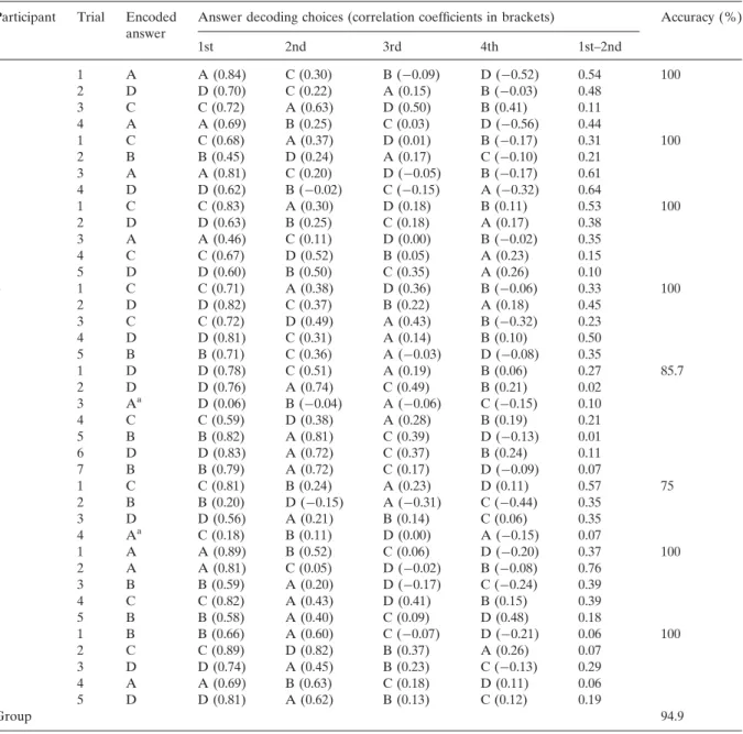
Participants’ answers were correctly decoded in 37 out of 39 answer encoding trials resulting in a mean accuracy of 94.9% (see Table 3). Thus, for six participants (75%) the decoding accuracy was 100% and also for the two remaining participants, the decoding accuracy was clearly above chance level (85.7 and 75%; chance level: 25%). Figure 5
shows answer encoding data for a representative trial demonstrating the robustness of the extracted single-trial fMRI time courses.
Results of the post hoc analysis
In order to disclose potential reasons for the two decoding errors that occurred, we explored all obtained data related to these two trials (online motion correction parameters, ROI time courses, and answer decoding values). The decoding error of participant 6 was very likely caused by severe
Table 3. Online answer decoding results
Participant Trial Encoded Answer decoding choices (correlation coefficients in brackets) Accuracy (%) answer 1st 2nd 3rd 4th 1st–2nd 1 1 A A (0.84) C (0.30) B (�0.09) D (�0.52) 0.54 100 2 D D (0.70) C (0.22) A (0.15) B (�0.03) 0.48 3 C C (0.72) A (0.63) D (0.50) B (0.41) 0.11 4 A A (0.69) B (0.25) C (0.03) D (�0.56) 0.44 2 1 C C (0.68) A (0.37) D (0.01) B (�0.17) 0.31 100 2 B B (0.45) D (0.24) A (0.17) C (�0.10) 0.21 3 A A (0.81) C (0.20) D (�0.05) B (�0.17) 0.61 4 D D (0.62) B (�0.02) C (�0.15) A (�0.32) 0.64 3 1 C C (0.83) A (0.30) D (0.18) B (0.11) 0.53 100 2 D D (0.63) B (0.25) C (0.18) A (0.17) 0.38 3 A A (0.46) C (0.11) D (0.00) B (�0.02) 0.35 4 C C (0.67) D (0.52) B (0.05) A (0.23) 0.15 5 D D (0.60) B (0.50) C (0.35) A (0.26) 0.10 4 1 C C (0.71) A (0.38) D (0.36) B (�0.06) 0.33 100 2 D D (0.82) C (0.37) B (0.22) A (0.18) 0.45 3 C C (0.72) D (0.49) A (0.43) B (�0.32) 0.23 4 D D (0.81) C (0.31) A (0.14) B (0.10) 0.50 5 B B (0.71) C (0.36) A (�0.03) D (�0.08) 0.35 5 1 D D (0.78) C (0.51) A (0.19) B (0.06) 0.27 85.7 2 D D (0.76) A (0.74) C (0.49) B (0.21) 0.02 3 Aa D (0.06) B (�0.04) A (�0.06) C (�0.15) 0.10 4 C C (0.59) D (0.38) A (0.28) B (0.19) 0.21 5 B B (0.82) A (0.81) C (0.39) D (�0.13) 0.01 6 D D (0.83) A (0.72) C (0.37) B (0.24) 0.11 7 B B (0.79) A (0.72) C (0.17) D (�0.09) 0.07 6 1 C C (0.81) B (0.24) A (0.23) D (0.11) 0.57 75 2 B B (0.20) D (�0.15) A (�0.31) C (�0.44) 0.35 3 D D (0.56) A (0.21) B (0.14) C (0.06) 0.35 4 Aa C (0.18) B (0.11) D (0.00) A (�0.15) 0.07 7 1 A A (0.89) B (0.52) C (0.06) D (�0.20) 0.37 100 2 A A (0.81) C (0.05) D (�0.02) B (�0.08) 0.76 3 B B (0.59) A (0.20) D (�0.17) C (�0.24) 0.39 4 C C (0.82) A (0.43) D (0.41) B (0.15) 0.39 5 B B (0.58) A (0.40) C (0.09) D (0.48) 0.18 8 1 B B (0.66) A (0.60) C (�0.07) D (�0.21) 0.06 100 2 C C (0.89) D (0.82) B (0.37) A (0.26) 0.07 3 D D (0.74) A (0.45) B (0.23) C (�0.13) 0.29 4 A A (0.69) B (0.63) C (0.18) D (0.11) 0.06 5 D D (0.81) A (0.62) B (0.13) C (0.12) 0.19 Group 94.9
aMisclassified answer trials.
head motion that could not be corrected success-fully by the online motion correction procedure. Using BrainVoyager QX post hoc, a more advanced motion correction procedure successfully coped with the head motion, in the end leading to the correct decoding of the given answer. When looking at the answer decoding value of the third
trial of participant 5 (r ¼ 0.06), it becomes clear that this value is extremely small (especially compared to the corresponding values of all other trials of this participant; r ¼ 0.7670.09 [mean71 s.d.]) — and in this sense constitutes an outlier that led to a misclassification. Thus, we assume that in this case the participant made an encoding error.
Fig. 5. Single-trial ROI time courses and corresponding RTCs. The figure provides the particular parameters used for coding the four different answer options. Moreover, single-trial time courses (black curves; red and blue curves in web version) generated by participant 7 during encoding of the answer option ‘‘A’’ are shown separately for the ‘motor imagery’ (rows 1 and 3) and the ‘mental calculation’ ROI (rows 2 and 4). Additionally, the RTCs corresponding to each answer option are displayed within the time course plots (gray curves) to demonstrate the close match of the data encoding the answer ‘‘A’’ with the particular RTC. The final column indicates the resulting correlation values and the automated ranking of the answer options according to their probability. Remarks: dashed vertical lines signify onsets and offsets of the general encoding periods; only time course segments relevant for decoding are shown (first 20 volumes skipped).
Discussion had not participated in any fMRI study before-hand, i.e., prior fMRI experience was not The present research shows that by using appro- mandatory for successful participation. All these priate experimental designs, participants are able facts argue for the robustness of the suggested to reliably generate differentiable fMRI brain procedure.
signals that can be used to encode at least four In our study, real-time fMRI data analysis distinct information units on a single-trial basis allowed for online decoding of a chosen answer, and in a reasonable amount of time, e.g., in order opening the possibility for back-and-forth com-to mocom-tor-independently answer multiple-choice munication within a single fMRI session. Note, questions. The participants’ answers were success- however, that our encoding technique can be fully extracted in 94.9% of the cases (chance level: beneficial even when real-time facilities (in terms 25%). Note that this high decoding accuracy was of data throughput and analysis) are not (yet) achieved in untrained volunteers of whom three available. Offline decoded answers might still be
of great importance. Moreover, a post hoc data analysis could result in additional gains in accuracy and might be advisable anyway, espe-cially when the patient’s answers would have a substantial impact on decisions, e.g., with respect to patient care. Another advantage of our method is that it requires very little effort and preparation time. Note also that the ‘localizer experiment’ needs to be conducted only once: The ROIs of the first fMRI session can be simply imported and used for communication experiments in later sessions.
Our study constitutes a ‘proof of concept’ working with nonclinical participants who mimicked the LIS patients’ limited behavioral capabilities by exclusively using thought processes for communication. Next, clinical trials in LIS patients are needed.
Potential clinical applications for patients with severe motor disabilities
By reviewing the relevant literature and present-ing a novel effective encodpresent-ing technique we could show that using fMRI-based BCIs constitutes a promising approach for the development of alternative communication and control tools for motor-disabled patients. Therewith, this direction considerably enriches the spectrum of already available non-fMRI-based BCI techniques
(Birbaumer et al., 2008). Since each brain imaging
method has its strengths and weaknesses, patients may differently benefit from one or the other technique. Thus, providing a method exploiting a complementary, namely hemodynamic (vs. elec-trophysiological) brain signal can have consider-able merits. In the following, we will shortly discuss potential clinical applications.
Online detection of consciousness
It is conceivable that the available real-time fMRI methods are also suited for online detection of consciousness in nonresponsive patients following acute brain damage. This would further extend the approach developed by Owen et al. (2006)
that relied on offline analyses. A short real-time
fMRI experiment to assess consciousness might be performed in the context of standard (anatomical) MRI diagnostics and — if successful — could be followed by a back-and-forth communication procedure. Additionally, the proposed procedures could be exploited for a further refinement of the diagnostics, e.g., by adaptive testing of cognitive functions (Iversen et al., 2008).
Communication and control
The suggested method for answering multiple-choice questions based on fMRI signals might offer a simple and effective possibility for LIS patients to communicate basic thoughts and needs in case no other alternative communication means are available (yet). Thus, especially patients in the acute phase of the LIS may benefit: Since the introduced method is grounded on relatively basic experimental and statistical principles and MRI scanners constitute standard clinical equipment, the techniques are easy to apply and can be readily transferred to clinical sites. Basic commu-nication in an early stage of the LIS could give patients confidence and may therefore prevent the development of depression and loss of general communication or cognitive abilities. In this context, an immediately usable communication approach as shown here to be feasible with real-time fMRI could serve as ‘first-aid’ intervention.
Promising paths for future hemodynamically based BCI developments
One principle goal in BCI research is to increase the number of correctly decoded information units within a certain time interval, thus improving the efficiency and accuracy of the BCI method. Moreover, a BCI system should be patient-friendly, easy-to-handle, and flexible. The current developmental state of BCIs exploiting hemody-namic brain signals leaves room for improvements in any of these respects. Therefore, we will, in the following sections, propose possible promising paths for future research in this field.
Increasing efficiency
Although gains in the efficiency and the accuracy of information transfer are more closely linked to, respectively, the encoding and decoding aspect of the discussed techniques, multiple interdependen-cies exist as, e.g., more sophisticated decoding methods can open up advanced possibilities for more efficient information encoding.
We think that the currently achieved degree of freedom in generating differential single-trial brain signals — that can be decoded online — can be significantly increased. For instance, more mental tasks and temporal variations of their execution can be included in the design, e.g., in order to encode single letters (Sorger et al., 2007). However, more sensitive decoding procedures have to be developed or implemented (see below) to reliably disentangle very similar but still distinctive brain activation patterns online. These more sophisticated methods in turn may contri-bute to the decrease of necessary encoding time. Another possibility to increase the communica-tion efficacy is the implementacommunica-tion of adaptive procedures, like automatic word completion or the use of ‘communication trees’.
Increasing accuracy
In order to improve decoding accuracy, it might be beneficial to follow a (coarse) multivariate approach by, e.g., selecting more than one region per mental task as this would most probably increase the robustness of the classification. How-ever, this could result in a time-consuming ROI selection process that would need to be overcome by implementing automated ROI selection proce-dures. Additionally, it might be advantageous to use individually determined (vs. standard) reference time courses (Handwerker et al., 2004), especially in brain-damaged patients for whom the BOLD signal might differ from that of healthy humans. More-over, the following more sophisticated data analysis techniques might be implemented:
1. real-time independent component analysis (Esposito et al., 2003), e.g., to automatically
detect artifacts (caused by motion or unde-sired thought processes during encoding) and 2. real-time multivariate analysis techniques, such as real-time multi-voxel pattern analysis (LaConte et al., 2007), e.g., support vector machines (Lee et al., 2009a), that might help to increase the sensitivity to detect more subtle spatial differences of brain activation patterns. Customizing
One important aspect in further developing fMRI-based BCI methods for communication and control would be the design of more patient-friendly procedures. This implicates that the particular communication procedures should be individually tailored for each participant and might involve the following aspects:
1. elaborating the individual degrees of free-dom in generating differentiable brain signals for each participant resulting in patient-tailored communication protocols (ranging from two-class BCI based on multiple trials up to multi-class BCI based on a single-trial level),
2. improving BOLD signal quality, e.g., through training of general mental imagery abilities, meditation (Eskandari and
Erfa-nian, 2008) or neurofeedback training
(Hwang et al., 2009),
3. developing encoding aids based on nonvi-sual, e.g., auditory (Kubler et al., 2009;
Nijboer et al., 2008) or tactile sensory
modalities to allow for communication in case of impaired vision,
4. considering individual preferences and parti-cular abilities of the patient (e.g., when choosing the mental tasks).
Mobility
Finally and maybe most importantly, the devel-oped real-time fMRI-based methods should be transferred to portable high-density fNIRS sys-tems allowing extending the use of hemodynamic brain signals for communication and control beyond clinical settings.
After overcoming the current challenges, hemo-dynamic BCI techniques might become beneficial even for patients in the incomplete and classical LIS state. Although these patient groups still do have some form of residual muscle control, BCIs based on the hemodynamic response (or any other BCI type) could constitute an alternative means of interaction, e.g., in situations of mus-cular fatigue. Moreover, it is conceivable that the suggested techniques may provide considerably more degrees of freedom and would therewith allow for more effective communication com-pared to other non-BCI-based solutions.
Abbreviations
BCI(s) brain–computer interface(s) BOLD blood oxygenation level–dependent ECoG electrocorticography
EEG electroencephalography
(f)MRI (functional) magnetic resonance imaging
(f)NIRS (functional) near-infrared spectro-scopy
ICoR intracortical recordings LIS ‘locked-in’ syndrome MEG magnetoencephalography ROI(s) region(s)-of-interest RTC(s) reference time course(s) 2D two-dimensional
3D three-dimensional Acknowledgements
The authors gratefully acknowledge the support of the BrainGain Smart Mix Programme of the Netherlands Ministry of Economic Affairs and the Netherlands Ministry of Education, Culture and Science. This research was also supported by the Fonds de la Recherche Scientifique (FRS), Eur-opean Commission (DECODER, DISCOS, Mind-bridge and COST), McDonnell Foundation and Mind Science Foundation. OG and AM are Research Fellow at FRS and SL is Senior Research Associate at FRS. Further, we would like to thank Lars Riecke for comments on the manuscript.
References
Allison, B. Z., Wolpaw, E. W., & Wolpaw, J. R. (2007). Brain-computer interface systems: Progress and prospects. Expert Review of Medical Devices, 4, 463–474.
Bagarinao, E., Nakai, T., & Tanaka, Y. (2006). Real-time functional MRI: Development and emerging applications. Magnetic Resonance in Medical Sciences, 5, 157–165. Bauer, G., Gerstenbrand, F., & Rumpl, E. (1979). Varieties of
the locked-in syndrome. Journal of Neurology, 221, 77–91. Birbaumer, N., & Cohen, L. G. (2007). Brain-computer
interfaces: Communication and restoration of movement in paralysis. The Journal of Physiology, 579, 621–636. Birbaumer, N., Ghanayim, N., Hinterberger, T., Iversen, I.,
Kotchoubey, B., Kubler, A., et al. (1999). A spelling device for the paralysed. Nature, 398, 297–298.
Birbaumer, N., Murguialday, A. R., & Cohen, L. (2008). Brain-computer interface in paralysis. Current Opinion in Neurology, 21, 634–638.
Birbaumer, N., Weiskopf, N., Weber, C., Kubler, A., Goebel, R., Caria, A., et al. (2006). An fMRI–Brain–computer-interface for the conditioning of the fear circuit in psychopaths. 12th Annual Meeting of the Organization for Human Brain Mapping. Florence, Italy.
Bruno, M., Bernheim, J. L., Schnakers, C., & Laureys, S. (2008). Locked-in: Don’t judge a book by its cover. Journal of Neurology, Neurosurgery, and Psychiatry, 79, 2. Buch, E., Weber, C., Cohen, L. G., Braun, C., Dimyan, M. A.,
Ard, T., et al. (2008). Think to move: A neuromagnetic brain-computer interface (BCI) system for chronic stroke. Stroke, 39, 910–917.
Coyle, S., Ward, T., Markham, C., & McDarby, G. (2004). On the suitability of near-infrared (NIR) systems for next-generation brain-computer interfaces. Physiological Measurement, 25, 815–822.
Dahmen, B., Sorger, B., Sinke, C. B. A., & Goebel, R. (2008). When the brain takes BOLD ‘steps’: Controlling differential brain activation levels via real-time fMRI-based neuro-feedback training. 14th Annual Meeting of the Organization for Human Brain Mapping (Vol. 41, p. 43). Melbourne: Elsevier.
deCharms, R. C. (2007). Reading and controlling human brain activation using real-time functional magnetic resonance imaging. Trends in Cognitive Sciences, 11, 473–481. deCharms, R. C. (2008). Applications of real-time fMRI.
Nature Reviews Neuroscience, 9, 720–729.
deCharms, R. C., Maeda, F., Glover, G. H., Ludlow, D., Pauly, J. M., Soneji, D., et al. (2005). Control over brain activation and pain learned by using real-time functional MRI. Proceedings of the National Academy of Sciences of the United States of America, 102, 18626–18631.
Eskandari, P., & Erfanian, A. (2008). Improving the perfor-mance of brain-computer interface through meditation practicing. Conference of the IEEE Engineering in Medicine and Biology Society, 662–665.
Esposito, F., Seifritz, E., Formisano, E., Morrone, R., Scarabino, T., Tedeschi, G., et al. (2003). Real-time
independent component analysis of fMRI time-series. Neuroimage, 20, 2209–2224.
Felton, E. A., Wilson, J. A., Williams, J. C., & Garell, P. C. (2007). Electrocorticographically controlled brain-computer interfaces using motor and sensory imagery in patients with temporary subdural electrode implants. Report of four cases. Journal of Neurosurgery, 106, 495–500.
Friston, K. J., Fletcher, P., Josephs, O., Holmes, A., Rugg, M. D., & Turner, R. (1998). Event-related fMRI: Characterizing differential responses. Neuroimage, 7, 30–40.
Goebel, R., Sorger, B., Birbaumer, N., & Weiskopf, N. (2005). Learning to play BOLD Brain Pong: From individual neurofeedback training to brain-brain interactions. 11th Annual Meeting of the Organization for Human Brain Mapping. Toronto.
Goebel, R., Sorger, B., Kaiser, J., Birbaumer, N., & Weiskopf, N. (2004). BOLD brain pong: Self regulation of local brain activity during synchronously scanned, interacting subjects. 34th Annual Meeting of the Society for Neuroscience. San Diego.
Gosseries, O., Demertzi, A., Noirhomme, Q., Tshibanda, J., Boly, M., de Beeck, M. O., et al. (2008). Functional neuroimaging (fMRI, PET and MEG): What do we measure? Revue Medicale de Liege, 63, 231–237.
Handwerker, D. A., Ollinger, J. M., & D’Esposito, M. (2004). Variation of BOLD hemodynamic responses across subjects and brain regions and their effects on statistical analyses. Neuroimage, 21, 1639–1651.
Hochberg, L. R., Serruya, M. D., Friehs, G. M., Mukand, J. A., Saleh, M., Caplan, A. H., et al. (2006). Neuronal ensemble control of prosthetic devices by a human with tetraplegia. Nature, 442, 164–171.
Hwang, H. J., Kwon, K., & Im, C. H. (2009). Neurofeedback-based motor imagery training for brain-computer interface (BCI). Journal of Neuroscience Methods, 179, 150–156. Irani, F., Platek, S. M., Bunce, S., Ruocco, A. C., & Chute, D.
(2007). Functional near infrared spectroscopy (fNIRS): An emerging neuroimaging technology with important applications for the study of brain disorders. The Clinical Neuropsychologist, 21, 9–37.
Iversen, I. H., Ghanayim, N., Kubler, A., Neumann, N., Birbaumer, N., & Kaiser, J. (2008). A brain-computer interface tool to assess cognitive functions in completely paralyzed patients with amyotrophic lateral sclerosis. The Clinical Neuropsychologist, 119, 2214–2223.
Karim, A. A., Hinterberger, T., Richter, J., Mellinger, J., Neumann, N., Flor, H., et al. (2006). Neural internet: Web surfing with brain potentials for the completely paralyzed. Neurorehabilitation and Neural Repair, 20, 508–515. Kubler, A., Furdea, A., Halder, S., Hammer, E. M., Nijboer,
F., & Kotchoubey, B. (2009). A brain-computer interface controlled auditory event-related potential (p300) spelling system for locked-in patients. Annals of the New York Academy of Sciences, 1157, 90–100.
Kubler, A., Kotchoubey, B., Hinterberger, T., Ghanayim, N., Perelmouter, J., Schauer, M., et al. (1999). The thought translation device: A neurophysiological approach to
communication in total motor paralysis. Experimental Brain Research, 124, 223–232.
Kubler, A., & Neumann, N. (2005). Brain-computer inter-faces — The key for the conscious brain locked into a paralyzed body. Progress in Brain Research, 150, 513–525. LaConte, S. M., Peltier, S. J., & Hu, X. P. (2007). Real-time
fMRI using brain-state classification. Human Brain Mapping, 28, 1033–1044.
Laureys, S., Owen, A. M., & Schiff, N. D. (2004). Brain function in coma, vegetative state, and related disorders. Lancet Neurology, 3, 537–546.
Laureys, S., Pellas, F., Van Eeckhout, P., Ghorbel, S., Schnakers, C., Perrin, F., et al. (2005). The locked-in syndrome: What is it like to be conscious but paralyzed and voiceless? Progress in Brain Research, 150, 495–511. Lebedev, M. A., & Nicolelis, M. A. (2006). Brain-machine
interfaces: Past, present and future. Trends in Neurosciences, 29, 536–546.
Lee, J. H., Marzelli, M., Jolesz, F. A., & Yoo, S. S. (2009a). Automated classification of fMRI data employing trial-based imagery tasks. Medical Image Analysis, 13, 392–404. Lee, J. H., Ryu, J., Jolesz, F. A., Cho, Z. H., & Yoo, S. S.
(2009b). Brain-machine interface via real-time fMRI: Preliminary study on thought-controlled robotic arm. Neuroscience Letters, 450, 1–6.
Leon-Carrion, J., van Eeckhout, P., & Dominguez-Morales Mdel, R. (2002). The locked-in syndrome: A syndrome looking for a therapy. Brain Injury, 16, 555–569.
Leuthardt, E. C., Miller, K. J., Schalk, G., Rao, R. P., & Ojemann, J. G. (2006). Electrocorticography-based brain computer interface — The Seattle experience. IEEE Transactions on Neural Systems and Rehabilitation Engineer-ing, 14, 194–198.
Luu, S., & Chau, T. (2009). Decoding subjective preference from single-trial near-infrared spectroscopy signals. Journal of Neural Engineering, 6, 016003.
Majerus, S., Gill-Thwaites, H., Andrews, K., & Laureys, S. (2005). Behavioral evaluation of consciousness in severe brain damage. Progress in Brain Research, 150, 397–413. Menon, R. S., & Kim, S. G. (1999). Spatial and temporal limits
in cognitive neuroimaging with fMRI. Trends in Cognitive Sciences, 3, 207–216.
Monti, M. M., Coleman, M. R., & Owen, A. M. (2008). ‘Brain Reading’ with real-time fMRI: Communication via detection of brain states in the absence of motor response. 14th Annual Meeting of the Organization for Human Brain Mapping, (Vol. 1, p. 133). Melbourne: Elsevier.
Naito, M., Michioka, Y., Ozawa, K., Ito, Y., Kiguchi, M., & Kanazawa, T. (2007). A communication means for totally locked-in ALS patients based on changes in cerebral blood volume measured with near-infrared light. IEICE Transactions on Information and Systems, E90-D, 1028–1037.
Nijboer, F., Sellers, E. W., Mellinger, J., Jordan, M. A., Matuz, T., Furdea, A., et al. (2008). A P300-based brain-computer interface for people with amyotrophic lateral sclerosis. Journal of Clinical Neurophysiology, 119, 1909–1916.
Ogawa, S., Lee, T. M., Kay, A. R., & Tank, D. W. (1990). Brain magnetic resonance imaging with contrast dependent on blood oxygenation. Proceedings of the National Academy of Sciences of the United States of America, 87, 9868–9872. Oldfield, R. C. (1971). The assessment and analysis of
handedness: The Edinburgh inventory. Neuropsychologia, 9, 97–113.
Owen, A. M., Coleman, M. R., Boly, M., Davis, M. H., Laureys, S., & Pickard, J. D. (2006). Detecting awareness in the vegetative state. Science, 313, 1402.
Pfurtscheller, G., Guger, C., Muller, G., Krausz, G., & Neuper, C. (2000). Brain oscillations control hand orthosis in a tetraplegic. Neuroscience Letters, 292, 211–214.
Plum, F., & Posner, J. B. (1966). The Diagnosis of stupor and coma: Edited book title. Philadelphia, PA: Davis, F.A. Ramsey, N. F., van de Heuvel, M. P., Kho, K. H., & Leijten, F.
S. (2006). Towards human BCI applications based on cognitive brain systems: An investigation of neural signals recorded from the dorsolateral prefrontal cortex. IEEE Transactions on Neural Systems and Rehabilitation Engineer-ing, 14, 214–217.
Rota, G., Sitaram, R., Veit, R., Erb, M., Weiskopf, N., Dogil, G., et al. (2009). Self-regulation of regional cortical activity using real-time fMRI: The right inferior frontal gyrus and linguistic processing. Human Brain Mappings, 30, 1605–1614.
Scharnowski, F., Weiskopf, N., Mathiak, K., Zopf, R., Studer, P., Bock, S. W., et al. (2004). Self-regulation of the BOLD signal of supplementary motor area (SMA) and parahippo-campal place area (PPA): fMRI-neurofeedback and its behavioural consequences. 10th Annual Meeting of the Organization for Human Brain Mapping. Budapest, Hungary. Scherer, R., Graimann, B., Huggins, J. E., Levine, S. P., & Pfurtscheller, G. (2003). Frequency component selection for an ECoG-based brain-computer interface. Biomedizinische Technik (Berlin), 48, 31–36.
Schnakers, C., Majerus, S., Goldman, S., Boly, M., Van Eeckhout, P., Gay, S., et al. (2008). Cognitive function in the locked-in syndrome. Journal of Neurology, 255, 323–330. Schwartz, A. B., Cui, X. T., Weber, D. J., & Moran, D. W. (2006). Brain-controlled interfaces: Movement restoration with neural prosthetics. Neuron, 52, 205–220.
Sitaram, R., Caria, A., Veit, R., Gaber, T., Rota, G., Kuebler, A., et al. (2007a). FMRI brain-computer interface: A tool for neuroscientific research and treatment. Computational Intelligence and Neuroscience, 25487.
Sitaram, R., Zhang, H., Guan, C., Thulasidas, M., Hoshi, Y., Ishikawa, A., et al. (2007b). Temporal classification of
multichannel near-infrared spectroscopy signals of motor imagery for developing a brain-computer interface. Neuro-image, 34, 1416–1427.
Sorger, B., Bareither, I., Weiskopf, N., Rodriguez, E. F., Birbaumer, N., & Goebel, R. (2004). Voluntary modulation of regional brain activity to different target levels based on real-time fMRI neurofeedback. 34th Annual Meeting of the Society for Neuroscience. San Diego.
Sorger, B., Dahmen, B., Reithler, J., & Goebel, R. (2007). BOLD communication: When the brain speaks for itself. 13th Annual Meeting of the Organization for Human Brain Mapping (Vol. 36, p. 37). Chicago: Elsevier.
Talairach, G., & Tournoux, P. (1988). Co-planar stereotaxic atlas of the human brain. New York: Thieme.
Vanzetta, I. (2006). Hemodynamic responses in cortex investigated with optical imaging methods. Implications for functional brain mapping. Journal of Physiology (Paris), 100, 201–211.
Villringer, A., & Chance, B. (1997). Non-invasive optical spectroscopy and imaging of human brain function. Trends in Neurosciences, 20, 435–442.
Weiskopf, N., Klose, U., Birbaumer, N., & Mathiak, K. (2005). Single-shot compensation of image distortions and BOLD contrast optimization using multi-echo EPI for real-time fMRI. Neuroimage, 24, 1068–1079.
Weiskopf, N., Mathiak, K., Bock, S. W., Scharnowski, F., Veit, R., Grodd, W., et al. (2004a). Principles of a brain-computer interface (BCI) based on real-time functional magnetic resonance imaging (fMRI). IEEE Transactions on Bio-Medical Engineering, 51, 966–970.
Weiskopf, N., Scharnowski, F., Veit, R., Goebel, R., Birbau-mer, N., & Mathiak, K. (2004b). Self-regulation of local brain activity using real-time functional magnetic resonance imaging (fMRI). Journal of Physiology (Paris), 98, 357–373. Weiskopf, N., Sitaram, R., Josephs, O., Veit, R., Scharnowski, F., Goebel, R., et al. (2007). Real-time functional magnetic resonance imaging: methods and applications. Magnetic Resonance Imaging, 25, 989–1003.
Weiskopf, N., Veit, R., Erb, M., Mathiak, K., Grodd, W., Goebel, R., et al. (2003). Physiological self-regulation of regional brain activity using real-time functional magnetic resonance imaging (fMRI): Methodology and exemplary data. Neuroimage, 19, 577–586.
Wijesekera, L. C., & Leigh, P. N. (2009). Amyotrophic lateral sclerosis. Orphanet Journal of Rare Diseases, 4, 3. Yoo, S. S., Fairneny, T., Chen, N. K., Choo, S. E., Panych, L.
P., & Park, H. (2004). Brain-computer interface using fMRI: Spatial navigation by thoughts. Neuroreport, 15, 1591–1595.