DOI: 10.5897/JMPR2015.6027 Article Number: 504BD8F57747 ISSN 1996-0875
Copyright © 2016
Author(s) retain the copyright of this article http://www.academicjournals.org/JMPR
Journal of Medicinal Plants Research
Full Length Research Paper
Terpenoids from Phaulopsis imbricata (Acanthaceae)
Archile Bernabe Ouambo Kengne
1, Mathieu Tene
1*, Alembert Tiabou Tchinda
2, Pierre Tane
1and Michel Frédérich
31
Laboratory of Natural Products Chemistry, Chemistry Department, University of Dschang, P. O. Box 67 Dschang, Cameroon.
2
Laboratory of Phytochemistry, Institute of Medical Research and Medicinal Plant Studies, (IMPM), P.O. Box 6163, Yaoundé, Cameroon.
3
Laboratory of Pharmacognosy, Interfaculty Centre for Research on Medicine (CIRM), Department of Pharmacy, University of Liège, B36, B-4000, Liège, Belgium.
Received 8 December, 2015; Accepted 16 February, 2016
The whole plant of Phaulopsis imbricata (Forssk.) Sweet (Acanthaceae) was collected at Bansoa, Cameroon, shade dried and extracted by maceration in methanol. This study was carried out to isolate secondary metabolites from this plant species that has not been investigated so far. Two lupane-type triterpenoids, one β-type carotenoid, one eudesmane-type sesquiterpenoid, and one sterol glycoside were isolated from the dried methanol extract using solvent partitioning, column chromatography and re-crystallization. They were identified as lupeol, betulin, (all-E)-lutein, cryptomeridiol, and sitosterol
3-O-β-D-glucopyranoside, respectively. The structures of the isolated compounds were elucidated on the
basis of spectroscopic methods including 1D- and 2D- nuclear magnetic resonance (NMR), infrared (IR) and mass spectrometry (MS). This is the first report of these compounds from the genus Phaulopsis. To the best of our knowledge, P. imbricata is also the first species of the genus to be phytochemically studied.
Key words: Phaulopsis imbricata, chromatography, spectroscopy, terpenoids, lupeol, betulin, (all-E)-lutein, cryptomeridiol.
INTRODUCTION
The genus Phaulopsis (Acanthaceae) comprises about 22 species encountered in tropical Africa and Asia (Ke et al., 2011). Phaulopsis imbricata (Forssk.) Sweet (syn.
Aetheilema anisophyllum R.Brown) is an erect herb of
about 1 m high mainly distributed in West Africa (Kayode and Omotoyinbo, 2008; Brummet, 2005). It has been used as medicinal plant for the treatment of pain, arthritis, rheumatism, skin diseases, diarrhoea, dysentery,
stomachache, nausea and sores (Burkill, 1985). It is also used as chewing stick for oral and dental healthcare (Kayode and Omotoyinbo, 2008). Another species, P.
fascicepala, has been reported to possess antioxidant
activity (Adesegun et al., 2009).
Many workers have isolated terpenoids from some species of Acanthaceae (Berrondo et al., 2003; Tamokou et al., 2011; Sudhanshu et al., 2000). But, according to
*Corresponding author. E-mail: [email protected]. Tel: +237- 677 808 420. Fax: +237- 243 451 735.
Author(s) agree that this article remain permanently open access under the terms of the Creative Commons Attribution License 4.0 International License
our knowledge, no phytochemical study has been reported from the genus Phaulopsis. This study approach has been to investigate the chemical constituents of P.
imbricata. We now report, herein, the isolation and
structural identification of two triterpenoids (lupeol and betulin), one carotenoid [(all-E)-lutein], one sesquiterpenoid (cryptomeridiol), and one sterol glycoside (sitosterol 3-O-β-D-glucopyranoside) from the methanol extract of the whole plant of P. imbricata.
MATERIALS AND METHODS General
Melting points (m.p.) were recorded with a Reichert microscope and uncorrected. 1H NMR (500 MHz) and 13C NMR (125 MHz) with APT program were recorded at room temperature in CDCl3, using a Bruker DMX 500 spectrometer. The chemical shifts (δ) are reported in parts per million (ppm) with the solvent signals as reference relative to TMS (δ = 0) as internal standard, while the coupling
constants (J values) are given in Hertz (Hz). COSY, NOESY, HSQC and HMBC experiments were recorded with gradient enhancements using sine shape gradient pulses. The Infra red (IR) spectra were recorded with a Shimadzu FTIR-8400S Infrared spectrophotometer and Gas chromatography-Mass spectrometry (GC-MS) data were obtained with an Agilent 6890N Network GC system/5975 Inert XL Mass Selective Detector at 70 eV and 20°C. The GC column (VARIAN, USA) was a CP- Sil 8 CB LB/MS, chromapack capillary column (0.25 mm × 30 m, film thickness 0.25 μm). The initial temperature was 50°C for 1 min, and then heated at 10°C/min to 300°C. For the carrier gas, helium was used with a flow rate of 1.20 ml/min. Kovat’s retention index (KI) was determined using a calibration curve of n-alkanes. Column chromatography was run on Merck silica gel 60 (70 to 230 mesh) (MERCK, Germany) and gel permeation on Sephadex LH-20 (SIGMA-ALDRICH, St. Louis, MO, USA) while thin layer chromatography (TLC) was carried out on silica gel GF254 pre-coated plates (MERCK, Germany) with detection accomplished by spraying with 50% H2SO4 followed by heating at 100oC, or by visualizing with a UV lamp at 254 and 365 nm.
Plant
The whole plant of P. imbricata was collected from Bansoa, West Region, Cameroon, in January, 2010. Authentication was done by Mr Paul Mezili, a retired Botanist of the Cameroon National Herbarium, Yaoundé, where the voucher specimen (No. 19290 / SRF / Cam) is deposited.
Extraction and isolation of compounds
The fresh plant of P. imbricata was dried under shadow for two weeks. The powdered material (2 kg) was extracted three times by maceration with MeOH (10 L) at room temperature (three days for each time). Evaporation of solvent under vacuum afforded 109 g of crude extract. A portion of this extract (105 g) was successively extracted with n-hexane, EtOAc and n-butanol. TLC analysis showed that the n-hexane and EtOAc extracts (21 and 3 g, respectively) were qualitatively the same. They were thus combined and a portion (23 g) subjected to silica gel (70 to 230 mesh) column chromatography eluted with gradient of n-hexane-EtOAc (100:0, 9:1, 4:1, 7:3, 1:1 and 0:100) followed by gradient of EtOAc-MeOH
(100:0, 19:1, 9:1 and 0:100). Sixty-six fractions of 300 ml each were collected and combined on the basis of TLC profile to give six major fractions A to F (A: 1 to 9; B: 10 to 20; C: 21 to 27; D: 28 to 45; E: 46 to 53; F: 54 to 66). Fraction A (3.2 g) was purified on a silica gel column eluted with gradient of n-hexane-EtOAc (10:0, 9:1, 4:1 and 7:3). 25 fractions of 50 ml each were collected to afford compound
1 (31 mg) after re-crystallization of sub-fractions 11 to 19 (A2).
Fraction B (5 g) was chromatographed on Sephadex LH-20 column eluted with the isocratic n-hexane-CH2Cl2-MeOH (7:4:0.5). 21 fractions of 10 ml each were collected to afford compound 2 (14 mg) from sub-fraction B2 (fractions 8 to 17) and mixture of chlorophylls. Fraction C (2.5 g) was also passed through Sephadex LH-20 column chromatography eluted with the isocratic n-hexane-CH2Cl2-MeOH (7:4:0.5). 14 fractions of 10 ml each were collected to give compound 3 (11 mg) after re-crystallization of sub-fraction C1 (fractions 1 to 9) in n-hexane. Fraction D was also chromatographed on Sephadex LH-20 column eluted with the isocratic n-hexane-CH2Cl2-MeOH (7:4:0.5). 20 fractions of 10 ml each were collected yielding three sub-fractions. Sub-fraction D2 (9 to14) was purified through silica gel column chromatography eluted with the gradient CH2Cl2-MeOH (100:0, 49:1, 19:1, 9:1, 7:3 and 1:1) giving 10 mg of compound 4.
The n-butanol extract (20 g) was also subjected to silica gel (70 to 230 mesh) column chromatography eluted with the gradient EtOAc-MeOH (100:0, 19:1, 9:1, 4:1 and 0:100). Seventeen fractions of 300 ml each were collected and combined on the basis of TLC profile to give three major fractions G to I (G: 1 to 8; H: 9 to 11; I: 12 to 17). Fraction G (3.7 g) crystallized to afford compound 5 (101 mg). An attempt to purify fractions E, F, H, I and filtrate of G failed, affording only complex mixtures in each case.
RESULTS
Compound 1 was obtained as white pellets from MeOH. It had a melting point (m.p.) of 216°C and showed the following spectral characteristics:
IR (KBr) Ѵmax cm -1
: 3300, 3080, 2920, 1640, 1470, 1420, 1390, 1045, 885. EIMS: m/z (rel. int.) = 426[M]+ (22), 207 (51), 189 (84), 161 (48), 135 (91), 121 (98), 95 (100), 81 (86), 55 (65), 41 (38).
Compound 2 was obtained as white crystals from MeOH. m.p. 237-238 °C. It showed the following spectral characteristics:
IR (NaCl) Ѵmax cm -1
: 3456, 3080, 2925, 1639, 1480, 1460, 1345, 1060, 885. EIMS: m/z (rel. int.) = 442[M]+ (4), 207 (52), 189 (81), 135 (82), 121 (72), 95 (100), 81 (93), 55 (80). HR-EI-MS: m/z = 442.3786 [M]+ (calcd. 442.3811 for C30H50O2).
1
H- and 13C-NMR spectral data (Table 1).
Compound 3 was obtained as reddish crystals from n-hexane. m.p. 190-191°C. It showed the following spectral data:
IR (NaCl) Ѵmax cm-1: 3500, 2962, 2921, 1662, 1390. EIMS: m/z (rel. int.) = 568 [M]+ (1), 550 (12), 479 (3), 442 (5), 313 (3), 135 (80), 121 (75), 95 (100), 81 (90), 55 (83). HR-EI-MS: m/z = 568.4276 [M]+ (calcd. 568.4280 for C40H56O2). 1H- and 13C-NMR spectral data (Table 2).
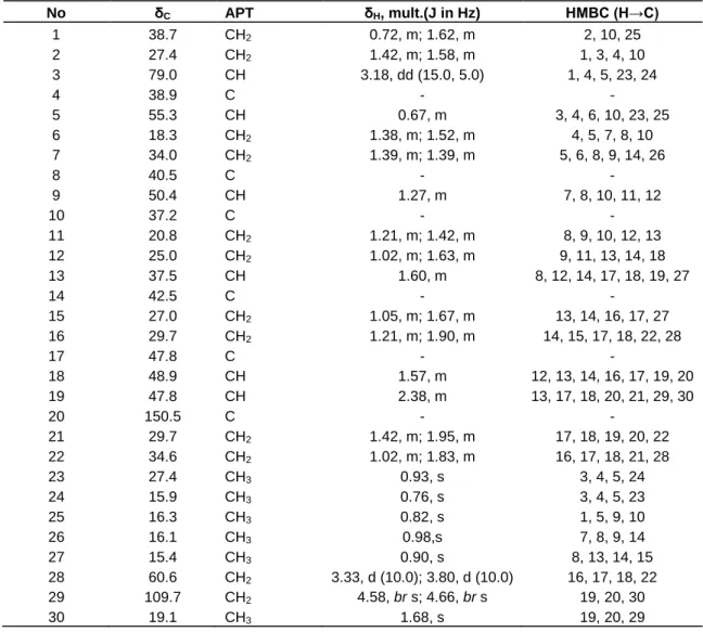
Table 1. 1H- and 13C/APT- NMR spectral data of betulin (2) (CDCl3,δ in ppm), and, selected HMBC data for 2. No δC APT δH, mult.(J in Hz) HMBC (H→C) 1 38.7 CH2 0.72, m; 1.62, m 2, 10, 25 2 27.4 CH2 1.42, m; 1.58, m 1, 3, 4, 10 3 79.0 CH 3.18, dd (15.0, 5.0) 1, 4, 5, 23, 24 4 38.9 C - - 5 55.3 CH 0.67, m 3, 4, 6, 10, 23, 25 6 18.3 CH2 1.38, m; 1.52, m 4, 5, 7, 8, 10 7 34.0 CH2 1.39, m; 1.39, m 5, 6, 8, 9, 14, 26 8 40.5 C - - 9 50.4 CH 1.27, m 7, 8, 10, 11, 12 10 37.2 C - - 11 20.8 CH2 1.21, m; 1.42, m 8, 9, 10, 12, 13 12 25.0 CH2 1.02, m; 1.63, m 9, 11, 13, 14, 18 13 37.5 CH 1.60, m 8, 12, 14, 17, 18, 19, 27 14 42.5 C - - 15 27.0 CH2 1.05, m; 1.67, m 13, 14, 16, 17, 27 16 29.7 CH2 1.21, m; 1.90, m 14, 15, 17, 18, 22, 28 17 47.8 C - - 18 48.9 CH 1.57, m 12, 13, 14, 16, 17, 19, 20 19 47.8 CH 2.38, m 13, 17, 18, 20, 21, 29, 30 20 150.5 C - - 21 29.7 CH2 1.42, m; 1.95, m 17, 18, 19, 20, 22 22 34.6 CH2 1.02, m; 1.83, m 16, 17, 18, 21, 28 23 27.4 CH3 0.93, s 3, 4, 5, 24 24 15.9 CH3 0.76, s 3, 4, 5, 23 25 16.3 CH3 0.82, s 1, 5, 9, 10 26 16.1 CH3 0.98,s 7, 8, 9, 14 27 15.4 CH3 0.90, s 8, 13, 14, 15 28 60.6 CH2 3.33, d (10.0); 3.80, d (10.0) 16, 17, 18, 22 29 109.7 CH2 4.58, br s; 4.66, br s 19, 20, 30 30 19.1 CH3 1.68, s 19, 20, 29
Compound 4 was obtained as white crystals from MeOH. m.p. 141 to 143°C. It showed the following spectral characteristics:
IR (NaCl) Ѵmax cm -1
: 3600-3200, 2920, 2848, 1456, 1377, 1238, 1166, 1049. EIMS: m/z (rel. int.) = 240 [M]+ (1), 222 (1), 204 (17), 189 (33), 149 (100), 123 (39), 109 (52), 95 (45), 43 (65). HR-EI-MS: m/z = 240.2094[M]+ (calcd. 240.2089 for C15H28O2).
1
H- and 13C-NMR spectral data (Table 3).
Compound 5 was obtained as white powder. m.p. 252-254 oC. IR (KBr) Ѵmax cm
-1
: 3500-3300, 2953,2866,1108.
DISCUSSION
Compound 1 was considered to be a triterpenoid due to a positive (violet coloration) Liebermann Burchard test. Its
electron impact mass spectrum (EIMS) showed molecular ion peak at m/z 426 (GC-MS). Analysis of the IR spectrum suggested that it contained a hydroxyl group (3300 cm-1) and a terminal double bond (3080, 1640, 885 cm-1). On the basis of TLC, m.p. and IR analyses, compound 1 was suggested to be lupeol, a lupane-type triterpenoid previously isolated in our laboratory (Tene et al., 2009). The identity of compound 1 was confirmed by comparison of its mass spectrum with that of lupeol in the NIST GC-MS library (NIST, USA).
Compound 2 showed a positive (violet coloration) Liebermann Burchard test and was suggested to be a triterpenoid. Its EIMS indicated a molecular ion peak at m/z 442 and a molecular formula C30H50O2 on the basis of its HR-EIMS (442.3793 [M]+). The IR spectrum showed vibration bands corresponding to hydroxyl group (3456 cm-1) and a terminal double bond (3080, 1639, 885 cm-1). Its 1H NMR spectrum showed five methyl singlets at δH
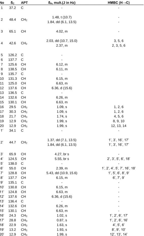
Table 2. 1H- and 13C/APT- NMR spectral data of (all-E)-lutein (3) (CDCl3,δ in ppm), and, selected HMBC data for 3. No δC APT δH, mult.(J in Hz) HMBC (H→C) 1 37.2 C - - 2 48.4 CH2 1.48, t (10.7) - 1.84, dd (6.1, 13.5) - 3 65.1 CH 4.02, m - 4 42.6 CH2 2.03, dd (10.7, 15.0) 3, 5, 6 2.37, m 2, 3, 5, 6 5 126.2 C - 6 137.7 C - 7 125.6 CH 6.12, m 8 138.5 CH 6.11, m 9 135.7 C - 10 131.3 CH 6.15, m 11 125.0 CH 6.63, m 12 137.6 CH 6.36, d (15.6) 13 136.5 C - 14 132.6 CH 6.26, m 15 130.1 CH 6.63, m 16 29.5 CH3 1.09, s 1, 2, 6 17 30.3 CH3 1.09, s 1, 2, 6 18 21.7 CH3 1.74, s 4, 5, 6 19 12.9 CH3 1.99, s 8, 9, 10 20 12.9 CH3 1.99, s 12, 13, 14 1’ 34.1 C - - 2’ 44.7 CH2 1.37, dd (7.1, 13.5) 1’, 3’, 16’, 17’ 1.84, dd (6.1, 13.5) 1’, 3’, 16’, 17’ 3’ 65.9 CH 4.27, br s - 4’ 124.5 CH 5.55, br s 2’, 3’, 5’, 6’, 18’ 5’ 138.0 C - - 6’ 55.0 CH 2.39, m 1’, 2’, 4’, 5’, 7’, 16’, 18’ 7’ 128.8 CH 5.43, dd (10.9, 15.6) 1’, 5’, 6’, 8’, 9’ 8’ 137.7 CH 6.15, m 6’, 7’, 9’ 9’ 135.1 C - - 10’ 130.8 CH 6.15, m - 11’ 124.8 CH 6.63, m - 12’ 137.6 CH 6.36, d (15.6) - 13’ 136.4 C - - 14’ 132.6 CH 6.26, m - 15’ 130.1 CH 6.63, m - 16’ 24.3 CH3 1.02, s 1’, 2’, 6’, 17’ 17’ 28.8 CH3 0.87, s 1’, 2’, 6’, 16’ 18’ 22.9 CH3 1.63, s 4’, 5’, 6’ 19’ 13.2 CH3 1.93, s 8’, 9’, 10’ 20’ 12.9 CH3 1.99, s 12’, 13’, 14’
Table 3. 1H- and 13C/APT- NMR spectral data of cryptomeridiol (4) (CDCl3,δ in ppm), and, selected HMBC data for 4.
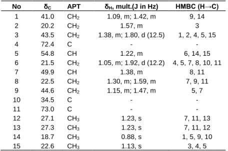
No δC APT δH, mult.(J in Hz) HMBC (H→C) 1 41.0 CH2 1.09, m; 1.42, m 9, 14 2 20.2 CH2 1.57, m 3 3 43.5 CH2 1.38, m; 1.80, d (12.5) 1, 2, 4, 5, 15 4 72.4 C - - 5 54.8 CH 1.22, m 6, 14, 15 6 21.5 CH2 1.05, m; 1.92, d (12.2) 4, 5, 7, 8, 10, 11 7 49.9 CH 1.38, m 8, 11 8 22.5 CH2 1.30, m; 1.59, m 7, 9, 11 9 44.6 CH2 1.15, m; 1.47, m 5, 7 10 34.5 C - - 11 73.0 C - - 12 27.1 CH3 1.23, s 7, 11, 13 13 27.3 CH3 1.23, s 7, 11, 12 14 18.7 CH3 0.88, s 1, 5, 9, 10 15 22.6 CH3 1.13, s 3, 4, 5
0.76, 0.82, 0.90, 0.93 and 0.98, a vinyl methyl group at δH 1.68 (3H-30, s), a double doublet of one oxygenated methine proton at δH 3.18 (H-3, dd, 5.0, 15.0), two doublets of one oxygenated methylene protons at δH 3.33 (H-28a, d, 10.0) and 3.80 (H-28b, d, 10.0) and two broad singlets of a terminal double bond, one proton each at δH 4.58 (H-29a) and 4.66 (H-29b) (Table 1). The 13C NMR/APT revealed 30 carbon signals including 6 methyl groups, 12 methylene groups, 6 methine carbons and 6 quaternary carbons (Table 1). The oxygenated carbon signals appeared at δC 60.6 (C-28) and 79.0 (C-3) and the double bond at δC 109.7 (C-29) and 150.5 (C-20), characteristics of triterpenes of the lup-20(29)-ene type (Mahato and Kundu, 1994). In the HSQC spectrum correlations observed between H-3 (δH 3.18) and C-3 (δC 79.0), H-28a (δH 3.33) and C-28 (δC 60.6), H-28b (δH 3.80) and C-28 (δC 60.6), H-29a (δH 4.58) and C-29 (δC 109.7), H-29b (δH 4.66) and C-29 (δC 109.7), suggested compound 2 to be a 3, 28-dihydroxylup-20(29)-ene. The positions of the two hydroxyl (3-OH and 28-OH) and vinylidene (C=CH2) groups were confirmed in the HMBC spectrum by correlations between H-3 (δH 3.18) and C-1 (δC 38.7), C-4 (δC 38.9), C-5 (δC 55.3), C-23 (δC 27.4), and C-24 (δH 15.9), H-28a (δH 3.33)/H-28b (δH 3.80) and C-16 (δC 29.7), C-17 (δC 47.8), C-18 (δC 48.9) and C-22 (δC 34.6), H-29a (δH 4.58)/H-29b (δH 4.66) and C-19 (δC47.8), C-20 (δC 150.5) and C-30 (δC 19.1). Further-more, correlations observed in the NOESY spectrum between H-3 (δH 3.18, dd, J = 5.0, 15.0 Hz) and H-5 (δH 0.67, m), and H-3 (δH 3.18) and H-23 (δH 0.93, s) suggested the 3-OH group to be β-oriented.
Compound 2 was identified to 3β,28-dihydroxylup-20 (29)-ene (betulin), a lupane-type triterpenoid previously reported from other sources (Tinto et al., 1992; Joshi et
al., 2013).
HR-EIMS of compound 3 showed the molecular ion at m/z 568.4276 [M]+ in agreement with the molecular formula C40H56O2 (13
o
of unsaturation). The IR spectrum showed bands for hydroxyl group (3500 cm-1) and double bond (1662 cm-1). 13C-NMR/APT indicated signals for forty carbon atoms including ten methyls, three methylenes, eighteen methines and nine quaternary carbons (see Table 2). Its 1H NMR spectrum showed signals for four angular methyl groups at δH 0.87 (3H-17’, s), 0.99 (3H-16 and 3H-17, s) and 1.02 (3H-16’, s), six vinyl methyl groups at δH 1.63 (3H-18’, s), 1.74 (3H-18, s), 1.93 (3H-19’, s), 1.99 (3H-19, 3H-20 and 3H-20’, s) and three methylene groups at δH 1.37 (H-2’α, dd, J = 7.1, 13.5 Hz), 1.48 (2α, t, J = 10.7 Hz), 1.84 (2α, H-2’α, each dd, J = 6.1, 13.5 Hz), 2.03 (H-4α, dd, J = 10.7, 15.0 Hz) and 2.37 (H-4α, m). Also on this spectrum, three aliphatic methine protons resonated respectively at δH 2.39 (H-6’, m), 4.02 (H-3, m) and 4.27 (H-3’, br s) (Table 2). The proton sequence of the compound was deduced from the 1H-1H COSY and the gradient HMQC spectra while the HMBC spectrum permitted the construction of the skeleton of 3. 1H- and 13C-NMR spectral data suggested the presence in the molecule of two different and six-membered aliphatic rings, each bearing one hydroxyl (C-3 / C-3’), one double bond (C-5, C-6 / C-4’, C-5’) and three methyl (C-16, C-17, C-18 / C-16’, C-17’, C-18’) groups. Each cyclohexenyl moiety is attached in one end of a symmetrical polyene system. But due to the structure difference of the two rings the molecule is not symmetric (Figure. 1 and Table 2). Signals of the polyene region of the 1H NMR spectrum were identical to that of the (all-E)-lutein, as previously described (Aman et al., 2005). The structure was determined as (3R, 3’R,
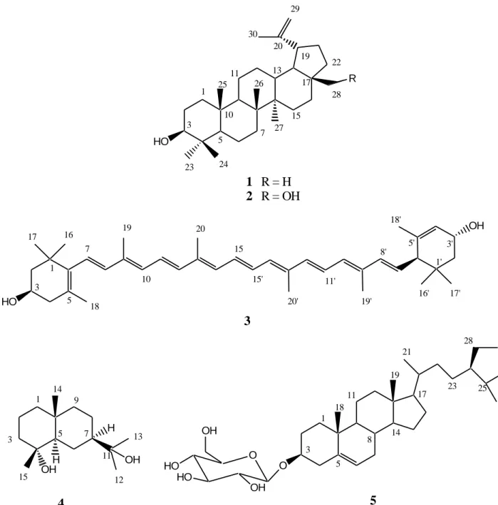
6’R)-HO R HO OH
1 R = H
2 R = OH
3
OH H OHH4
10 15 16 17 19 20 7 5 3 1 8' 1' 18' 18 17' 16' 3' 5' 19' 20' 11' 15' 29 19 17 15 11 7 10 5 3 1 13 23 24 27 26 25 28 22 30 20 13 11 15 9 7 5 3 1 12 14 O O HO HO OH OH5
3 5 17 28 25 21 18 19 23 8 14 11 1Figure 1. Structures of the isolated compounds.
β,β-carotene-3,3’-diol using 1
H, 13C/APT, HMBC, HSQC, 1
H-1H COSY and NOESY spectra. Its NMR data (see Table 2) agree with those reported in the literature (Srividya and Vishnuvarthan, 2014). (all-E)-Lutein (3) was previously isolated from Rhus leptodictya (Songca et al., 2012).
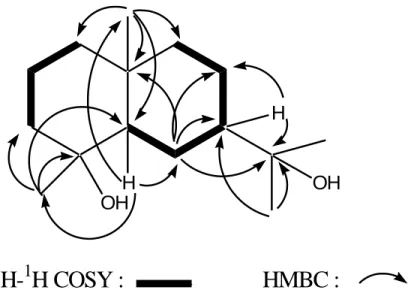
The EIMS of compound 4 showed a molecular ion peak at m/z 240 and a molecular formula C15H28O2 on the basis of its HR-EIMS (240.2094 [M]+). The IR spectrum showed a vibration band at 3400 cm-1 corresponding to hydroxyl group. Its 13C NMR/APT spectrum showed fifteen carbon signals including four methyls (δC 27.3, 27.1, 22.6, 18.7),
six methylenes (δC 44.6, 43.5, 42.0, 22.5, 21.5, 20.2), two methines (δC 54.8, 49.7), and three quaternary carbons (δC 73.0, 72.4, 34.5) two of which are oxygenated (Figure 1 and Table 3). Its 1H NMR spectrum showed signals for four tertiary methyl groups three of which are attached to carbons bearing oxygen atom, at δH 0.88 (3H-14, s), 1.13 (3H-15, s) and 1.23 (3H-12 and 3H-13, s). The two methine protons appeared respectively at δH 1.22 (H-5, m) and 1.38 (H-7, m). Other signals were attributed to the methylene protons of the molecule (Table 3). The proton sequence of the compound was deduced from the 1H-1H COSY and the gradient HMQC spectra while the HMBC
OH
OH
H
H
1
H-
1
H COSY : HMBC :
Figure 2. Selected correlations for compound 4spectrum permitted the construction of the skeleton of 4 (Figure 2). In the NOESY spectrum, correlations observed between H-5 (δH 1.22) and H-7 (δH 1.38), and between 3H-14 (δH 0.88) and 3H-15 (δH 1.13) permitted the attribution of configurations at the chiral centers of compound 4. The 1H and 13C NMR data (Table 3) were in good agreement with those reported in the literature for cryptomeridiol, an eudesmane-type sesquiterpenoid previously isolated from Artemisia pygmaea (Irwin and Geissman, 1973) and Blumea basalmifera (Ruangrunsi et al., 1985).
Compound 5 was identified to sitosterol 3-O- β-D-glucopyranoside after TLC, m.p. and IR analyses, compared to a referenced sample previously isolated in our laboratory (Tene et al., 2008). This compound is widely distributed in plant kingdom.
Conclusion
The phytochemical study of the whole plant of Phaulopsis
imbricata (Acanthaceae) afforded five known compounds
including lupeol, betulin, (all-E)-lutein, cryptomeridiol and sitosterol 3-O-β-D-glucopyranoside. To the best of our knowledge, this plant is phytochemically studied here for the first time. In addition, pure compounds are here reported for the first time from the genus Phaulopsis. The antimicrobial activity of the extracts and pure compounds from P. imbricata will be evaluated, with a view of verifying the hypothesis that this plant species is used by traditional healers for specific medicinal ends.
Conflict of interests
The authors have not declared any conflict of interests.
ACKNOWLEDGMENTS
The authors gratefully acknowledge financial support from the research grant committees of both the University of Dschang and the Cameroonian Ministry of Higher Education.
REFERENCES
Adesegun SA, Fajana A, Orabueze CI, Coker HAB (2009). Evaluation of antioxidant properties of Phaulopsis fascisepala C. B. Cl. (Acanthaceae). Evid. Based Complement. Altern. Med. 6(2):227-231. Aman R, Biehl J, Carle R, Conrad J, Beifuss U, Schieber A (2005).
Application of HPLC coupled with DAD, APcI-MS and NMR to the analysis of lutein and zeaxanthin stereoisomers in thermally processed vegetables. Food Chem. 92(4):753-763.
Berrondo LF, Gabriel FT, Fernandes SBO, Menezes FS, Moreira DL (2003). Dirhamnosyl flavonoid and other constituents from
Brillantaisia palisatii. Quim. Nova 26(6):922-923.
Brummet RK (2005). Vascular plants. Families and genera. Royal Botanical Gardens. pp. 20-25.
Burkill HM (1985). The useful plants of West Tropical Africa vol. 1; Edition 2; Families A-D. Royal Botanic Gardens, kew. 960p. Irwin MA, Geissman TA (1973). Sesquiterpene alcohols from Artemisia
pygmaea. Phytochemistry 12(4):849-852.
Joshi H, Saxena GK, Singh V, Arya E, Singh RP (2013). Phytochemical investigation, isolation and characterization of betulin from bark of
Betula utilis. J. Pharmacogn. Phytochem. 2(1):145-151.
Kayode J, Omotoyinbo MA (2008). Cultural erosion and biodiversity: conserving chewing stick knowledge in Ekiti State, Nigeria. Afr. Scientist 9(1):41-51.
Ke JC, Jiaqi H, Yunfei D, Wood JRI, Daniel TF (2011). Acanthaceae. In: Wu ZY, Raven PH, Hong DY (Eds), Flora of China, vol. 19; Science Press, Beijing, and Missouri Botanical Garden Press, St. Louis. pp. 429-430.
Mahato SB, Kundu AP (1994). 13C NMR spectra of pentacyclic triterpenoids-a compilation and some salient features. Phytochemistry 37(6):1517-1575.
Ruangrunsi N, Tantivatana P, Tappayuthpijarn P, Borris RP, Cordell GA (1985). Traditional medicinal plants of Thailand VI. Isolation of cryptomeridiol from Blumea balsamifera. J. Sci. Soc. Thailand 11(1):47-50.
Songca SP, Sebothoma C, Samuel BB, Eloff JN (2012). A biflavonoid and a carotenoid from Rhus leptodictya: isolation, characterization and antibacterial properties. Afr. J. Biochem. Res. 6(13):172-178. Srividya AR, Vishnuvarthan VJ (2014). Physical, chemical and
biological properties of lutein: A Review. World J. Pharm. Res. 3(5):105-118.
Sudhanshu S, Dharam CJ, Madan MG, Rajendra SB, Hari OM, Ram PS (2000). High performance thin layer chromatographic analysis of hepatoprotective diterpenoids from Andrographis paniculata.
Phytochem. Anal. 11(1):34-36.
Tamokou JD, Kuate JR, Tene M, Nwemeguela TJK, Tane P (2011). The antimicrobial activities of extract and compounds isolated from
Brillantaisia lamium. Iran. J. Med. Sci. 36(1):24-31.
Tene M, Ndontsa BL, Tane P, Tamokou J-D, Kuiate J-R (2009). Antimicrobial diterpenoids and triterpenoids from the stem bark of
Croton macrostachys. Int. J. Biol. Chem. Sci. 3(3):538-544.
Tene M, Tane P, Tamokou JD, Kuiate JR, Connolly JD (2008). Degraded diterpenoids from the stem bark of Neoboutonia mannii. Phytochem. Lett. 1(2):120-124.
Tinto WF, Blair LC, Alli A, Reynolds WF, McLean S (1992). Lupane triterpenoids of Salacia cordata. J. Nat. Prod. 55(3):395-398.