HAL Id: hal-03130775
https://hal.archives-ouvertes.fr/hal-03130775
Submitted on 3 Feb 2021
HAL is a multi-disciplinary open access
archive for the deposit and dissemination of
sci-entific research documents, whether they are
pub-lished or not. The documents may come from
L’archive ouverte pluridisciplinaire HAL, est
destinée au dépôt et à la diffusion de documents
scientifiques de niveau recherche, publiés ou non,
émanant des établissements d’enseignement et de
To cite this version:
Bertrand
Plainchont,
Jonathan
Farjon,
Nicolas
Giraud.
Magnetic
Field
Depen-dence of Spatial Frequency Encoding NMR. encyclopedia of magnetic resonance,
2016,
1 2 3 4 5 6 7 8 9 10 11 12 13 14 15 16 17 18 19 20 21 22 23 24 25 26 27 28 29 30 31 32 33 34 35 36 37 38 39 40 41 42 43 44 45 46 47 48 49 50 51 52 53 54 55 56 57 58 59 61 62 63 64 65 66 67 68 69 70 71 72 73 74 75 76 77 78 79 80 81 82 83 84 85 86 87 88 89 90 91 92 93 94 95 96 97 98 99 100 101 102 103 104 105 106 107 108 109 110 111 112 113 114 115 116 117 118 119
Magnetic Field Dependence of Spatial Frequency Encoding NMR
Bertrand Plainchont, Jonathan Farjon & Nicolas Giraud
Université Paris-Sud, Orsay Cedex, France
The influence of the strength of the static magnetic field on pulse sequences based on the concept of spatial frequency encoding is addressed. The evolution of the slice selection process occurring during encoded excitation and refocusing pulses, as well as pure shift and J-edited refocusing blocks, is studied both theoretically and experimentally on a model ABX1H spin system. It is shown that carrying out gradient-encoded pulse sequences at
higher field enhances the quality of the spin dynamics that is locally triggered in each encoded slice and results in spectra of better resolution and sensitivity.
Keywords:high-resolution NMR, spatial frequency encoding, selective pulses, magnetic field gradient, magnetic field, sensitivity, resolution, pure shift, J-edited
How to cite this article:
eMagRes, 2016, Vol 5: 1–6. DOI 10.1002/9780470034590.emrstm1528
Introduction
Spatial frequency encoding (SFE) NMR is at the root of a series of methodological developments that have paved the way to the acquisition of1H 1-D and 2-D experiments with unprecedented high resolution. This method is based on the general concept of gradient-encoded pulses, which have been extensively used for a wide range of applications in NMR spectroscopy, depending on whether the time evolution of a spin system (single scan or ultrafast NMR1), the local spin composition (imaging2, diffusion3), or more generally, any spin dynamics process need to be edited throughout the sample.4 In the latter case, a SFE is needed to control locally the selec-tivity of the excitation, the decoupling, or the refocusing of individual spin nuclei from each site of the analyzed molecule, in contrast to the spatial encoding of a time domain that is performed in ultrafast NMR.5 In this spirit, Zangger and Sterk had first shown that broadband homonuclear decoupling sequences based on a SFE can be performed efficiently by implementing a pure shift (also known as𝛿-resolved) refocus-ing block.6 More recently, we have shown that the selective refocusing of homonuclear scalar or residual dipolar couplings can also be encoded along the sample, according to the res-onance frequency of each coupling partner.7,8 We have also demonstrated that it is possible to combine pure shift and J-edited spin evolutions into fully tailored experiments and produce ultrahigh-resolution correlation spectra with exactly the desired analytical content.9,10 The high flexibility of SFE methods has also opened the way to an implementation of both pure shift and J-edited blocks into real-time acquisition schemes.11–13
In a recent paper, we have reported the acquisition of high-resolution gradient-encoded correlation experiments on an oligomeric saccharide sample showing a highly crowded1H spectrum.14We have experimentally shown that operating at
higher magnetic field leads to a better efficiency of pure shift as well as J-edited refocusing blocks based on a SFE, leading to noticeable resolution enhancements on this spectroscopically challenging sample. In the present work, we study the magnetic field dependence of SFE NMR on a model ABX1H spin system. Notably, we describe the evolution of spatial resolution and sensitivity when going to higher field. We use the theoretical formalism that we have introduced recently15 to simulate the spatial properties of the investigated irradiation schemes throughout the sample during a slice selection process.
Magnetic Field Dependence of the Slice
Selection Process
Excitation Step
SFE results from the simultaneous application of a selective pulse and a pulsed magnetic field gradient. The basic idea underlying this irradiation scheme is that proton spins with different resonance frequencies are selectively irradiated in different ‘slices’ of the sample. The key features of this process can be understood through the basic single gradient-encoded excitation pulse sequence depicted in Figure 1(a). In the follow-ing, 3-hydroxy-4,4,4-trichlorobutyric𝛽-lactone 1 (Figure 1b) is chosen as a model system to study the magnetic field dependence of the SFE. During a gradient-encoded pulse, the strength of the total magnetic field depends on the position z in the sample due to the application of a pulsed field gradient (Figure 1c). As a result, each proton site HA, HB, and HX will interact with the encoded selective pulse only around the position zA(respectively, zBand zX) that satisfies the relation:
𝜈A,B,X = 𝛾H 2π ⋅
{
1 2 3 4 5 6 7 8 9 10 11 12 13 14 15 16 17 18 19 20 21 22 23 24 25 26 27 28 29 30 31 32 33 34 35 36 37 38 39 40 41 42 43 44 45 46 47 48 49 50 51 52 53 54 55 56 57 58 59 61 62 63 64 65 66 67 68 69 70 71 72 73 74 75 76 77 78 79 80 81 82 83 84 85 86 87 88 89 90 91 92 93 94 95 96 97 98 99 100 101 102 103 104 105 106 107 108 109 110 111 112 113 114 115 116 117 118 119 (a) (c) PFG 1H HX HA 0 B(z) z HB trec. tacq. (b) 1
Figure 1. (a) The single gradient-encoded pulse sequence. The delays trec. and tacq.are the recovery and the acquisition times, respectively. The black ellipsoidal shape on the proton channel (1H) corresponds to a shaped
exci-tation pulse. The white rectangular bar on the pulsed field gradient (PFG) channel refers to the application of a rectangular-shaped z field gradient. (b) The structure of the model1H ABX spin system 1. (c) Schematic
rep-resentation of the spatial frequency encoding performed during the single gradient-encoded shaped pulse experiment shown in (a). The receiver coil is represented in orange in order to materialize the total height of the sam-ple that is detected. The z gradient coils are pictured in red. Yellow arrows symbolize the magnetic field gradient
where𝜈Aand𝜎
A(respectively,𝜈Band𝜎B, and𝜈Xand𝜎X) are the resonance frequency and the shielding of HA(respectively,
HBand HX), B
0the external static magnetic field generated by the superconducting magnet,𝛾Hthe gyromagnetic ratio of the proton, and Gzthe strength of the magnetic field gradient taken here along the z-axis.
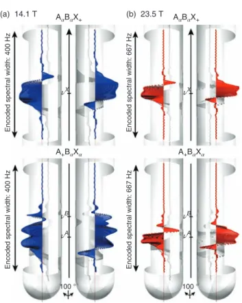
Figure 2 shows the spatial distribution of the magnetization corresponding to the coherences A+B𝛼X𝛼and A𝛼B𝛼X+of HA and HXcalculated at the end of a gradient-encoded excitation step, at 14.1 T (𝜈H= 600 MHz) and 23.5 T (𝜈H= 1 GHz). For each magnetic field strength B0, the strength of the encoding gradient GB0 is set so that the same range of chemical shifts, corresponding to the spectrum of the analyzed compound, is encoded along the virtual NMR sample. A larger spectral width
𝛾GB0l, where𝛾 is the gyromagnetic ratio and l the length of the sample that is detected by the receiver coil, is thus encoded along the sample at higher field. Moreover, the same E-Burp2 pulse of identical duration is used at both fields, which ensures the same spectral bandwidth 𝛺B. For HX first, the region around𝜈Xwithin which the corresponding transverse magne-tization is created is found to be thinner at 23.5 T than at 14.1 T, which is coherent with the fact that at first approximation the fraction of the sample that is selected by the gradient-encoded pulse is proportional to 𝛺B/(𝛾GB0l).16 We also remark that the shape of the phase distribution within the region encoded around𝜈Xis comparable at both fields, although being thinner at 23.5 T. Moreover for HA, it is apparent that at 14.1 T, a selec-tivity issue arises as transverse magnetization corresponding
(a) 14.1 T (b)
Encoded spectral width: 400 Hz
Encoded spectral width: 400 Hz
Encoded spectral width: 667 Hz
Encoded spectral width: 667 Hz
23.5 T AαBαX+ AαBαX+ A+BαXα νX νX νB νA νB νA 100 ° 100 ° A+BαXα
Figure 2. The spatial variation of the magnetization calculated at (a) 14.1 and (b) 23.5 T at the end of a gradient-encoded excitation pulse is repre-sented with 2 viewing angles inside the NMR tube, for (top) A𝛼B𝛼X+and (bottom) A+B𝛼X𝛼coherences. An E-Burp2 pulse of duration 60 ms was used for the selective irradiation. The virtual sample was divided into 401 slices of equal height around each region of interest, to which a position-dependent magnetic field value was assigned
to the A+B𝛼X𝛼term is observed not only around𝜈Abut also around𝜈B, which means that the gradient-encoded excitation pulse cannot select HA and HB in spatially resolved cross sections. For the simulation carried out at 23.5 T, the thinner region that is observed around𝜈A, combined with the greater difference between the Larmor frequencies of HA and HB, contributes to improve the selectivity of the excitation pulse. As a result, very few residual magnetization is detected around
𝜈B, which indicates that at 23.5 T the selectivity of the gradient-encoded pulse is good enough to excite HAand HBin distinct slices of the sample.
Furthermore, the 1H spectra resulting from the SFE described above can be reconstructed by adding the NMR signals that are locally encoded for each observable coherence of the model spin system 1. In Figure 3, the spatial distribution of NMR spectra generated locally by the gradient-encoded excitation pulse is shown as a pseudo-2-D plot at 14.1 and 23.5 T. At 14.1 T, we note that HAand HBare encoded in overlapped cross sections, as it is highlighted by the amplitude profile displayed within the NMR tube. The overall spectrum that is the sum of these subspectra shows distorted lines, which arises directly from this spatial resolution issue. Again, it is apparent that operating at higher field allows for solving this problem, as at 23.5 T two cross sections are clearly distinguishable at
1 2 3 4 5 6 7 8 9 10 11 12 13 14 15 16 17 18 19 20 21 22 23 24 25 26 27 28 29 30 31 32 33 34 35 36 37 38 39 40 41 42 43 44 45 46 47 48 49 50 51 52 53 54 55 56 57 58 59 61 62 63 64 65 66 67 68 69 70 71 72 73 74 75 76 77 78 79 80 81 82 83 84 85 86 87 88 89 90 91 92 93 94 95 96 97 98 99 100 101 102 103 104 105 106 107 108 109 110 111 112 113 114 115 116 117 118 119 (a) SUM (b) 14.1 T z z 23.5 T 5.0 3.8 3.7 3.6 Proton chemical shift (ppm)
5.0 3.8 3.7 3.6 Proton chemical shift (ppm)
Encoded spectral width ~ 2000 Hz
Encoded spectral width ~ 2000 Hz
νX νB νA νX νX νB νA νA νB νX νB νA
Figure 3. The amplitude profile of the NMR signal evaluated on the first point of each locally generated free induction decay is represented in cylin-drical coordinates at 14.1 T (a) and 23.5 T (b). For each magnetic field, the spatial distribution of virtual proton spectra is reported in pseudo-2-D plots where the vertical axis refers to the position z in the NMR sample. On these maps, blue and red contours are positive and negative, respectively
𝜈A and 𝜈B, respectively, leading to a 1H spectrum showing the classical roof effect expected for a second-order spin system.
In conclusion for this part, the implementation of a SFE at higher magnetic field allows for improving the spatial resolu-tion of the slice selecresolu-tion process, by selecting thinner cross sections throughout the sample.
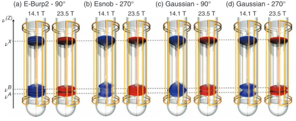
Influence of the Shape of the Gradient-encoded Pulse In a previous paper, we had shown that among the different shaped pulses designed for the purpose of selective irradiations, not all of them are suitable for undergoing a SFE.15 We had established that a key feature for yielding a proper slice selec-tion process is the ability for a given shape to maintain the dis-tribution of magnetization coherent along the encoded cross section. Figure 4 shows the evolution of the slice selection pro-cess simulated at 14.1 and 23.5 T for a series of excitation shaped pulses. Their duration was set so that they all have the same spectral bandwidth. For every shape, we observe a thinning of the encoded slices when going to higher field, as it can be mon-itored on the signals located at𝜈X. An improvement of the slice selection process is notably observed at higher field for E-Burp2 (90∘), Esnob (270∘), as well as Gaussian pulses calibrated for both 90∘ and 270∘, as it is illustrated by the presence at 23.5 T of two distinct slices at𝜈Aand𝜈B. We note that this was less predictable for the Gaussian 90∘ pulse that had been previously shown to suffer from a problem of phase coherence upon gradi-ent encoding. These results highlight the suitability of E-Burp2, Esnob, and Gaussian shapes for SFE NMR.
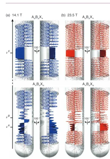
Influence ofB0on a Gradient-encoded Refocusing Pulse Gradient-encoded refocusing pulses are key actors of the spin dynamics that is locally triggered in each region of the sam-ple, because they have been shown to improve greatly the spatial selectivity of the refocusing blocks within which they are implemented. In order to evaluate the influence of B0on this kind of selective pulse, we have simulated the action of a gradient-encoded Reburpπ pulse on transverse magnetization initially taken along the y-axis and composed of selected coher-ences of HAand HX, at 14.1 and 23.5 T. In Figure 5, the spatial distribution of the resulting magnetization is shown for both magnetic fields. Similarly to what is observed for the excitation case, the cross sections within which the magnetization is properly refocused by the refocusing block are found to be thinner at higher field for both proton sites, whereas outside of these regions the initial transverse magnetization is fully dephased. We also note that the height of the cross section encoded at 𝜈A at 23.5 T allows for refocusing with a high
(a) E-Burp2 - 90°
14.1 T 23.5 T 14.1 T 23.5 T 14.1 T 23.5 T 14.1 T 23.5 T
(b) Esnob - 270° (c) Gaussian - 90° (d) Gaussian - 270°
νX
ν(Z)
νB
νA
Figure 4. The evolution of the amplitude profile generated by a gradient-encoded excitation step is represented at 14.1 and 23.5 T for (a) an E-Burp2 90∘ pulse of 60 ms, (b) an Esnob 270∘ pulse of 20 ms, (c) a Gaussian 90∘ pulse of 20 ms, and (d) a Gaussian 270∘ pulse of 20 ms
1 2 3 4 5 6 7 8 9 10 11 12 13 14 15 16 17 18 19 20 21 22 23 24 25 26 27 28 29 30 31 32 33 34 35 36 37 38 39 40 41 42 43 44 45 46 47 48 49 50 51 52 53 54 55 56 57 58 59 61 62 63 64 65 66 67 68 69 70 71 72 73 74 75 76 77 78 79 80 81 82 83 84 85 86 87 88 89 90 91 92 93 94 95 96 97 98 99 100 101 102 103 104 105 106 107 108 109 110 111 112 113 114 115 116 117 118 119 (a) 14.1 T 100 ° 100 ° 100 ° 100 ° AαBαX+ (b) 23.5 T AαBαX+ νX νB νA A+BαXα A+BαXα
Figure 5. The spatial variation of the magnetization calculated at the end of gradient-encoded Reburp 180∘ pulse of 60 ms at (a) 14.1 T and (b) 23.5 T, for (top) A𝛼B𝛼X+and (bottom) A+B𝛼X𝛼coherences
spatial selectivity the evolution of a coherence involving HA without affecting the spin state of HB, which is a prerequisite for decoupling these two strongly coupled protons.
From 1-D to 2-D Spatial Frequency Encoding
NMR
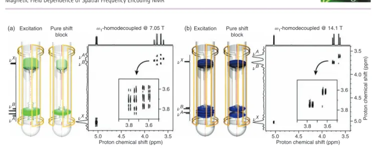
Following on from the above, the study of the magnetic field dependence of SFE NMR can be extended to the general case of a pulse sequence composed of several gradient-encoded pulses. As an illustration, we have acquired a 2-D 𝜔1-homonuclear decoupled experiments17at two different magnetic fields, on 10 mg of 1 dissolved in 700μl of deuterated chloroform. We remind that this pulse sequence implements a pure shift block during the indirect evolution delay, which yields a 2-D phased spectrum with only diagonal peaks showing a singlet structure in the indirect domain, and a normal multiplet structure in the direct domain. For this experimental part, we have chosen to monitor the resolution enhancement that can be achieved in the pure shift dimension when going from 7.05 T (𝜈H = 300 MHz) to 14.1 T. The resulting spectra are shown in Figure 6 together with the amplitude profiles that were calculated after the excitation step and at the end of the pure shift block of this pulse sequence. At 7.05 T, we observe that neither the
excitation step nor the pure shift block can achieve a slice selection process of sufficient spatial resolution for HAand HB. On the resulting 2-D spectrum, it is apparent both protons were encoded in the same cross section, as it is illustrated by the shape of the distorted correlation appearing around 𝜈A and𝜈B. Conversely, at 14.1 T, we note that the efficiency of the refocusing block allows for refining the slice selection process initially created during the excitation step, to a point where
HA and HB can be selectively decoupled. The better spatial resolution that is reached at this field can be straightforwardly correlated with the high spectral resolution achieved at𝜈Aand
𝜈B. The projection displayed along the indirect domain allows for observing the singlet structure of each correlation, which highlights the higher efficiency of the homonuclear decoupling sequence at 14.1 T than at 7.05 T.
Magnetic Field Dependence of Sensitivity
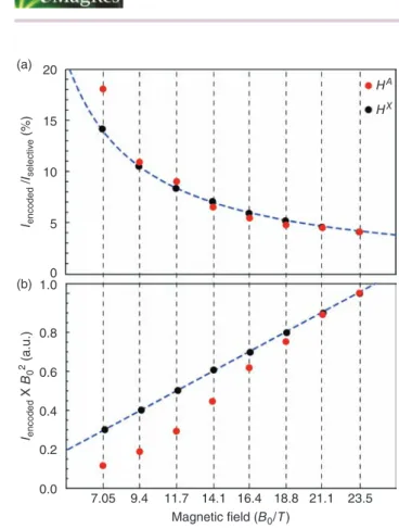
One frequent reservation that is sometimes expressed about the relevance of SFE techniques regards their low sensitivity. To tackle this problem, it is important to discuss here whether acquiring spatially encoded spectra at higher field has an overall beneficial effect on sensitivity. In this section, we aim thus at quantifying the relationship between the sensitivity of a gradient-encoded experiment and B0. We have studied, for eight different magnetic fields that are representative of the range of NMR spectrometers commercially available to date, the evolution of the sensitivity of a single gradient-encoded pulse experiment. The integration of the signal of HXand HA has been calculated for each magnetic field and normalized by the signal arising from the same semiselective excitation pulse without SFE (Figure 7). The resulting ratio Iencoded/Iselective provides a direct quantification of the fraction of the sample within which HXand HAare encoded, while allowing to throw off the contribution of the intrinsic probehead sensitivity and the Boltzmann population of the observed spin. Again, we have set the gradient strength so that the spectral width that is encoded along the virtual sample matches exactly the proton spectrum of 1. As expected, Iencoded/Iselectivevalues range from 4 to 18% and decrease with B0, which reflects the thinning of the encoded cross section with the strength of the magnetic field. Furthermore, as Iencoded is proportional to the spectral bandwidth of the encoded pulse, the resulting ratio should, to a first approximation, be proportional to B0−1. Figure 7(a) shows the corresponding trend curve described by Pell and Keeler16that we have adjusted to the ratio calculated at 23.5 T to account for the contribution of the shaped pulse to the slice selection process. For HX, the evolution of the ratios is in agreement with this trend curve. For HA, however, although the ratios obtained at higher field (14.1–23.5 T) follow the trend curve, those determined at lower field (7.05, 9.4, and 11.7 T) are higher than expected, which is explained by the fact that the slice selection induced by the SFE could not be operated properly, resulting in an overlap of the two slices within which
HAand HB are encoded, and hence the observed deviations. From this result, it is now possible to extrapolate the overall dependence of the sensitivity of a single gradient-encoded pulse experiment on B0. For the homonuclear case, and if we
1 2 3 4 5 6 7 8 9 10 11 12 13 14 15 16 17 18 19 20 21 22 23 24 25 26 27 28 29 30 31 32 33 34 35 36 37 38 39 40 41 42 43 44 45 46 47 48 49 50 51 52 53 54 55 56 57 58 59 61 62 63 64 65 66 67 68 69 70 71 72 73 74 75 76 77 78 79 80 81 82 83 84 85 86 87 88 89 90 91 92 93 94 95 96 97 98 99 100 101 102 103 104 105 106 107 108 109 110 111 112 113 114 115 116 117 118 119
(a) Excitation Pure shift (b)
block
ω1-homodecoupled @ 7.05 T Excitation ω1-homodecoupled @ 14.1 T
5.0 4.5 4.0 3.5
3.6 3.6
3.8 3.8
Proton chemical shift (ppm)
5.0 4.5 4.0 3.5
3.5
4.0
4.5
5.0
Proton chemical shift (ppm)
Proton chemical shift (ppm)
Pure shift block 3.6 3.8 3.8 3.6 νX νX νX νB νA νA νB νX νB νA νA νB
Figure 6. The amplitude profiles calculated after the gradient-encoded excitation pulse and at the end of the pure shift block of a 2-D1H𝜔
1-homonuclear
decoupled experiment simulated on 1 at (a) 7.05 T and (b) 14.1 T. The corresponding experimental 2-D spectra acquired are also shown at both fields. E-Burp2 and Reburp pulses of 60 ms were used for the gradient-encoded excitation and the pure shift block, respectively. The strength of the gradient coil was set so as to encode along the sample a spectral width of 6.4 and 6.3 ppm at 7.05 and 14.1 T, respectively
assume comparable linewidths and probe performances, a rough estimate of the dependence of S/N on B0is18
S∕N ∝ n ⋅ 𝛾H5∕2⋅ B3∕20 (2) where n is the number of observed proton nuclei in the sam-ple. The value of the integration that can be derived from
Iencoded to account for (i) the influence of the magnetic field on the Boltzmann population of the observed proton spins, (ii) the signal induced in the receiver coil at detection, and (iii) the noise has been calculated for different B0s (Figure 7b). On the one hand, we note that the overall sensitivity of the spatially frequency-encoded experiment increases with the strength of the magnetic field, despite the selection of thinner slices upon the gradient-encoded excitation step. On the other hand, for HA, the NMR signal acquired at lower field is weaker than what could be expected from the ideal curve adjusted to the sensitivity measured at 23.5 T, which underlines again the spatial resolution issue previously described at lower field. These results highlight the relevance of implementing spatially frequency-encoded pulse sequences at higher magnetic field, in so far as it contributes to enhance the three inextricably linked features that are the dispersion of the resonance frequencies (i.e., the spectral resolution), the spatial resolution of the slice selection process, and the sensitivity – although this latter point is modulated by the slice selection issue.
Conclusion
In this article, we have described the magnetic field dependence of NMR pulse sequences based on a SFE of the sample. We have highlighted the evolution of the key features that drive sensitivity and resolution in gradient-encoded pulse sequences, from the basic irradiation schemes, to state-of-the-art refo-cusing blocks that are nowadays routinely implemented in high-resolution experiments dedicated to the extraction of1H chemical shifts and/or homonuclear couplings. We have shown that operating at higher field leads to a better spatial resolu-tion of the slice selecresolu-tion process, together with a moderate
sensitivity enhancement of the resulting data. The combination of SFE techniques with high magnetic fields shades a new light on the trade-off that has classically to be found between sensi-tivity and resolution in SFE NMR. Indeed, the main limitation of high-resolution techniques based on this method regards the sensitivity penalty that has to be paid when protons with very close chemical shifts have to be decoupled, because it implies the use of highly selective pulses.19We remark that operating at higher field amounts to increasing the chemical shift dispersion while keeping the same selectivity for the gradient-encoded pulses, which is a way to address this issue without sacrificing sensitivity. The implementation of SFE methods on high field spectrometers should thus be particularly suitable for the analysis of larger molecular systems showing overcrowded1H spectra.
Acknowledgment
This work was supported by the French Research Agency (ANR-2011-JS08-009-01).
Biographical Sketches
Bertrand Plainchont. b. 1985. MSc, Université de Strasbourg (France), 2009. PhD directed by J.-M. Nuzillard, Université de Reims Champagne-Ardenne (France), 2012. Postdoctoral research fellow at Université Paris-Sud, France. About 10 publications dealing with methodological developments for the simplification of complex liquid-state NMR spectra, computer-assisted structure elucidation and verification, and spin dynamics simulation.
Jonathan Farjon. b. in 1977, carried out a master in Chemistry at the Paris South University in 2000. PhD in chemistry, Paris South Uni-versity 2003. Since 2009, research associate of the National Center for Scientific Research at the Paris South University. 23 publications on NMR in liquid and oriented media, methodology, and their applica-tions in bio(in)organic chemistry. Actual interest is dedicated to highly resolved, sensitive, and quantitative techniques as well as fast NMR.
Nicolas Giraud. b. 1977. BSc, Ecole Normale Supérieure de Lyon (France) 2001. PhD, Chemistry, Université Claude Bernard Lyon I, Lyon (France), 2005. Currently Reader in Chemistry at the Université
1 2 3 4 5 6 7 8 9 10 11 12 13 14 15 16 17 18 19 20 21 22 23 24 25 26 27 28 29 30 31 32 33 34 35 36 37 38 39 40 41 42 43 44 45 46 47 48 49 50 51 52 53 54 55 56 57 58 59 61 62 63 64 65 66 67 68 69 70 71 72 73 74 75 76 77 78 79 80 81 82 83 84 85 86 87 88 89 90 91 92 93 94 95 96 97 98 99 100 101 102 103 104 105 106 107 108 109 110 111 112 113 114 115 116 117 118 119 (a) HA HX 20 15 10 5 0 1.0 Iencoded X B0 2 (a.u.) Iencoded /Iselective (%) 0.8 0.6 0.4 0.2 0.0 7.05 9.4 11.7 14.1 16.4 18.8 21.1 23.5 Magnetic field (B0/T) (b)
Figure 7. (a) The simulated sensitivity ratio between the integration of the signal resulting from a selective excitation with (Iencoded) and without spa-tial frequency encoding (Iselective) at eight different magnetic fields (from 7.05 to 23.5 T), for HAand HX. The trend curve corresponding to the ideal
behavior of this integration ratio modeled by Keeler and Pell is plotted as a dashed line. (b) The evolution of the integration of the gradient-encoded signal ponderated by B02, as a function of B0, for HAand HX. The trend
curve corresponding to the ideal evolution of this integration is plotted as a dashed line
Paris-Sud, France. About 25 publications on the development and the applications of liquid- and solid-state NMR techniques. Current research interest: development of new high-resolution NMR methods for the structural and dynamic analysis of complex molecular systems in chemistry and biology.
Related Articles
Modern NMR Pulse Sequences in Pharmaceutical R&D; emrstm1402
Pure Shift NMR Spectroscopy; Field Gradients and Their emrstm1362
emrstm0164
Application; Radiofrequency Pulses: Response of Nuclear emrstm0443
Spins; Gradient Coil Systems; Radiofrequency Gradient Pulses; emrstm0192
emrstm0442
Selective Pulses; Shaped Pulses emrstm0486
emrstm0493
References
1. L. Frydman, T. Scherf, and A. Lupulescu, Prog. Nucl. Magn. Reson. Spectrosc., 2002, 99, 15858. DOI: 10.1073/pnas.252644399.
2. M. K. Stehling, R. Turner, and P. Mansfield, Science, 1991, 254, 43. 3. C. S. Johnson Jr, Prog. Nucl. Magn. Reson. Spectrosc., 1999, 34, 203. 4. S. Berger, Prog. Nucl. Magn. Reson. Spectrosc., 1997, 30, 137. DOI:
10.1016/S0079-6565(97)00003-4.
5. A. Tal and L. Frydman, Prog. Nucl. Magn. Reson. Spectrosc., 2010, 57, 241. 6. K. Zangger and H. Sterk, J. Magn. Reson., 1997, 124, 486. DOI:
10.1006/jmre.1996.1063.
7. N. Giraud, L. Beguin, J. Courtieu, and D. Merlet, Angew. Chem. Int. Ed., 2010,
49, 3481. DOI: 10.1002/anie.200907103.
8. D. Merlet, L. Beguin, J. Courtieu, and N. Giraud, J. Magn. Reson., 2011, 209, 315. DOI: 10.1016/j.jmr.2011.01.030.
9. N. Giraud, D. Pitoux, J. M. Ouvrard, and D. Merlet, Chem. Eur. J., 2013, 19, 12221. DOI: 10.1002/chem.201302005.
10. D. Pitoux, B. Plainchont, D. Merlet, Z. Hu, D. Bonnaffé, J. Farjon, and N. Giraud,
Chem. Eur. J., 2015, 21, 9044. DOI: 10.1002/chem.201501182.
11. N. H. Meyer and K. Zangger, Angew. Chem. Int. Ed., 2013, 52, 7143. DOI: 10.1002/anie.201300129.
12. N. Gubensak, W. M. F. Fabian, and K. Zangger, Chem. Commun., 2014, 50, 12254. DOI: 10.1039/C4CC05892E.
13. N. Lokesh, S. R. Chaudhari, and N. Suryaprakash, Chem. Commun., 2014, 50, 15597. DOI: 10.1039/C4CC06772J.
14. D. Pitoux, Z. Hu, B. Plainchont, D. Merlet, J. Farjon, D. Bonnaffé, and N. Giraud,
Magn. Reson. Chem., 2015, 53, 836. DOI: 10.1002/mrc.4281.
15. B. Plainchont, D. Pitoux, G. Hamdoun, J. M. Ouvrard, D. Merlet, J. Farjon, and
N. Giraud, Submitted, 2016. AQ1 16. A. J. Pell and J. Keeler, J. Magn. Reson., 2007, 189, 293. DOI: 10.1016/j.jmr.
2007.09.002.
17. N. Giraud, M. Joos, J. Courtieu, and D. Merlet, Magn. Reson. Chem., 2009, 47, 300. DOI: 10.1002/mrc.2387.
18. R. R. Ernst, G. Bodenhausen, and A. Wokaun, Principles of Nuclear Magnetic
Resonance in One and Two Dimensions, 2nd edn, Clarendon Press: Oxford,
1987.
19. M. Foroozandeh, R. W. Adams, M. Nilsson, and G. A. Morris, J. Am. Chem. Soc., 2014. DOI: 10.1021/ja507201t.
1 2 3 4 5 6 7 8 9 10 11 12 13 14 15 16 17 18 19 20 21 22 23 24 25 26 27 28 29 30 31 32 33 34 35 36 37 38 39 40 41 42 43 44 45 46 47 48 49 50 51 52 53 54 55 56 57 58 59 61 62 63 64 65 66 67 68 69 70 71 72 73 74 75 76 77 78 79 80 81 82 83 84 85 86 87 88 89 90 91 92 93 94 95 96 97 98 99 100 101 102 103 104 105 106 107 108 109 110 111 112 113 114 115 116 117 118 119
QUERIES TO BE ANSWERED BY AUTHOR
IMPORTANT NOTE: Please mark your corrections and answers to these queries directly onto the proof at the relevant place. DO NOT mark your corrections on this query sheet.
Queries: