HAL Id: hal-02890128
https://hal.archives-ouvertes.fr/hal-02890128
Submitted on 9 Jul 2020
HAL is a multi-disciplinary open access
archive for the deposit and dissemination of
sci-entific research documents, whether they are
pub-lished or not. The documents may come from
teaching and research institutions in France or
abroad, or from public or private research centers.
L’archive ouverte pluridisciplinaire HAL, est
destinée au dépôt et à la diffusion de documents
scientifiques de niveau recherche, publiés ou non,
émanant des établissements d’enseignement et de
recherche français ou étrangers, des laboratoires
publics ou privés.
improved excited state properties
Lea Gimeno, Errol Blart, Jean-Noël Rebilly, Marina Coupeau, Magali Allain,
Thierry Roisnel, Alexis Quarré de Verneuil, Christophe Gourlaouen, Chantal
Daniel, Yann Pellegrin
To cite this version:
Lea Gimeno, Errol Blart, Jean-Noël Rebilly, Marina Coupeau, Magali Allain, et al.. Non symmetrical
sterically challenged phenanthroline ligands and their homoleptic copper(I) complexes with improved
excited state properties. Chemistry - A European Journal, Wiley-VCH Verlag, 2020, 26 (51),
pp.11887-11899. �10.1002/chem.202001209�. �hal-02890128�
FULL PAPER
Non symmetrical sterically challenged phenanthroline ligands
and their homoleptic copper(I) complexes with improved excited
state properties
Lea Gimeno,
[a]Errol Blart,
[a]Jean-Noël Rebilly,
[b],* Marina Coupeau,
[a]Magali Allain,
[c]Thierry Roisnel,
[d]Alexis Quarré de Verneuil,
[a]Christophe Gourlaouen,
[e],* Chantal Daniel,
[e]and Yann Pellegrin
[a],*
Abstract: A strategy is presented to improve the excited state
reactivity of homoleptic copper-bis(diimine) complexes CuL2+ by increasing the steric bulk around Cu(I) while preserving their stability. Substituting the phenanthroline on position 2 by a phenyl group allows the implementation of stabilizing intramolecular π stacking within the copper complex, while tethering a branched alkyl chain on position 9 provides enough steric bulk to rise the excited state energy E00. Two novel complexes are thus studied and compared to symmetrical models. The impact of breaking the symmetry of phenanthroline ligands on the photophysical properties of the complexes is analyzed and rationalized thanks to a combined theoretical and experimental study. The importance of fine-tuning the steric bulk of the N-N chelate in order to stabilize the coordination sphere is demonstrated. Importantly, the excited state reactivity of the newly developed complexes is improved as demonstrated in the frame of a reductive quenching step, evidencing the relevance of our strategy.
Introduction
Copper(I) complexes of the general formula [Cu(NN)2]+ where NN
is a diimine ligand bearing bulky substituents in α of the nitrogen atoms are appealing luminophores in the frame of photosensitive coordination compounds.[1] Indeed, they exhibit photo-physical
properties which are similar to those of benchmark [Ru(bpy)3]2+.[1a,
1c, 2]. In particular they are luminescent upon excitation in the Metal
to Ligand Charge Transfer (MLCT), provided that encumbering groups are appended in α of the chelating nitrogen atoms. During
excitation (equation 1), the metal formally shifts from Cu(I) (favored tetrahedral geometry) to Cu(II) (favored square-planar geometry).
[CuIL
2]+ + hv → [CuIIL•-L]+ (equation 1)
The coordination sphere relaxes in the [CuIIL•-L]+ excited state by
flattening which opens several channels for efficient and counter-productive extinction of the luminescence.[3] Consequently, the
excited state energy levels dynamically shift to lower energy on a typical 10-100 ps time scale, favoring non radiative de-excitation of the excited states (gap law). Besides, flattening of the transient copper(II) ion opens a fifth binding site for a ligand (e.g. a solvent molecule) leading again to the very fast so-called “exciplex quenching” of the excited state.[4] Encumbering groups limit the
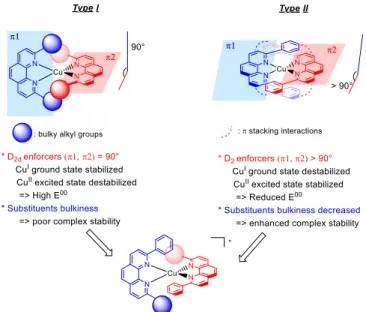
extent of the photo-induced flattening, thus promoting the luminescence of copper(I) complexes. Consequently considerable efforts have been made to improve the luminescence of copper(I) complexes by increasing the steric bulk around Cu(I).[5] The latter can be brought by either aliphatic (type
I complexes, figure 1) or aromatic (type-II complexes) substituents. Impressive luminescence quantum yields have been obtained with type-I complexes by Castellano and co-workers with [Cu(dipp)2]+, [Cu(tmdsbp)2]+,[5b, c, 6] or Burstyn and co-workers
with [Cu(dtbp)2]+[5a, 7] (where dipp stands for
2,9-diisopropyl-1,10-phenanthroline, tmdsbp stands for 2,9-di-secbutyl-3,4,7,8-tetramethyl-1,10-phenanthroline and dtbp for 2,9-ditert-butyl-1,10-phenanthroline). Remarkably, [Cu(dtbp)2]+ featured even
better photophysical parameters than [Ru(bpy)3]2+ (luminescence
lifetime of 3.26 µs vs. ca. 1 µs respectively) but the stability of the coordination scaffold was sacrificed. Indeed, the copper(I) complex is so sterically burdened that it quantitatively decomposes in presence of a weak ligand like acetonitrile.[5a]
Type-II [Cu(dpp)2]+ reported by Sauvage and co-workers (and
parent complexes) also proved very luminescent[8] even at room
temperature in presence of strongly coordinating species (e.g. water or acetonitrile): the complex is rigidified and shielded by the phenyl groups, π-stacked to the vicinal phen ligand.[9] But the
latter interactions substantially distort the coordination cage entailing a decrease of the singlet state energy E00.[10] This in turn
impact negatively the reactivity of the excited state, by virtue of the Rehm and Weller equation.
[a] Lea Gimeno, Marina Coupeau, Alexis Quarre de Verneuil, Dr. Errol Blart, Dr. Yann Pellegrin
Université de Nantes, CNRS CEISAM UMR6230 F-44000 Nantes, France E-mail: yann.pellegrin@univ-nantes.fr [b] Dr. Jean-Noël Rebilly ICMMO, UMR 8182
Université Paris Sud, Université Paris Saclay Orsay 91405 cedex, France
[c] Magali Allain
Laboratoire MOLTECH-Anjou, UMR CNRS 6200 Univ Angers, SFR MATRIX
2 Bd Lavoisier, 49045 Angers Cedex, France [d] Thierry Roisnel
Université de Rennes CNRS
Institut des Sciences Chimiques de Rennes UMR6226
F-35000 Rennes, France
[e] Dr. Christophe Gourlaouen, Pr. Chantal Daniel
Laboratoire de Chimie Quantique Institut de Chimie UMR 7177 CNRS-Université de Strasbourg, 4, Rue Blaise Pascal CS 90032, F-67081 Strasbourg Cedex, France
Supporting information for this article is given via a link at the end of the document.
Accepted
FULL PAPER
Figure 1. Illustration of the strategy developed in this contribution
Summarily, very bulky aliphatic groups like tert-butyl in α of the nitrogen atoms endow the corresponding copper(I) complex with long lived, intense luminescence and high E00 while aromatic
groups confer improved stability to the coordination cage, maintain significant luminescence lifetime and quantum yield, but lead to a smaller E00.
In this contribution, we explore a strategy to implement intramolecular π-stacking while maintaining the tetrahedral geometry to secure high E00. Our approach is based on
non-symmetrical phen ligands substituted by one phenyl group on one side and one bulky branched alkyl chain (isopropyl: ligand L1; or tert-butyl: ligand L2) on the other.
Figure 2. Structures of the complexes investigated therein.
The resulting homoleptic copper complexes, C1 and C2 (figure 2) are a “fusion” of [Cu(dpp)2]+ with [Cu(dipp)2]+ and [Cu(dtbp)2]+,
respectively, and would benefit from the stabilizing intramolecular phenyl-phenanthroline π-stacking interactions while the steric bulk provided by the alkyl chains (isopropyl or tert-butyl) contribute reaching a high E00. We describe the synthesis and full
optical and electrochemical characterization of C1 and C2. The structural and optical properties of the two complexes have been analyzed by means of density functional theory (DFT). Our goals are 1) to find out if intramolecular π-stacking can take place within the complex despite the presence of bulky ramified chain on the same ligand; 2) to assess the stability of the corresponding coordination scaffold vs. the steric strain; 3) to observe if the
energy of the excited state can be increased and how it impacts the reactivity of the excited state.
Results and Discussion
The synthesis of ligands L1 and L2 was performed in two steps, by addition of the corresponding organolithium reagents on anhydrous phenanthroline and re-aromatization with MnO2
(scheme 1). Other synthetic pathways have been envisioned (for instance, an improved Povarov approach has been successfully developed to synthesize ligand L1)[11] but the organolithium route
proved to be more efficient in our hands. The synthesis of the three corresponding copper(I) complexes proceeded smoothly, by mixing two equivalents of ligand with one equivalent of tetrakis(acetonitrile)copper hexafluorophosphate in degassed dichloromethane.
Scheme 1. Synthesis of ligands L1, L2 and of complexes C1 and C2. The bright red powders were characterized by 1H-NMR and mass
spectrometry, all the analyses confirm the proposed structures. Monocrystals of ligand L2 and complex C2 were obtained by slow diffusion of hexane in a dichloromethane solution of the corresponding molecule, and their structures could be resolved by X-ray diffraction. Relevant parameters are reported in table ST1 (supporting information). The structure of L2 (figure S1, supporting information) reveals as expected a very sterically strained environment around chelating nitrogen atoms. The phenyl group tethered to C12 is slightly twisted with respect to the
phenanthroline plane (twist angle = ca. 25.3°), due to repulsion between protons H11 and H14 and possibly crystal packing forces.
The structure of complex C2 is given in figure 3 (ORTEP view available in figure S2). Basically, the CuN4 coordination sphere is
a flattened tetrahedron; the τ4 parameter for C2 is almost exactly
the average between [Cu(dpp)2]+ and [Cu(dtbp)2]+ (figure S6).
However C2’s structure is closer to a I complex than a type-II, despite the presence of the aryl group in α of the nitrogen atom.
Figure 3. Two views for the structure of complex C2.
Accepted
FULL PAPER
Rather unfavorable angles between phenyl groups C and E and phenanthroline B and A respectively (aromatic cycles and relevant atom nomenclature is given in figure S3) are measured (more than 30° vs. ca. 10° in the case of Cu(dpp)2+)[9a] and the
distance between the aromatic cores is larger (more than 4 Å) meaning that the extent of intramolecular π stacking is likely smaller than for [Cu(dpp)2]+ in the crystal phase. A relevant way
to quantitatively estimate how distorted from the ideal D2d
symmetry copper complexes are, consists in calculating θx, θy and
θz angles, respectively the rocking, waggling and flattening angles
(see figures S4-6 in supporting information for an illustration of those angles).[12] For a perfectly tetrahedral geometry, θ
x = θy = θz
= 90°; this is roughly the case for [Cu(dtbp)2]+ (table ST1, figure
S6). For [Cu(dpp)2]+, the intramolecular stacking interactions lead
to a strongly distorted structure (θx = 104.6, θy = 69.8, θz= 100.2).
On the other hand, C2’s structure is not perfectly tetrahedral since θz is significantly different from 90° revealing the coordination
sphere is substantially flattened in the ground state, but to a lesser extent than [Cu(dpp)2]+, as exemplified by θx and θy values which
are close to 90°. All in all, the θx,y,z parameters of C2 are closer to
those of plain [Cu(dmp)2]+.[12] This means that the steric bulk
imposed by the tert-butyl group limits the development of intramolecular π stacking within the complex in the crystal phase. Nevertheless, let us pinpoint that the phenyl group is more twisted with respect to the phenanthroline plane onto which it is tethered (C on A, E on B) passing from the free ligand to the complex. This could be a proof that some interaction between C and B (and E and A) is taking place. CH-π interactions are possible in this frame (Figure S7). Accordingly, crystal packing could account for this observation too. Importantly, no significant intermolecular interaction is observed in the structure. In solution, it is interesting to notice that the chemical shifts assigned to hydrogen atoms on the phenyl groups tethered on phenanthroline are step wisely shielded when passing from [Cu(dpp)2]+ to C1 then C2 indicating
that the orientation of the bulky aromatic groups is different (table ST2), resulting from a mixture of steric constraints, π-stacking and possible CH-π interactions.
UV-Vis absorption
UV-Vis spectra were recorded for C1 and C2 in dichloromethane solutions (figure 4) and experimental data are gathered in table 1. Both complexes exhibit the same absorption features, namely intense ligand centered π-π* transitions below 370 nm and a broad band assigned to the MLCT spanning between 400 and 500 nm and responsible for the bright orange color of all complexes. A shoulder is monitored above 500 nm for all complexes but is more prominent for C1 than C2. For symmetry reasons, the S0→S1 transition is forbidden in the case of tetrahedral copper(I)
complexes and the MLCT peaking at ca. 460 nm is thus a S0→Sn
(n > 1) transition. When the geometry moves away from tetrahedral, the S0→S1 transition becomes partially allowed.
The intensity of the S0 to S1 transition thus allows to estimate the
degree of flattening in a copper(I) complex in its ground state.[5h, 13][Cu(dpp)
2]+ shows a rather significant S0 to S1 transition in the
shape of a broad shoulder,[14] whereas no such spectral feature
can be detected for [Cu(dtbp)2]+ (virtually perfectly tetrahedral).
This indicates a significant symmetry descent from D2d to D2, in
line with the favorable π-stacking interactions between the phenyl and the adjacent phenanthroline.[15]
Figure 4. Electronic absorption spectra for complexes C1 (black trace), C2 (blue trace), [Cu(dipp)2]+ (green trace) and [Cu(dpp)2]+ (red trace) recorded in
dichloromethane at room temperature. Average concentrations 0.05 mM.
All other [Cu(NN)2]+ complexes are located in between those two
extremes, featuring shoulders of variable intensity at the stem of the MLCT. In our case, one can learn from normalized absorption spectra (figure S8) that the structure of C1 is substantially more flattened in the ground state than C2 or benchmark [Cu(dipp)2]+.
This proves that the phenyl substituent in ligand L1 is capable of intramolecular π-stacking with the vicinal phenanthroline. On the other hand, the tert-butyl group in L2 imposes a stronger steric bulk which restrains the flattening in C2 and limits the possibility of π-π interactions. This corroborates the observations made on the solid state where the 30° angle and the distance between phenyl C and phenanthroline B (figure S3) are more in line with CH-π interactions (figure 3).
Electrochemistry
The electrochemical behavior of complexes C1 and C2 was explored by cyclic voltammetry in dichloromethane and dimethylformamide (DMF) solutions, in presence of tetrabutylammonium hexafluorophosphate as supporting electrolyte. The voltammograms are given in figures S9 and S10; and related data are reported in table 1. One mono-electronic, pseudo-reversible oxidation wave was monitored and assigned to the CuII/CuI couple for both complexes (E
1 = 0.49 and 0.55 V vs.
ferrocenium/ferrocene redox couple (Fc+/Fc) for C1 and C2
respectively). Generally speaking, C1, C2 and [Cu(dpp)2]+ are
oxidized at similar potentials, significantly less positive than E1(Cu(dipp)2+). Indeed, bulky substituents enforce a D2d symmetry
which stabilizes the copper(I) ion and induce a more anodic oxidation potential.[5d, 5h, 16] Phenyl substituents generate
intramolecular π-stacking and entail a descent to D2 symmetry
and thus a shift of the oxidation potential to less positive values vs. [Cu(dipp)2]+.[5a] C2 is undeniably more sterically burdened than
C1 because of the tert-butyl group, which translates into E1(C2) >
E1(C1).
Accepted
FULL PAPER
Table 1. UV-Vis and electrochemical data for complexes C1, C2, [Cu(dpp)2]+
and [Cu(dipp)2]+ in dichloromethane. All data recorded at room temperature.
Electrochemical potential values reported vs. the Fc+/Fc couple taken as internal
standard.
Complexes λmax/nm εʎmax/L.mol-1cm-1 E
1 (ΔE)/V (mV)[a] E2 (ΔE)/V (mV)[b]
C1 443, 540 3990, 1000 0.49 (120) -2.06 (130)
C2 450 3850 0.55 (110) -2.08 (90)
[Cu(dpp)2]+ 439, 550 3800, 1800 0.53 (80) -2.07 (120) [Cu(dipp)2]+ 453 7260 0.65 (120) -2.10 (75)
[a] Degassed dichloromethane solutions, sweep rate for electrochemical measurements = 100 mV.s-1. [b] Degassed DMF solutions, sweep rate for
electrochemical measurements = 1 V.s-1. E
1 = (Ep,a1 + Ep,c1)/2, E2 = (Ep,a2 +
Ep,c
2)/2; ΔE = Ep,a1 – Ep,c1 or Ep,a2 – Ep,c2, where Ep,a1, Ep,c1, Ep,a2 and Ep,c2 are
anodic and cathodic peak potentials for electrochemical processes 1 and 2, respectively.
Both C1 and C2 are expected to be less flattened than [Cu(dpp)2]+,
but nevertheless, E1([Cu(dpp)2]+) > E1(C1). Rationalizing the
order of their close oxidation potentials is made particularly difficult by the interplay of both steric and electronic effects. No reduction wave was observed within the solvent electro-activity window of dichloromethane. In DMF however, a pseudo-reversible wave was monitored below -2 V vs. Fc+/Fc as internal
standard for all complexes and was assigned to the addition of an extra electron on one phenanthroline ligand. All potential values E2 are very close to each other despite the differences between
the electronic effects from branched alkyl chains (electron rich) and the aromatic groups (electron poor).
Excited state properties
Complexes were studied by steady state fluorescence and time-correlated single photon counting in degassed dichloromethane. All complexes are luminescent at room temperature in dichloromethane dilute solutions. Steady state emission spectra are given in figure 5, while decay traces are given in figures S11 and S12. Emission maximum wavelengths λem, luminescence
quantum yields ɸlum, lifetimes τ and E00 values (energy difference
between singlet excited state and the ground state in ground vibrational states, estimated by the tangent method)[5b] are
reported in table 2.
Concerning model complexes [Cu(dpp)2]+ and [Cu(dipp)2]+, bulky
isopropyl groups in the latter tend to favour D2d ground state
geometry and a stabilized environment for copper(I) affording short emission wavelength and large E00. Conversely,
intramolecular π-stacking in [Cu(dpp)2]+ entail the flattening of the
coordination cage towards D2 symmetry, destabilization of the
copper(I) ion and thus longer emission wavelength and smaller E00. All figures of merit for C1 and C2 are comprised between
those of the model complexes. ʎem (C1) is red shifted compared
to ʎem([Cu(dipp)2]+) precisely because of the intramolecular
π-stacking interaction entailing some significant distortion of the coordination sphere away from D2d; conversely the steric bulk
from the branched isopropyl chain tends to oppose to this ground state distortion, forcing C1 in a less distorted geometry than [Cu(dpp)2]+, hence ʎem (C1) is blue shifted compared to
ʎem(Cu(dpp)2+) and E00 is larger. These results are comparable
with those obtained by Miller et al. where dpp-like ligands were progressively encumbered with substituents, gradually limiting the π-stacking interaction within the corresponding complexes.[5h]
C2’s emission wavelength is further blue-shifted due to the even greater steric strain brought by tert-butyl (compared to isopropyl), with similar ʎem and E00. Conclusively, increasing the steric bulk
with one ramified alkyl chain on one side of phenanthroline while keeping a phenyl group on the other allowed to increase E00
compared to [Cu(dpp)2]+ and maintain significant π-stacking
interaction at least for the case of C1.
Figure 5. Emission spectra for complexes C1 (black trace), C2 (blue trace) and Cu(dpp)2+ (red trace) recorded in dilute dichloromethane solutions (ca. 0.05
mM) at room temperature.
C1 and C2 exhibit similar emission quantum yields than model
[Cu(dpp)2]+, but unexpectedly short lifetimes, especially for C2
(table 2). As a matter of fact, the emission lifetimes for C2 and simple Cu(dmp)2+ are comparable, although the latter is less
sterically strained. In order to deepen the study, we extracted the radiative and non-radiative rate constants (respectively kr and knr)
from our experimental data (table 2). C2 exhibits the second best kr (behind [Cu(dipp)2]+), which can be assigned to its blue shifted
absorption and emission bands compared to C1 or [Cu(dpp)2]+,
because kr is proportional to the cube of the radiative energy
gap.[5h, 17] The reason for the weak emission properties of C2 are
therefore not grounded in kr. knr is however larger for C1 and
especially C2. A similar behavior was observed with the copper(I) complex [Cu(MeMap)2]+ (where MeMap is a non-symmetrical
phenanthroline ligand bearing one anisyl and one methyl group in α of the chelating nitrogen atoms)[18] and seems related to the
non-symmetrical nature of the ligands.
As reported in previous works, a phen ligand substituted by an aromatic and an aliphatic groups provides a very bulky environment on one side of the corresponding complex and at the same time a less bulky environment on the other.[19] This can be
exacerbated in the excited state and interactions of the exposed copper(I) center with solvent molecules or counter-anions would thus be favored.
Accepted
FULL PAPER
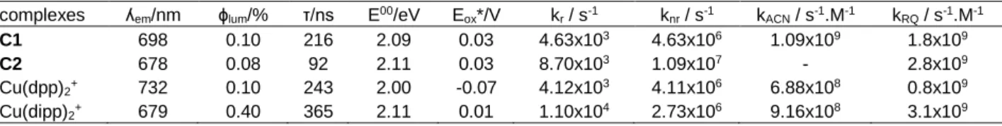
Table 2. Excited state parameters for complexes C1, C2, Cu(dpp)2+ and Cu(dipp)2+. Potentials are given vs. Fc+/Fc.
complexes ʎem/nm ɸlum/% τ/ns E00/eV Eox*/V kr / s-1 knr / s-1 kACN / s-1.M-1 kRQ / s-1.M-1
C1 698 0.10 216 2.09 0.03 4.63x10³ 4.63x10⁶ 1.09x10⁹ 1.8x10⁹
C2 678 0.08 92 2.11 0.03 8.70x10³ 1.09x10⁷ - 2.8x10⁹
Cu(dpp)2+ 732 0.10 243 2.00 -0.07 4.12x10³ 4.11x10⁶ 6.88x10⁸ 0.8x10⁹
Cu(dipp)2+ 679 0.40 365 2.11 0.01 1.10x10⁴ 2.73x10⁶ 9.16x10⁸ 3.1x10⁹
Such a process is particularly prominent with coordinating species (exciplex quenching) but has been monitored with non-coordinating molecules too.[20] Generally speaking, the solvent is
known to have an influence on the ground and excited state properties of copper(I) complexes,[21] and this influence could be
stronger when non-symmetrical ligands are coordinated to Cu(I) and lead to large knr. Tentatively, we recorded the luminescence
decays of C1, C2, [Cu(dpp)2]+ and [Cu(dipp)2]+ in dichloromethane
while progressively adding acetonitrile, well known to quench the emission of the copper complexes by exciplex formation, in order to appreciate how affected C1 and C2 are compared to classical [Cu(dipp)2]+ and [Cu(dpp)2]+. Following a Stern-Volmer analysis
we could retrieve the quenching constants kACN (figure S13, table
2) of the emission by acetonitrile. The exciplex quenching is undeniably more efficient for C1 than [Cu(dipp)2]+ or [Cu(dpp)2]+,
the latter being as previously observed the most resilient.[8a] C2
proved to be too unstable in presence of acetonitrile to allow recording spectra in the same conditions, but preliminary analysis of the data shows a very efficient emission quenching too. The enhanced exciplex quenching for C1 and C2 likely contributes to their particularly high knr.
Computational chemistry Structures
The presence of phenyl moieties in the C1 and C2 complexes generates several [Cu(dpp)2]+-type conformers, the structures of
which have been fully optimized at the DFT level. The structure of [Cu(dipp)2]+ complex belongs to the D2d symmetry group. For
[Cu(dpp)2]+, two conformers are possible, one of D2 symmetry and
one of S4 symmetry (Figure 6). The D2 structure is significantly
more stable than the S4 one (G = 2.3 kcal mol-1 computed at the
DFT level) in accordance with experimental observations.[9a] The
origin of the greater stability of the D2 structure can be attributed
to -stacking interactions as shown by the analysis of Non-Covalent Interactions (NCI) (figure 6). Whereas in the D2 structure,
π-stacking is enhanced by the interaction between the phenyl groups of one ligand and the phenanthroline moiety of the second one, it is weakened in the S4 structure where the two moieties are
less parallel. Furthermore, to ensure a greater parallelism between these moieties, the D2 structure is already flattened in
the ground state. C1 and C2 also exhibit conformers due to different orientations of the phenyl rings. In C1, the two computed structures have the same energy (G = 0.0 kcal mol-1). For C2,
the structure issued from the X-ray data (figure 3) is slightly less stable than the conformer (G = -0.6 kcal mol-1 computed at the
DFT level, Figure 6). Such variations are the result of the competition between the stabilizing -stacking interactions generated by the phenyl moieties and the steric congestion induced by the isopropyl and tert-butyl groups. In order to discuss
the theoretical results in light of those two possible conformers, we will name RX the structures similar to what is pictured in Figure 3 (and calculated as presented in Figure 6, left side) and conf the second computed structure (calculated as presented in figure 6, right side) for [Cu(dpp)2]+, C1 and C2 complexes.
[Cu(dpp)2]+
C2
RX, D2 Conf, S4
Figure 6. NCI analysis performed on the RX (top left) and conf (top right) structures of the [Cu(dpp)2]+ complex and on the RX (bottom left) and conf
(bottom right) structures of C2. In green are the basins corresponding to Van der Waals forces and in red area of steric congestion. All the structures have been fully optimized in the electronic ground state with GAUSSIAN (see computational details).
Absorption
The absorption spectra were computed on the DFT/B3LYP optimized structures (Table 3) of [Cu(dipp)2]+, [Cu(dpp)2]+, C1 and
C2 complexes and of their conformers (Figure 7). As observed
experimentally, the visible domain is assigned to MLCT transitions and the UV-Visible domain to transitions localized on the ligands. The computed spectra of [Cu(dipp)2]+ is very similar
to the experimental one with an intense band at 485 nm. Two singlet states are present at higher wavelength (505 and 522 nm) with no intensity for symmetry reasons. A shoulder is present at lower wavelength (433 nm).
The agreement between experimental (figure 3) and theoretical results (figure 7) is less good for the three other complexes in their RX structure. We retrieve the drop of the main S0→Sn band
intensity and the appearance of smaller bands. For [Cu(dpp)2]+,
the overall structure of the spectrum is well reproduced though being red shifted as compared to the experimental one. The main absorbing band is located at 454 nm. Four absorbing singlet
Accepted
FULL PAPER
states are located at 549, 582, 584 and 628 nm generating the band between 570 to 650 nm.
Table 3. Geometric parameters of the ground state of [Cu(dipp)2]+, [Cu(dpp)2]+,
C1 and C2 complexes and of their conformers. Distances are in Angstroms and angles in degrees. For each complex are given the relative stabilities (E) between the RX and conf structures from ADF calculations.
State [Cu(dipp)2]+ [Cu(dpp)2]+ C1 C2
RX conf RX conf RX conf
Cu-N1A 2.061 2.063 2.095 2.146 2.088 2.031 2.037 Cu-N2A 2.061 2.063 2.095 2.024 2.058 2.180 2.162 Cu-N1B 2.061 2.063 2.095 2.146 2.088 2.031 2.037 Cu-N2B 2.061 2.063 2.095 2.024 2.058 2.180 2.162 z 90.0 -118.7 90.0 107.2 67.9 106.2 73.4 E - 0.0 3.8 1.0 0.0 0.7 0.0
Figure 7. Theoretical absorption spectra in the visible domain for complexes C1 (black trace), C2 (blue trace), [Cu(dipp)2]+ (green trace) and [Cu(dpp)2]+ (red
trace) on the RX structures
The theoretical spectrum of C1 mostly reproduces the characteristics of the experimental one; the main band is located at 466 nm with again smaller intensity transitions between 500-600 nm and corresponding to the lowest singlet excited states calculated at 569, 532, 504 and 500 nm. The absorption spectrum of C2 is characterized by three peaks at 445, 510 and 570 nm. The most intense band at 510 nm is generated by two singlet states calculated at 506 and 512 nm. The presence of accessible conformers may explain the discrepancies between theoretical and experimental absorption spectra.
The spectra computed on the conf structures present significant shift of the bands. For the [Cu(dpp)2]+ complex, the conf structure
leads to tetrahedral arrangement around the copper cation and the spectrum is very similar to that of [Cu(dipp)2]+ complex,
illustrating again the structural impact on the optical properties (figure S14). Moreover, these results prove as well that the conf structure of [Cu(dpp)2]+ does not participate in its absorption
spectrum. For C1 and C2 complexes, the differences between the RX (Figure 7) and conf spectra (Figures S15 and S16) are less drastic though significant, revealing that both structures are substantially participating in the overall optical properties. As for our previous work on the [Cu(phenX2)2]+ complexes (where
phenX2 stands for 2,9-diX-1,10-phenanthroline and X = Cl, Br or
I),[22] the present complexes are highly flexible and present
several minima that can contribute to the absorption spectrum. The limitation of the static approach, which does not reflect this flexibility, explains the difference between the experimental and theoretical results. The influence of the dynamics on the absorption spectrum of C2, which are known to strongly influence the latter, are likely responsible for the differences between the experimental and calculated spectra. Importantly, the calculations match with the experimentally observed trend that the extinction coefficients of the S0→Sn transitions decrease when phenyl
groups are tethered in α of the nitrogen atoms of phenanthroline.[23]
Emission spectra
Nuclear evolution, mainly the z angle (as defined in figure S4)
and the Cu-N distances, and electronic re-localization underlie excited state relaxation. Delocalization of the excited electron over the two phenanthroline ligands corresponds to D2 point
group for [Cu(dipp)2]+ and [Cu(dpp)2]+ complexes and to C2 point
group for C1 and C2 complexes. Localization on one phenanthroline only leads to C2 point group for [Cu(dipp)2]+ and
[Cu(dpp)2]+ complexes and to C1 point group for C1 and C2
complexes (see figure S17 and table ST3).
[Cu(dipp)2]+
For [Cu(dipp)2]+, two symmetry point groups were considered, D2
allowing the flattening of the complex and C2 allowing the
electronic localization on one of the ligand (figure S17 and table ST3). We retrieve similar results as for the [Cu(phenX2)2]+
complexes series.[22] The lowest energy structures belong to the
C2 symmetry with singlet and triplet states of MLCT character
(Figure 8, right). However, the lowest singlet and triplet states of D2 symmetry are very close in energy (Table 4) and correspond
to transition states on the C2 potential energy profile joining the
two minima. Whereas the triplet emission wavelength (C2 and D2)
are underestimated as compared to the experimental values, the emission wavelength of the lowest singlet state of D2 symmetry
(first singlet of B1 symmetry) agrees with the experiment (calculated 673 nm in table 4 vs. measured 679 nm in table 2). Similarly to the [Cu(phenX2)2]+ complexes and as observed
experimentally in usual homoleptic[5b, 22, 24] and heteroleptic
copper(I) complexes[25] a thermally activated delayed
fluorescence (TADF) is proposed. Indeed, SOC and ΔEST are
favorable to an efficient re-population of the singlet potential energy surface (PES) after relaxation in the lowest triplet PES. Molecular oscillatory motion between the two minima of C2
symmetry characterizes the dynamics of the system in both PES. This results in a structure that is, in average, of D2 symmetry,
which can be considered as the emitting one. This is further supported by the respective oscillator strength of the singlet states in the C2 (S1A) and D2 (S1B1) symmetry. There is one order of
magnitude in favor of the D2 structure (Figure 8).
Accepted
FULL PAPER
Table 4. Computed emission wavelength (in nm), Singlet-Triplet energy splitting (EST in eV) at the geometry of the triplet state and Spin-Orbit Coupling (SOC
in cm-1) at the same geometry for [Cu(dipp)
2]+ complex in the D2 and C2 point
groups, energetic barrier (in cm-1) between the D
2 geometry and the C2 minima,
flattening of structure (z in degrees) and oscillator strength of the singlet state.
Symmetry D2 C2
State S1B1 T1A S1A T1A
em 673 861 760 914 EST 0.332 0.336 SOC 18.2 56.8 barrier 736 502 0 0 Cu-NA 2.039 2.017 1.992 1.992 Cu-NB 2.039 2.017 2.116 2.063 z -110.0 -108.9 110.1 110.9 fosc 7.9 10-2 1.7 10-2
Figure 8. Electronic density difference between ground and excited states of T1a states of D2 symmetry (left) and of C2 symmetry (right) of the [Cu(dipp)2]+
complex. In red and green are the electron depleted and enriched zones respectively.
[Cu(dpp)2]+
The structures of the excited states generated by the conf structure of [Cu(dpp)2]+ complex are much less stable than those
from the RX conformer and will not be detailed further (See table ST4). The RX structures are already flattened in the ground state (z=-119.7°) and almost no extra flattening is observed in the
excited state geometries which justifies its particularly good photophysical behavior.[26] Similarly to [Cu(dipp)
2]+ complex, the
triplet emission wavelengths are incompatible with the experimental data (Table 5, Table 2) while the singlet emission wavelength (D2 symmetry) reproduces rather well the experiment
leading to a nearly identical photophysical mechanism.
For [Cu(dipp)2]+ and [Cu(dpp)2]+ complexes only two geometrical
distortions are observed upon excitation. We observe a variation of the z angle due to the flattening of the ligand and variation of
the Cu-N distances. Those distances are reduced for the ligand accepting the excited electron and lengthened for the inert ligand. No rocking nor wagging is observed.
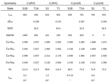
C1 and C2
Complexes C1 and C2 in their excited states are characterized by several conformers close in energy with similar emission wavelengths (table 6 and table 7). The singlet and triplet emission wavelengths generated by the structures of C1 symmetry disagree
with the experimental data. The same conclusion can be drawn for the emission from the structures of C2 symmetry in their triplet
state. However, the emission wavelength computed for the S1B
singlet states of C2 symmetry is in very good agreement with the
experiment regardless of the considered conformer RX or conf. As for [Cu(dpp)2]+, C1 and C2 present a flattening in the ground
state (Table 3) due to intramolecular π-stacking interactions. The extra flattening generated by the excitation is small but larger for the RX structures (around 7°) than for the conf structures (around 3°).
Table 5. Emission wavelength (in nm), Singlet-Triplet energy splitting (EST in
eV) at the geometry of the triplet state and Spin-Orbit Coupling (SOC in cm-1) at
the same geometry for [Cu(dpp)2]+ complex in the D2 and C2 point groups,
energetic barrier (in cm-1) between the D
2 geometry and the C2 minima,
flattening of structure (z in degrees) and oscillator strength of the singlet state.
Symmetry D2 C2
State S1B1 T1A S1A T1A
em 698 905 754 949 EST 0.331 0.353 SOC 15.3 49.5 Barrier 252 445 0 0 Cu-NA 2.041 2.007 1.995 1.988 Cu-NB 2.041 2.007 2.106 2.055 z -120.7 -121.3 -121.0 -120.9 fosc 6.3 10-2 2.8 10-2
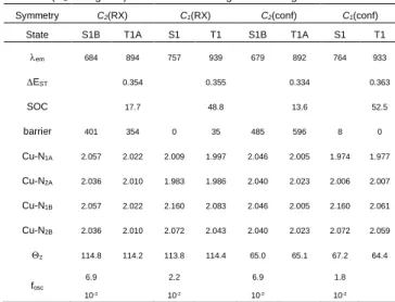
Table 6. Emission wavelength (in nm), Singlet-Triplet energy splitting (EST in
eV) at the geometry of the triplet state and Spin-Orbit Coupling (SOC in cm-1) at
the same geometry for C1 complex in the C2 and C1 point groups, energetic
barrier (in cm-1) between all geometries and the RX C
1 minima, flattening of
structure (z in degrees) and oscillator strength of the singlet state.
Symmetry C2(RX) C1(RX) C2(conf) C1(conf)
State S1B T1A S1 T1 S1B T1A S1 T1 em 684 894 757 939 679 892 764 933 EST 0.354 0.355 0.334 0.363 SOC 17.7 48.8 13.6 52.5 barrier 401 354 0 35 485 596 8 0 Cu-N1A 2.057 2.022 2.009 1.997 2.046 2.005 1.974 1.977 Cu-N2A 2.036 2.010 1.983 1.986 2.040 2.023 2.006 2.007 Cu-N1B 2.057 2.022 2.160 2.083 2.046 2.005 2.160 2.061 Cu-N2B 2.036 2.010 2.072 2.043 2.040 2.023 2.072 2.059 z 114.8 114.2 113.8 114.4 65.0 65.1 67.2 64.4 fosc 6.9 10-2 2.2 10-2 6.9 10-2 1.8 10-2
The asymmetry in the substituents of the phenanthroline ligand is reflected in the different Cu-N distances. For C1, the Cu-N1
distances are longer than the Cu-N2 distances. This is reversed
for C2. This is easily explained; in C2 the longer Cu-N2 distance
is due to the steric repulsion generated by the tert-butyl moiety which is larger than isopropyl. The evolution of these distances in
C1 and C2 in the excited state depends if the molecule is excited
from the RX or from the conf structure. The most noticeable point is the evolution of the Cu-N2B distances in the conf conformer of
D2 C2
Accepted
FULL PAPER
C2. It rises up to 2.4 Å in the C1 point group symmetry (Table 7)
potentially opening the way for partial ligand decoordination. Table 7. Emission wavelength (in nm), Singlet-Triplet energy splitting (EST in
eV) at the geometry of the triplet state and Spin-Orbit Coupling (SOC in cm-1) at
the same geometry for C2 complex in the C2 and C1 point groups, energetic
barrier (in cm-1) between all geometries and the RX C
1 minima, flattening of
structure (z in degrees) and oscillator strength of the singlet state.
Symmetry C2(RX) C1(RX) C2(conf) C1(conf)
State S1B T1A S1 T1 S1B T1A S1 T1 em 662 830 815 925 650 781 793 910 EST 0.339 0.270 0.287 0.250 SOC 35.5 63.4 32.9 56.9 barrier 1460 891 421 239 933 823 0 0 Cu-N1A 2.096 2.057 1.995 2.002 2.006 1.982 1.980 1.971 Cu-N2A 2.045 2.027 1.994 2.003 2.149 2.138 1.993 1.995 Cu-N1B 2.096 2.057 2.212 2.176 2.006 1.982 2.057 2.026 Cu-N2B 2.045 2.027 2.130 2.060 2.149 2.138 2.421 2.390 z 111.0 111.5 98.8 113.4 68.3 71.6 74.8 72.0 fosc 5.2 10-2 1.5 10-3 5.6 10 -2 6.9 10-3
The presence of a flexible non symmetrical ligand generates several minima on the lowest excited singlet and ground and lowest excited singlet and triplet states PES. This conformational flexibility has to be taken into account to reproduce the experimental result, as the different conformers will be present in the solution due to their close stability. This affects the calculation of the absorption and emission spectra and may be the source of the large band observed for the emission. The excitation can localize on one of the two ligands. However, the energy of the symmetric structure, which is the transition state between the two minima, is so low that in reality the complexes freely oscillate in between those two minima. As a consequence, in average the structure is symmetric and explains the very good agreement between the computed and experimental emission wavelengths for the symmetric structures for all complexes. The value computed for the triplet-singlet splitting (EST), between 0.3-0.4
eV and the spin-orbit coupling (SOC) values are compatible with the TADF mechanism but from a structure that is not an energy minimum. From this point of view the rather large ΔEST is
compensated not only by SOC but also by vibronic coupling effects.[27] The presence of phenyl rings on the phenanthroline
ligand induces a flattening of the structure in the ground state, which is almost identical in the excited states: no further flattening is observed. This invalidates the hypothesis that the non-symmetrical nature of the ligands is responsible for extra flattening of the coordination sphere upon excitation.
The computed energies of the excited state (see table ST5) correlate well with those measured by the tangent method whether the conf or RX structures are considered for C1 and C2. This confirms that increasing the steric burden around the copper ion allows indeed to rise the energy gap between S1 and the
ground state.
The theoretical results have shown that the decrease in absorption for [Cu(dpp)2]+, C1 and C2 is due to the initial flattening
of the complexes induced by the phenyl moieties. We also put in
evidence that dynamically the excited state is delocalized over two ligands. However, none of the discussed above characteristics are responsible for the emission intensity drop of
C1, C2 and [Cu(dpp)2]+ compared to [Cu(dipp)2]+. One last
movement has to be considered. Upon excitation, the initially D2d
[Cu(dipp)2]+ complex flattens generating two degenerated
conformers (see figure S18). We found a transition state connecting these two conformers on the lowest triplet PES with a relatively low associated energy barrier (computed G = 3.5 kcal.mol-1). At this geometry, in which
z is almost 90°, the
coupling between S0 and S1 is very small (it is strictly nil in the
ground state D2d structure). The emission of [Cu(dipp)2]+ is
relatively intense because it arises from a flattened conformation. The observed stabilizing interactions between the ligand for [Cu(dpp)2]+, C1 and C2 (which hold while shifting from one energy
well to the other) may lower this barrier so that dynamically, an orthogonal structure significantly contributes to the emission properties. As a consequence, the reverse intersystem crossing channel would be less favorable, decreasing the TADF efficiency and leading to the less intense emission for these complexes compared to that of [Cu(dipp)2]+ complex (for which such structure
is too high in energy).
Overall, complexes C1 and C2 stand out in the series of complexes by the fact that many different but equally probable structures are in dynamic equilibrium, and this opens many non-radiative pathways which entail a significant increase of knr and
thus weaker emission quantum yields.
Stability studies
Given the lability of the copper(I)-phenanthroline coordination sphere, the stability of the complexes is strongly impacted by the steric bulk around the metal ion. For example, [Cu(dtbp)2]+ is
quantitatively destroyed in presence of one equivalent of acetonitrile[5a] thanks to a strong steric relief. We thus began by
recording the 1H NMR spectrum of C1 and C2 in deuterated
acetonitrile. C1’s spectrum featured the expected signals of the homoleptic complex, and the spectra remained unchanged for several weeks (figure S19). On the other hand, the 1H NMR
spectrum of C2 revealed the latter is in equilibrium with other species (figure S20). Indeed, the UV-Vis spectrum of C2 in dichloromethane is strongly affected by the addition of aliquots of CH3CN (figure S21), the MLCT significantly collapsing upon
increasing the acetonitrile’s concentration while some weak absorbance over the visible range confirms the formation of a [Cu(L2)(CH3CN)]+ species following equation 2:
[Cu(L2)2]+ + 2CH3CN → [Cu(L2)(CH3CN)2]+ + L2 (equation 2)[19, 28]
This is not unexpected since the tert-butyl group leads to longer Cu-N2A and Cu-N2B distances and limits the extent of stabilizing
intramolecular π-stacking.
Interestingly, C1’s spectrum was slightly affected by acetonitrile too revealing some instability for dilute solutions, while [Cu(dipp)2]+ and [Cu(dpp)2]+ spectra remained roughly idle in the
same conditions (figures S22 and S23). This prompted us to inquire further about the behaviour of our complexes in presence of nucleophiles.
Accepted
FULL PAPER
Stopped flow analysisMonitoring the real time decomplexation of copper(I) complexes by cyanide by stopped flow is a valuable tool to study the accessibility of copper(I) towards nucleophilic attacks, and how shielded is the latter by the groups tethered in α of the nitrogen atoms of phenanthroline. The process is based on the chemical equation:
Cu(NN)2+ + 4 CN- →Cu(CN)43- + 2NN (equation 3)
The huge complexation constant of Cu(CN)43- (1028 vs. 1010-12 for
Cu(NN)2+ complexes) is the main driving force for this quantitative
demetallation reaction.[29] Complexes bearing non-symmetrical
phenanthroline ligands have previously shown enhanced kinetics towards cyanide assisted demetallation compared to symmetrical systems.[19] Willing to assess if the behaviour of C1 and C2 in
those conditions would follow the same trend, the decay of the MLCT with time in presence of excess CN- in a 1:1 (v/v)
dichloromethane:acetonitrile mixture was monitored by stopped flow. [Cu(dipp)2]+ and [Cu(dpp)2]+ were studied in the same
conditions for comparison purpose.
C2 proved to be too unstable in the solvent mixture and was
therefore not further examined. In all other cases, decays are monoexponential with measured decay constants kobs (figure
S24). In our experimental conditions (large excess of tetrabutylammonium cyanide) the degradation kinetics can be considered as pseudo first order in complex. Plots of kobs vs. [CN-]
yield straight lines (figure S25). The intersection with vertical axis affords the constant kD insensitive to [CN-] and assigned to the
natural dissociation kinetics of the complexes in the analysis medium.[18-19] The slopes of the straight lines yields the constant
kCN assigned to cyanide assisted demetallation. Significant values
of kCN indicate that cyanide is involved in the rate determining step
(figure S26). Constants are given in table 8 and experimental details can be found in the SI (table ST6).
Table 8. Dissociation rate constants of copper(I) bis(diimine) complexes C1, [Cu(dpp)2]+ and [Cu(dipp)2]+ in acetonitrile, with [Bu4NPF6] = 0.05 M; T = 25.0 ±
0.2 °C.
Complexes kD (s-1) kCN(M-1s-1)
Cu(dpp)2+ 0.976 ± 0.07 11.50 ± 0.08
Cu(dipp)2+ 0.30 ± 0.04 229 ± 4
C1 14 ± 5 8526 ± 551
In line with previously published results,[19, 29] [Cu(dpp)
2]+ is the
more resilient species vs. cyanide demetallation thanks to the intramolecular π-stacking interactions between the phenyl and the vicinal phenanthroline: the copper(I) ion is well protected in this rigid cavity, as incidentally evidenced by its good photophysical properties in strongly coordinating medium. [Cu(dipp)2]+ is deprived of stabilizing interactions and therefore
more prone to react with cyanide, as indicated by a ca. 20 times larger kCN compared to [Cu(dpp)2]+. As previously observed for
complexes bearing non-symmetrically substituted phenanthroline ligands, C1 features a larger kCN than [Cu(dpp)2]+ by almost two
orders of magnitude, revealing that the non-symmetrical nature of
L1 promotes cyanide nucleophilic attack on the copper ion.[19]
Less intuitively, kCN is also much larger for C1 than for [Cu(dipp)2]+
despite an intermediate steric hindrance. However, this is in line
with the behavior hypothesized in the literature: the non-symmetrical nature of the ligands provides a protected side by stacking of the phenyl group which simultaneously opens a site on the other side of the complexes, explaining their enhanced reactivity towards cyanide (and acetonitrile).
kD values reflect the fact that acetonitrile can behave as a
competitor ligand, leading to partial dissociation of the complexes. kD increases in the order [Cu(dipp)2]+ < [Cu(dpp)2]+ << C1: this
means that C1 dissociates faster than [Cu(dpp)2]+ and
[Cu(dipp)2]+ in acetonitrile, which is in line with the conclusions
from the cyanide induced decomplexation. The fact that [Cu(dpp)2]+ dissociates faster than [Cu(dipp)2]+ is unexpected; we
propose that the electron-withdrawing character of phenyl depletes the σ-donating power of dpp compared to dipp (where electron rich isopropyl groups have the opposite effect) and this could translate into the (slightly) faster dissociation of [Cu(dpp)2]+.
As a conclusion, the observation that complexes bearing non symmetrical phenanthroline are more prone to nucleophilic attack than classical complexes with symmetrically substituted ligands can be relevantly compared to their enhanced exciplex quenching. This fragility in the ground state is likely exacerbated in the excited state.
Reactivity
Using the Rehm and Weller equation, it is possible to estimate the reductive and oxidative power of photosensitizers promoted in their excited state, namely E(Cn+/Cn*) and E(Cn*/Cn-) (or E
ox*)
for C1 and C2. Considering the case of Eox*, the Rehm and Weller
equation gives:
Eox* = E(Cn/Cn-) + E00 (equation 4)
Where E(Cn/Cn-) is the reduction potential of complexes Cn (E
2
in table 1) and E00 is the energy of the excited state. Both C1 and
C2 exhibit more positive Eox* than [Cu(dpp)2]+, and comparable to
[Cu(dipp)2]+. Thus, the driving force for photo-induced electron
transfers is more favorable for C1 and C2 than for the model complex [Cu(dpp)2]+. In order to test this assertion, we decided to
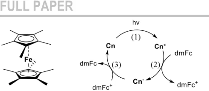
try and implement reductive quenching with C1 and C2 with decamethylferrocene (dmFc) as electron donor (figure 9). The latter is an excellent donor (E(dmFc+/dmFc) = -0.57 V vs. Fc+/Fc)
which has been successfully used by McMillin et al. in what are, to the best of our knowledge, the only articles ever reporting reductive quenching with homoleptic [Cu(NN)2]+ complexes
(namely [Cu(dipp)2]+ and [Cu(dpp)2]+).[10, 30] Indeed, the latter are
particularly poor photo-oxidants.[1a] Yet, successfully performing
reductive quenching with plain homoleptic copper(I) complexes is a valuable endeavor leading to the photo-generation of very reductive species [Cu(NN)2]0 (more precisely [CuI(NN)(NN•-)]0)
paving the way towards low-cost, visible light-activated photo-reduction processes of difficult substrates or catalysts. We however highlight that the reversibility of the dmFc+/dmFc couple
prevents accumulation of Cn- upon photoexcitation (figure 9, step
3).
Accepted
FULL PAPER
Figure 9. The reductive quenching cycle in the case of Cn (n = 1 or 2) as photosensitizer and dmFc as electron donor.
Using the same experimental protocol than McMillin et al.,[30] we
monitored the impact of increasing quantities of dmFc in dichloromethane solutions of C1, C2, [Cu(dpp)2]+ and [Cu(dipp)2]+
on their luminescence lifetime and extracted bimolecular quenching rates kQ using Stern-Volmer formalism (figure S27).
Importantly, the quenching of the excited state of all copper complexes may occur via two mechanisms: energy transfer (“EnT” from MLCT excited state to low lying iron(II) based dd states, kinetic constant kEnT) and electron transfer (from dmFc to excited
copper complexes, namely reductive quenching, kinetic constant kRQ). Thus, kQ = kRQ + kEnT. As previously demonstrated, kEnT can
be considered similar for plain ferrocene and dmFc since the energies of the final quencher d-d state are similar.[30-31] Electron
transfer processes between ferrocene and copper complexes being thermodynamically uphill, one can roughly estimate kEnT (kQ
= kEnT when the quencher is plain ferrocene Fc) by monitoring
copper complexes luminescence quenching by plain ferrocene (figure S28, table ST7). As previously observed, quenching by energy transfer is one order of magnitude less efficient than quenching by photo-induced electron transfer, making the latter the dominant phenomenon in presence of dmFc. Thus, we could estimate kRQ and the results are reported in table 2: C1 and C2’s
emissions are more efficiently quenched by dmFc than [Cu(dpp)2]+, meaning that C1 and C2 are more efficient
photo-oxidants than Cu(dpp)2+, kRQ being twice and three times larger
for C1 and C2 respectively than kRQ([Cu(dpp)2]+). Interestingly
kRQ(C1) is smaller than kRQ([Cu(dipp)2]+) or kRQ(C2) although
those three complexes have comparable photo-oxidative powers from the sole thermodynamic point of view (see Eox* table 2). This
is certainly due to kinetic factors. Indeed, the latter are believed to be particularly prominent for reductive quenching,[8c, 30] because
the complex shifts from a flattened excited state toward a tetrahedral reduced copper(I) complex (figure 9, step 2) whereas it shifts from a flattened excited state to a flattened oxidized copper(II) complex in the frame of oxidative quenching. The reorganization energy is thus supposed to be a particularly important factor to take into account here; given our results, C1 and C2 should thus display higher reorganization energies. However, computed distortion energies do not reflect the observed trend (table ST5) as [Cu(dipp)2]+ exhibits larger
reorganization energies than C1 and C2 for the computed states (due to the flattening movement contribution which is more prominent that in the three other complexes). Work is ongoing to rationalize this observation.
Conclusions
The use of non-symmetrical phenanthroline ligands bearing one phenyl and one ramified alkyl chain to respectively stabilize and sterically strain corresponding copper(I) complexes is reported. Tethering an isopropyl or a tert-butyl chain and a phenyl group in α of the nitrogen atoms allowed to implement intramolecular π stacking interactions and rise the complex excited state energy at the same time by ca. 100 meV compared to symmetrical [Cu(dpp)2]+. Calculations reveal that the stacking interactions lead
to a more rigid coordination sphere for C1 and C2, the extent of computed photo-induced flattening being smaller than for [Cu(dipp)2]+ where no stacking takes place. The stacking was
nevertheless not sufficient to stabilize the coordination sphere when using tert-butyl instead of isopropyl substituents, because of the strong steric strain imposed by the tertiary ramified alkyl chain.
The unsymmetrical nature of the phenanthroline ligand promotes nucleophilic attacks on the copper centre in C1 and C2 in their ground states. We extend this statement to the excited state, exciplex quenching being more efficient for non-symmetrical complexes, and thus rationalize their rather weak photophysical parameters (emission quantum yield and lifetime). Simulation of the emission wavelengths was achieved with TD-DFT calculations. Importantly, the fitting required to take into account that the electron in the MLCT excited state oscillates between the two ligands, leading to an overall symmetrical potential well. The increased energy of the excited state for C1 and C2 lead to better photo-oxidizing power, which materialized in improved reductive quenching kinetics compared to [Cu(dpp)2]+.
Experimental Section
General: chemicals were purchased from Sigma-Aldrich or Fisher Scientific and used as received. Thin-layer chromatography (TLC) was performed on aluminium sheets precoated with Merck 5735 Kieselgel 60F254. Column chromatography was carried out with Merck 5735 Kieselgel 60F (0.040-0.063 mm mesh). 1H spectra were recorded on an
AVANCE 300 UltraShield BRUKER. Chemical shifts for 1H NMR spectra
are referenced relative to residual protium in the deuterated solvent (CDCl3
δ = 7.26 ppm). NMR spectra were recorded at room temperature, chemical shifts are written in ppm and coupling constants in Hz. Mass spectrometry was performed with a JEOL JMS-700 B/E spectrometer. Electrochemical measurements were made under an argon atmosphere in CH2Cl2 with 0.1
M Bu4NPF6. Cyclic voltammetry experiments were performed by using an
Autolab PGSTAT 302N potentiostat/galvanostat. A standard three-electrode electrochemical cell was used. Potentials were referenced vs. reversible ferrocenium/ferrocene couple. The working electrode was a glassy carbon disk and the auxiliary electrode was a Pt wire. UV-visible absorption spectra were recorded on an Analytik Jena spectrophotometer, using 1 cm path length cells. Emission spectra were recorded on a Fluoromax-3 Horiba spectrofluorimeter (1 cm quartz cells). E00 values were measured by the tangent method: a tangent to the emission spectrum was drawn on the blue side of the latter and the intersection with the baseline gives wavelength λ00, used to calculate E00 = 1240/λ00.
Luminescence decays were recorded with a DELTAFLEX time correlated single photon counting system (HORIBA) on degassed dichloromethane solutions. X-ray single-crystal diffraction data for L1 were collected at 150K on a Rigaku Oxford Diffraction SuperNova diffractometer equipped with Atlas CCD detector and micro-focus Cu-Kα radiation (λ = 1.54184 Å). The structure was solved by direct methods and refined on F2 by full matrix least-squares techniques using SHELX programs (G. M. Sheldrick
2013-Accepted
FULL PAPER
2016, SHELXS 2013/1 and SHELXL 2016/4). All non-H atoms were refined anisotropically and multiscan empirical absorption was corrected
using CrysAlisPro program (CrysAlisPro, Agilent Technologies,
V1.171.38.46, 2015). The H atoms were included in the calculation without refinement. CCDC-1972177 contains the supplementary crystallographic data for this paper. Crystallographic data: C22H20N2, M = 312.40, colorless
prism, 0.326 x 0.298 x 0.254 mm3, orthorhombic, space group P212121, a = 9.7972(1) Å, b = 11.6086(2) Å, c = 15.1268(2) Å, V = 1720.40(4) Å3, Z = 4, ρcalc = 1.206 g/cm3, μ = 0.543 mm-1, F(000) = 664, θmin = 4.802 °, θmax = 76.289°, 6751 reflections collected, 3339 unique (Rint = 0.0252), parameters / restraints = 221 / 0, R1 = 0.0419 and wR2 = 0.1117 using 3278 reflections with I>2σ(I), R1 = 0.0445 and wR2 = 0.1155 using all data, absolute structure parameter = 0.2(3), GOF = 1.041, -0.250 < Δρ < 0.387 e.Å-3. X-ray single-crystal diffraction data for C2 were collected at 150K on a D8 VENTURE Bruker AXS diffractometer equipped with a (CMOS) PHOTON 100 detector, Mo-Kα radiation (λ = 0.71073 Å, multilayer monochromator). Crystallographic data: (C44H40CuN4•F6P•2(C7H8)); M =
1017.57. monoclinic P 21/n (I.T.#14), a = 14.9886(15), b = 19.446(2), c = 17.4437(15) Å, β = 105.329(3) °, V = 4903.3(8) Å3. Z = 4, d = 1.378 g.cm-3, μ = 0.545 mm-1. The structure was solved by dual-space algorithm using the SHELXT program,[32] and then refined with full-matrix
least-squares methods based on F2 (SHELXL).[33] All non-hydrogen atoms were
refined with anisotropic atomic displacement parameters. H atoms were finally included in their calculated positions and treated as riding on their parent atom with constrained thermal parameters. A final refinement on F2 with 11112 unique intensities and 639 parameters converged at ωRF2 = 0.1231 (RF = 0.0500) for 8360 observed reflections with I > 2σ(I). Stopped Flow absorption spectrophotometry was performed on a BioLogic SFM-4000 coupled to a J&M Tidas diode array spectrometer. Experiments were at least triplicated for kinetic fits. Monoexponential fits were performed using the Biokine software (BioLogic).
2-(tert-butyl)-1,10-phenanthroline (1) Under argon, the 1,10-phenanthroline (1.00 g, 5.56 mmol) is dissolved into dry toluene (48 mL). The solution is cooled to 0°C before addition dropwise of tert-butyllithium (1.7 M, 4.9 mL). The resulting solution was allowed to warm to room temperature and stir for 18 h. Water (10 mL) was then added followed by a dichloromethane extraction. Organics phases are stirred with activated manganese dioxide (12.00 g, 139.00 mmol) for 6h. The mixture was dried over magnesium sulfate, filtrated and evaporated. The orange oil is chromatographed on silica column (0 to 5% of methanol in dichloromethane) to obtain 931 mg (71%) of the product. 1H NMR
spectrum (300MHz, CDCl3) δ= 9.23 (dd, 4JH,H=1.8Hz, 3JH,H=4.5Hz, 1H), 8.24 (dd, 4JH,H=1.8Hz, 3JH,H = 8.1Hz, 1H), 8.19 (d, 3JH,H = 8.4Hz, 1H), 7.78 (m, 3H), 7.62 (dd, 3JH,H= 4.5Hz, 3JH,H= 8.1Hz, 1H), 1.61 (s, 9H). 13C NMR spectrum (75MHz, CDCl3) δ= 169.80 (s), 150.38 (s), 146.53 (s), 145.00 (s), 136.10 (s), 128.93 (s), 126.72 (s), 126.41 (s), 125.62 (s), 122.43 (s), 119.95 (s), 38.66 (s), 30.51 (s). HRMS (ESI+) for C16H16N2 [M+H]+, m/z 237.1399 found, 237.1392 calc.
2-isopropyl-1,10-phenanthroline (2) was synthesized in a similar manner to that of (1) using the following reagents: 1,10-phenanthroline (200 mg, 1.11 mmol), isopropyllithium (0.7 M, 2.4 mL) and manganese dioxyde (2.40 g, 27.8 mmol). Yield: 174 mg (71%). 1H NMR spectrum
(400MHz, CDCl3) δ= 9.25 (dd, 4JH,H = 1.6Hz, 3JH,H = 4.4Hz, 1H), 8.25 (m, 2H), 7.78 (AB, JAB= 15.6Hz, 2H), 7.64 (m, 2H), 3.69 (sept, 3JH,H = 7.2Hz, 1H), 1.49 (d, 3JH,H = 7.2Hz, 6H). 13C NMR spectrum (100MHz, CDCl 3) δ= 206.82 (s), 150.34 (s), 145.41 (s), 136.58 (s), 135.99 (s), 126.47 (s), 125.56 (s), 122.61 (s), 120.05 (s), 37.71 (s), 30.89 (s), 23.16 (s). HRMS (ESI+) for C 15H14N2 [M+H]+, m/z 223.1243 found, 223.1235 calc.
2-isopropyl-9-phenyl-1,10-phenanthroline (L1) was synthesized in a similar manner to that of (1) using the following reagents: (2) (174 mg, 0.78 mmol), phenyllithium (1.9 M, 0.62 mL) and manganese dioxide (1.69 g, 19.50 mmol). Yield: 224 mg (96%). All characterization data are in agreement with the ones published by Kavita et al.[11]
2-(tert-butyl)-9-phenyl-1,10-phenanthroline (L2) was synthesized in a similar manner to that of (1) using the following reagents: (1) (100 mg, 0.42 mmol), phenyllithium (2.0 M, 0.32 mL) and manganese dioxide (912 mg, 10.5 mmol). Yield: 117 mg (89%). 1H NMR spectrum (300MHz, CDCl
3) δ= 8.48 (m, 2H), 8.30 (d, 3JH,H = 8.7Hz, 1H), 8.18 (d, 3JH,H = 8.7Hz, 1H), 8.15 (d, 3JH,H = 8.4Hz, 1H), 7.77 (m, 3H), 7.57 (m, 2H), 7.47 (m, 1H), 1.64 (s, 9H). 13C NMR spectrum (75MHz, CDCl 3) δ= 156.34 (s), 139.43 (s), 136.84 (s), 136.05 (s), 129.39 (s), 128.82 (s), 127.42 (s), 126.99 (s), 126.08 (s), 125.34 (s), 120.08 (s), 119.24 (s), 38.76 (s), 30.36 (s). HRMS (ESI+) for C22H20N2 [M+H]+, m/z 313.1714 found, 313.1705 calc.
Anal. Calcd for C22H20N2 .0.05 CH2Cl2 . 0.05 C5H12 : C, 83.63; H, 6.51; N,
8.75. Found: C, 83.31; H, 6.19; N, 8.53.
[Cu(L1)2]PF6 (C1) Under argon atmosphere, [Cu(ACN)4]PF6 (58 mg,
0.16mmol) was dissolved in 4mL of degassed dichloromethane. This solution was transferred into a vial that contains L1 (92 mg, 0.31mmol). The red resulting solution was stirred overnight at room temperature. The mixture was precipitate in hexanes to obtain a red powder (100%, 248mg)
1H NMR spectrum (300MHz, CDCl 3) δ= 8.59 (d, 3JH,H= 8.4Hz, 2H), 8.52 (d, 3JH,H= 8.4Hz, 2H), 8.04 (s, 4H), 7.94 (d, 3JH,H= 8.4Hz, 2H), 7.72 (d, 3JH,H= 8.4Hz, 2H), 7.24 (m, 4H), 6.69 (t, 3JH,H= 7.5Hz, 2H), 6.37 (d, 3JH,H= 7.8Hz, 4H), 3.21 (t, 3JH,H = 6.9Hz, 2H), 1.13 (d, 3JH,H= 7.2Hz, 6H), 0.92 (d, 3JH,H= 6.9Hz, 6H). 13C NMR spectrum (125MHz, CDCl 3) δ= 166.42 (s), 157.67 (s), 143.77 (s), 142.85 (s), 139.03 (s), 137.85 (s), 137.81 (s), 128.63 (s), 128.38 (s), 128.26 (s), 127.36 (s), 127.10 (s), 126.81 (s), 126.19 (s), 124.83 (s), 121.89 (s), 39.57 (s), 29.70 (s), 22.67 (s), 21.91 (s). HRMS (ESI): m/z calcd for C42H36CuN4+ : 659.2236 [M]+; found 659.2250.
UV/Vis(CH2Cl2): MLCT ʎmax(ε)= 443 nm (3987 L.cm-1.mol-1); fluorescence
(CH2Cl2): λex= 443nm; λem= 698nm.
Anal. Calcd for C42H36CuF6N4P.0.9 CH2Cl2.0.15 C5H12: C, 58.78; H, 4.47;
N, 6.28. Found C, 58.56; H, 4.82; N, 6.66.
[Cu(L2)2]PF6 (C2) was synthesized in a similar manner to that of (C1)
using the following reagents: [Cu(ACN)4]PF6 (28 mg, 0.075mmol) and L1
(47 mg, 0.15mmol). Yield: (100%, 125mg). 1H NMR spectrum (300MHz,
CDCl3) δ= 8.60 (d, 3JH,H= 8.4Hz, 2H), 8.47 (d, 3JH,H= 8.7Hz, 2H), 8.07 (AB, JAB = 12.6Hz, 4H), 7.99 (d, 3JH,H= 8.7Hz, 2H), 7.87 (d, 3JH,H= 8.1Hz, 2H), 7.14 (m, 4H), 6.65 (m, 2H), 6.32 (m, 4H), 1.24 (s, 18H). 13C NMR spectrum (75MHz, CDCl3) δ= 168.57 (s), 157.28 (s), 143.09 (s), 139.21 (s), 137.95 (s), 137.80 (s), 128.85 (s), 128.56 (s), 128.38 (s), 127.50 (s), 126.90 (s), 126.55 (s), 125.39 (s), 124.21 (s), 38.52 (s), 30.56 (s). HRMS (ESI): m/z calcd for C44H40CuN4+ : 687.2549 [M]+; found 687.2541. UV/Vis (CH2Cl2):
MLCT ʎmax(ε)= 450 nm (3851 L.cm-1.mol-1); fluorescence (CH2Cl2): λex=
450nm; λem= 695nm.
Anal. Calcd for C44H40CuF6N4P.0.45 CH2Cl2: C, 61.26; H, 4.73; N, 6.43.
Found: C, 61.27; H, 4.85; N, 6.50.
Computational details All calculations were performed using ADF 2019[34] package at the DFT level of theory with B3LYP functional.[35] The
all-electrons slater type TZP basis set described all atoms.[36] Scalar
relativistic ZORA Hamiltonian was employed.[37] Van der Waals forces
were treated through the introduction of Grimme’s corrections (grimme3).[38] Solvent corrections for dichloromethane were introduced
through a PCM model.[39] Structures were fully optimized and the
absorption spectrum was computed by mean of TD-DFT[40] on these
structures with the inclusion of the Tamm-Dancoff approximation.[41]
Spin-Orbit Coupling was computed by a perturbative approach. The excited states (singlet or triplet) structures were computed by the same approach. All calculations were first performed retaining the symmetry point group of the ground state. Then symmetry was broken to allow complete relaxation of the structures. For the study of Non-Covalent interactions, the complexes were reoptimized with GAUSSIAN 09 (D01)[42] package at DFT
level of theory (B3LYP functional)[43] using 6-31+G** basis set[44] for all
atoms (the f polarization were deleted). Again, dispersion corrections were introduced through Grimme’s correction GD3 and solvent introduced through a PCM model of dichloromethane. These calculations were performed on the ground state and on the lowest triplet state of the
![Figure 4. Electronic absorption spectra for complexes C1 (black trace), C2 (blue trace), [Cu(dipp) 2 ] + (green trace) and [Cu(dpp) 2 ] + (red trace) recorded in dichloromethane at room temperature](https://thumb-eu.123doks.com/thumbv2/123doknet/8195434.275237/4.892.479.817.183.398/figure-electronic-absorption-spectra-complexes-recorded-dichloromethane-temperature.webp)
![Table 1. UV-Vis and electrochemical data for complexes C1, C2, [Cu(dpp) 2 ] + and [Cu(dipp) 2 ] + in dichloromethane](https://thumb-eu.123doks.com/thumbv2/123doknet/8195434.275237/5.892.71.434.196.330/table-uv-vis-electrochemical-data-complexes-dipp-dichloromethane.webp)
![Table 3. Geometric parameters of the ground state of [Cu(dipp) 2 ] + , [Cu(dpp) 2 ] + , C1 and C2 complexes and of their conformers](https://thumb-eu.123doks.com/thumbv2/123doknet/8195434.275237/7.892.76.422.259.683/table-geometric-parameters-ground-state-dipp-complexes-conformers.webp)
![Table 8. Dissociation rate constants of copper(I) bis(diimine) complexes C1, [Cu(dpp) 2 ] + and [Cu(dipp) 2 ] + in acetonitrile, with [Bu 4 NPF 6 ] = 0.05 M; T = 25.0 ± 0.2 °C](https://thumb-eu.123doks.com/thumbv2/123doknet/8195434.275237/10.892.71.406.792.880/table-dissociation-rate-constants-copper-diimine-complexes-acetonitrile.webp)