HAL Id: dumas-01891152
https://dumas.ccsd.cnrs.fr/dumas-01891152
Submitted on 9 Oct 2018HAL is a multi-disciplinary open access archive for the deposit and dissemination of sci-entific research documents, whether they are pub-lished or not. The documents may come from teaching and research institutions in France or abroad, or from public or private research centers.
L’archive ouverte pluridisciplinaire HAL, est destinée au dépôt et à la diffusion de documents scientifiques de niveau recherche, publiés ou non, émanant des établissements d’enseignement et de recherche français ou étrangers, des laboratoires publics ou privés.
Serum amyloid A as a marker of disease activity in giant
cell arteritis
Anaïs Dartevel
To cite this version:
Anaïs Dartevel. Serum amyloid A as a marker of disease activity in giant cell arteritis. Human health and pathology. 2018. �dumas-01891152�
AVERTISSEMENT
Ce document est le fruit d'un long travail approuvé par le
jury de soutenance et mis à disposition de l'ensemble de la
communauté universitaire élargie.
Il n’a pas été réévalué depuis la date de soutenance.
Il est soumis à la propriété intellectuelle de l'auteur. Ceci
implique une obligation de citation et de référencement
lors de l’utilisation de ce document.
D’autre part, toute contrefaçon, plagiat, reproduction illicite
encourt une poursuite pénale.
Contact au SID de Grenoble :
bump-theses@univ-grenoble-alpes.fr
LIENS
LIENS
Code de la Propriété Intellectuelle. articles L 122. 4
Code de la Propriété Intellectuelle. articles L 335.2- L 335.10
http://www.cfcopies.com/juridique/droit-auteur
1
UNIVERSITÉ GRENOBLE ALPES UFR DE MÉDECINE DE GRENOBLE
Année : 2018
SERUM AMYLOID A AS A MARKER OF DISEASE ACTIVITY
IN GIANT CELL ARTERITIS
THÈSE
PRÉSENTÉE POUR L’OBTENTION DU TITRE DE DOCTEUR EN MÉDECINE DIPLÔME D’ÉTAT
Anaïs DARTEVEL
THÈSE SOUTENUE PUBLIQUEMENT À LA FACULTÉ DE MÉDECINE DE GRENOBLE Le 5 octobre 2018
DEVANT LE JURY COMPOSÉ DE
Président du jury : Mme. le Professeur Laurence BOUILLET, directrice de thèse Membres :
M. le Professeur Christophe CHIQUET M. le Professeur Patrice FAURE
M. le Professeur Bertrand TOUSSAINT M. le Docteur Maxime LUGOSI
Mme. le Docteur Candice TROCME
L’UFR de Médecine de Grenoble n’entend donner aucune approbation ni improbation aux opinions émises dans les thèses ; ces opinions sont considérées comme propres à leurs auteurs.
6
REMERCIEMENTS :
Mme. le Professeur Laurence BOUILLET, directrice de thèse
Merci Laurence de me faire l’honneur de présider ce jury. Merci de m’avoir guidé tout au long de ma formation et de m’avoir soutenu dans mes choix professionnels. Merci de m’avoir encadrer pour ce travail de thèse.
M. le Professeur Christophe CHIQUET
Merci de participer à ce jury et de me faire l’honneur de juger mon travail. Merci au service d’ophtalmologie pour son implication dans l’inclusion des patients. Veuillez recevoir l’expression de ma sincère gratitude et de mon profond respect.
M. le Professeur Patrice FAURE
Merci de participer à ce jury et de me faire l’honneur de juger mon travail. Veuillez recevoir l’expression de ma sincère gratitude et de mon profond respect
M. le Professeur Bertrand TOUSSAINT
Merci de votre aide et de vos conseils pour mettre en route ce travail de thèse. Veuillez recevoir l’expression de ma sincère gratitude et de mon profond respect.
M. le Docteur Maxime LUGOSI
Merci pour tes conseils et le partage de tes connaissances en médecine. Merci de ton soutien tout au long de l’internat, notamment moral avec nos séances de discussion le soir dans le service mais même après à chaque fois que je frappais à ton bureau. Tu as toujours été disponible pour moi et je t’en remercie.
Mme. le Docteur Candice TROCME
Merci de votre aide dans la dernière ligne droite de ce travail. Merci de participer à ce jury et de me faire l’honneur de juger mon travail. Veuillez recevoir l’expression de ma sincère gratitude et de mon profond respect.
A Adèle, qui me remplit de bonheur depuis sa venue au monde.
A Benjamin, qui à la force de me supporter et de me soutenir tous les jours. A notre amour. A mes parents, merci pour le soutien que vous m’avez toujours apporté. Je n’y serais jamais arrivé sans vous.
A mon frère, Adrien, merci de ta présence à mes côtés. Merci d’avoir agrandi la famille avec des personnes formidables, Marie-Anne et Gatien.
A Aline, pour nos séances papotages et ragots. A mes beaux-parents.
7
Au Dr Dols, merci de m’avoir aidé pour les statistiques.
A Gaëtan, qui m’a fait apprécier la médecine et la clinique avant tout !
A Annick et Barbara, merci pour mon semestre passé au 3ème C et de m’avoir enseigné vos connaissances en infectiologie.
A Laure, merci de m’avoir tant appris sur la néphrologie et merci de nos soirées sushis dans le service.
Merci aux médecins de pneumologie (Sébastien, Christel, le Pr Pison) mais également mes co-internes (Louis-Marie et Pauline) pour m’avoir appris à manier la VNI.
Au Pr Mouthon, merci pour m’avoir accepter dans son service et pris sous son aile pour mon travail de mémoire de DES.
Au Dr Besson, merci pour m’avoir tant appris sur les maladies métaboliques.
A Olivier et Mathieu, merci de m’avoir fait connaître les maladies inflammatoires neurologiques.
A Carole et Nicolas, merci de m’avoir pris sous leurs ailes et soutenu lorsque j’ai décidé de faire le DESC de réanimation.
A Agnès, Clémence, Rébecca et Claire, merci de m’avoir fait apprécier la réanimation médicale.
A toute l’équipe de réanimation de Chambéry où j’aurais appris que la traumatologie peut également être intéressante…
Merci à toutes les secrétaires rencontrées lors de mon internat, notamment Suzanne, pour supporter mes dictées très très rapides et parfois incompréhensibles.
A Julie et Flore, merci pour votre soutien et amitié lors de la P1.
A Clotilde et Estelle, merci pour les années fac passées avec vous et pour vos conseils maternité et suite de couche.
A Juliette et Mathilde, merci pour votre soutien en D4.
A Clémence et Florent, merci pour vos avis de pharmacologies.
A Alexis, merci pour avoir enfin trouver quelqu’un qui est plus potins (et LDP) que moi, à Thibaud, pour avoir essayer de m’apprendre l’intubation.
A Brune, merci pour nos moments détente après la tension ambiante au 3ème A.
A mes co-internes rencontrées en neurologie, Pauline, pour nos papotages autour du thé, à Fanny, pour nos balades avis neurologie, à Anne, malgré des débuts difficiles une très belle rencontre.
8
A mes co-internes de réanimation, pour s’être soutenu et surtout très amusé durant ce semestre. A mes co-internes et médecin de Cochin, plus particulièrement Caroline et Marion, pour m’avoir accueillit et m’avoir fait oublier le manque de montagne à Paris.
A Amélie, merci d’avoir remplit de discussion en tout genre les 6 mois de trajet train.
A tous mes co-internes de médecine interne, Chloé, Aldric, Catherine, Aude, Aurélie, Romain, Nicolas, Salomé, Mélodie, Benoit.
A Nicolas, merci de m’avoir aider dans ce travail, sans toi les patients auraient été perdus.. A Florian et Céline, merci pour les soirées jeux et escape games.
A Claire, Mathieu, Bénédicte, Sylvain et Michèle, merci pour m’avoir soutenu pendant mes quelques mois au laboratoire.
9
SERUM AMYLOID A AS A MARKER OF DISEASE ACTIVITY IN GIANT CELL ARTERITIS
RÉSUMÉ :
Introduction: L'artérite à cellules géantes (ACG) est une vascularite systémique avec un taux de rechute important et parfois des complications sévères. Le sérum amyloïde A (SAA) est une protéine de la phase aigue de l’inflammation et pourrait intervenir dans sa pathogénie. L'objectif de cette étude est de déterminer si le SAA est un marqueur d’activité de la maladie.
Méthodes: Entre octobre 2015 et janvier 2018, les patients présentant un diagnostic ou une rechute de ACG ont été inclus. Le dosage de SAA a été réalisé à l’inclusion puis tous les 3 mois pendant un an. A chaque point de l’étude les patients ont été séparés en 3 groupes selon leur statut clinique: maladie active, maladie inactive ou infection.
Résultats: Vingt patients ont été inclus: 15 nouvellement diagnostiqués et 5 rechutes. Il y a eu 21 situations cliniques avec une maladie active, 46 avec une maladie inactive et 7 infections. Le taux de SAA était significativement plus élevé avec une maladie active (84.5, IR: 30.7-220 mg/l) par rapport à une maladie inactive (23.85, IR: 14.6-35.3 mg/l), p = 0.01. Il n'y avait pas de différence statistique entre les taux de SAA entre maladie active et infections, p = 0.09.
Conclusion: Nous avons montré que le SAA est un marqueur d’activité de la maladie. La deuxième étape serait de déterminer si le SAA est plus spécifique que la vitesse de sédimentation des érythrocytes ou la protéine C réactive et pourrait donc constituer un test de diagnostic potentiel.
MOTS CLÉS :Artérite à cellules géantes, sérum amyloide A FILIÈRE : médecine interne
10
Abstract
Introduction: Giant cell arteritis (GCA) is a systemic vasculitis with important relapses and sometimes severe complications. Serum amyloid A (SAA) is a protein of the acute phase of inflammation and could be involved in the pathophysiology of GCA. The aim of this study is determining if SAA is a marker of disease activity.
Methods: Patients with diagnosis or relapse of GCA were included between October 2015 and January 2018 in our center. SAA dosage was realized at baseline then every 3 months for one year. Along the study, patients were separated in 3 groups according to clinical status: active disease, inactive disease or infections.
Results: Twenty patients were included: 15 newly diagnosed and 5 relapses. There have been 21 clinical situations with active disease, 46 with inactive disease and 7 infections. SAA level were significantly higher during active disease (84.5, IR: 30.7-220 mg/l) compared with patients in inactive disease (23.85, IR: 14.6-35.3 mg/l), p = 0.01. There were no statistically difference between SAA level in active disease and infections, p = 0.09.
Conclusion: We have shown that SAA is a marker of disease activity. The second step is determinate if SAA is more specific than erythrocyte sedimentation rate or C-protein reactive and could be a potential diagnostic test.
11
Introduction
Giant cell arteritis (GCA) is a systemic vasculitis affecting large and medium-sized blood vessels characterized by the granulomatous involvement of the aorta and its major branches [1]. It is a rare disease that touches person over 50 with an incidence increasing with age and a peak between 70 and 79 years old [2]. GCA affects more often women [2]. Typical clinical signs are unusual headaches, constitutional symptoms, jaw claudication, scalp tenderness and abnormal temporal arteries [2-4]. GCA can be associated with polymyalgia rheumatic (PMR) in around 50% of cases [3]. Morbidity is linked to visual impairment due to ischemic complications, but also to cerebrovascular accidents and aortic aneurism [4]. Mortality is increased in the early years of disease mainly from aortic dissection or heart disease [5,6]. The first-line treatment is glucocorticoids [3] but recently tocilizumab, an anti-IL-6 receptor, showed its effectiveness [7,8]. Despite treatment, relapses are frequent [9].
The pathophysiology can be divided into 3 phases: dendritic cells activation, recruitment, activation and polarization of T cells then recruitment of macrophages and vascular remodeling [10-12]. After dendritic activation, T lymphocytes are recruited and polarized in Th1 and Th17 with IFN gamma and IL-17 cytokine secretion [10-12]. Then IFN gamma recruits and activates macrophages [10-12]. IL-17 induces pro-inflammatory cytokine production by macrophages such as TNF-alpha, IL-6 and IL-1 [10-12]. Macrophage chronic activation is responsible of giant cells. Next, these cells product metalloproteinase (MMP), platelet-derived growth factor and vascular endothelial growth factor which contribute to internal elastic media destruction, proliferation, migration of myofibroblasts and neoangiogenesis [10-12]. Recently, one study has found a key role of acute serum amyloid A (A-SAA) in GCA pathophysiology. Indeed, they proved that A-SAA is directly secreted in inflammatory temporal artery [13]. A-SAA treatment in temporal artery explants induces IL-6 and IL-8 secretion and myofibroblasts outgrowth, MMP-9 activation and angiogenic tube formation [13].
12
Serum amyloid A (SAA) is an apolipoprotein encoded by 4 genes: SAA1, SAA2, SAA3 and SAA4 [14-15]. Proteins coded by SAA1 and 2 are acute phase protein SAA (A-SAA) because the serum concentration of these SAA isotypes can be increase up to 1000 times in cases of inflammation [16]. A-SAA synthesis is doing mainly by hepatocytes but also by different types of cells during inflammation including macrophages. A-SAA rapidly binds to high-density lipoprotein (HDL) [14-15]. This synthesis is induced by cytokines synergy (IL-1β, IL-6 and TNF-alpha), toll like receptors (TLR) or glucocorticoids [14-15]. Its part in inflammation is controversy but A-SAA can stimulate the expression of pro and anti-inflammatory cytokines and chemokines principally via binding to TLR2 [14-15]. A-SAA has proangiogenic properties via upregulation of vascular endothelial growth factor receptor 2 and induction of cell growth and angiogenesis mediated by TLR2 [17-18]. A-SAA plays also a part in atherosclerosis and cardiovascular disease [19-20].
SAA can be implicating in GCA pathogenesis, so this protein could be a potential marker of disease activity. The objective of this study was to explore SAA as marker of disease activity in GCA.
13
Methods Study design
This prospective study took place between October 2015 and January 2018 at the Grenoble University Hospital in internal medicine, ophthalmology and neurology services. The Grenoble University Hospital Ethics Committee approved the study. Inclusion criteria were newly diagnosed or relapse of GCA, diagnosis according to American College of Rheumatology classification with at least 3 criteria amount 5: age older or equal than 50, new onset of localized headache, temporal artery tenderness or decreased temporal artery pulse, elevated erythrocyte sedimentation rate (ESR) more important or equal than 50 mm/hour, and biopsy sample including an artery, showing necrotizing arteritis, characterized by a predominance of mononuclear cell infiltrates or a granulomatous process with multinucleated giant cells. If temporal arteritis biopsy was normal, GCA diagnosis had to be validated by an independent committee. Relapse is defined by a clinic relapse with clinical feature recidivism with or without inflammatory syndrome. Exclusion criteria were chronic dialysis, corticosteroid for more than 8 days except for patients with low doses for PMR and in context of GCA relapse, and other diseases that can lead to inflammatory syndrome (solid neoplasia, chronic inflammatory disease, chronic infection). Patients were followed every 3 months for one year. At inclusion, blood samples were collected; clinical signs were collected with a standardized questionnaire; aortic imaging was realized by injected scanner or positron emission tomography (PET) scanner, and an ophthalmological examination with fundus was realized. Then, every 3 months blood samples and clinical signs were collected. At each time of the study, patients were separated in 3 groups according to clinical status: active disease, inactive disease or infections. Active disease was defined by clinical signs of disease with or without inflammatory syndrome (ESR>50 mm/hour, CRP>5 mg/dl), inactive disease was defined by absence of clinical signs and inflammatory syndrome, infections were defined by inflammatory syndrome, fever and organ infection.
14
Sample collection
Blood samples (15 ml) were collected from consecutive GCA patients satisfying the inclusion criteria. Then blood samples were collected every 3 months along the study and tested for Count Blood Cell (CBC), ESR, CRP and SAA and fibrinogen (every 6 months). Blood samples from patients treated by tocilizumab have not been analyzed for SAA because tocilizumab (anti-IL-6) can influence dosage.
SAA dosage
Blood samples were conserved and frozen in laboratory. SAA will be assayed by nephelometric technique on the BN ProSpect® II (Siemens) PLC.
Statistical analysis
Clinical characteristics on inclusion were described for quantitative data by means and standard deviations or by median and interquartile range as appropriate. Qualitative data were described by numbers and percentages. Association of SAA dosages with clinical status was analyzed with a linear mixed model. P-values of <0.05 were considered statistically significant. All analyses were performed using Stata version 14 (Stata Corporation, College Station, TX, USA)”.
15
Results
Baseline characteristics
During the inclusion period, 26 patients met the inclusion criteria. Among these patients 4 were excluded (2 because GCA has been eliminated a posteriori, 1 for chronic dialysis, 1 for solid tumor) and 2 refuse to give their consent. Finally, fifteen patients with newly diagnosed and 5 with relapse were successively enrolled after they provided written informed consent, in accordance with the Declaration of Helsinski (figure 1). Patients were classifying as P1 to P20.
Figure 1: Study enrollment
26 eligible patients
20 patients included for analysis:
- - 3 deaths (2 just after inclusion and 1 at 9 months) - - 2 lost to follow-up (at 6 and 9 months) 2 excluded because GCA has been eliminated 1 excluded for dialysis 1 excluded for solid tumor 2 refused to participate
16
Median [interquartile range: IR] age for patients included was 76 [82-69]. Sex ratio was 3:1 (female to male). Patients’ clinical characteristics and aortic imaging are detailed in table 1 and 2 respectively. Physician decided treatment and management without defined protocol. All newly patient diagnosed with GCA were treated with corticosteroids at 0,7 or 1mg/kg (after three 500 mg bolus of corticosteroids for 4 patients with visual impairment) and patients with relapse were treated by resumption of corticosteroids for 1, increase corticosteroids for 1 and increase corticosteroids and tocilizumab for 3 patients (P7, 8 and 16).
Table 1: Clinical characteristics of 20 patients included.
Variables Data Age (median) 76 (69-82) Women 15 (75) Relapse 5 (25) Headache 12 (60) Constitutional symptoms 17 (85) Fever 4 (20)
Abnormal temporal arteries 9 (45)
Arthralgias 13 (65) Scalp tenderness 8 (40) Jaw claudication 7 (35) Cough 4 (20) Vascular murmur 3 (15) Stroke 1 (5) Amaurosis 1 (5) Diplopia 6 (30)
Permanent visual loss 4 (20)
AION 3 (15)
CRAO 2 (10)
Positivity of temporal biopsy (20
patients) 10 (50)
Data are presented as No (%) or median (Q1-Q3) unless otherwise indicated AION: Anterior Ischemic Optic Neuropathy
17
Table 2: Results of aortic imaging of 20 patients included.
Variables Data
Injected scanner
Normal 11/13 (84.6)
Aortic dissection 2/13 (15.4) Pet-scanner (median days after
start of treatment: 8)
Normal 5/10 (50)
Aortic fixation 5/10 (50) Data are presented as No (%)
Sample collection
Blood samples were obtained for 3 patients newly diagnosed before corticosteroids and for 4 patients with relapse before corticosteroids increase or tocilizumab introduction and after start of corticosteroids for others (13 patient; median days 3 [IR: 1.5-5]). Ten blood samples are missing because patients have forgotten.
Patient follow-up
During follow-up, 3 patients died: 1 by pneumonia one month after inclusion (patient treated by tocilizumab, P7), 1 by aortic dissection a few days after inclusion (patient treated by tocilizumab, P16), 1 by myocardial infarction at 11 months (P4). Two patients were lost to follow-up after 6 and 9 months (P8 and P5 respectively).
There have been 5 relapses. These relapses were treated by increasing corticosteroids for 2 (P19 and 20 after 6 months), tocilizumab for 2 (P18 after 3 months and P15 after 6 months) and methotrexate for 1 (P10 at 9 months). There have been many complications during follow-up: 3 patients had at least one osteoporotic fracture, 7 infections in 6 patients including 2 severe infections after introduction of tocilizumab: 1 pneumonia causing death and 1 septic shock due to dermohypodermitis.
18
SAA level according to clinical status
There have been 21 clinical situations with dosage of SAA in active disease: all patients were considered as active at inclusion and P10 who relapsed at 9 months (Unfortunately only one patient who relapsed had SAA dosage before therapeutic modification), 59 in inactive disease and 7 infections cases (P9, P10 and P11 at 3 months, P10 at 6 months, P2 at 9 months and P11 and P15 at 12 months).
Serum amyloid-A levels for each patient along the study are represented in figure 2. At inclusion, the 2 patients with the highest SAA level were patients with aortic dissection (P14 and P16).
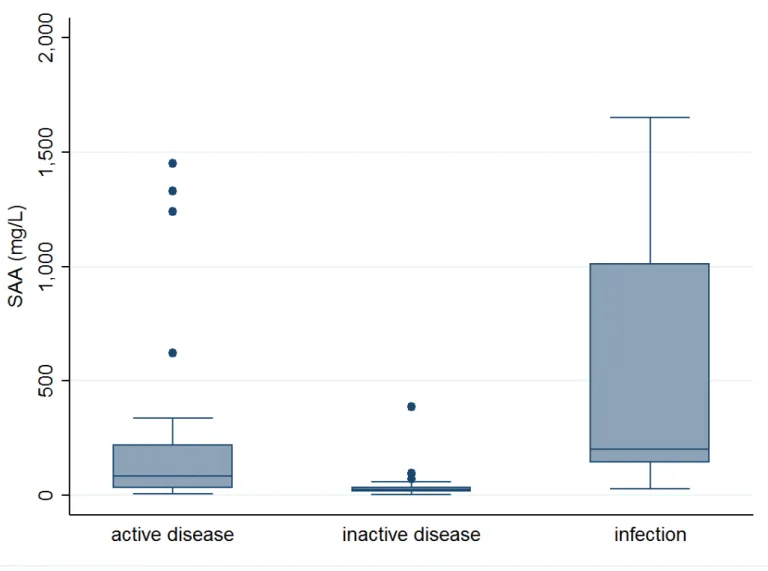
SAA levels were significantly higher in active disease (84.5, IR: 30.7-220 mg/l) compared with patients with inactive disease (23.85, IR: 14.6-35.3 mg/l), p = 0.01. SAA was higher during infections (202, IR: 143-1010 mg/l) compared with inactive disease, p <0.01. There were no statistically difference between SAA levels in active disease and infections, p = 0.09 (figure 3).
19
20
Figure 3: SAA level according to clinical status.
Box plot of SAA levels of GCA on the basis of clinical status (active disease: 21; inactive disease: 59; infections: 7)
CRP and fibrinogen level according to clinical status
In active disease, CRP median were 39 mg/l (IR: 12-80 mg/l; normal < 3 mg/l) and fibrinogen median were 7 g/l (IR: 5-7 g/l; normal between 2.1 and 4.1 g/l). In inactive disease, CRP median were 3 mg/l (IR: 3-7.75 mg/l) and fibrinogen median were 4 g/l (IR: 4-4 g/l)
21
Discussion
In our study, we were able to show that SAA level significantly distinguished GCA patients with active disease from GCA patients with inactive disease. Moreover, patients with aortic dissection, the clinical sign demonstrating the disease high activity, have the highest SAA level. Our results are in accordance with the latest date from literature. Indeed, a recent study found an elevation of 83-fold of SAA median values of GCA untreated patients compared to healthy controls [21]. This study as well as another finds a significant SAA elevation in patients with relapse compared in patients without relapse [22]. In Takayasu disease, the second large vessels vasculitis, SAA also showed his interest. Indeed two different studies indicated a SAA level significantly higher in active Takayasu disease versus non-active Takayasu disease and controls [23,24]. In inflammatory arthritis, it had already been proved that SAA is a better disease activity biomarker than ESR and C-reactive protein (CRP) [25].
On the other hand, there were no statistically difference between SAA level in active disease and infections. In fact, despite a tendency to higher SAA level in infections, we could not demonstrate a significant difference. Actually, it could be interesting to differentiate an infection and a GCA because some symptoms could be identical. This non-significant result can be caused by lack of power of our study that included only 20 patients.
There are several limitations in this work. Firstly, we did not realize SAA dosage on controls which could have been healthy patients or patients initially suspected of GCA. Indeed, this could have been interesting for validate our results and find out if SAA could be a good diagnosis test. Secondly, patients were included only in internal medicine, ophthalmology and neurology services. No patients were included in geriatric or vascular disease services. This may correspond to a selection bias. Thirdly, more than half of initially blood samples are realized after corticosteroids treatment, which could modify SAA level and disease status (inactive versus active). Finally, the number of patients included is low and there are many lost to follow-up or
22
deaths. In view of these limitations, it could be necessary to validate our results with a larger cohort and controls.
In conclusion, SAA is really interesting in GCA. Indeed, this protein could intervene in GCA pathophysiology [13] and participate to bloods vessels modification and destruction by this proangiogenic property. Moreover, A-SAA is able to induce IL-6 synthesis, a major pro-inflammatory cytokine in GCA.
We have shown that SAA is a marker of disease activity. The second step is determinate if SAA is more specific than ESR or CRP and if it could be a potential diagnostic test.
THÈSE SOUTENUE PAR : Anaïs Dartevel
TITRE : SERUM AMYLOID A AS A MARKER OF DISEASE ACTIVITY IN GIANT CELL ARTERITIS
CONCLUSION :
L'artérite à cellules géantes (ACG) est une vascularite systémique avec un taux de rechute important et parfois des complications sévères. Le sérum amyloïde A (SAA) est une protéine de la phase aigue de l'inflammation et pourrait intervenir dans sa pathogénie. L'objectif de cette étude est de déterminer si le SAA est un marqueur d'activité de la maladie.
Entre octobre 2015 et janvier 2018, les patients présentant un diagnostic ou une rechute de ACG ont été inclus. Le dosage de SAA a été réalisé à l'inclusion puis tous les 3 mois pendant un an. A chaque point de l'étude les patients ont été séparés en 3 groupes selon leur statut clinique: maladie active, maladie inactive ou infection.
Vingt patients ont été inclus: 15 nouvellement diagnostiqués et 5 rechutes. Il y a eu 21 situations cliniques avec une maladie active. 59 avec une maladie inactive et 8 infections. Le taux de SAA était significativement plus élevé avec une maladie active (84.5, IR: 30.7-220 mg/I) par rapport à une maladie inactive (23.85, IR: 14.6-35.3 mg/1), p = 0,01. Il n'y avait pas de différence statistique entre les taux de SAA entre maladie active et infections, p = 0,09.
Nous avons montré que le SAA est un marqueur d'activité de la maladie. La deuxième étape serait de déterminer si le SAA est plus spécifique que la vitesse de sédimentation des érythrocytes ou la protéine C réactive et pourrait donc constituer un test de diagnostic potentiel.
VU ET PERMIS D'IMPRIMER Grenoble, le :
LE DOYEN LE PRÉSIDENT DE LA THÈSE
25
References
1. Jennette JC, Falk RJ, Bacon PA, Basu N, Cid MC, Ferrario F, et al. 2012 revised International Chapel Hill Consensus Conference Nomenclature of Vasculitides. Arthritis Rheum. 2013 Jan;65(1):1–11. Epidémiology of giant cell arteritis and polymyalgia Rheumatica
2. Gonzalez-Gay MA, Vazquez-Rodriguez TR, Lopez-Diaz MJ, Miranda-Filloy JA, Gonzalez-Juanatey C, Martin J, et al. Epidemiology of giant cell arteritis and polymyalgia rheumatica. Arthritis Rheum. 2009 Oct 15;61(10):1454–61.
3. Sherwin JC, De Smit E, Hewitt AW. Giant-cell arteritis and polymyalgia rheumatica. N Engl J Med. 2014 23;371(17):1652.
4. Myklebust G, Gran JT. A prospective study of 287 patients with polymyalgia rheumatica and temporal arteritis: clinical and laboratory manifestations at onset of disease and at the time of diagnosis. Br J Rheumatol. 1996 Nov;35(11):1161–8.
5. Baslund B, Helleberg M, Faurschou M, Obel N. Mortality in patients with giant cell arteritis. Rheumatology (Oxford). 2015 Jan;54(1):139–43.
6. Li L, Neogi T, Jick S. Mortality in Patients With Giant Cell Arteritis: A Cohort Study in UK Primary Care. Arthritis Care Res (Hoboken). 2018 Aug;70(8):1251–6.
7. Villiger PM, Adler S, Kuchen S, Wermelinger F, Dan D, Fiege V, et al. Tocilizumab for induction and maintenance of remission in giant cell arteritis: a phase 2, randomised, double-blind, placebo-controlled trial. Lancet. 2016 May 7;387(10031):1921–7.
8. Stone JH, Tuckwell K, Dimonaco S, Klearman M, Aringer M, Blockmans D, et al. Trial of Tocilizumab in Giant-Cell Arteritis. N Engl J Med. 2017 27;377(4):317–28.
9. Alba MA, García-Martínez A, Prieto-González S, Tavera-Bahillo I, Corbera-Bellalta M, Planas-Rigol E, et al. Relapses in patients with giant cell arteritis: prevalence, characteristics, and associated clinical findings in a longitudinally followed cohort of 106 patients. Medicine (Baltimore). 2014 Jul;93(5):194–201.
26
10. Ly K-H, Régent A, Tamby MC, Mouthon L. Pathogenesis of giant cell arteritis: More than just an inflammatory condition? Autoimmun Rev. 2010 Aug;9(10):635–45.
11. Ly K-H, Liozon E, Fauchais A-L, Vidal E. [Pathophysiology of giant cell arteritis]. Rev Med Interne. 2013 Jul;34(7):392–402.
12. Martinez-Taboada VM, Alvarez L, RuizSoto M, Marin-Vidalled MJ, Lopez-Hoyos M. Giant cell arteritis and polymyalgia rheumatica: role of cytokines in the pathogenesis and implications for treatment. Cytokine. 2008 Nov;44(2):207–20.
13. O’Neill L, Rooney P, Molloy D, Connolly M, McCormick J, McCarthy G, et al. Regulation of Inflammation and Angiogenesis in Giant Cell Arteritis by Acute-Phase Serum Amyloid A. Arthritis & Rheumatology (Hoboken, NJ). 2015 Sep;67(9):2447–56. 14. De Buck M, Gouwy M, Wang JM, Van Snick J, Proost P, Struyf S, et al. The
cytokine-serum amyloid A-chemokine network. Cytokine Growth Factor Rev. 2016;30:55–69. 15. Ye RD, Sun L. Emerging functions of serum amyloid A in inflammation. J Leukoc Biol.
2015 Dec;98(6):923–9.
16. Gabay C, Kushner I. Acute-phase proteins and other systemic responses to inflammation. N Engl J Med. 1999 Feb 11;340(6):448–54.
17. Lv M, Xia Y, Li B, Liu H, Pan J, Li B, et al. Serum amyloid A stimulates vascular endothelial growth factor receptor 2 expression and angiogenesis. J Physiol Biochem. 2016 Mar;72(1):71–81.
18. Connolly M, Rooney PR, McGarry T, Maratha AX, McCormick J, Miggin SM, et al. Acute serum amyloid A is an endogenous TLR2 ligand that mediates inflammatory and angiogenic mechanisms. Ann Rheum Dis. 2016;75(7):1392–8.
19. Johnson BD, Kip KE, Marroquin OC, Ridker PM, Kelsey SF, Shaw LJ, et al. Serum amyloid A as a predictor of coronary artery disease and cardiovascular outcome in women: the National Heart, Lung, and Blood Institute-Sponsored Women’s Ischemia Syndrome Evaluation (WISE). Circulation. 2004 Feb 17;109(6):726–32.
27
20. Lewis KE, Kirk EA, McDonald TO, Wang S, Wight TN, O’Brien KD, et al. Increase in serum amyloid a evoked by dietary cholesterol is associated with increased atherosclerosis in mice. Circulation. 2004 Aug 3;110(5):540–5.
21. Burja B, Feichtinger J, Lakota K, Thallinger GG, Sodin-Semrl S, Kuret T, et al. Utility of serological biomarkers for giant cell arteritis in a large cohort of treatment-naïve patients. Clin Rheumatol. 2018 Aug 24;
22. Hocevar A, Rotar Z, Jese R, Semrl SS, Pizem J, Hawlina M, et al. Do Early Diagnosis and Glucocorticoid Treatment Decrease the Risk of Permanent Visual Loss and Early Relapses in Giant Cell Arteritis: A Prospective Longitudinal Study. Medicine (Baltimore). 2016 Apr;95(14):e3210.
23. Nair AM, Goel R, Hindhumati M, Jayakanthan K, Visalakshi J, Joseph G, et al. Serum amyloid A as a marker of disease activity and treatment response in Takayasu arteritis. Rheumatol Int. 2017 Oct;37(10):1643–9.
24. Ma J, Luo X, Wu Q, Chen Z, Kou L, Wang H. Circulation levels of acute phase proteins in patients with Takayasu arteritis. J Vasc Surg. 2010 Mar;51(3):700–6.
25. Cunnane G, Grehan S, Geoghegan S, McCormack C, Shields D, Whitehead AS, et al. Serum amyloid A in the assessment of early inflammatory arthritis. J Rheumatol. 2000 Jan;27(1):58–63.
29
SERUM AMYLOID A AS A MARKER OF DISEASE ACTIVITY IN GIANT CELL ARTERITIS
RÉSUMÉ :
Introduction: L'artérite à cellules géantes (ACG) est une vascularite systémique avec un taux de rechute important et parfois des complications sévères. Le sérum amyloïde A (SAA) est une protéine de la phase aigue de l’inflammation et pourrait intervenir dans sa pathogénie. L'objectif de cette étude est de déterminer si le SAA est un marqueur d’activité de la maladie.
Méthodes: Entre octobre 2015 et janvier 2018, les patients présentant un diagnostic ou une rechute de ACG ont été inclus. Le dosage de SAA a été réalisé à l’inclusion puis tous les 3 mois pendant un an. A chaque point de l’étude les patients ont été séparés en 3 groupes selon leur statut clinique: maladie active, maladie inactive ou infection.
Résultats: Vingt patients ont été inclus: 15 nouvellement diagnostiqués et 5 rechutes. Il y a eu 21 situations cliniques avec une maladie active, 46 avec une maladie inactive et 7 infections. Le taux de SAA était significativement plus élevé avec une maladie active (84.5, IR: 30.7-220 mg/l) par rapport à une maladie inactive (23.85, IR: 14.6-35.3 mg/l), p = 0.01. Il n'y avait pas de différence statistique entre les taux de SAA entre maladie active et infections, p = 0.09.
Conclusion: Nous avons montré que le SAA est un marqueur d’activité de la maladie. La deuxième étape serait de déterminer si le SAA est plus spécifique que la vitesse de sédimentation des érythrocytes ou la protéine C réactive et pourrait donc constituer un test de diagnostic potentiel.
MOTS CLÉS :Artérite à cellules géantes, sérum amyloide A FILIÈRE : médecine interne