Université de Montréal
Effects of exercise training on intra-uterine growth
restriction in an animal model
par
Aida Kasaei Roodsari
Département de kinésiologie
Mémoire présenté au Département du kinésiologie
en vue de l’obtention du grade de maîtrise
en science de l'activité physique
[Septembre, 2015]
© Aida Kasaei Roodsari, 2015
i
Résumé
Selon l'OMS, la retard de croissance intra-utérine (RCIU; 10% en dessous du poids normal pendant la grossesse) affecte 5-10% des grossesses et est une cause principale de la morbidité et de la mortalité périnatales. Dans notre étude précédente sur un modèle de souris transgénique de prééclampsie (R+A+), nous avons constaté que l’entraînement physique (ExT) avant et pendant la grossesse réduisait la pression artérielle maternelle et empêchait la RCIU en améliorant le développement placentaire. Dans le cadre de mon projet, nous avons confirmé les bénifices de l’ExT dans un modèle de RCIU (souris déficiente en p57Kip2 (p57-/+). Ainsi, nous avons observé la présence de RCIU, d’une masse placentaire réduite, d’une augmentation de la pathologie placentaire ainsi qu’une plus petite taille des portées chez les souris p57-/+ sédentaire. L’ExT prévient la RCIU ainsi que tous les paramètres mentionnés ci-haut. Nous avons observé que l'expression du facteur de croissance de l’endothélium vasculaire, un régulateur clé de l'angiogenèse lors de la croissance placentaire, était réduite dans le placenta des souris p57-/+ et normalisée par l’ExT. Nous avons également trouvé que l'expression en ARN dans le placenta de 2 facteurs inflammatoires (interleukine-1β et MCP-1) était augmenté chez les souris sédentaires p57-/+ alors que ceci n’était pas présent chez les souris entraînées, ce qui suggère que l'inflammation placentaire peut contribuer à la pathologie placentaire. Toutefois, contrairement aux souris R+A+, le système rénine-angiotensine placentaire chez les souris p57-/+ était normale et aucun effet de l’ExT a été observé. Ces résultats suggèrent que l’ExT prévient la RCIU en normalisant la pathologie placentaire, l’angiogenèse et l’inflammation placentaire.
Mots-clés: RCIU, entraînement physique, p57Kip2, angiogenèse, inflammation, système rénine-angiotensine
ii
Abstract
Intrauterine growth restriction (IUGR) refers to the condition in which the baby's estimated weight is below the 10th percentile of babies of the same gestational age. It affects 5-10% of pregnancies and is a major cause of perinatal morbidity and mortality. In our previous studies in a transgenic preeclampsia mouse model (R+A+ mice), we found that exercise training (ExT) before and during pregnancy reduced maternal blood pressure (BP), and prevented IUGR by improving placental development. Here, we confirm the benefits of ExT in a mouse model of IUGR without preeclampsia (p57kip2 KO (p57-/+). We confirmed the presence of IUGR, reduced placental mass, increased placental pathology, smaller litter size and increased number of non-viable pups per litter in sedentary p57-/+ mice. ExT prevented IUGR as well as normalized all the mentioned parameters. The expression of the vascular endothelial growth factor (VEGF), a key regulator of angiogenesis required for normal placental development, was reduced in pregnant p57-/+ mice and normalized by ExT. The expression of 2 inflammatory factors (interleukin-1β and MCP-1 mRNA) in placenta was elevated in KO sedentary mice and MCP-1 was normalized by ExT, proposing that placental inflammation may contribute to placental pathology. However, in contrast to R+A+ mice, the placental RAS in p57-/+ mice was found to be normal and there was no effect of ExT. Taken together, these results suggest that exercise training prevents intrauterine growth restriction by improving angiogenesis, placental alterations and placental inflammation.
iii
Table of Contents
List of tables ... vii
List of figures ... vii
List of abbreviations ... viii
Dedication ... xi
Acknowledgment ... xii
Chapter 1: ... xiii
Introduction ... xiii
1. Intrauterine growth restriction (IUGR) ... 14
1.1. Definition and epidemiology ... 14
1.2. IUGR risk factors: ... 15
1.2.1. Maternal nutrition ... 15 1.2.2. Multiple births ... 15 1.2.3. Smoking ... 16 1.2.4. Adolescent pregnancies ... 16 1.2.5. Substance abuse ... 16 1.2.6. Inter-pregnancy interval ... 17 1.2.7. High altitude ... 17 1.2.8. Congenital infections ... 18
iv
1.2.10. Preeclampsia and eclampsia ... 18
1.3. Etiology ... 20
1.3.1. Normal placentation ... 20
1.3.2. Placental dysfunction in IUGR ... 21
1.3.3. Histopathology of placental injury ... 24
1.4. Classification... 24
1.5. IUGR and diseases later in life ... 25
1.5.1. IUGR and metabolic syndrome ... 25
1.5.2. IUGR and reproduction problems ... 27
1.6. Clinical diagnosis of IUGR:... 27
1.6.1. Serum biochemistry ... 27
1.6.2. Uterine artery doppler: ... 28
1.7. Evaluating fetal wellbeing ... 29
1.7.1. Fetal heart rate monitoring ... 29
1.7.2. Biophysical profile ... 29
1.7.3. Doppler velocimetry of blood flow ... 30
1.7.4. Histopathological and molecular diagnostics ... 30
1.8. Clinical management of IUGR ... 31
2. Exercise training ... 34
v
2.1.1. Theoretic concerns regarding the effects of exercise on pregnancy ... 34
2.2. Positive effects of exercise training on normal pregnancies ... 36
2.2.1. Maternal wellbeing ... 36
2.2.2. Fetal benefits... 38
2.2.3. Preeclampsia and gestational diabetes ... 38
2.2.4. Obesity ... 39
2.3. General recommendations for exercise in pregnancy and post-partum period according to ACOG ... 40
2.4. Exercise training and IUGR: ... 42
2.5. Hypothesized protective mechanisms implicated in the prevention of IUGR by exercise training ... 42
2.5.1. Enhanced placental development and vascularity ... 42
2.5.2. Prevention and/or reduction of oxidative stress ... 43
2.5.3. Reduction of inflammation ... 44
2.5.4. Improving of endothelial dysfunction ... 44
3. Animal models of IUGR... 45
3.1 Fetal intervention models:... 46
3.2. Maternal interventions ... 46
3.2.1. Surgical methods ... 46
vi
3.3. Environmental restrictions ... 48
3.4. Genetic interventions ... 49
3.5. P57 kip2 knock-out mouse model: ... 51
Chapter 2: ... 53
Article ... 53
Abstract ... 56
Introduction ... 58
Materials and methods ... 60
Results ... 62 Comment ... 64 Acknowledgements ... 67 Chapter 3: ... 76 Discussion ... 76 References ... 83
vii
List of tables
Table 1: Fetal growth restriction risk factors ... 19
Table 2: Recommendations for sport activities during pregnancy ... 41
Table 3: Some IUGR animal model species ... 50
Article tables:
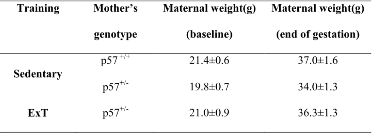
Table 1: Primer sequences……….68Table 2: Maternal weight………..….68
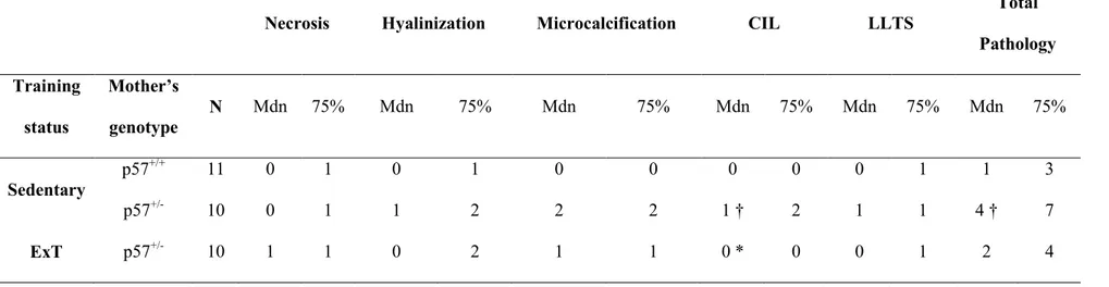
Table 3: Placental pathology………..………69
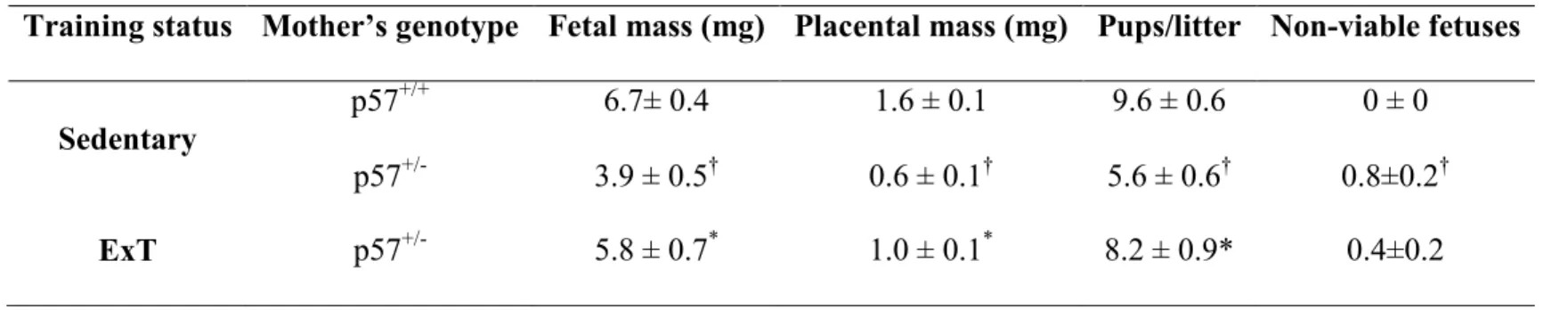
Table 4: Fetal, placental mass and litter size……….…70
List of figures
Figure 1: structure of placenta and placental villi . ... 23Article figures:
Figure 1: Placental VEGF mRNA expression ………..…72Figure 2 :Placental Inflammatory factors………..73
Figure 3 : Local placental RAS……….….74
viii
List of abbreviations
1. ACE2: Angiotensin-converting enzyme 2 2. AC: Abdominal circumference
3. Ach; Acetylcholine
4. ACOG: American college of obstetricians and gynecologists 5. AEDF: Absent end diastolic flow
6. AFP: Alpha-feto protein
7. AGA: Appropriate for gestational age 8. AGTR1: Angiotensin II receptor type 1 9. BMI: Body mass index
10. BP: Blood pressure 11. BPP: Biophysical profile
12. BWS: Beckwith-wiedemann syndrome 13. CHD: Coronary heart disease
14. CDK: cyclin dependent kinase 15. CDA: Canadian diabetes association 16. DV: Ductus venosus
17. CRH: corticotropin releasing hormone 18. EFW: Estimated fetal weight
ix 20. FGR: Fetal growth restriction
21. GDM: Gestational diabetes mellitus 22. GPX: Glutathione peroxidase
23. hCG: Human chorionic gonadotropin 24. HIV: Human immune deficiency virus 25. HPA: Hypothalamic-pituitary-adrenal 26. IGF: Insulin growth factor
27. IL-1β: Interleukin 1β
28. IUGR: Intrauterine growth restriction 29. KO: Knockout
30. LTPA: Leisure time physical activity 31. Mas-R: Mas receptor
32. MCA: Median cerebral artery 33. METs: Metabolic equivalents
34. PAPA-A: Pregnancy associated protein-A 35. PCOS: Polycystic ovarian syndrome 36. PE: Preeclampsia
37. P-GR: Plasma glutathione reductase 38. PCR: Polymerase chain reaction 39. PIH: Pregnancy-induced hypertension 40. PLGF: Placental growth factor
41. RAS: Renin-angiotensin system 42. RI: Resistance index
x 43. ROS: Reactive oxygen species
44. SGA: Small for gestational age 45. SNP: Sodium nitroprusside 46. SOD: Superoxide dismutase
47. sFlt-1: soluble fms-like tyrosine kinase 48. UA: Uterine artery
49. UPD: uniparental disomy
50. VEGF: vascular endothelial growth factor 51. WHO: World health organization
xi
Dedication
To my mother and father for their
Encouragement and support to complete this work.
Thank you!
xii
Acknowledgment
My sincere thanks go to Dr. Julie Lavoie whose expertise, understanding, and patience, added considerably to my graduate experience. Without her guidance and precious support it would not be possible to conduct this research.
I am sincerely thankful to my labmates, Paul Tan, Zulaykho Shamansurova, Alexandre Garneau and Olga Asaftei for all their helps, compassion and for everything I learnt while working with them. I also wish to express my gratitude to Sonia Kajla, and Zhenhong Li for their kindness and also their technical help at research.
I am grateful to all my friends in CRCHUM, particularly, to Ju-Jing Tan for his kind help and support during writing my memoir.
Last but not the least, I would like to thank my family: my parents and my brothers for supporting me spiritually throughout writing this memoir.
xiii
Chapter 1:
14
1. Intrauterine growth restriction (IUGR)
1.1. Definition and epidemiology
Intrauterine growth restriction (IUGR)/Fetal growth restriction (FGR) refers to a condition in which a fetus is unable to reach its genetically determined potential size. In fact, it is a
pathological reduction in an expected pattern of fetal growth that occurs in utero. IUGR is thus a major cause of perinatal morbidity and mortality1. Traditionally, in North America the standard definition of IUGR is a birth weight below the 10th percentile for gestational age. However, adverse consequences and mortality are also increased in infants with birth weights between 10th and 15th percentile. Conversely, many neonates whose weights are below the 10th percentile are healthy which are known as small for Gestational Age (SGA) babies 2. Indeed, the SGA group includes fetuses that are constitutionally but not pathologically small and may reflect a normal pattern in a given population 1. Several definitions of IUGR are accepted in different areas of the world. In Europe, for example, an abdominal circumference (AC) below the 10th or 5th percentile is the preferred diagnostic criteria. Published definitions include: weight at birth <2500 g, EFW (estimated fetal weight) <10th percentile, AC <10th percentile, EFW <10th percentile with abnormal Doppler indices in the umbilical artery or middle cerebral artery, and AC <10th percentile with abnormal umbilical artery or middle cerebral artery Doppler studies. Other
diagnostic criteria utilize the fetus as a control for itself 3, or use customized fetal growth standards 4.
The prevalence of IUGR is about 5%-10% in the general obstetric population. Studies show that each year 18 million babies are born with low birth weight worldwide, half of which are born in
15
Asia 5. Fetal Growth restriction is the second main cause of perinatal morbidity and mortality while prematurity is the leading cause 6.
1.2. IUGR risk factors:
Generally, the population at high risk for IUGR are women with low socioeconomic status, low weight before pregnancy, low weight gain during pregnancy, history of preterm delivery, stillbirth and
previous pregnancies affected by IUGR 7-11. Other risk factors are described as follow (Table 1).
1.2.1. Maternal nutrition
The specific responses of fetal growth to acute under nutrition at different points in pregnancy are still unclear. One study on the birth reports during the Dutch famine of 1944-1955 showed that only if the famine exposure happens late in pregnancy will result in low birth weight and declined crown-to-heel length 12In contrast another revealed that, mothers of appropriate for gestational age (AGA) infants ate more servings of carbohydrate rich food and fruit, and were more likely to have taken folate and
vitamin supplements than mothers of SGA infants at the time of conception. There was also an
association between Iron supplementation when taken in the last month of pregnancy and a diminished risk of SGA 13. These results suggest that malnutrition in early or late pregnancy may result in small for gestational age infants.
1.2.2. Multiple births
Newborns from multiple births are generally smaller than singletons. An analysis of birth weight and gestational age in twins and triplets in Norway showed that the intrauterine growth of both male and female twin diverged considerably from singletons starting at approximately 30 weeks of
16
twins 15. In a retrospective study of multi-fetal pregnancies it was revealed that birth weight of quadruplets and quintuplets was significantly lower than triplets 16.
1.2.3. Smoking
Smoking has been considered as one of the important risk factors for IUGR. One study on the impact of maternal exposure to environmental tobacco smoke on IUGR in a sample of 6866 singleton births represented a significant decrease in the mean birth weight of infants of active smoking mothers. This reduction was minimal but still present for mothers who stopped smoking after recognizing their pregnancy. Also, environmental tobacco smoke exposure in 1797 of 5507 non-smoking mothers decreased the mean birth weight of their infants by 53g17. Another study in Sweden demonstrated that babies of smoking mothers were at an increased risk for decreased head circumference, <32 cm 17, 18.
1.2.4. Adolescent pregnancies
It is reported that infants of adolescent mothers experienced almost twice the rates of preterm delivery (21.3%) and low birth weight (12.6%) compared to older mothers aged between 20 and 39 years 19. Generally, adolescents most likely to become pregnant are those with insufficient nutritional status and unfavorable socio-economic status which may contribute to the higher rate of IUGR observed in this population 20.
1.2.5. Substance abuse
It is well known that alcohol and drug consumption are harmful to the developing embryo and fetus, and in the majority of cases cause IUGR. What we know as fetal alcohol syndrome includes pre- and postnatal growth deficiency, a “characteristic” facial appearance, microcephaly, mental retardation, and occasional major malformations 21. In a 1-year study on all live singleton infants whose mothers were exposed to cocaine, it was observed that low birth weight (<2500 g) was more common among
17
associated with a decrease in birth weight (154 g), length (1.02 cm), head circumference (0.69 cm), and duration of gestation (0.74 weeks). The birth-weight deficits were larger for infants born from mothers who used cocaine in combination with other drugs (195 g) and for infants born to mothers who
specifically admitted using crack (200 g) 22. Another study in china showed that mothers who had abused narcotics and heroin had SGA babies in 27.5% of pregnancies. The babies born to drug addicted mothers were on average 629 g lighter which was significantly different from the infants of the non-addicted mothers 23.
1.2.6. Inter-pregnancy interval
A shortened interval between pregnancies is associated with adverse perinatal consequences 24. A research in Utah, USA showed that infants conceived 18–23 months after a previous live birth had the lowest risks of negative perinatal outcomes. However, in this study shorter intervals were related to higher risks for low birth weight 25.
1.2.7. High altitude
High altitude seems to reduce birth weight independently of other factors. It was found in a study in Colorado that birth weights at high altitude (2744–3100 m) were reduced due to IUGR 26. The association between ethnicity and high altitude was also assessed in a study done in Tibet. Tibetans experience less altitude-associated IUGR than Chinese and have reduced levels of prenatal and
postnatal mortality. When comparing the link between birth weight and altitude among these and other high-altitude populations, the results showed that those who have been living the longest at high altitude had the least altitude-associated IUGR. In general, the pregnant Tibetans had higher umbilical artery blood flow velocity and distributed a higher portion of common iliac blood flow to the umbilical artery compared to the Chinese women 27. This might propose the occurrence of an evolutionary adaptation 28.
18
1.2.8. Congenital infections
Infections acquired in utero may often cause IUGR such as rubella 29, cytomegalovirus 30, herpes virus 31 and toxoplasma gondi which is less common than the others 32. Moreover, HIV-Infected infants often suffer from IUGR 33.
1.2.9. Genetic and chromosomal factors
Chromosomal abnormalities are found in up to 7% of neonates with IUGR , which is over 10 times higher than in AGA (appropriate for gestational age) infants34 . Moreover, the genomic imprinting, through which several genes in the human genome are differentially expressed based on whether they are located on the maternal or paternal chromosome, may play a role in embryonic and fetal growth. This has led to the theory that genomic imprinting regulates embryonic and fetal growth 35. Silver-Russell syndrome which represents an extreme syndrome of IUGR and dysmorphic features, as well as maternal uniparental disomy (UPD: the inheritance of both chromosomes of a chromosome pair from only one parent) of human chromosome 7 has been observed in approximately 10% of these cases 36. Other known imprinted genes where IUGR is the most common feature are maternal UDP14, maternal UDP20 and paternal UDP6q24 37. Also p57 kip2 which is a paternally imprinted gene 38 has been related to severe growth restriction 39.
1.2.10. Preeclampsia and eclampsia
FGR can be related to preeclampsia (PE) as a result of impaired trophoblast invasion into the placental bed. In normal pregnancy, occlusion of the spiral arterioles by the endovascular trophoblast at the implantation site and the anatomical destruction of the distal spiral arteriole contribute to improved uterine blood flow. Failed interstitial invasion of spiral arterioles may lead to failure in local angiogenic and systemic cardiovascular adaptation signals that could be the main reason for early onset of IUGR and PE 40.
19
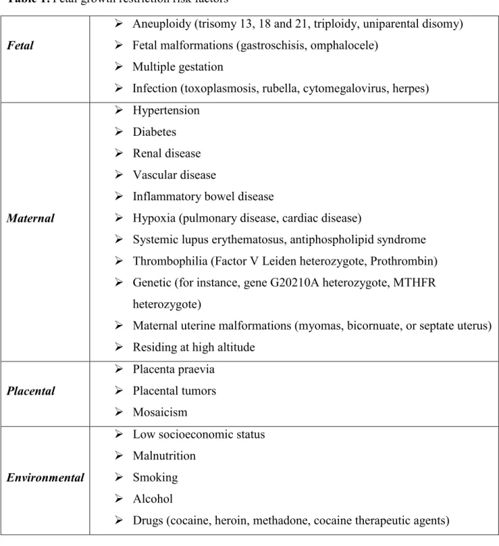
Table 1:Fetal growth restriction risk factors
Fetal
Aneuploidy (trisomy 13, 18 and 21, triploidy, uniparental disomy) Fetal malformations (gastroschisis, omphalocele)
Multiple gestation
Infection (toxoplasmosis, rubella, cytomegalovirus, herpes)
Maternal
Hypertension Diabetes Renal disease Vascular disease
Inflammatory bowel disease
Hypoxia (pulmonary disease, cardiac disease)
Systemic lupus erythematosus, antiphospholipid syndrome Thrombophilia (Factor V Leiden heterozygote, Prothrombin) Genetic (for instance, gene G20210A heterozygote, MTHFR
heterozygote)
Maternal uterine malformations (myomas, bicornuate, or septate uterus) Residing at high altitude
Placental
Placenta praevia Placental tumors Mosaicism
Environmental
Low socioeconomic status Malnutrition
Smoking Alcohol
20
1.3. Etiology
Growth and development of the fetus/embryo are complex biological procedures which are affected by different factors such as genetic, epigenetic, maternal age, environmental factors, etc. 41. These factors can influence the size and efficiency of the placenta, uteroplacental transfer of nutrients and oxygen from mother to fetus, the endocrine environment of the fetus, and metabolic pathways 42-44. Normal fetal growth consists of two phases. The first phase or embryonic life which starts from fertilization till the end of the 8th week includes proliferation, organization and differentiation of the embryo while the second phase, which we know as fetal life, starts at the end of week 8 and involves continuing growth and functional maturation of the different tissues and organs of the fetus 45. The maternal-fetal-placental unit acts in harmony to fulfil the needs of the fetus, while supporting the physiological changes of the mother. The fetus has an inherent growth potential which results in a healthy newborn with appropriate size in normal situation. Studies showed that in IUGR, placentation is impaired 46, 47. Dysregulation of endocrine-related factors such as, growth factor deficiencies, mainly insulin and the insulin-like growth factors (IGF) or their signaling pathway, often induce IUGR 45, 48. Moreover, assessment of uterine, placental and umbilical blood flow shows that in growth restricted fetuses blood flows are decreased on both sides of the placenta. It is also reported that there is less placental exchange of essential nutrients such as amino acids in IUGR fetuses both in vitro and in vivo 49.
1.3.1. Normal placentation
Placentation starts with implantation of the blastocyst in the uterine epithelium and differentiation into embryonic and extra-embryonic tissues 50. The trophectoderm of the blastocyst is the
21
necessary for successful embryonic development as it is integral in the transfer of nutrients from the mother to the child. 51. Uteroplacental circulation is not fully established until the end of the first trimester. One proposed theory based on the observations of ex vivo histologic analysis of hysterectomy specimens of first-trimester placentas to explain the uteroplacental circulation states that trophoblasts invade decidual spiral arteries and form trophoblastic plugs. These trophoblastic plugs obstruct maternal blood flow into the intervillous space and prevent flow until the end of first trimester of pregnancy (10–12 weeks) 52. Thus, human placental development during the first ten weeks of gestation occurs in a low oxygen environment with a PO2 (oxygen pressure)
measured at < 15 mmHg 53. The plugs then loosen and permit continuous maternal blood flow into the intervillous space 52. As a result, the low oxygen state changes and the pressure increases, so that the developing villous tree of the chorioallantoic placenta is then exposed to maternal blood with higher oxygen content.
1.3.2. Placental dysfunction in IUGR
Normal placental development is necessary for normal fetal growth. Failure of one or more of the components of the placentation process may result in pregnancy complications like preeclampsia, IUGR and placental abruption. There is considerable evidence showing that placentation is deficient in IUGR 46, 47. Histologically, the features that can be observed in placenta from IUGR fetuses include: damage to branching angiogenesis with long unbranched intermediate and terminal villi, altered cytotrophoblast proliferation, trophoblast apoptosis, fibrin deposition, syncytial knotting and bridging, and enhanced villous maturation 54. It is assumed that the reduced secretion of placental growth hormones (PGH) and IGF-1 which are some of the important
22
In IUGR, absence of endovascular trophoblast invasion of the myometrial segments of the spiral arterioles produces a high resistance vasculature in these arterioles. This lack of transformation leads to hypoperfusion, hypoxia, re-perfusion injury, oxidative stress and ultimately, to signs of villous tree maldevelopment in the second half of the pregnancy 56. Villous cytotrophoblasts in IUGR have an augmented sensitivity to cell death in hypoxic situations when compared to normal pregnancies 57-59. Although apoptosis is considered to be a normal part of villous trophoblast turnover and syncytiotrophoblast formation from cytotrophoblast 60, 61, in pregnancies complicated with IUGR this is augmented and produces an increase in syncytial knots 62. This is also the case in the villi in IUGR and has been detected by the expression of cleavage products of caspase 62-64. In IUGR, excess injury of the villous trophoblast layer decreases the functional mass of
syncytiotrophoblast and restricts the capacity of the villi to transport nutrients. Furthermore, the microscopic injury has functional effects on placental permeability, as α-fetoprotein and small molecular weight compounds are able to pass between the maternal and fetal circulations 65, 66.
23
24
1.3.3. Histopathology of placental injury
There are several stimuli and mediators that may contribute to the observed injury to the chorioallantoic villi but the most important one is oxidative stress 68. Oxidative stress can be caused by ischemia in the placenta as a result of the insufficiently developed spiral arterioles. Indeed the production of reactive oxygen species (ROS) which promote oxidative stress
contributes to tissue injury in many diseases including IUGR 69. Studies show that the placentas of pregnancies complicated with IUGR demonstrate obvious signs of oxidative stress 70. Moreover, hypoxia, ischemia, or both may contribute to placental injury via mechanisms other than ROS generation, as variable organ blood flow also activates the complement cascade 68, 71, 72.
Dysregulated complement activation in non-pregnant patients mediates immunological injury in the heart, lung and kidney, and recent data indicate that it also plays a role in abnormal human pregnancy 73. For example, the kidneys of women with preeclampsia show deposition of complement split products in their glomeruli 74.
1.4. Classification
Growth during intrauterine life occurs during different stages. For example, growth in length happens in early prenatal life (during the 3rd, 4th and 5th month) while weight gain develops later in prenatal life (during the last 2 months of gestation) 45. Depending on the time of adverse
intrauterine environment, IUGR fetuses are classified as symmetric or asymmetric. In symmetric growth restriction, the entire fetus body is proportionally small (small weight, length, and head circumference). In this situation, the adverse environment has happened early in pregnancy which can be as a result of genetic factors or congenital infections, syndromes, or toxic effects in early gestation 75. On the other hand, in asymmetrical IUGR, adverse intrauterine environment happens
25
later during gestation. In this case, most of the energy is conducted for the maintenance of vital organs such as the brain and heart whereas liver, muscle and fat are less developed which results in smaller weight but normal length 76. In asymmetrical IUGR , we observe normal head
circumference but small abdominal circumference, skinny limbs, and thinned skin as a result of decreased liver size, muscle mass and subcutaneous fat77.
1.5. IUGR and diseases later in life
Intrauterine life adapts the fetus to become mature and also overcome postnatal insults. During this period, the fetus goes through critical stages of tissue growth and elevated cell division. If the organism is affected in these critical periods it may have an important impact on organ development 78. It has been reported in animals and also in some human cases that IUGR is associated with increased prevalence of many adult diseases 79. In addition, results of a study by Park et al. demonstrated that low protein diet during pregnancy causes long-lasting changes in the liver and skeletal muscle mitochondria in the offspring 80.
1.5.1. IUGR and metabolic syndrome
According to the literature, there is an association between IUGR and the development of metabolic syndrome later in life, a condition associated with obesity, arterial hypertension, hypercholesterolemia, impaired glucose tolerance, and diabetes mellitus type 2 81-90. There is a hypothesis to explain this condition which is known as the thrifty phenotype or Barker’s
hypothesis. It proposes that in the case of an impaired intrauterine environment, such as nutrient restriction, the intrauterine milieu creates a “reprogramming” of the endocrine–metabolic status of the fetus in order to reach short-term survival benefits, although, it may be harmful in the long
26
term 81-84, 91, 92. Barker was able to demonstrate that, the smaller the birth weight or the weight at 1 year of age, the greater was the prevalence of metabolic syndrome in adult life.
According to the thrifty phenotype, the connection of low birth weight and insulin resistance or diabetes in adulthood may be a result of fetal malnutrition due to poor nutritional reserves of the mother, not adequate flow of the blood in uterus, or destruction of nutrients in the placenta 93. Hypothalamic-pituitary-adrenal (HPA) axis overacts by changing its set point in response to the adverse intrauterine environment, which can lead to increased cortisol levels 94-98. This situation is similar to what can be seen with chronic stress 96, 99. The HPA over-activation has been observed in both experimental animals and newborns with IUGR and both showed elevated cortisol levels in umbilical cord blood 100, 101. This increase in cortisol during intrauterine life causes endothelial damage which contributes to the development of cardiovascular diseases. Furthermore, growth restricted babies have a reduced muscle mass and this deficiency will persist because the crucial period for muscle growth is at ∼30 weeks in utero, and there is little cell replication after birth. As such, if they gain weight rapidly in childhood, they are more likely to increase their fat mass rather than their muscle mass, leading to a disproportionately high fat mass in later life resulting in an increase in the development of obesity and insulin resistance 85. Ozanne et al. found decreased expression of specific insulin-signaling proteins, such as, protein kinase C (PKC) zeta, p85alpha, p110beta and GLUT4, in low birth weight subjects compared to controls 102 and it was also shown that, children born small for gestational age have reduced adiponectin levels, an adipokine with insulin-sensitizing and antiatherogenic properties 103, which may increase the risk for developing diabetes type 2.
Moreover, studies in Europe, North America, and India have shown association between coronary heart disease and small size at birth 104, as well as a study in Finland which reported that the
27
cumulative incidence of hypertension requiring medication was 20.2% in those weighing <3 kg at birth compared to 12.3% in those weighing >4 kg 105.
1.5.2. IUGR and reproduction problems
Some studies have demonstrated a link between IUGR and adrenarche (early sexual maturation), elevated prevalence of functional ovarian hyper-androgenism as well as the development of polycystic ovary syndrome 106-109. Moreover, there has been reports of decreased uterine volume and smaller fraction of ovarian follicles in girls born with IUGR, 110, 111 which both may have a negative effect on fertility. Although it has not been clarified yet, there seems to be an association between IUGR and reproductive function
1.6. Clinical diagnosis of IUGR:
Abnormal fetal growth is suspected when there is a subnormal uterine size detected by abdominal palpation and direct measurement of the symphyseal-fundal distance 112. Indeed, abdominal palpation has a sensitivity of 30% for detecting SGA fetuses, while the symphysis-fundal distance has a
sensitivity of 27-86% and specificity of 80–93% 113. Ultrasound has also been used for accurate pregnancy dating and for the diagnosis of IUGR, although it is reported that IUGR remains undetected in about 30% of routinely scanned cases and it is falsely detected in 50% of cases 114.
1.6.1. Serum biochemistry
In SGA and IUGR affected pregnancies, biochemical markers have been proposed. An increased maternal Serum alpha-fetoprotein (AFP) is correlated with an elevated risk of low birth weight 115. Also low levels of maternal serum pregnancy-associated plasma protein A (PAPP-A) (at the lowest 5th percentile) are associated with higher risk of a SGA infant 116. There are also other placental markers
28
like human chorionic gonadotropin (hCG), ADAM12 (A Disintegrin and Metalloprotease), placental protein 13 (PP13), serum soluble Fas (sFas) and placental growth factor (PlGF), amongst others. However, studies have shown that all of these markers are below the detection rate warranted for large population screening 117-122. In low-risk populations, a combination which includes PP13, PAPP-A, ADAM12, activin A, or inhibin A, measured in the first or early second trimester and uterine artery Doppler in the second trimester, reveal sensitivities of 60%–80% and specificities >80%. Studies are still required to estimate the full potential of evaluating combining multiple markers and ultrasound in screening for IUGR.
1.6.2. Uterine artery doppler:
Since the 1980s, many progresses have been made in the utilisation of uterine artery Doppler in obstetrical practice, particularly, in the detection of maternal perfusion abnormalities in PE and IUGR 123-125. Since trophoblastic invasion was thought to be completed by the second trimester of pregnancy, most of the studies were performed between 20 and 24 weeks of gestation. However, some believe that trophoblastic invasion peaks in the first trimester, so it would be more appropriate to screen for growth restriction in that period 126. Although, the sensitivity of this test is not that high (24% and 16%
according to some reports) 127, 128, women with a high uterine artery resistance index (RI), are 5.5 times more likely to have IUGR 128. In more recent studies, they attempted to add serum biochemistry factors like PAPPA in order to increase detection rates. Unfortunately, sensitivity of these tests still remains low 117, 129.
29
1.7. Evaluating fetal wellbeing
1.7.1. Fetal heart rate monitoring
Fetal heart rate analysis is extensively used to detect pregnancies at high risk. It aims to determine fetal well-being by estimating the fetal heart rate baseline, variability, and periodic changes. A normal reactive test is likely to reflect adequate oxygenation of the fetal central nervous system. Since it is interpreted by visual inspection, it is prone to a significant intra-observer and inter-observer variation and therefore, there is a high rate of false positive. In premature fetuses, particularly those with IUGR, interpretation is challenging 130. Computerized fetal heart rate analysis was introduced to decrease discrepancies in the interpretation. However, it was showed that the results from computerised cardiotocographic analysis agreed closely with visual assessment 113.
1.7.2. Biophysical profile
Ensuring fetal well-being and determining the optimal timing for delivery of an IUGR fetus is the primary goal of fetal specialists. However, the optimal method to use for fetal testing is also debatable; in the United States, the most frequently used test is the biophysical profile, whereas in Europe,
cardiotocography (computerized fetal heart rate monitoring) is the preferred method 131.
The fetal biophysical profile (BPP) is a method for assessing fetal asphyxia and/or chronic hypoxia and is the most acceptable method of fetal well-being evaluation in the North America. It consists of a number of measurements including the amniotic fluid volume, fetal tone, fetal movements, fetal
breathing movements, and fetal heart rate monitoring. In a normal situation, each parameter has a value of two out of total points of ten 132-134.
30
1.7.3. Doppler velocimetry of blood flow
Doppler blood flow has significantly changed the management of IUGR. We use it to assess vascular resistance and end organ function. There are three types: Doppler assessment of the umbilical artery (UA), middle cerebral artery (MCA) and ductus venous (DV). When the UA blood flow from the fetus to the placenta is determined, the placental vessel resistance can be evaluated. MCA detects fetal cerebral blood flow and DV reflects alterations in fetal cardiac function. In severe cases of IUGR the DV is abnormal 135, 136. UA is the most frequently used Doppler test in women diagnosed with IUGR. It has the ability to distinguish IUGR caused by placental problems from SGA fetuses. Indeed,
monitoring IUGR pregnancies with UA Doppler decreases the mortality rate and lowers the need for antepartum admissions, labor induction, and Caesarean deliveries 137, 138, as when IUGR is diagnosed, clinical management is performed by more frequent surveillance of fetal weight (every 2 weeks), along with UA and if available MCA and DV and in case of observation of adverse conditions like no change in fetal growth and decline in amniotic fluid index or fetal tone or gross movements more intensive surveillance (e.g., 2 to 3 times per week) or admission to hospital and delivery planning will be considered 139.
1.7.4. Histopathological and molecular diagnostics
Currently, sampling of amniotic fluid, fetal blood, maternal blood, and feto-placental or
transabdominally obtained placental tissues is possible. Placental villi from human IUGR pregnancies show distinctive alterations in “hypoxic trophoblast signature transcripts”, for example, upregulation of transcripts for VEGF, connective tissue growth factor, follistatin-related protein, N-Myc downstream-regulated gene1, and adipophilin (ADRP), and downregulation of human placental lactogen and PHLDA2 140 have been shown. For instance, dysregulation of transcripts like CRH, IGF1, IGF2,
31
AGTR1, leptin, and sFlt have also been described 141. These techniques are novel; nevertheless, the potential combination of fetal biophysical testing and informatics-based molecular analysis may prove useful in the future management of IUGR 11.
1.8. Clinical management of IUGR
Currently, there are no standard prenatal therapies which are designed to specifically improve fetal growth or reverse the complications of IUGR. For management of IUGR, It is important to improve nutrition, stop smoking, avoid drug use, and control maternal disorders such as hypertension and renal dysfunction. If there is an infectious disease, it should be treated. Sonography is essential to identify fetal malformations particularly if lethal and offer fetal karyotyping. Previous studies demonstrated that administration of glucose or amino acids, and low-dose aspirin to the mother did not show a significant impact on perinatal outcomes 142-144. It was also observed that smoking cessation and antimalarial therapy appeared to prevent IUGR, but they were not effective if IUGR was already established. Particularly, some studies propose that balanced energy/protein supplements may be beneficial in reducing the risk of IUGR 145, 146. Experimental evidence from humans and animal models indicate that amino acid transport from mother to fetus and fetal amino acid metabolism are disturbed in IUGR 147. As we know, accretion of amino acids into proteins is an essential component of fetal growth.
Therefore, maternal protein supplementation to improve fetal growth is an attractive therapeutic option, especially when fetal growth is failing. Although this is supported by some studies 148-150, there have been some reports of adverse effects of protein supplementation on pregnancies with an increased risk of preterm and SGA delivery. Human trials generally show that increased maternal energy intake (in the context of malnutrition), without high amounts of dietary protein, improve fetal weight (though not necessarily lean mass) without significant adverse effects 151.
32
Timing the delivery of the growth restricted fetus is important. Currently there is no test which dictates the optimum time of delivery. When IUGR pregnancy is at full term (≥37 weeks), delivery is favored as there is no evidence that delaying delivery has benefits 152, 153. At 34 to 37 weeks, the rate of
significant neonatal morbidity is low, therefore, delivery is not a complex issue 154. IUGR at 34 or more weeks gestation (late onset) is typically characterized by milder placental dysfunction and often may not produce an elevation in the umbilical artery Doppler resistance indexes 155. When IUGR is detected before 34 weeks of gestation, decision to deliver is more difficult and is individualized 156. Delivery of the IUGR fetus before 34 weeks gestation is associated with high rates of newborn morbidity and mortality. In the absence of clear indications for delivery, the emphasis should be on safely prolonging the pregnancy 156-158. The decreased perinatal mortality that is found for each week that the IUGR fetuses remains in utero should be taken into account when a decision to deliver babies with less than 30 weeks age is made 157. Factors like abnormal biophysical or modified biophysical profile score, oligohydramnios, repetitive FHR (fetal heart rate) decline are strong indicators that delivery is reasonable or warranted when IUGR is identified at or after 34 weeks of gestation. Also, a decrease in maternal perception of fetal movement indicates the need for further evaluation of the fetus 159.
After 34 weeks, the IUGR fetus in a singleton or twin pregnancy that develops either oligohydramnios or AEDF (Absent end diastolic flow) in the UA should be delivered proximate to the diagnosis of these complications. In singleton pregnancies in which the IUGR fetus has normal amniotic fluid volume, Doppler studies, and biophysical testing, the fetus is likely constitutionally small and may be managed expectantly until 38-39 weeks. If Doppler testing becomes abnormal indicating a placental etiology, delivery by 36-37 weeks is reasonable. In any of these scenarios, biophysical (weekly BPP or twice weekly modified BPP) and Doppler testing is warranted until delivery. When it comes to mode of delivery, there is contradictory evidence in the literature regarding the best mode of delivery of the
33
growth-restricted fetus. A vaginal delivery is rarely attempted when biophysical assessment of fetal status is not reassuring before labor because there is an increased risk of fetal hypoxia. Even when biophysical parameters are reassuring, clinicians vary in their decisions 156. One study indicated that caesarean delivery for SGA fetuses was associated with a lower rate of respiratory distress syndrome, neonatal seizures, and death, but these trends were not statistically significant 160. Certainly, other factors such as the gestational age, cervical status, fetal presentation, and maternal medical complications may influence the choice of delivery 50.
34
2. Exercise training
2.1. Exercise training and normal pregnancies
Regular exercise training for non-pregnant women has many benefits which are well recognized. Studies have shown no harm in doing exercise for the pregnant women and the fetus 161. However, there are theoretic concerns regarding the effects of exercise during pregnancy, which are listed below.
2.1.1. Theoretic concerns regarding the effects of exercise on pregnancy
2.1.1.1. Teratogenic effect: One of the concerns about exercise during pregnancy is increasing the
risk of teratogenic effect 162. As far as we know, the metabolic rate increases during both exercise and pregnancy which results in higher heat production. Normally, fetal temperature is 0.5 to 1.0°C above maternal levels as a result of fetoplacental metabolism which
generates additional heat. Theoretically, by doing exercise training during pregnancy, an increase in maternal core temperature may decrease fetal heat dissipation to the mother. Some data suggest a teratogenic potential when maternal temperatures rise above 39.2°C (102.6°F), especially in the first trimester 162. According to these studies, pregnant women should perform exercise in thermoneutral conditions. However human studies are limited.
2.1.1.2. Hemodynamic: During exercise training, blood is diverted from abdominal viscera,
including the uterus, to supply exercising muscle. The splanchnic blood flow can decrease to 50 percent and makes theoretic concerns about fetal hypoxemia 163. However,
measurements of the effect of exercise on fetal heart rate showed either no significant change or short-term increases of five to 15 beats per minute 164. There is report of fetal bradycardia during vigorous exercise in untrained women performing near maximal
35
capacity which was resolved in less than two minutes. In the same women, submaximal exercise up to 70 percent of maximal aerobic capacity did not induce any fetal bradycardia 165, 166.
2.1.1.3. Energy demand: Both exercise and pregnancy are associated with high energy
consumption. The competing energy demands of the exercising mother and the growing fetus raise the theoretic concern that excessive exercise might adversely affect fetal development. However, in clinical studies, there has been no significant difference in maternal weight during the first and second trimester of gestation among women who train during pregnancy compared to sedentary women. At the same time, some data propose that continuous exercise in the second and third trimesters is related with reduced maternal and fetal weight gain 167, However, the overall weight gain during pregnancy remains well within normal limits in exercising mothers 168. Apparently, if pregnant women adjust their calorie intake to their energy demand, there should not be less fetal weight gain.
2.1.1.4. Oxygen demand: During pregnancy and exercise, adaptive changes happen in the pulmonary
system. Pregnant and non-pregnant women have an equivalent respiratory frequency while resting. However, mild increases in tidal volume and oxygen consumption are noted in pregnant women probably as an adaptive mechanism to the increased oxygen requirement of the fetus 169. With mild exercise, pregnant women have a greater increase in respiratory frequency and oxygen consumption to meet their greater oxygen demand. As exercise increases to moderate and maximal levels, they show a reduction in respiratory frequency, lower tidal volume and maximal oxygen consumption. The oxygen demand at high levels of activity seems to overwhelm the adaptive changes that occur at rest. This may be because of the obstructive effect of an enlarged uterus on diaphragmatic movement. However, several studies have shown a decreased maximal voluntary exercise performance in pregnant women 170, 171.
36
2.1.1.5. Labor and outcomes: There are some theoretic concerns about premature labor in women who
exercise in late pregnancy. It is well known that exercise training increases circulating levels of norepinephrine and epinephrine 162. Norepinephrine increases both the strength and the
frequency of uterine contractions. Nevertheless, epinephrine has an inhibiting effect on uterine activity. Runners often have complaints of contractions during exercise, but actual
measurements with external tokodynamometry have not indicated consistent changes in uterine contractility. Moreover, there is no evidence that supports an elevation in preterm labor related to exercise training 172, also, no significant difference in maternal weight gain, infant birth weight, length of gestation, length of labor or Apgar scores was found 173.
2.2. Positive effects of exercise training on normal pregnancies
2.2.1. Maternal wellbeing
Generally speaking, recent proofed guidelines indicate that regular maternal exercise is an important component of a healthy pregnancy 174. Exercise training has been reported to have a positive effect on the experience of discomfort during pregnancy. In a study, women who exercised during three months before pregnancy felt during the first trimester than those who did not exercise (such as having less musculoskeletal discomfort, mood stability and decreased dyspnea, etc). Exercise in the first and second trimesters was associated with feeling better in the third trimester 175. Another study on the effect of structured non-endurance antepartum exercise on pregnancy outcomes showed no adverse effect labor outcomes in the exercising group. They had significantly shorter first and second stages of labor compared to the sedentary group and they were less likely to need oxytocin augmentation and had spontaneous vaginal deliveries 176. According to another study, continuing weight-bearing exercise during pregnancy helps to maintain the mother’s fitness in the long-term and also reduces cardiovascular risks in
37
the premenopausal period 177. It has also been proved that women with structured, supervised exercise training during gestation have 15% reduced risk for C-section compared to the non-trained group of study 178. In the context of a normal and healthy pregnancy, the American College of Obstetrics and Gynecology (ACOG) guidelines encourages continuation of pre-pregnancy exercise activities and recommend that sedentary women start exercising during pregnancy. The intensity, duration and frequency of exercise should start at a level that does not result in pain, shortness of breath or excessive fatigue. Exercise may then progress at a rate that avoids significant discomfort. Patients should be counseled to perform frequent
self-assessments of physical conditioning and well-being, including hydration, caloric intake, quality of rest and presence of muscle or joint pain. It should be emphasized that decreases in exercise performance are common, especially later in pregnancy. The goal is to obtain the maximal benefits of the mentioned benefits derived from exercise, while ensuring that there is no adverse effects on the mother or the fetus (Table 2) 179. Also according to ACOG, regardless of physiological alterations during pregnancy which allow for the increased metabolic demands of the mother and fetus, women can benefit from regular exercise training during gestation as it has been demonstrated to result in marked maternal benefits including improved maternal cardiovascular and metabolic adaptations 180, limited pregnancy weight gain 181, decreased musculoskeletal discomfort 182, mood stability 183, 184 and decreased risk of dyspnea 185. Many studies have reported elevated levels of stress and depressed mood during pregnancy. One study evaluated the outcomes of leisure time physical activity (LTPA) during pregnancy and its association to psychological well-being. When comparing exercisers to non-exercisers in each trimester, they found that exercisers had significantly less depressed mood, daily hassles, state-anxiety and pregnancy-specific stress in the first and second trimester. Women who exercised in the third trimester reported less anxiety in that trimester compared to non-exercisers. The results
38
showed that in healthy pregnant women, even low-intensity regular aerobic exercise may be potentially effective as a low-cost method of enhancing psychological well-being 186. There is also data demonstrating improved placental development with exercise training. Although exercise during pregnancy can cause an intermittent reduction in oxygen and substrate delivery to the fetus while performing exercise, but it is probable that regular sessions of exercise training improves oxygen and substrate delivery at rest 187. Women who start training in early pregnancy have elevated placental volumes and growth rates 188, as well as a decreased fraction of non-functional tissue and an increased volume of villous tissue 189.
2.2.2. Fetal benefits
Some reported fetal benefits include decreased fat mass, improved stress tolerance and advanced neurobehavioural maturation 190. Barakat et al. reported that low intensity resistance training performed during the second and third trimester of pregnancy does not have a negative impact on the newborn's body size or overall health 191.
2.2.3. Preeclampsia and gestational diabetes
Several studies have found a reduced frequency of PE and pregnancy-induced hypertension (PIH) in women who participated in low- and moderate-intensity during physical activities 180, 192, 193. In addition, epidemiological studies demonstrated that exercise training may be
advantageous in prevention of gestational diabetes (GDM), specifically in obese women with BMIs that are more than 33 194. The prevalence of GDM in Canada may be higher than previously thought, ranging up to 4% in the general population 195 and as high as 18% in the Aboriginal population 196, 197. Exercise is currently considered a complementary therapy for women with GDM. The Canadian Diabetes Association (CDA) recommends physical activity for women with GDM; however, the frequency, intensity, type, and duration of activity should
39
be based on each individual’s condition 195. The ACOG (2001) recommends that women with GDM who lead an active lifestyle be encouraged to continue an exercise program approved for pregnancy. These vague recommendations make it difficult for health professionals to give proper advice other than to increase physical activity. In one study where they examined the etiology of GDM in Saskatchewan, it was found that women who were the most physically active had the lowest prevalence of GDM 196. It was also demonstrated that women who participated in any recreational physical activity within the first 20 weeks of gestation experienced a 48% reduction in the risk of GDM 198.
2.2.4. Obesity
Generally, exercise training can decrease the risk of obesity. Women who are overweight or obese have an increased risk of complications, including polycystic ovarian syndrome (PCOS) 199, menstrual irregularity, and infertility 200, that reduce the probability of conception. Clark et al. showed that regular exercise training is effective in restoring fertility in obese women 201. In addition, overweight and obese women have an increased risk of maternal and fetal
complications such as gestational diabetes, preeclampsia, increased risk for delivering at or before 32 weeks gestation, which contribute to longer hospitalization 202 and higher delivery costs 203. In fact, the risk of maternal and fetal complications increases with the degree of obesity. The incidence of preeclampsia doubles with every 5–7 kg/m2 increase in
pre-pregnancy BMI 204. The risk of gestational diabetes also increases progressively in overweight, obese, and morbidly obese women 205, 206. Overweight and obese women are more likely to deliver large for gestational age and macrosomic infants 207. Infants of obese women are more likely to experience neonatal intensive care unit admission 208 and caesarean section 203. In fact, infants from morbidly obese mothers (BMI ≥ 40 kg/m2) are twice as likely to demonstrate fetal distress and low APGAR (activity, pulse, grimace, appearance, and respiration) scores 206.
40
Regular exercise training, which includes exercise conducted before and during pregnancy, may act through several mechanisms to prevent obesity-related pregnancy complications. First, performing exercise before pregnancy may induce weight loss, resulting in a healthier BMI, which may prevent the risk of the obesity-related complications described above. Second, it has been suggested that regular aerobic exercise initiated during pregnancy may prevent gestational diabetes and preeclampsia 209. Lowering the incidence of these 2 conditions among overweight and obese women may also prevent the resulting complications and adverse pregnancy
outcomes associated with them. Third, exercise during pregnancy may assist women in preventing excessive weight gain 210. Excessive gestational weight gain is associated with increased post-partum weight retention, and hence prenatal exercise may also be beneficial to facilitate return to pre-pregnancy weight after delivery 211.
2.3. General recommendations for exercise in pregnancy and
post-partum period according to ACOG
Recreational and competitive athletes with uncomplicated pregnancies can remain active during pregnancy and should modify their usual exercise routines as medically
indicated. The information on strenuous exercise is scarce; however, women who engage in such activities require close medical supervision.
Previously inactive women and those with medical or obstetric complications should be evaluated before recommendations for physical activity during pregnancy are made. Exercise during pregnancy may provide additional health benefits to women with gestational diabetes.
A physically active woman with a history of or at risk for preterm labor or fetal growth restriction should be advised to reduce her activity in the second and third trimesters.
41
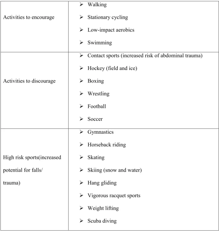
Table 2: Recommendations for sport activities during pregnancy
Activities to encourage Walking Stationary cycling Low-impact aerobics Swimming Activities to discourage
Contact sports (increased risk of abdominal trauma) Hockey (field and ice)
Boxing Wrestling Football Soccer
High risk sports(increased potential for falls/
trauma)
Gymnastics Horseback riding Skating
Skiing (snow and water) Hang gliding
Vigorous racquet sports Weight lifting
42
2.4. Exercise training and IUGR:
Based on the reported data, the link between exercise training during pregnancy and birth weight is evident. Although there are studies stating that there is no relationship between maternal physical activity and fetal birth weight 161, others have suggested that babies from recreational athletes have lower body fat compared with offspring of sedentary mothers 161. It has been shown that the rate of macrosomia and gestational diabetes in women who performed submaximal intensity exercise starting from week 6-8 of gestation is lower than the control group. The results also demonstrated that
participation in moderate-intensity aerobic exercise did not increase the risk of IUGR 175. Interestingly, Dr. Lavoie’s group has demonstrated that IUGR in the context of preeclampsia can be prevented by exercise training when performed both before and during gestation in different mouse models of this disease. This was related to a reduction in blood pressure and placental development normalisation in the exercise group compared to sedentary counterparts 212, 213.
2.5. Hypothesized protective mechanisms implicated in the prevention
of IUGR by exercise training
2.5.1. Enhanced placental development and vascularity
Abnormal placental development is a central cause of fetal growth-restriction. Insufficient trophoblastic invasion of the uterine spiral arteries in early pregnancy may contribute to an incomplete loss of
sensitivity to vasoconstrictors in utero-placental vessels, causing intermittent hypoxia and reperfusion 68. Conversely, regular physical activity in early pregnancy stimulates placental growth. Women who start training in early pregnancy have elevated placental volumes and growth rates 188, as well as a decreased fraction of non-functional tissue and an increased volume of villous tissue 189. Interestingly, these adaptations are still noticeable at term even if the mother stopped training by 20 weeks gestation,
43
indicating that early pregnancy is a critical period for placental development. Additionally, if the mother continues to exercise until term a slightly additional increase in placental volume and surface area will be observed 189. Improved placental growth and vascularity enhances its perfusion and transport capacity, and this may prevent reductions in fetal substrate and oxygen supplies during intermittent decreases in placental blood flow which may be associated with IUGR 188.
2.5.2. Prevention and/or reduction of oxidative stress
Regular physical training in non-pregnant rats, has been shown to augment antioxidant defense systems in heart, liver and muscle, which restricts cellular damage caused by oxidative stress related to acute bouts of exercise. Also, studies in animal models have shown that exercise training up-regulates antioxidants in skeletal muscles and growing evidence indicates that endurance exercise training promotes an elevation in both total SOD (superoxide dismutase) and GPX (glutathione peroxidase) activity in skeletal muscles. In this regard, it appears that high-intensity exercise training is generally more effective then low-intensity exercise in the up-regulation of muscle SOD and GPX activities 214, 215. In a study consisting of a 16 week aerobic exercise training program with an individualized intensity in healthy men and women, the activity of superoxide dismutase in erythrocytes (E-SOD), glutathione peroxidase in whole blood (GSH-Px), and glutathione reductase in plasma (P-GR) were measured and GSH-Px and P-GR activity were found to be increased without any alteration in E-SOD activity 216. In another study among a large group of Spanish women, with two categories of leisure time physical activity according to their intensity: low (<=6 METs) and high (>6 METs), a direct relationship between the amount and intensity of regular leisure physical activity and endogenous antioxidant enzymes was observed. Low intensity exercise training was associated with high SOD levels and high intensity exercise with high peroxidase levels. These results suggest a modulatory effect of leisure physical activity intensity on the anti-oxidative balance 217. Although no differences could be observed in erythrocyte antioxidant enzyme activities (SOD, glutathione peroxidase, and
44
catalase) between active and sedentary pregnant women before delivery, SOD and catalase activities were dramatically elevated 1 h post-partum in trained women which seemed to inhibit labor-induced increases in malondialdehyde (an indicator of lipid peroxidation) 218. These results propose that regular exercise training may enhance maternal antioxidant responses to augmented oxidative stress in normal pregnancies 218, which may prevent or improve endothelial dysfunction and thus IUGR. Indeed, in experimental animal models of atherosclerosis, hypercholesterolemia, hypertension, and diabetes, associations between oxidative stress and impaired endothelial function have been demonstrated 219-221.
2.5.3. Reduction of inflammation
There are many evidences supporting the anti-inflammatory effect of regular exercise training 222 in non-pregnant individuals and patients with heart failure 223 and coronary artery disease 224. It has been shown in healthy men and women that plasma levels of sTNF-R1, sTNF-R2, (soluble tumor necrosis factor receptors 1, 2) interleukin-6, and C-reactive protein are decreased with physical training 225. Exercise training can also attenuate interleukin-1, interleukin-6, and interferon-γ, while increasing the anti-inflammatory cytokine interleukin-10 224. Since there have been reports of increased inflammatory factors in IUGR, such as IL6 226, if exercise training has similar anti-inflammatory effects in pregnant women, this could prevent or decrease the inflammatory response that may contribute to the
development of IUGR.
2.5.4. Improving of endothelial dysfunction
It is proved that regular exercise training improves endothelial function in non-pregnant individuals with endothelial dysfunction 227, plus, it can favorably modify some risk factors of endothelium
dysfunction like blood pressure 228. Aerobic exercise has been demonstrated to raise local endothelium-dependent dilation in patients with endothelial dysfunction caused by aging 229 and type2 diabetes 230. It has also been shown that in heart failure patients large muscle mass exercise improves systemic
45
endothelial function which was measured by assessing the response to acetylcholine (ACh) and sodium nitroprusside (SNP) 230. If similar results are observed in women at risk for IUGR, training-induced correction of disease-related endothelial dysfunction may prevent the main pathological process leading to this disease.
3. Animal models of IUGR
Many of our knowledge regarding the short- and long-term effects of IUGR comes from animal studies. A number of animal models of maternal malnutrition and placental insufficiency have been developed over recent years to investigate the causes and consequences of IUGR. A variety of species have been studied, including: rodents, sheep and primates; and both, maternal dietary manipulations or surgical interventional techniques have been employed 231-237. We use animal models as they better reproduce the human condition compared to in vitro studies. However, in spite of the advances made using “in vitro” models to study some aspects of pregnancy, the IUGR condition as a whole is more properly represented in vivo. Still other features of pregnancy, such as the development of the uteroplacental circulation, fetal growth velocity and fetal development have no in vitro counterpart. Moreover, when new therapies arrive, although they are first tested extensively in vitro, they must present a clean reproductive toxicology panel in vivo 238 before they can be considered for use in humans, hence animal experiments are necessary. The majority of experimental fetal growth restriction studies are performed in rats and mice 239 (Table 3). We chose to study a mouse model because in mice, environment and genetic background can be easily controlled. Furthermore, their gestation has many characteristics which are common to human pregnancy 240, which makes them a good model of the disease. Generally, animal models of IUGR fall into three categories when divided by method of intervention: fetal intervention, maternal intervention and genetic models.
46
3.1 Fetal intervention models:
The hypoxic chick is the primary model of fetal intervention 241-243. Since many IUGR in human pregnancies are caused by placental insufficiency, there is no change in the health status of the mother. As such, the main advantage of this model is the ability to investigate the effects of hypoxia in the fetus in isolation, without any maternal effects. 244.
3.2. Maternal interventions
3.2.1. Surgical methods
There is a range of maternal interventions for inducing IUGR in animal models. The most frequent and oldest intervention is uterine artery ligation which was first introduced by Wigglesworth. In this
method, the uterine arteries of the pregnant rat are permanently ligated near the cervical end of the arterial arcade at day 17 of pregnancy. It causes utero-placental insufficiency which involves altered intrauterine environment characterized by hypoxia, reduced growth factor and hypoglycemia 245. Uterine artery ligation has been shown in other species to cause IUGR like guinea pigs and sheep 246-250. Similarly, uterine artery embolization in sheep also results in IUGR 251-253. Although these methods induce IUGR, the lack of an intact uteroplacental circulation in these models makes them less useful for testing maternal therapies that target uterine blood flow or the placental barrier directly.
Since bilateral uterine artery ligation obstructs blood supply, 30% of the fetuses die or go through partial resorption 254. As a result, it causes severe maternal outcomes such as necrotic uterus, ectopic pregnancy, abortion, etc. Therefore unilateral ligation is preferred to provide chronic placental insufficiency. This procedure in guinea pigs is performed at mid-gestation. In about one-third of the cases, fetal death occurred, in another third, fetuses with less than 60% of normal weight were observed and in the remainder all fetuses were in the normal weight range. It produces fetuses that are growth