Severe Skin Lesions Cause Patients With Inflammatory Bowel Disease to
Discontinue Anti-Tumor Necrosis Factor Therapy
JEAN-FRANCOIS RAHIER,* SÉBASTIEN BUCHE‡ LAURENT PEYRIN-BIROULET,§ YORAM BOUHNIK,|| BERNARD DUCLOS¶, EDOUARD LOUIS,# PAVOL PAPAY,** MATTHIEU ALLEZ,‡‡ JACQUES COSNES,§§ ANTOINE CORTOT,|||| DAVID LAHARIE, ¶¶ JEAN-MARIE REIMUND,## MARC LÉMANN,‡‡ EMMANUEL DELAPORTE,‡ JEAN-FRÉDÉRIC COLOMBEL,|||| and the GROUPE D'ETUDE THÉRAPEUTIQUE DES AFFECTIONS INFLAMMATOIRES DU TUBE DIGESTIF (GETAID)
*Department of Gastroenterology, Cliniques Universitaires UCL Mont Godinne, Yvoir, Belgium; ‡ Lille 2 University and Department of
Dermatology, Hôpital Claude Huriez, CHU Lille, France; §Department of Gastroenterology, CHU, Université Henri Poincaré Nancy 1, Nancy, France; || Department of Gastroenterology, Hôpital Beaujon, Paris, France; ¶Department of Gastroenterology, CHU, Strasbourg,
France; # Department of Gastroenterology, CHU, Liège, Belgium; ** Department of Gastroenterology, Medical University, Vienna, Austria; ‡‡ Department of Gastroenterology, Hôpital Saint Louis, Paris, France; §§Department of Gastroenterology, Hôpital Saint Antoine, Paris,
France; |||| Lille 2 University and Department of Gastroenterology, Hôpital Claude Huriez, CHU Lille, France; ¶¶Department of
Gastroenterology, CHU, Bordeaux, France; and ##Department of Gastroenterology, CHU, Caen, France
Abstract
BACKGROUND & AIMS: Psoriasiform and eczematiform lesions are associated with anti-tumor necrosis
factor (TNF)-α therapies. We assessed clinical characteristics, risk factors, and outcomes of skin disease in patients with inflammatory bowel diseases that presented with psoriasiform and eczematiform lesions induced by anti-TNF-α agents.
METHODS: We studied 85 patients (69 with Crohn's disease, 15 with ulcerative colitis, and 1 with
indeterminate colitis; 62 women) with inflammatory skin lesions (62 psoriasiform and 23 eczematiform lesions).
RESULTS: Twenty-four patients had a history of inflammatory skin lesions and 15 had a familial history of
inflammatory skin disease. Locations of eczematiform lesions varied whereas scalp and flexural varieties were mostly psoriasiform. Skin lesions emerged but inflammatory bowel disease was quiescent in 69 patients following treatment with any type of anti-TNF-α agent (60 with infliximab, 20 with adalimumab, and 5 with certolizumab). Topical therapy resulted in partial or total remission in 41 patients. Patients with psoriasiform lesions that were resistant to topical therapy and that changed anti-TNF-α therapies once or twice developed recurring lesions. Overall, uncontrolled skin lesions caused 29 patients to stop taking TNF-α inhibitors.
CONCLUSIONS: Inflammatory skin lesions following therapy with TNF-α inhibitors occurred most frequently
among women and patients with a personal or familial history of inflammatory skin disease; lesions did not correlate with intestinal disease activity. Recurring and intense skin lesions caused 34% of patients in this study to discontinue use of anti-TNF-α agents.
Keywords : Skin Eruptions ; Dermatological Complications ; Side Effects of Anti-TNF-α Therapy. Abbreviations used in this paper : CD, Crohn's disease ; DLQI, Dermatology Life Quality Index ; IBD,
inflammatory bowel disease ; IC, indeterminate colitis ; IFN, interferon ; IQR, interquartile range ; PDC, plasmacytoid dendritic cell ; TNF, tumor necrosis factor ; UC, ulcerative colitis ; UV, ultraviolet.
Anti-tumor necrosis factor (TNF) agents are used in a wide variety of immune-mediated diseases. Their efficacy has been proven in the treatment of inflammatory bowel disease (IBD) including both luminal and fistulizing Crohn's disease (CD) and ulcerative colitis (UC).1-6 The anti-TNF agents are globally safe6 although some concerns have been raised regarding an increased risk of opportunistic infections7 and lymphoproliferative diseases8 in a small percentage of patients. Attention has recently focused on a wide spectrum of skin lesions, the most common being eczematiform and psoriasiform eruptions (inflammatory lesions) arising in patients treated with anti-TNF agents. A recent literature survey identified 127 psoriatic skin lesions induced by anti-TNF agents in a variety of patients,9 most of them with rheumatic disorders. These lesions appear paradoxical because TNF is a pivotal molecule in the physiopathology of psoriatic skin lesions and anti-TNF agents have been approved for treatment of moderate to severe psoriasis for 5 years.10 The risk factors and natural history of these lesions, their impact on the treatment of IBD, and the impact on skin disease of switching the anti-TNF are poorly
documented.
multicentric series of patients with IBD presenting with psoriasiform and eczematiform lesions induced by anti-TNF agents (infliximab, adalimumab, and certolizumab).
Methods Study Design
We conducted a retrospective study between January 2004 and September 2009 of patients with new onset or exacerbation of eczematiform or psoriasiform lesions during treatment with anti-TNF agents for IBD observed in the GETAID centers (35 centers in Belgium, France, and Switzerland) and 1 center from Austria. Furthermore, from January 2006 to January 2010, all patients treated with anti-TNF agents in the department of
hepatogastroenterology of Lille and presenting with eczematiform or psoriasiform lesions were referred to a single dermatologist and consecutively recorded. Eczematiform lesions were defined as the association of xerosis and pruriginous ill-limited plaques with etythematous or squamous macules or vesicles. Psoriasiform lesions were defined as well delimited scaly erythematous plaques. Nail involvement (nail pitting and nail discoloration) and palmoplantar pustulosis were also recorded as psoriasiform lesions.
Data from each patient were collected using a standardized questionnaire. Patients were eligible if they experienced new onset, reactivation, or worsening of skin lesions (psoriasiform or eczematiform) with confirmation by a local dermatologist while being treated with anti-TNF agents. All cases were then reviewed simultaneously by a gastroenterologist JFR) and a single expert dermatologist (SB) prior to inclusion in the study. Histological analysis was not necessary for inclusion. Cases with imprecise dermatological diagnosis or absence of dermatological follow-up were excluded.
Variables
Clinical data included age, sex, smoking status, disease duration, IBD phenotype according to Montreal classification, type and dosage of the anti-TNF agent, concomitant medication, and disease activity (based on physician's appreciation) at the time of diagnosis of skin lesions. Information related to skin diseases included personal or familial (first-degree relatives) history of atopy (including atopic dermatitis, asthma, allergic rhinitis, and conjunctivitis) and/or psoriasis, time from starting anti-TNF agent to onset of skin lesions (the time to onset was considered the time the patient was regularly treated with the anti-TNF agent), morphological description, extent and topography of skin lesions, dermatological treatments, and histopathological information when available. With the Psoriasis Area and Severity Index (PASI) score being rarely applicable in our cases of psoriasiform lesions, the severity of the skin disease was assessed using the Dermatology Life Quality Index (DLQI).11 All follow-up information regarding skin complications and specific therapy was collected. Patients were categorized as responders or nonresponders to topical dermatological treatments. A favorable response was defined as a reduction of more than 50% of the lesions covering surface or a drop of the DLQI below 10. Persistent alteration of DLQI (above 10), withdrawal, or switching of anti-TNF agent for dermatological reasons was identified as nonresponse to topical treatments. Date and reason for discontinuation of anti-TNF agents (dermatological or nondermatological reasons), subsequent improvement of skin lesions after anti-TNF
discontinuation, and the impact of switching the anti-TNF treatment on skin lesion outcome were also recorded.
Results
Description of the Population
A total of 85 patients (23 males and 62 females) with IBD (69 CD, 15 UC, and 1 indeterminate colitis [IC]) were included. Demographic and clinical characteristics of patients are given in Table 1. Skin lesions were overall observed in patients who received infliximab (n = 60), adalimumab (n = 20), and certolizumab pegol (n = 5) therapy. At the time of onset of skin lesions, 79 patients were on maintenance scheduled therapy but 10 patients on infliximab had previous episodic treatment. No lesions occurred during high dose treatment and 6 patients developed skin lesions during the induction period. An estimate of the incidence of skin lesions was obtained from Lille Centre where, over the last 4 years, 562 patients initiated an anti-TNF treatment (341 infliximab and 221 adalimumab). During this period, 28 patients (5%) developed an inflammatory skin lesion: 2% developed psoriasiform lesions, and 3% eczematiform lesions.
Psoriasiform lesions were observed in 62 patients. None of them had psoriatic articular involvement. The median age at the time of occurrence of skin lesions was 32 years (interquartile range [IQR], 24 to 39) and the female to male ratio was 2:1. Twenty-eight were active smokers. The median time between start of anti-TNF therapy and the onset of psoriasiform lesions was 17 months (range, 1-60) for infliximab, 12 months for adalimumab (range, 0.5-29), and 4.5 months for certolizumab pegol (Table 2). Fourteen patients (23%) had a previous history of psoriasis before treatment with any anti-TNF agent (Table 1). The others were diagnosed as a new onset of psoriasiform eruption (incident psoriasiform eruption). Among patients with incident psoriasiform lesions, 8 (17%) reported a history of familial psoriasis, 2 of familial atopy, and 6 patients (13%) had personal history of atopy.
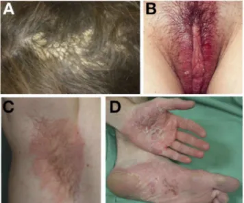
Figure 1. (A) Psoriasiform lesions of the scalp (tinea amiantacea form) induced by infliximab. (B) Flexural
psoriasiform lesions of the pubic region induced by infliximab. (C) Flexural psoriasiform lesions of axillae folds induced by adalimumab. (D) Palmo-plantar pustulosis induced by adalimumab.
Skin lesions were distributed in single (n = 12) or multiple sites (n = 50) (Table 2). The most frequently affected areas were the scalp (Figure 1A) and flexures (Figure 1B and C), respectively, in 40 (64%) and 27 (43%) patients. Combination of scalp and flexures involvement was observed in 21 (34%) patients. Palmoplantar psoriasiform lesions were observed in 22 patients including palmoplantar pustulosis (Figure 1D). The well-known "predilection" sites of psoriasis with the exception of scalp (the region sacralis and the extensor surfaces of the elbows and knees) were affected in 20 patients. Nail involvement was observed in 2 patients. Others localizations — trunk, face, members (with the exception of knees and elbows) — were observed in 11 patients. Distribution of skin lesions varied according to presence or absence of personal history of psoriasis. About half of patients with previous history of psoriasis were affected in the "predilection" sites (43%) and scalp (50%) whereas patients with no previous history had predominantly lesions of the scalp (69%) and flexures (46%). Histological data were obtained in 16 patients. Skin biopsies confirmed typical features of psoriasis, ie,
parakeratosis, agranulosis, epidermal hyperplasia, elongation of the rete ridges, dilated capillaries in the dermis, and perivascular infiltration in 12 patients and a lichenoid pattern in 4 patients. No difference regarding distribution of psoriasiform lesions was found according to IBD type, CD and UC phenotype, disease activity, and the anti-TNF received (data not shown).
Table 1. Demographic, Clinical Characteristics, and Treatment of 85 Patients With Inflammatory Bowel
Psoriasiform lesions (n = 62)
Eczematiform lesions (n = 23) Demographic characteristics of the population
Male, n (%) 20 (32) 3 (13)
Median age, y (IQR) 32 (24-39) 31 (23-39)
Current smoker, n (%) 28 (45) 10 (43)
Previous personal history of psoriasisa/atopyb, n (%) 14 (23)a 10 (43)b
New onset of skin lesion, n (%) 48 (77) 13 (57)
Personal history of atopy, n 6 -
Personal history of psoriasis, n - 1
Familial history of atopy, n 2 4
Familial history of psoriasis, n 8 1
IBD phenotype CD, n (%) 52 (84) 17 (74) UC, n (%) 9 (14) 6 (26) IC, n (%) 1 (2) - Age of diagnosis, n (%)c A1 15 (29) 3 (18) A2 34 (65) 14 (82) A3 3 (6) - Disease location, n (%)c L1 7 (13) 5 (30) L2 17 (33) 2 (11) L3 28 (54) 10 (59) L4 14 (27) 4 (24) Disease behavior, n (%)c B1 35 (67) 8 (47) B2 11 (21) 6 (35) B3 6 (12) 3 (18) p+ 28 (54) 8 (47) Extent of UC, nc E1 1 1 E2 5 1 E3 3 4 Disease activity Quiescent, n (%) 49 (79) 20 (87) Treatment
Anti-TNF treatment used at the time of skin lesion, n (%)
Infliximab 45(73)d 15 (65)e
Adalimumab 15(24)f 5 (22)g
Certolizumab 2 (3) 3 (13)
Use of immunomodulators at onset of skin lesion, n (%) 31 (50) 10 (43)
Corticosteroids 5 (8) -
Azathioprine 22 (35) 7 (30)
Methotrexate 4 (7) 3 (13)
No use of immunomodulators, n (%) 31 (50) 13 (57)
CD, Crohn's disease; IBD, inflammatory bowel disease; IC, indeterminate colitis; IQR, interquartile range; TNF, tumor necrosis factor; UC, ulcerative colitis.
aPsoriasis. bAtopy.
cAccording to Montreal classification.
dEight patients had episodic infliximab infusion in the past. eTwo patients had episodic infliximab infusion in the past. fEight patients received infliximab infusion in the past. gTwo patients received infliximab infusion in the past.
The outcome of patients is given in Figure 2. All patients were treated with topical corticosteroids, keratolytics (salicylic acid, urea), emollients, and vitamin D analogues. Ultraviolet (UV) therapy (UVA or narrow band
UVB) was used in 10 cases and effective in 5. Of the 62 patients, 25 (40%) had a good response to topical treatment and 37 had no response. Presence or absence of previous personal history of psoriasis did not influence response to topical treatment (data not shown). Nonresponders to topical treatment were considered for
withdrawal or switching of the anti-TNF agent. Addition of methotrexate to anti-TNF was unsuccessful in 4 patients. First anti-TNF therapy was stopped in 18 patients (16 for dermatological reasons). Twenty-six patients were switched (19 for dermatological reasons and 7 for nondermatological reasons) from first anti-TNF to a second anti-TNF. Improvement of skin lesions was observed in only 1 patient. Recurrence of skin lesions was observed and easily managed in 7 patients with an initially good response to local treatment but 1 quit the anti-TNF therapy. In 8 patients, the second anti-anti-TNF therapy was continued despite uncontrolled skin lesions because the benefit/risk ratio was considered strong enough. The second anti-TNF was stopped in 7 patients allowing complete regression of skin lesions. Two patients were unsuccessfully switched to a third anti-TNF. Overall a definitive withdrawal of anti-TNF therapies due to uncontrolled skin lesion was required in 25/62 (40%) patients allowing a complete regression of skin lesions in 24 patients within a median time of 3 months (range, 1-10). In Lille Centre, a definitive withdrawal of anti-TNF therapy due to uncontrolled skin lesion was observed in 27% of patients with psoriasiform lesions. One patient had ongoing skin lesions 8 months after anti-TNF
discontinuation. In patients with scalp localization, rate of abandon of the anti-TNF therapies due to uncontrolled skin disease was even higher approaching 48% (19/40).
Figure 2. Outcomes of psoriasiform lesions and effect of switching the anti-tumor necrosis factor (TNF) therapy.
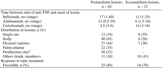
Table 2. Main Characteristics of the Skin Lesions Observed in 85 Patients with Inflammatory Bowel Disease
Psoriasiform lesions, n = 62
Eczematiform lesions, n = 23 Time between start of anti-TNF and onset of lesion
Infliximab, mo (range) 17 (1-60) 11 (1-25) Adalimumab, mo (range) 12 (0.5-29) 6 (1.5-10) Certolizumab, mo (range) 4.5 (3-6) 14 (3-16) Distribution of lesions, n (%) Single site 12 (19) 8 (35) Scalp 40 (65) 6 (26) Flexural varieties 27 (44) 7 (30) Palmo plantar 22 (35) - Predilection sitesa 20 (32) -
Others (trunk, members) 11 (18) 10 (43)
Response to topic treatment
Favorable, n (%) 25 (40) 16 (70)
TNF, tumor necrosis factor.
a
Predilection sites (except scalp) are the region sacralis and the extensor surfaces of the elbows and knees.
Eczematiform Lesions
Eczematiform lesions were observed in 23 patients. The median age at the time of occurrence of skin lesions was 31 years (IQR, 23-39) and the female to male ratio was 6:7. Ten patients were active smokers. The median time between start of anti-TNF therapy and the onset of eczematiform lesions was 11 months (range, 1-25) for infliximab and 6 months for adalimumab (range, 1.5-10), and 14 (range, 3-16) for certolizumab (Table 2). Of the 23 patients who developed eczematiform lesions, 10 (44%) had a previous history of atopy (Table 1). The other 13 patients were incident cases of atopy. Among them, 4 (31%) reported a history of familial atopy, 1 patient a personal history of psoriasis, and 1 of familial psoriasis.
Eczematiform lesions were equally distributed on the scalp, trunk, members, face, and flexures (Figure 3A and
B). There was no predominantly affected area (Table 2). Histological data were available in a minority of
patients (n = 4), and were consistent with eczema features, ie, spongiform dermatitis with edema in the epidermis between keratinocytes and perivascular lymphoid infiltrate. No difference regarding topography of eczematiform lesions was found according to IBD type, CD, and UC phenotype, disease activity, and the anti-TNF received (data not shown).
The outcome of patients is given in Figure 4. All patients were treated with topical steroids and emollients. In 1 case, topical tacrolimus was successfully used. Of the 23 patients, 16 (70%) had a good response to topic treatments and 7 patients had no response (30%). Nonresponders to topic treatment were considered for definitive withdrawal or switching the anti-TNF agent. First anti-TNF therapy was continued in 14 patients and stopped in 4 patients allowing complete regression. Five patients were switched (3 for dermatological reasons) from the first anti-TNF to a second anti-TNF. Improvement of skin lesions was observed in only 1 patient. One patient was lost to follow-up. The second anti-TNF therapy was continued in 2 patients and stopped in 1. One patient was unsuccessfully switched to a third anti-TNF. Overall a definitive withdrawal of anti-TNF therapies due to an uncontrolled skin lesion was required in 4/23 patients (17%) allowing a complete regression of skin lesions in 3 patients (data missing for 1 patient) within a median time of 3 months (range, 1-10). In Lille Centre, withdrawal was needed in 18% of patients with eczematiform lesions.
lower left member induced by adalimumab.
Figure 4. Outcomes of eczematiform lesions and effect of switching the anti-tumor necrosis factor (TNF)
therapy.
Discussion
Eczematiform and psoriasiform eruptions are, after infections, the most frequent dermatological adverse events in patients receiving anti-TNF-α therapy.12,13 Their prevalence in patients with IBD is unknown. Recent data from the rheumatologic literature indicates that the incidence rate of new onset psoriasis in patients treated with anti-TNF is around 1.04 (95% confidence interval, 0.97-1.54) per 1000 person-years.14 As psoriasis may be associated with CD,15 psoriasiform lesions may appear more frequently in this population. Fidder and colleagues found eczematiform or psoriasiform lesions in 9% of 743 IBD patients treated with infliximab.16 More recently, Esmailzadeh et al found an incidence rate of new onset eczema in 16% of patients receiving infliximab for various inflammatory disorders including 59 patients with IBD.17
them (SB). We believe that this methodology avoided misclassification of psoriasiform or eczematiform lesions. On the other hand, it probably reflects the most severe cases of skin lesions because patients who consulted once and had no follow-up were considered as having minor skin lesions and excluded from the analysis.
Skin manifestations were observed with the 3 anti-TNF agents that are used in IBD but a majority of patients were treated with infliximab. This most probably reflects the earlier approval of this drug in Europe. Data from the rheumatologic literature indicated that adalimumab induces a significantly higher rate of incident
psoriasiform eruptions compared with etanercept or infliximab.14 Whether this is true in IBD is unknown. It is generally accepted that the development or worsening of psoriasiform or eczematiform lesions during treatment with anti-TNF therapy can occur at any time from days to years after drug initiation. We observed a median time of 17 months for psoriasiform lesions and 11 months for eczematiform lesions after infliximab initiation. With adalimumab, time to onset was slightly shorter for both types of lesions (12 months and 6 months). This shorter time to onset of skin lesions with adalimumab was also observed in anti-TNF naive patients. However, a wide range was seen in each situation, and the small number of patients developing skin lesions with adalimumab makes it difficult to draw a conclusion. In a large series of 120 cases in the literature the mean time to appearance of the psoriasiform and eczematiform lesions for all TNF-α inhibitors (infliximab, etanercept, and adalimumab) was 9.5 months and 4 months for adalimumab.18
In our series, occurrence of psoriasiform or eczematiform lesions was associated with female gender and previous or familial history of psoriasis and/or atopy. Female predominance is well known in autoimmune diseases and may be related to hormonal influence and/or sex-related genes.19,20 These lesions might be gender-related and seem to occur more frequently in women. Among patients with incident psoriasiform lesions, 17% had a familial history of psoriasis and 31% of patients with incident eczematiform lesions had a familial history of atopy. This could suggest a genetic susceptibility. Common susceptibility genes have been identified in CD and psoriasis.21 Ten patients (43%) with eczematiform lesions and 28 patients (45%) with psoriasiform lesions were smokers. Palmoplantar lesions, which can be triggered by smoking,22 were observed in 22 patients of whom 15 (68%) were smokers. This close relationship has also been observed for other forms of psoriasis.23
In patients with psoriasiform lesions, 2 sites were predominantly affected: the scalp and the flexures with no correlation with IBD phenotype, anti-TNF therapy, or disease activity. Flexural psoriasis is generally an
uncommon phenotype affecting 2%-6% of patients with psoriasis.24 Also named inverse psoriasis, it involves the axillae, groins, gluteal clefts, and submammary folds but all the flexures can be affected. This phenotype which has already been reported in patients with CD25,26 appears less frequent in patients treated with anti-TNFs for rheumatologic disorders in whom the most frequent presentation is palmoplantar with or without plaque like-psoriasis.27
One of the most important findings is that a definitive withdrawal of anti-TNF therapies was necessary in 40% of patients with psoriasiform lesions and 17% with eczematiform lesions despite various therapeutic manipulations. Response to dermatological treatment was higher for eczematiform lesions (70%) than for psoriasiform lesions (40%). This poor response rate is probably biased by the severity of lesions collected in this cohort. Detailed data from Lille reported a definitive withdrawal of anti-TNF therapy in 27% of patients with psoriasiform lesions and 18% of patients with eczematiform lesions. Extrapolating data from this center indicates that overall, when initiating anti-TNF therapies, the risk of developing an inflammatory skin lesion is around 5% and the definitive need to withdraw treatment due to uncontrolled skin lesion is around 1%. In the early years, switching might have been tempted rapidly after a failure of topical treatment. Switching from 1 TNF inhibitor to another did not improve psoriasiform lesions in 25/26 patients suggesting a class-related side effect. On the other hand,
definitive resolution of skin lesions following anti-TNF discontinuation was observed in almost all patients as already reported.27 The effect of TNF inhibition on skin lesions is thus reversible.
The pathogenesis of psoriasiform and eczematiform eruptions occurring under anti-TNF treatment remains poorly understood. They appear paradoxical because tumor necrosis factor α plays a key role in IBD and psoriasis28 and anti-TNF therapies can dramatically improve the outcomes of both diseases.6,28-30 Some
hypotheses have been formulated. Plasmacytoid dendritic cells (PDCs) might play a key role in the induction of psoriasiform lesions by anti-TNFs.31 PDCs are natural interferon (IFN)-α producing cells. They infiltrate the skin of psoriatic patients and become activated to produce IFN-α early during disease formation.32 TNF inhibits PDC maturation from hematopoietic progenitors as well as IFN-α production.33 Subsequently, inhibition of TNF may allow unlimited and unregulated production of IFN-α by PDCs. Increased IFN-α expression was found in the dermal vasculature and in perivascular lymphocytic infiltrate of lesional skin of patients with TNF antagonist therapy.33 Altered lymphocyte trafficking during TNF inhibition may also participate in this process through the expression of chemokine receptor CXCR3 ligands.34
There are some limitations of this study. The main limitation concerns the selection of patients. This paper reports on a selected cohort of patients presenting with severe psoriasiform and eczematiform skin lesions seen in various centers. Thus inclusion into the study was biased by severity of skin lesions. However we believe that these patients with more severe skin lesions are those that pose problems to the clinicians in their daily practice. Therefore, the withdrawal rate, which is derived from cases at all participating centers might be biased by the severity of the cases collected. Secondly, our work is not an epidemiological study examining the incidence of skin lesions in patients with IBD treated with anti-TNF therapy. We only gave an estimate of the rate of patients presenting with such lesions in the Lille Centre even though this should not be seen as true incidence data. Lastly, the data presented are descriptive, but provide new information about increased frequency among those with familial/personal history of inflammatory skin lesions, an altered distribution of psoriasiform lesions in IBD patients not seen in the rheumatologic population, the varying distribution of lesions depending on a personal history of psoriasis, and the failure of resolution after switching the anti-TNF agents.
In conclusion, psoriasiform and eczematiform skin lesions are a significant side effect of anti-TNF therapies. In our experience, they frequently occur in women and patients with a familial history of psoriasis and/or atopy lesions. Because the most severe forms can lead to cessation of anti-TNF therapy they need to be carefully managed with the help of a dermatologist. All patients should be referred to a dermatologist for clinical evaluation and biopsy of skin lesions if indicated. We recommend thorough treatment of the skin lesions by dermatologists with topical corticosteroids, emollients, keratolytics, vitamin D analogues, and phototherapy. Skin lesion resolution should be achieved in most cases. We would not advise a change in the TNF antagonists if the lesions are unresponsive to conventional treatment although a larger trial should address this question more specifically. Future research is needed to further explore epidemiological, clinical, and fundamental aspects of this vexing paradoxical reaction.
Conflicts of interest
These authors disclose the following: Jean-François Rahier received lecture fees from speaking at continuing medical education events from Abbott Laboratories and Schering-Plough; Laurent Peyrin Biroulet has received consulting fees from Abbott Laboratories and UCB Pharma; Yoram Bouhnik received lecture fees from Abbott Laboratories and Schering-Plough Corporation; Edouard Louis declared consulting fees or paid advisory board for Abbott, Schering-Plough, and UCB; Pavol Papay received honoraria as consultant for Abbott, Centocor, and UCB Pharma; Matthieu Allez has served or received honoraria for teaching activities from Abbott
Pharmaceuticals, Schering-Plough, and UCB Pharma; Jacques Cosnes received consulting fees from Abbott Laboratories and lecture fees from speaking at continuing medical education events from Abbott Laboratories and UCB Pharma; Antoine Cortot has served as consultant for and received honoraria from Ferring and Schering-Plough; David Laharie received honoraria from Abbott Pharmaceuticals and Schering-Plough; Jean-Marie Reimund received lecture fees for speaking at continuing medical education events from Abbott
Laboratories and grants for basic research from Ferring Laboratories and UCB Pharma; Marc Lémann declared consulting fees or paid advisory boards for Abbott Laboratories, Cellerix SL, Centocor, Schering-Plough Corporation, UCB Pharma, and received lecture fees from Abbott Laboratories, Schering-Plough Corporation, and UCB Pharma; Emmanuel Delaporte received lecture fees for Abbott, Schering-Plough, and Wyeth; and Jean-Frédéric Colombel has served as a consultant for and received honoraria and research grants from Centocor, Inc, Schering-Plough, UCB Pharma, and Abbott Pharmaceuticals. The remaining authors disclose no conflicts.
Acknowledgments
In addition to the authors, the following investigators participated in the study: Pierre Desreumaux (CHU Lille), Vered Abitbol (Hôpital Cochin Paris), Arnaud Boureille (CHU Nantes), Hedia Brixi-Benmansour (CHU Reims), Benoît Coffin (Hôpital Louis-Mourier Paris), Mathurin Flamant (CHU Nantes), Eric Lerebours (CHU Rouen), Jacques Moreau (CHU Toulouse), Stéphane Nancey (CHU Lyon), Anne Laure Pelletier (Hôpital Bichat Paris), and Guillaume Savoye (CHU Rouen).
The corresponding author had full access to all of the data and takes full responsibility for the veracity of the data and analysis.
This work was conducted in conformity with ethical and human principles of research.
1. Hanauer SB, Feagan BG, Lichtenstein GR, et al. Maintenance infliximab for Crohn's disease: the accent I randomised trial. Lancet 2002;359:1541-1549.
2. Colombel JF, Sandborn WJ, Rutgeerts P, et al. Adalimumab for maintenance of clinical response and remission in patients with Crohn's disease: the CHARM trial. Gastroenterology 2007;132: 52-65.
3. Schreiber S, Khaliq-Kareemi M, Lawrance IC, et al. Maintenance therapy with certolizumab pegol for Crohn's disease. N Engl J Med 2007;357:239-250.
4. Sands BE, Blank MA, Diamond RH, et al. Maintenance infliximab does not result in increased abscess development in fistulizing Crohn's disease: results from the Accent II study. Aliment Pharmacol Ther 2006;23:1127-1136.
5. Rutgeerts P, Sandborn WJ, Feagan BG, et al. Infliximab for induction and maintenance therapy for ulcerative colitis. N Engl J Med 2005;353:2462-2476.
6. Peyrin-Biroulet L, Deltenre P, de Suray N, et al. Efficacy and safety of tumor necrosis factor antagonists in Crohn's disease: meta-analysis of placebo-controlled trials. Clin Gastroenterol Hepatol 2008;6:644-653.
7. Toruner M, Loftus EV Jr., Harmsen WS, et al. Risk factors for opportunistic infections in patients with inflammatory bowel disease. Gastroenterology 2008;134:929-936.
8. Siegel CA, Marden SM, Persing SM, et al. Risk of lymphoma associated with combination anti-tumor necrosis factor and immunomodulator therapy for the treatment of Crohn's disease: a meta-analysis. Clin Gastroenterol Hepatol 2009;7:874-881. 9. Ko JM, Gottlieb AB, Kerbleski JF. Induction and exacerbation of psoriasis with TNF-blockade therapy: a review and analysis of 127 cases. J Dermatol Treat 2009;20:100-108.
10. Smith CH, Anstey AV, Barker JN, et al. British association of dermatologists' guidelines for biologic interventions for psoriasis 2009. Br J Dermatol 2009;161:987-1019.
11. Finlay AY, Khan GK. Dermatology Life Quality Index (DLQI)-a simple practical measure for routine clinical use. Clin Exp Dermatol 1994;19:210-216.
12. Flendrie M, Vissers WH, Creemers MC, et al. Dermatological conditions during TNF-alpha-blocking therapy in patients with rheumatoid arthritis: a prospective study. Arthritis Res Ther 2005;7:R666-R676.
13. Lee HH, Song IH, Friedrich M, et al. Cutaneous side-effects in patients with rheumatic diseases during application of tumour necrosis factor-alpha antagonists. Br J Dermatol 2007;156: 486-491.
14. Harrison MJ, Dixon WG, Watson KD, et al. Rates of new-onset psoriasis in patients with rheumatoid arthritis receiving anti-tumour necrosis factor alpha therapy: results from the British Society for Rheumatology Biologies Register. Ann Rheum Dis 2009;68:209-215. 15. Delaporte E. [Immune-mediated inflammatory diseases and psoriasis]. Ann Dermatol Venereol 2008;135;Suppl 4:S269-S274.
16. Fidder H, Schnitzler F, Ferrante M, et al. Long-term safety of infliximab for the treatment of inflammatory bowel disease: a single-centre cohort study. Gut 2009;58:501-508.
17. Esmailzadeh A, Yousefi P, Farhi D, et al. Predictive factors of eczema-like eruptions among patients without cutaneous psoriasis receiving infliximab: a cohort study of 92 patients. Dermatol Basel Switzerland 2009;219:263-267.
18. Wollina U, Hansel G, Koch A, et al. Tumor necrosis factor-alpha inhibitor-induced psoriasis or psoriasiform exanthemata: first 120 cases from the literature including a series of six new patients. Am J Clin Dermatol 2008;9:1-14.
19. Whitacre CC. Sex differences in autoimmune disease. Nat Immunol 2001;2:777-780.
20. Zandman-Goddard G, Peeva E, Shoenfeld Y. Gender and autoimmunity. Autoimmun Rev 2007;6:366-372.
21. Ho P, Bruce IN, Silman A, et al. Evidence for common genetic control in pathways of inflammation for Crohn's disease and psoriatic arthritis. Arthritis Rheum 2005;52:3596-3602.
22. Hagforsen E, Awder M, Lefvert AK, et al. Palmoplantar pustulosis: an autoimmune disease precipitated by smoking? Acta Derm Venereol 2002;82:341-346.
23. Naldi L, Mercuri SR. Smoking and psoriasis: from epidemiology to pathomechanisms. J Invest Dermatol 2009;129:2741-2743.
Dermatol 2005;15:13-17.
25. Avila Alvarez A, Garcia-Alonso L, Solar Boga A, et al. Flexural psoriasis induced by infliximab and adalimumab in a patient with Crohn's disease [in Spanish]. An Pediatr (Barc). 2009;70:278-281.
26. Peramiquel L, Puig L, Dalmau J, et al. Onset of flexural psoriasis during infliximab treatment for Crohn's disease. Clin Exp Dermatol 2005;30:713-714.
27. Collamer AN, Guerrero KT, Henning JS, et al. Psoriatic skin lesions induced by tumor necrosis factor antagonist therapy: a literature review and potential mechanisms of action. Arthritis Rheum 2008;59:996-1001.
28. Mease P. TNFalpha therapy in psoriatic arthritis and psoriasis. Ann Rheum Dis 2004;63:755-758.
29. Gottlieb AB, Evans R, Li S, et al. Infliximab induction therapy for patients with severe plaque-type psoriasis: a randomized, double-blind, placebo-controlled trial. J Am Acad Dermatol 2004;51:534-542.
30. Papp KA, Tyring S, Lahfa M, et al. A global phase III randomized controlled trial of etanercept in psoriasis: safety, efficacy, and effect of dose reduction. Br J Dermatol 2005;152:1304-1312.
31. Palucka AK, Blanck JP, Bennett L, et al. Cross-regulation of TNF and IFN-alpha in autoimmune diseases. Proc Natl Acad Sci U S A 2005;102:3372-3377.
32. Nestle FO, Conrad C, Tun-Kyi A, et al. Plasmacytoid predendritic cells initiate psoriasis through interferon-alpha production. J Exp Med 2005;202:135-143.
33. de Gannes GC, Ghoreishi M, Pope J, et al. Psoriasis and pustular dermatitis triggered by TNF-{alpha} inhibitors in patients with rheumatologic conditions. Arch Dermatol 2007;143:223-231.
34. Seneschal J, Milpied B, Vergier B, et al. Cytokine imbalance with increased production of interferon-alpha in psoriasiform eruptions associated with antitumour necrosis factor-alpha treatments. Br J Dermatol 2009;161:1081-1088.