THE JOURNAL OF BIOIDGICAL CHEMISTRY
0 1994 by The American Society for Biochemistry and Molecular Biology, Inc Vol. 269, No. 26, Issue of July 1, pp. 17448-17453, 1994 Printed in U.S.A.
Cold Adaptation
of
Proteins
PURIFICATION,
CHARACTERIZATION,
AND SEQUENCE OFTHE HEAT-LABILE
SUBTILISINFROM
THEANTARCTIC PSYCHROPHILE
BACILLUS TA41*(Received for publication, March 7, 1994, and in revised form, April 26, 1994) Stephane Davail, Georges Feller$, Emmanuel Narinx, and Charles Gerday
From the Laboratory of Biochemistry, Institute of Chemistry B6, University of Liege, B-4000 Liege, Belgium
The gene of subtilisin 541, an alkaline protease se- creted by the psychrophile Bacillus TA41, encodes for a preproenzyme of 419 amino acids residues. The nucle- otide sequence and
5-
and COOH-terminal amino acid sequencing of the purified enzyme indicate that the ma- ture subtilisin 541 is composed of 309 residues with a predictedM,
= 31,224. Subtilisin 541 shares most of its properties with mesophilic subtilisins (structure of the precursor, 52% amino acid sequence identity, alkaline pH optimum, broad specificity, Ca" binding) but is char- acterized by a higher specific activity on macromolecu- lar substrate, by a shift of the optimum of activity to- ward low temperatures, and by a low thermal stability. The enzyme also differs by an acidic PI (5.3) and the presence of one disulfide bond. It is proposed that the psychrophilic enzyme possesses a more flexible molecu- lar structure when compared to mesophilic and thermo- philic subtilases in order to compensate for the reduc- tion of reaction rates at low temperatures. The model of subtilisin 541 indeed reveals several features able to in- duce a more flexible, heat-labile conformation: the oc- currence of four extended surface loops, a very hydro- philic surface through 11 extra Asp residues, and the lack of several salt bridges and aromatic-aromatic inter- actions. The low affinity of the Cal calcium binding site (K,,,,,=
lo-' M), resulting possibly from one chelatingside chain substitution and the stacking of Gly residues, also reflect a less compact conformation. The difference of free energy of stabilization between subtilisin 541 and a mesophilic subtilisin suggests that the balance of exo- and endothermically formed weak bonds is critical for the enzyme flexibility.
Environmental factors are key determinants in the process of adaptation and evolution of living organisms. In this respect, analysis of the structure-stability relationships in proteins from extremophiles provides valuable insights on the molecu- lar strategies adopted in response to environmental stress such as extremes of pH, pressure, ionic strength, or temperature (Jaenicke, 1991; Di Prisco, 1991). I t is anticipated that the understanding of such molecular adaptations will give access to the physical basis of the forces driving the folding of a polypep- tide chain. Among the microorganisms, thermophiles have been
CT915/02 (to S. D. and C. G.) and by Research Grant 2.4526.92 (to C. G.)
*This work was supported by EC Allocation Grant ERBSCU from the Fonds de la Recherche Fondamentale et Collective. The costs page charges. This article must therefore of publication of this article were defrayed in be hereby marked "aduertise-
part by the payment of ment" in accordance with 18 U.S.C. Section 1734 solely to indicate this fact.
The nucleotide sequencels) reported in this paper has been submitted to the GenBankmIEMBL Data Bank with accession number(s) X63533.
f To whom correspondence should be addressed. Tel.: 32-41563343; Fax: 32-41563364.
the most widely studied in an effort to identify the structural features allowing their proteins to retain a folded conformation a t elevated temperatures. These enzymes are characterized by the strengthening of one or a combination of noncovalent in- teractions such as salt bridges, hydrophobic and weakly polar interactions, hydrogen bonding, charge-dipole interactions in &-helices, proline residues in loops, or ion binding (Jaenicke, 1991; Fontana, 1991).
At the other end of the biological temperature scale, psy- chrophiles have to cope with the reduction of the enzymatic reaction rates at often sub-zero temperatures. Psychrophilic enzymes are thought to have evolved toward a high conforma- tional flexibility, which is responsible for a n increased catalytic efficiency associated with a low stability (Hochachka and Som- ero, 1984). The study of these enzymes is still fragmental, and the molecular basis of adaptations to low temperatures re- mains largely unknown. Cloning of genes from psychrophilic bacteria in Escherichia coli results in the expression of ther- molabile recombinant enzymes active at temperatures close to 0 "C, demonstrating the intrinsic character of these properties (Feller et al., 1991, 1992; Davail et a l . , 1992; Rentier-Delrue et
a l . , 1993). In most cases, however, the analysis of psychrophilic protein sequences has been impaired by insufficient homology with their mesophilic or thermophilic counterparts. A critical step will be the availability of the refined crystallographic structure of such enzymes, which presently is lacking.
Here, we report the characterization of subtilisin S41, a n alkaline protease secreted by the Antarctic psychrophile Bacil- lus TA41. This enzyme was selected because 50 subtilisin-like proteases, or subtilases, have been characterized and se- quenced (Siezen et al., 1991). Moreover, the three-dimensional structures of subtilisin BPN' (McPhalen and James, 19881, subtilisin Carlsberg (Bode e t a l . , 19871, subtilisin Savinase (Betzel et al., 19921, thermitase (Gros et al., 1989), and protein- ase K (Betzel et al., 1988) have been reported. The large amount of data available for subtilisins, as illustrated by over 450 site-directed mutants constructed, and their industrial im- portance as detergent additives make them attractive model enzymes for structure-stability studies.
EXPERIMENTAL PROCEDURES
Source and Purification of Subtilisin S41-The strain Bacillus sp. TA41 was isolated from sea water at the Dumont d'Urville Antarctic Station (6O04O'S; 40"Ol'E) and was grown at 4 "C for 5 days in 6 liters
of marine broth containing 5 g/liter Bacto-peptone, 1 g/liter yeast ex-
tract, 15 g/liter sea salts, pH 7.6. After centrifugation of the culture at 12,000 x g for 15 min, the supernatant was concentrated up to 200 ml and diafiltrated against 20 mM Tris, 1 mM CaCl,, pH 8.5, using a Mini- tan tangential flow ultrafiltration unit (Millipore) fitted with PTGC membranes (10 kDa molecular mass limit). The sample was loaded on
a phenyl-Sepharose CL-4B column (5 x 15 cm) equilibrated in the above mentioned buffer and eluted with a linear gradient of isopropanol (0- 15%, 500-500 ml). Fractions containing proteolytic activity were pooled, inhibited when required by diisopropyl fluorophosphate, and dialyzed against 2 x 2 liters of 20 mM Tris, 1 mM CaCl,, pH 8.5. For further experiments, the purified protease was either conditioned in the appro-
Subtilisin S41 from the Psychrophile
Bacillus TA41
17449
priate buffers by gel filtration on PDlO column (Pharmacia Biotech Inc.) or lyophilized. Subtilisin Carlsberg was from Sigma.
Enzyme Assay-Proteolytic activity was routinely assayed at 25 "C using 1 mM N-succinyl-Ala-Ala-Pro-Phe-p-nitroanilide (AAPF)' as sub- strate in 50 mM Tris, 1 mM CaCl,, pH 8.5. Activities toward the synthetic substrate were recorded in a thermostated Uvicon 860 Spectrophotom- eter (Kontron) and calculated on the basis of an extinction coefficient for p-nitroaniline of 8,480 M - ~ cm-' at 412 nm (DelMar et al., 1979). For
comparative studies, azocasein was used as a nonspecific substrate. The assay was carried out at 25 "C for 15 min in a reaction mixture con- taining 250 pl of 50 m Tris, 1 II~M CaCl,, 200 m NaC1, pH 8.5, 100 pl of 3% azocasein in the buffer, and 150 pl of enzyme. The reaction was stopped by addition of 500 pl of 10% trichloroacetic acid. Proteolytic activity was calculated on the basis of an extinction coefficient of 900 cm" at 366 nm for the chromophore.
Analytical ProceduresSDS-polyacrylamide gel electrophoresis and
isoelectric focusing were run essentially as described by the supplier of
the electrophoresis equipment (Hoeffer Scientific Instruments). Cal- cium bound to subtilisin S41 was measured using a Perkin-Elmer 303 atomic absorption spectrophotometer after gel filtration on PDlO col- umn eluted with 25 mM NH,HCO,, lyophilization, and solubilization in high punty water. Activation kinetics by calcium titration was per- formed in 50 mM Tris, 1 m EGTA, 1 mM AAPF, pH 8.5. The desired free Ca2+ concentration was set by addition of 100 mM CaC1, according to a program developed by Robertson et al. (1982). Sulhydryl titration by
5,5'-dithiobis(2-nitrobenzoic acid) was made in denaturing conditions before and after reduction of the proteins with sodium borohydride (Habeeb, 1973). Active site titration was performed with N-trans-cin- namoylimidazole according to Bender et al. (1966) using an extinction coefficient of 21,000 M-' cm-' at 310 nm in 50 mM piperazine-HC1,2 m CaCl,, pH 6. The energy of activation (E,) was determined from the slope (-E,IR) of Arrhenius plots, and the thermodynamic activation parameters of the proteolytic reaction were calculated according t o the following equations.
AG* = A H * - TAS* 0%. 1)
M* = E , - RT (Eq. 2)
AS* = 2.303 R(log kc,, - 10.753 - log T
+
E,/2.303 RT) (Eq. 3) Cleavage sites of oxidized insulin B-chain were determined by digesting the peptide with 2% subtilisin S41 at 4 "C and 20 "C for 2 h in 20 mTris, 1 mM CaCl,, pH 8.5, for 2 h. The digest was submitted t o three Edman degradation cycles, and the released 3-[4-(4'-dimethylamino-
azobenzenel-2-thiohydantoin amino acids were analyzed by two-dimen- sional thin layer chromatography. Cloning of the subtilisin-encoding gene from Bacillus TA41 in E. coli has been described previously (Davail et al., 1992). The nucleotide sequence was determined on both strands using the pGEM Single Strand System (Promega) and Seque- nase (U. S. Biochemical Corp.). The NH,-terminal amino acid sequence of the native subtilisin S41 was determined using a pulsed liquid phase protein sequenator (Applied Biosystems 477A) equipped with an on-line 120A phenylthiohydantoin analyzer. Partial amino acid sequences of a CNBr peptide (purified on a Sephadex G50 column) and of a tryptic peptide (purified by high performance liquid chromatography) were also determined. The COOH-terminal amino acid sequence was investigated by carboxypeptidase Y digestion (Klemm, 1984) of 20 nmol of purified
subtilisin S41. The released amino acids were analyzed using a Dionex DC300 amino acid analyzer equipped with a Waters model 440 dual wavelength colorimeter and a high performance liquid chromatography polystyrene sulfonic column (Waters, 0.4 x 25 cm).
Molecular Modeling-Atomic coordinates were obtained from the Brookhaven Protein Data Base for subtilisin BPN' (code 2SNI; McPhalen and James (1988)), subtilisin Carlsberg (1CSE; Bode et al.
(1987)), and thermitase (1TEC; Gros et al. (1989)). Structures were superimposed using the program FRODO (Jones, 1978) on an Evans and Sutherland PS330 system. The subtilisin S41 model was con- structed from the subtilisin BPN' coordinates; insertions 42a, 42b, and 75a were built from thermitase coordinates. Amino acid replacements were generated in the low energy conformation, and bad contacts were corrected according t o the superimposed models. The amino acid num- bering corresponds to that of mature subtilisin BPN'.
The abbreviation used is: AAPF, N-succinyl-Ala-Ala-Pro-Phe-p- nitroanilide.
pea'+ PH
Kacapp, for Caa+ and of the pH optimum of subtilisin S41. Left,
FIG. 1. Determination of the apparent dissociation constant
calcmm-Induced activation following CaC1, titration in 20 mMTris, 1
mM EGTA, 2 m AAPF, pH 8.0; pCa2+ = -log[Ca2+l. Right, effect of pH on activity toward the macromolecular substrate azocasein ).( and the peptidic synthetic substrate AAPF (0).
RESULTS
Effect of Tkmperature on Growth and Protease Secretion-
The strain Bacillus TA41 is able to grow from 0 "C to about 25 "C. However, temperatures higher than that ofAntarctic sea water (-2 to 4 "C) lead to 50-60% inhibition of growth and protease secretion in the culture supernatant. Growth and al- kaline protease secretion occurred concomitantly, and no spor- ulation or secretion stimulation in the late stationary phase was observed.
Purification and Characterization of Subtilisin S41-
Alkaline protease production by Bacillus TA41 at 4 "C amounts to 7 mgll and represents about 50% of the total exoproteins. A protocol based on supernatant ultrafiltration and on hydropho- bic chromatography has been devised, leading to 65-70% re- covery of the protease in a fairly pure state as judged by SDS- gel electrophoresis, isoelectric focusing, and limited NH,- terminal sequencing.
The molecular mass of the purified subtilisin S41 has been estimated at 30,000 Da by gel filtration on a calibrated Seph- adex GlOO column. On SDS-gel electrophoresis, the apparent molecular mass is 35 kDa, but this can be related t o anomalous SDS binding as reported for other subtilisins (Wells et al., 1983). The calculated extinction coefficient was eZBO = 37,849 M - ~ cm". The isoelectric point of the native enzyme has been found at pH 5.3. This value is drastically lower than that of meso-
philic subtilisins which have PI between 9 and 11 (Markland and Smith, 1971; Betzel et al., 1992). Calcium assay deter- mined by atomic absorption indicated that S41 is a metallopro- tein containing 1.5-1.8 Ca2+/mol of enzyme. These figures are consistent with the low occupancy of the Ca2 calcium binding site in subtilisins (Betzel et al., 1992). EGTA-mediated removal of Ca2+ ions results in the reversible inactivation of subtilisin S41. This allowed the determination of an apparent dissocia- tion constant Kd!app) = 1.3 M by activation kinetics following calcium titration (Fig. 1). This Kdcapp, value relates to the high affinity Ca2+ binding site because occupancy of the low affinity site (Ca2) of subtilisins is not required for activity (Pantoliano
et al., 1988). Like other subtilisins, the protease of Bacillus
TA41 is relatively resistant to detergent-induced denaturation: SDS and Triton X-100 at 1% concentration result in 62% and
36% inhibition of the activity, respectively.
Substrate Specificity, Kinetic Parameters, and Temperature Dependence-The optimum pH value for activity of subtilisin
S41 was found near pH 9 using either a macromolecular or a synthetic substrate (Fig. 1). It is worth mentioning that 85% of residual activity still remained at pH 11, as also reported for subtilisins Carlsberg and BPN' (Markland and Smith, 1971). Subtilisins have a very broad substrate specificity (Svend- sen, 1976). The B-chain of oxidized insulin has been used for
determining the specificity of the alkaline protease from Bacil-
lus TA41. As shown in Fig. 2, subtilisin S41 has four major
17450
Subtilisin ,941
from
the Psychrophile Bacillus TA41
Phe-Va1-Asn-Glu-Hir-Leu-CySOJI-Gly-Ser-His-teu-Val-Glu-Ala-leu-Iyr-~eu-Val-CySO~H-6ly-6lu-Arg-Gly-Phe-Phe-~yr-Thr-Pra-lys-AlaI l l 1
I4
t l'
1 "
1
'
I l t
1
I LB C 11
4
FIG. 2. Hydrolysis of the B-chain of oxidized insulin by subtilases. Hydrolysis by subtilisin Carlsberg (A) (Svendsen, 1976), subtilase Ib
from S. fradiae (B), and subtilisin S41 (C). Heavy arrows indicate the main site of splitting, and light arrows locate bonds split after completion of the hydrolysis.
TABLE I
Relative activities of subtilisins S41 a n d Carlsberg toward synthetic chromogenic substrates
Relative activity Subtilisin S41 Subtilisin Carlsberc Substrate N-Succinyl-Ala-Ala-Pro-Leu-pNa" 100 N-Succinyl-Ala-Ala-Pro-Phe-pNa 75 N-Succinyl-Ala-Ala-Ala-pNa 1.1 N-Succinyl-Gly-Gly-Phe-pNa 0.7 N-Succinyl-Ala-Ala-Val-pNa < N-Succinyl-Phe-pNa < N-Benzoyl-kg-pNa < p-Nitrophenyl acetate 0.6 p-Nitrophenyl butyrate 6.0
"pNa: p-nitroanilide; <, not detectable.
% 100 130 0.5 0.7 < C < < 0.9 60
2
..-. +w,:
20 30 60 W O 40 80 Tirnehin) TernperatweF'C)FIG. 3. Thermal stability and thermodependence of the activ- ity of subtilisin 541 (0) and subtilisin Carlsberg (I). Left, enzymes were incubated a t 50 "C in 20 mM Tris, 1 mM CaCl,, pH 8.0, and residual activities of timed aliquots were recorded using AAPF as substrate. Right, specific activities were recorded at increasing temperatures us- ing azocasein as substrate.
TABLE I1
Kinetic a n d thermodynamic activation parameters of proteolysis at 15 "C
Parameter Subtilisin S41 Subtilisin Carlsberg kc,, W 1
E , ( k J mol")"
AG* ( k J mol-I) AH* ( k J mol")
AS* (J mol" K-')
LI Experimental energy of activation below 15 "C.
25.4
*
0.3 (3) 5.4 -c 0.1 (3) 38.5 f l(16) 48.5 f 1 (14)62
*
1 66 -c 1 36 -c 1 46 2 1-92
*
3 -70 f 2His' and Le~''-Tyr'~ corresponds to major cleavage sites of mesophilic subtilisins, whereas splitting of bonds C y ~ ' ~ - G l y ~ ~ and PheZ4-Phez5 by these enzymes is only reached after comple- tion of the hydrolysis. The cleavage specificity of subtilisin S41
is also very close to that of protease Ib from Streptomyces fra-
diae. Increasing the incubation temperature from 4 "C t o 20 "C does not modify the specificity of the psychrophilic enzyme. The side chain specificity of subtilisin S41 for the hydrolysis of short synthetic substrates does not differ markedly from the pattern of subtilisin Carlsberg (Table
I).
Subtilisin S41 also requires a proline residue in position P2 for optimal activity but an ali- phatic residue (Leu) in positionP1
leads to faster hydrolysis than with an aromatic side chain (Phe) at that position. How- ever, the AAPF/L peptides, originally designed for chymotryp- sin assay (DelMar et al., 1979), are relatively poor substrates for subtilisin S41, which displays 60-70% lower k,,JKm values than subtilisin Carlsberg between 5 "C and 25 "C.The denaturation curves of subtilisins S41 and Carlsberg
M K R S G K I P ~ T A H L A V T L H H P A I G V S A N R G N ~ ~ N E K P R V L V D S ~ Q N N L K
- 1 1 0 -80 -60
NVKEPYCVHWDFACEGF~TNNEKQFNALQNNKNLTVJ~KVPELEIATATNKPEALYNAM
signal peptide propeptide
- 4 0 -20 - 1 hl hZ
.*.*.*
.*.*** b l...
b2...
---AQSV----PYGVSPI-KAPALHSGSNVKVAVIgSGIDSSHPDL--KVA~SM - - - A Q l l r - - - - P Y C I P L I - K A D K V P A 4 G F G I ~ S R ~ L - - N W G C A S P - - - A Q S V - - - - P Y C I S P I - K P ~ ~ S ~ A V I ~ S G I D S S H P D L - - ~ ~ S F - - - A Q S V - - - - P W G I S R V - 9 P ~ G L T C S G V K V A V ~ C I - S ~ D L - - N I R G G A S F YTPNoPYFSSRPYCWKI-PAPQAW-DIAEGSGAKIAIVDTGVQSNAPDLAGKWGGYDF ---APTV----PYGIPLI-KADKV~PGYKWLNVKVGIIPTGIAAS~L--KWGGASF - A A S Q S T - - - - P W G I I I l \ I Y N N S N L T S T S G - G A C I N I A ~ P D L S ~ ~ F 50 10 20 30 40 h3...
b3 h4....
VPSETNP---FQDNNS~CTRVAGllrAAI-NNSIGVLGVAPSASLYAVKVLGADGSCQYSW..***..
VACEAY----NTDGN~GWVAWAAL-DWITCVLGVAPSVSLYAVKVLNSSGS~YSC VPS~NP---YQDGSS~CTRVA~IAAL-NSIGVUNAPSASLYAVKVW)STGSCQYSW VPGEPST--- P D Q I I ~ ~ A ~ I A A L - N S I ~ A P S ~ Y A V K V S S V S C E S Y - - - - N T D G N C H C T R V A G l l r A A L - D ~ ~ G V ~ N V S L Y A I K V L N S S G ~ S A V D N D S T P - - - - Q N G N ~ ~ C A G I A A A ~ S T G I A ( ; T A P K A S I L A V R V L D N S C S ~ A T V ~ ~ N S C G S A ~ ~ G S ~ G V A P ~ L W A Y ~ D G S G Y ~ D 60 b4 70 h5 8 0 90 b5 I00... ...
...
I A P G ~ A G N G H - - - H V A N L S L G S P S P S A T L S A ~ R ~ W ~ S G N S G - - A G IVSGIEWA~CL---DVINHSLGGPSGSTALKPAVDKAYASGI~AAAGNSGSSGS V A N G I T Y A A D p G A - - - W I S L S L ~ G N S C ~ ~ A ~ K C S ~ A A A G N A G N - - - I A E A I R H A G D P A T A ~ W I ~ L G S S G B S S L I T N A V D L I I A A A G N S G P K H : 1 1 0 120 130 140 I50 I 60 b6 b7...
SSTVCYPGKYPSVIAVGAVDSSNPR"----AS~SVGPE---LD~~S ~ I G Y P A W D S V I A V C A V D S N S N R " - - - - A S ~ S V ~ - - - ~ ~ C SSTVGYPAWPSTIAVGAVNSSNPR---ASPSSAGSE---LDVHAPGVS PNTIGYPAWDSVIAVWLVDSNKNR"----~~SV~---LBVMAffiVS S - - I S Y P A R Y A N A H A V G A ~ ~ - - - ~ ~ Y G A G - - - L D I V A ~ -TAPNYPAWSNAIAVASTDPNDNX---SS~TYG~---VDVAAffiSS - - S I C Y P G A L V N A V A V A A L I ~ ~ V ~ ~ S R G H K A *.**** I 71) I RO W S T Y P ~ ~ S L N ~ ~ H A S P H V A W L A A L I ~ ~ L S A S Q ~ S S T A ~ L G - D S ~ I Y S ~ S T Y A S L S ~ ~ H A T P H V A C V A C L ~ - - G R S A S N I R A A I ~ ~ K I S G M ; P ( W 21 0 220- 230 240 250 260 W S 1 W P D G G Y A T I S m S H A S P H A A G L A A K I W A P S P A A S ~ ~ ~ S ~ I ~ G N S b8 h9...
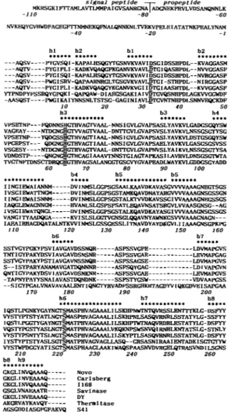
CKGLINVQAAAQ--- NOVO CKGLINVEAAAQ--- Carlsberg GKCLINVQAAAQ--- I 1 6 8 GSGLVNAEAATI--- Savinase GKCLINVEAAAQ--- DY AKGRVAYKAVQY--- Thernitase AGSGDDIASGFCFAKVQ S41FIG. 4. Primary structure of preprosubtilisin 541 from Bacil-
lus TA41. Upper panel, structure of the signal peptide and of the propeptide derived from amino acid and nucleotide sequences. Amino acids are numbered from the first residue of the mature enzyme. Lower panel, multiple amino acid sequence alignment of subtilisin BPN' from Bacillus amyloliquefaciens (Wells et al., 1983), subtilisin Carlsberg from
B. licheniformis (Jacobs et al., 19851, subtilisin I168 from Bacillus sub- lentus (Betzel et al., 19921, subtilisin DY from B. subtilis DY (Nedkov et tilis 168 (Stahl and Ferrari, 1984), subtilisin Savinase from Bacillus al., 19831, thermitase from Thermoactinomyces vulgaris (Meloun et al., 19851, and subtilisin S41 from Bacillus TA41. Numbering and second- Active site residues Asp3', His64, and SerzZ1 are underlined. ary structures correspond to the reference enzyme, subtilisin BPN'.
(Fig. 3 ) clearly illustrate the low thermal stability of the psy- chrophilic enzyme. The half-time of inactivation ( t i ) of subtili- sin S41 at 50 "C is 10 times lower when compared with the mesophilic enzyme. The difference in free energy of stabiliza- tion between both enzymes, estimated by the relation AAG =
2.3RTlog(til/tiz), amounts to 6-7 k J mol-'. The thermodepen- dence curves of the proteolytic activity of the psychrophilic and the mesophilic enzymes are shown in Fig. 3. One can observe a shift of the apparent optimal temperature of activity of about
Subtilisin S41 from the Psychrophile Bacillus
TA41
17'45
1
bon tracing of the subtilisin 541
FIG. 5. Stereo diagram of the a-car-
model. The positions of the active site Hise4, and S e F ) . The main features of
side chains residues are indicated (AspA2,
the subtilisin S41 molecule are shown. The seven amino acid residue insertions in surface loops are located by arrows (pointed to the first residue t o be in- serted). The position of Cys4' and of Cy@, engaged in a disulfide linkage, is given, as well as the location of T h P , the Ca2+ co- ordinating ligand specific of the psychro-
philic subtilisin.
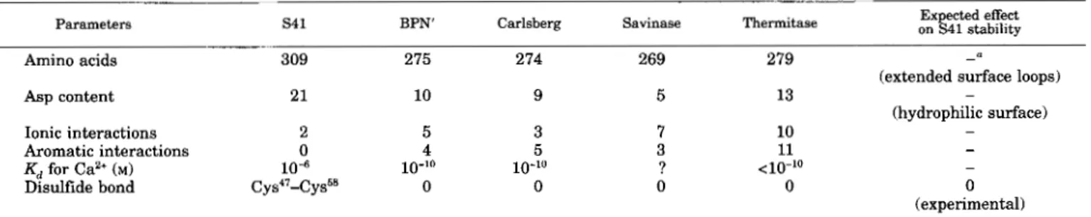
TABLE I11
Main structural features related to subtilisin S41 thermal lability
Parameters S4 1 BPN' Carlsberg Savinase Thermitase on Expected effect S41 stability
Amino acids 309 275 274 269 279
Asp content 21 10 9 5 13
Ionic interactions 2 5 3
Aromatic interactions 0 4 5
Kd for Ca2+ (M) 104 10-10 10-10
Disulfide bond Cys"-Cys58 0 0
7 10
3 11
0
? <10"0
0 - denotes an expected decrease of stability.
-
(extended surface loops) (hydrophilic surface) - - - 0 (experimental)
20 "C toward low temperatures. Following active site titration by 1-trans-cinnamoylimidazole, the specific activity of subtili- sin ,941 toward azocasein between 5 and 20 "C was found to be 4 times higher than that of subtilisin Carlsberg and BPN'. The thermodependence curves have been used t o construct Arrhe- nius plots and to calculate activation energy parameters of the proteolytic reaction (Table 11). The lower free energy of activa- tion (AG') of subtilisin S41 when compared with subtilisin Carlsberg correlates with its high specific activity, but the con- tribution of the enthalpy term ( A R ) and of the entropy term
(TAS')
to AG* also differs in both enzymes.Sequence a n d Homology Modeling-The primary structure of the preprosubtilisin S41 deduced from nucleotide and amino acid sequences is given in Fig. 4. The potential signal peptidase cleavage site (Ala31) has been located by homology with the known cleavage site of subtilisin I168 (Ala-Gln-Ala"
1
Ala-Gly- Lys) identified by Wong and Doi (1986). The cleavage site of the propeptide has been determined by NH,-terminal sequencing of the purified mature subtilisin S41. This propeptide, com- posed of 79 amino acid residues in the case of prosubtilisin ,941, is considered a s a n intramolecular chaperone characterized by two consensus sequences (Shinde and Inouye, 1993). These sequences are found between residues -57 and -47 (motif 1) and between residues -32 and -7 (motif 2). The kinetics of amino acid release by carboxypeptidase Y from the purified subtilisin S41 identified the carboxyl-terminal sequence Lys- Val-Gln-COOH and indicate that no COOH-terminal process- ing occurs during preprosubtilisin S41 maturation.The mature subtilisin S41 is composed of 309 amino acid residues with a predicted
M,
of 31,224. This value is somewhat higher than the mean M, (27,000) of mesophilic subtilisins and arises from seven insertions, as shown by sequence alignment with five subtilisins and thermitase (Fig. 4). The amino acid sequence of subtilisin S41 shows 52% residue identity with the reference sequences and 71% residue similarity (G=A=S, S=T, V=L=I, F=W=Y, E=D=R=K, Q=N). Conserved amino acids oc-cur essentially in the secondary structures of the reference enzymes and in regions bearing functional residues pertaining to the active site (Asp", HisM, S e P ' ) a n d to the Ca2+ binding sites. This degree of homology allowed the building of a three- dimensional structure model of subtilisin S41 that follows the pattern of known subtilisin structures (Fig. 5).
This model was analyzed in order to identify the structural features typical of the psychrophilic enzyme. The main results are summarized in Table 111. Whereas the conserved amino acid residues of subtilisin S41 pertain to the structurally con- served regions defined by Siezen et al. (19911, the specific in- sertions of the Bacillus TA41 enzyme belong to the variable structures of subtilases located on surface loops of the molecule. Subtilisin S41 is also characterized by a high number of charged amino acids mainly arising from 11 extra aspartic acid residues (Table 111). These extra charges are located at the surface of the molecule and are most probably responsible for the anomalous isoelectric point recorded for the native enzyme. Subtilases possess conserved electrostatic and aromatic inter- actions involved in their folding stabilization (Teplyakov et al., 1990; Siezen et al., 1991). As shown in Table IV, subtilisin 541 only retains two conserved salt bridges and no aromatic inter- actions. The high affinity calcium binding site (Cal) has been modelled on the basis of the known site geometry of subtilisins. Table V indicates that the calcium ligands are conserved except that is replaced by Thr in the psychrophilic enzyme. The protein ligands of the low affinity calcium binding site (Ca2) are the main chain carbonyl of Gly"j9, Leu'71, and Ala'74, whereas the positive charge of stabilizes this site in the absence of the Ca2+ ion (Teplyakov et al., 1990).
Subtilisin 541 possesses 1 cystine residue in contrast t o other subtilisins. DTNB titration of the urea-denatured enzyme in- deed failed to reveal any free -SH group, whereas about 2 sulfhydryl groups are detected after sodium borohydride reduc- tion. This indicates that Cys47 and Cys5' are engaged in a di- sulfide linkage, which bonds the extremities of a protruding
17452
Subtilisin
S41
from the Psychrophile Bacillus
TA41
loop between
p
sheet 2 and helix 3. In order t o assess theinfluence of this disulfide bridge on subtilisin S41 stability, the native enzyme has been incubated with 0, 20, and 100 mM
dithiothreitol a t 45 "C (not shown). Because the activity decay of the three samples are identical, the bridge does not provide significant extra stability to the enzyme.
DISCUSSION
Subtilisin S41 secreted by the Antarctic psychrophile Bacil-
lus TA41 shares most of its properties with other subtilisins from bacilli species. However, this enzyme displays a higher specific activity, a shift of the optimum of activity toward low temperatures, and a weak thermal stability, which are all com- mon properties of cold adapted enzymes. Hochachka and Som- ero (1984) postulated that these enzymes are characterized by a more flexible structure allowing fast conformational changes during catalysis in order to compensate for the reduction of reaction rates at low temperatures. Thermal instability is then
regarded as the consequence of the flexible structure of cold- active enzymes. The thermodynamic parameters for the en- zyme-substrate complex formation (Table 11) indeed agree with the fact that the activated complex will be reached through a
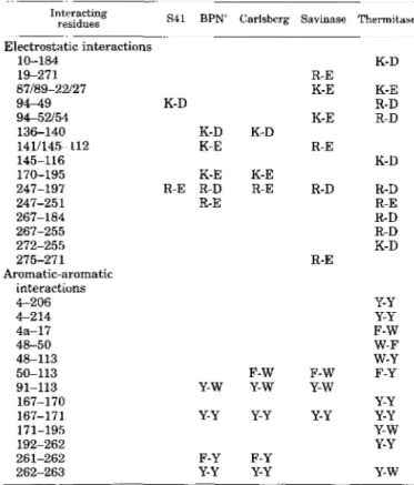
TABLE IV
Electrostatic and aromatic-aromatic interactions in the psychrophilic subtilisin S41, the mesophilic subtilisins,
and the thermophilic thermitase
Interacting
residues S41 BPN' Carlsberg Savinase Thermitase
Electrostatic interactions 10-184 87189-22127 19-271 94-49 94-52/54 136-140 1411145-112 145-116 170-195 247-197 247-251 267-255 272-255 267-184 275-271 Aromatic-aromatic 4-206 interactions P 2 14 4a-17 48-113 50-113 91-113 167-170 167-171 192-262 261-262 262-263 48-50 171-195 K-D K-D K-E K-E R-E R-D R-E Y-W Y-Y F-Y Y-Y K-D K-E R-E F-W Y-w Y-Y F-Y Y-Y R-E K-D K-E K-E R-D K-E R-D R-E K-D R-D R-D R-E R-D R-D K-D R-E Y-Y Y-Y F-W W-F w-Y F-W F-Y Y-W Y-Y Y-Y Y-Y Y-W Y-Y Y-w
minimum of entropy change and that less heat content would be associated with a more flexible protein structure.
All amino acids pointing their side chain toward the active site of subtilisin S41 are strictly conserved in respect to other subtilisins. This indicates that the catalytic cavity per se is not
affected by the molecular adaptations required for catalysis at low temperatures. One can reasonably assume that slight modifications of the active site geometry will deeply alter the enzyme reaction mechanism. However, two notable amino acid substitutions in substrate binding sites at the rim of the cata- lytic cavity are worth mentioning. Subtilisin S41 lacks which severely restricts the mobility of a substrate binding loop in all subtilisins. Conversely, an extra proline in this loop
(Pro131) has been involved in the stability of savinase (Betzel et
al., 1992). In addition, the psychrophilic enzyme has an Ala
residue at position 104, instead of an aromatic or branched aliphatic side chain, that reduces the steric hindrance at the entrance of the active site. Whereas both substitutions can be expected to lower the energy needed for conformational adjust- ments during catalytic events, they might be related to sub- strate specificity rather than to cold adaptation.
More direct evidences of structural flexibility are found at the level of subtilisin S41 conformation. As shown in Table IV,
several salt bridges and aromatic interactions conserved in subtilisins are lacking within the Bacillus TA41 enzyme. Be- cause the number of interactions decreases in the order ther- mophile > mesophile > psychrophile, there is little doubt that the removal of ion pairs and weakly polar interactions contrib- utes to increase the flexibility of the cold-adapted enzyme. Sub- tilisin 541 displays four long specific amino acid insertions located in surface loops. In addition, a surprisingly large num- ber of polar residues, mainly Asp, are found on the external shell of the protein providing a very hydrophilic surface. These two features, namely the length and the polarity of the surface loops, give rise to improved solvent interactions, reduce the compactness of the molecule, and can destabilize the psychro- philic enzyme.
Calcium contributes to the thermal stability of subtilisins by binding at specific sites and hence reduces the flexibility of the protein and its susceptibility to partial unfolding (Pantoliano et
al., 1988). The dissociation constant Kd of the high affinity Ca2+
binding site of subtilisins is close to 10"' M but rises to M in
the case of the psychrophilic enzyme. The weak coordination of the Ca2+ ion is a n additional insight of a less compact confor- mation. The predicted geometry of the high affinity calcium binding site (Table V) suggests that substitution of the con- served by Thr, which can only provides coordination via the main chain carbonyl, is responsible for this effect. In addi- tion, T h P may be less firmly oriented in the Ca2+ binding site as a result of the unusual stacking of Gly around this residue. On the other hand, in thermitase the replacement of the che- lating side chain carbonyl oxygen of Gln2 by a negatively charged ligand (the carboxylate oxygen of Asp2) has been in- volved in the greater Ca2+ affinity (Kd < 10"' M) and stability of TABLE V
Protein ligands of the high affinity calcium binding site (Cal) in subtilases and predicted Ca2+ coordination in subtilisin S41
Ca2+ ligands are as follows: 0, main chain carbonyl; O"&, side chain oxygen; Os, unidentate coordination by side chain carboxylate; 061s2,
bidentate coordination by side chain carboxylate.
~ ~ ~ ~ ~
Observed
BPN' Carlsberg Savinase Thermitase
os1s2
0 6 1 6 * Asp4'Oel Gln2 0" Gln2 0" Gln2
os's2
Os Asp2
0 Leu7s 0 Leu75 0 Leu75
o
va175Os' Os'
0 T h P
o
1 1 ~ 7 9Os'
0 Thr7'
0 ValE1 0 Val" 0 Val'' 0 Iles'
0 6 1 8 2 AS 41 P
o
1 1 ~ 7 9 08' Predicted (5411 0" Gln2os's2
ASP4' 0 0 Thr77 0 Ser7' 0 Val8'Subtilisin S41
from
the Psychrophile Bacillus TA41
17453
this enzyme (Gros et al., 1989).
The difference in free energy of stabilization between sub- tilisins S41 and its mesophilic counterpart is small (6-7 kcal mol”) and is close to the formation enthalpy of one weak bond a t 37 “C. One should remember that electrostatic interactions form exothermically and are thus stabilized by a decrease of temperature, whereas hydrophobic interactions form endother- mically and are destabilized at low temperature. According to these thermodynamic properties, the lack of several usually conserved salt bridges and weakly polar (aromatic) interactions in subtilisin S41 are elements that can preserve the appropri- ate protein flexibility a t low temperatures. However, the num- ber of weak bonds involved is probably less critical than the balance between endo- and exothermically formed interactions. It is interesting to note that enzymes from thermophilic mi- croorganisms reinforce the same type of weak interactions in order to gain thermal stability (Jaenicke, 1991; Fontana, 19911, showing that there is a continuum in the strategy of protein adaptation to temperature. Subtilisin S41 is therefore a n ap- propriate candidate for site-directed mutagenesis experiments devoted to the analysis of structure-stability relationships.
Acknowledgments-We acknowledge the “Expeditions Polaires F r a q a i s e s ” for support and facilities offered a t t h e Dumont d’Urville Antarctic Station. We thank Drs. R. Cesar, J. Van Beeumen (Rijksuni- versiteit Gent), 0. Dideberg, and E. Fonze for valuable collaboration on
these experiments and N. Gerardin-Otthiers, S. Collin, and R. March- and for expert technical assistance.
REFERENCES
Bender, M. L., Begue-Canton, M. L., Blakeley, R. L., Brubacher, L. J., Feder, J.,
Gunter, C. R., Kezdy, F. J., Killheffer, J . V., Marshall, T. H., Miller, C. G., Roeske,
R. W., and Stoops, J. K. (1966) J . Am. Chem. Soc. 88, 5890-5913
Betzel, C., Pal, G. P., and Saenger, W. (1988) Eur J. Biochem. 178, 155-171
Betzel, C., Klupsch, S., Papendorf, G., Hastrup, S., Branner, S., and Wilson, K. S .
(1992) J. Mol. Bid. 223, 427445
Bode, W., Papamokos, E., and Musil, D. (1987) Eur J . Biochem. 166,673-692
Davail, S., Feller, G., Narinx, E., and Gerday, C. (1992) Gene (Amst.) 119,143-144
DelMar, E. G., Largman, C., Brodrick, J. W., and Goekas, M. C. (1979) Anal.
di Prisco, G. (ed) (1991) Life Under Extreme Conditions: Biochemical Adaptations,
Feller, G., Thiry, M., and Gerday, Ch. (1991) DNA Cell Biol. 10, 381388 Feller, G., Lonhienne, T., Deroanne, C., Libioulle, C., Van Beeumen, J., and Gerday,
Fontana, A. (1991) in Life Under Extreme Conditions: Biochemical Adaptations (di
Gros, P., Betzel, C., Dauter, Z., Wilson, K. S., and Hol, W. G. J. (1989) J . Mol. Biol. Habeeb, A. F. S. A. (1973jAnal. Biochem. 5 6 , 6 0 4 5
Hochachka, P. W., and Somero, G. N. (1984) Biochemical Adaptations, Princeton
Jacobs, M., Eliasson, M., Uhlen, M, and Flock, J. I. (1985) Nucleic Acids Res. 13,
Jaenicke, R. (1991) Eur J . Biochem. 202, 715-728 Jones, T. A. (1978) J . Appl. Crystallogr. 11, 268-272
Klemm, P. (1984) in Methods in Molecular Biology (Walker, J. M., ed) Vol. 1, pp.
Markland, F. S., and Smith, E. L. (1971) in The Enzymes (Boyer, P. D., ed) 3rd Ed.,
McPhalen, C. A,, and James, M. N. G. (1988) Biochemistry 27,65824598 Meloun, B., Baudys, M., Kostka, V., Hausdorf, G., Frommel, C., and Hohne, W. E. Nedkov, P., Oberthur, W., and Braunitzer, G. (1983) Hoppe-Seyler’s Z. Physiol.
Biuchem. 99,316-320
Springer-Verlag, Berlin
C. (1992) J. Biol. Chem. 267, 5217-5221
Prisco, G., ed) pp. 89-113, Springer-Verlag, Berlin
210,347-367
University Press, Princeton, NJ
8913-8926
255-259, The Humana Press, Clifton, NJ
Vol. 3, pp. 561-608, Academic Press, New York
(1985) FEBS Lett. 183, 195-200
Chem. 364, 1537-1540
~~
Pantoliano, M. W., Whitlow, M., Wood, J. F., Rollence, M. L., Finzel, B. C., Gilliland,
Rentier-Delrue, F., Mande, S . C., Moyens, S., Terpstra, P., Mainfroid, V., Goraj, K.,
G. L., Poulos, T. L., and Bryan, P. N. (1988) Biochemistry 27, 8311-8317
Robertson, S . P., Potter, J. D., and Rouslin, W. (1982) J. Biol. Chem. 267,1743-1748
Hol, W. G. J., and Martial, J. A. (1993) J . Mol. Biol. 229,85-93
Shinde, U., and Inouye, M. (1993) Dends Biochem. Sci. 18, 442-446
Siezen, R. J., de Vos, W. M., Leunissen, J . A. M., and Dijkstra, B. W. (1991) Protein Stahl, M. L., and Ferrari, E. (1984) J . Bacteriol. 158,411418
Svendsen, I. (1976) Carlsberg Res. Commun. 41, 237-291
Teplyakov, A. V., Kuranova, I. P., Harutyunyan, E. H., Vainshtein, B. K., Frommel,
Wells, J. A., Ferrari, E., Henner, D. J., Estell, D. A,, and Chen, E. Y. (1983) Nucleic
Wong, S . L., and Doi, R. H. (1986) J . Biol. Chem. 261, 10176-10181 Eng. 4, 719-737
C., Hohne, W. E., and Wilson, K. S . (1990) J. Mol. Biol. 214, 261-279