Université de Montréal
Characterization of two sorting nexins:
sorting nexin-11 and sorting nexin-30
par Michel Cameron
Département de Pharmacologie, Faculté de Médecine
Thèse présentée à la Faculté de Médecine en vue de l’obtention du
grade de Doctorat en Pharmacologie
Octobre, 2014
ii
L’identification du jury
Président : Dr Éric Thorin
Membre du jury : Dr Gregor Andelfinger (Directeur de recherche)
Membre du jury : Dr Sylvain Chemtob (Co-directeur de recherche)
Membre du jury : Dr Stéphane Lefrançois
iii
Résumé français
À l’intérieur de la cellule sillonnent d’innombrables molécules, certaines par diffusion et d’autres sur des routes moléculaires éphémères, empruntées selon les directives spécifiques de protéines responsables du trafic intracellulaire. Parmi celles-ci, on compte les sorting nexins, qui déterminent le sort de plusieurs types de protéine, comme les récepteurs, en les guidant soit vers des voies de dégradation ou de recyclage. À ce jour, il existe 33 membres des sorting nexins (Snx1-33), tous munies du domaine PX (PHOX-homology). Le domaine PX confère aux sorting nexins la capacité de détecter la présence de phosphatidylinositol phosphates (PIP), sur la surface des membranes lipidiques (ex : membrane cytoplasmique ou vésiculaire). Ces PIPs, produits de façon spécifique et transitoire, recrutent des protéines nécessaires à la progression de processus cellulaires. Par exemple, lorsqu’un récepteur est internalisé par endocytose, la région avoisinante de la membrane cytoplasmique devient occupée par PI(4,5)P2. Ceci engendre le recrutement de SNX9, qui permet la progression de
l’endocytose en faisant un lien entre le cytoskelette et le complexe d’endocytose.
Les recherches exposées dans cette thèse sont une description fonctionnelle de deux sorting nexins peux connues, Snx11 et Snx30. Le rôle de chacun de ces gènes a été étudié durant l’embryogenèse de la grenouille (Xenopus laevis). Suite aux résultats in vivo, une approche biomoléculaire et de culture cellulaire a été employée pour approfondir nos connaissances.
Cet ouvrage démontre que Snx11 est impliqué dans le développement des somites et dans la polymérisation de l’actine. De plus, Snx11 semble influencer le recyclage de récepteurs
iv
membranaires indépendamment de l’actine. Ainsi, Snx11 pourrait jouer deux rôles intracellulaires : une régulation actine-dépendante du milieu extracellulaire et le triage de récepteurs actine-indépendant. De son côté, Snx30 est impliqué dans la différentiation cardiaque précoce par l’inhibition de la voie Wnt/β-catenin, une étape nécessaire à l’engagement d’une population de cellules du mésoderme à la ligné cardiaque. L’expression de Snx30 chez le Xénope coïncide avec cette période critique de spécification du mésoderme et le knockdown suscite des malformations cardiaques ainsi qu’à d’autres tissus dérivés du mésoderme et de l’endoderme.
Cet ouvrage fournit une base pour des études futures sur Snx11 et Snx30. Ces protéines ont un impact subtil sur des voies de signalisation spécifiques. Ces caractéristiques pourraient être exploitées à des fins thérapeutiques puisque l’effet d’une interférence avec leurs fonctions pourrait être suffisant pour rétablir un déséquilibre cellulaire pathologique tout en minimisant les effets secondaires.
Mots clés : Sorting nexin, trafic intracellulaire, embryogenèse, cardiogénèse, somitogenèse, Wnt/β-catenin, actin
v
English Summary
The intracellular milieu is housed by countless numbers of intracellular molecules travelling by diffusion or along transient paths, which are regulated by specific trafficking proteins. Among these traffic regulators are the sorting nexins that determine the fate of internalized proteins by directing toward a defined path, which can lead to either degradation or recycling. To date, 33 sorting nexins (Snx1-33) have been identified, which all share a common characteristic, the presence of a PX (PHOX-homology) domain. The PX domain is a phosphatidylinositol phosphate (PIP)-binding domain, which helps bring sorting nexins to PIP-enriched areas of lipid membranes. For example, during receptor endocytosis, the surrounding membrane becomes transiently occupied by PI(4,5)P2. PI(4,5)P2 is then recognized by Snx9, which
contributes to the progression of endocytosis by linking the receptor complex to the actin cytoskeleton.
The research presented in this thesis is the first to investigate the functions for two sorting nexins, Snx11 and Snx30, during embryogenesis and endosomal protein trafficking. Results obtained from knockdown experiments in the frog (Xenopus laevis) were combined with data from cell culture and biomolecular experiments to propose a function for these two proteins. The data presented here suggest that Snx11 is involved in somitogenesis, and regulates actin-dependent and -inactin-dependent processes. Snx11 could serve as a scaffolding protein, linking the extra-cellular matrix to the actin cytoskeleton and could also function in actin-independent receptor recycling. On the other hand, Snx30 is implicated in early cardiogenesis and promotes the commitment of a population of mesoderm cells to the cardiac lineage. It does so through the inhibition of Wnt/β-catenin signaling but the underlying mechanism is still unclear.
vi
Expression of Snx30 in Xenopus coincides with this critical period of cardiac specification and knockdown of Snx30 results in cardiac malformations as well as other defects in mesoderm- and endoderm-derived tissue. In addition, data from both Xenopus and HEK293T cell culture show that knockdown of Snx30 increases Wnt/β-catenin signaling.
This work provides the basis for future studies on Snx11 and Snx30. Interestingly, Snx11 and Snx30 seem to act as fine-tuners of signaling pathways. These proteins could potentially become interesting therapeutic targets due to their specificity and relatively subtle impact when knocked-down. As such, interference with their function could be useful to re-balance a cellular disequilibrium while minimizing side effects.
Key words: Sorting nexin, protein trafficking, embryogenesis, cardiogenesis, somitogenesis, Wnt/β-catenin, actin
vii
Table of Contents
L’identification du jury ... ii
Résumé français ... iii
English Summary ... v
Acknowledgements ... xiv
Chapter 1: The cellular biology of protein trafficking ... 1
1.1 – Endocytosis ... 2
1.2 – Clathrin-mediated endocytosis ... 5
1.3 – Clathrin-independent endocytosis ... 9
1.4 – The endosomal system ... 13
1.5 – Receptor degradation and recycling ... 18
1.6 – Sorting nexins ... 21
1.7 – The PX domain ... 23
1.8 – Phosphatidylinositol phosphates ... 27
1.9 – SNX-BARs ... 30
viii
1.11 – SNX-other ... 35
1.12 – RABs ... 36
1.13 – The cytoskeleton ... 38
1.14 – Vesicular transport of proteins ... 41
1.15 – Actin and membrane dynamics ... 46
1.16 – Embryogenesis ... 49
1.17 – Cardiogenesis ... 54
1.18 – Wnt signaling ... 63
1.18.1 – Endocytosis and Wnt/β-catenin signalling ... 68
1.19 – Somitogenesis ... 72
1.20 – Model organism: Xenopus laevis ... 75
1.21 – Disorders related to endosomes and sorting nexins ... 77
1.22 – Wnt/β-catenin signaling and disease ... 78
Chapter 2: Journal article #1 (submitted to the Journal of Biological Chemistry) ... 80
Chapter 3: Journal article #2 (submitted to Traffic) ... 122
ix
Characterization of two new sorting nexins ... 158
SNX30 and Wnt/β-catenin signaling ... 160
The dual functions of SNX11 ... 164
x
Abbreviations
AAA: ATPases associated with various cellular activities ANTH: AP180 amino-terminal homology
APC: Adenomatosis polyposis coli APCDD: APC downregulated BAR: Bin/Amphyphysin/Rvs BMP: Bone morphogenetic protein CHC: Clathrin heavy chain
CIE: Clathrin-independent endocytosis
CI-M6PR: Cation-independent mannose-6-phosphate receptor CLC: Clathrin light chain
CME: Clathrin-mediated endocytosis DAG: Diacylglycerol
DKK: Dickkopf-related protein DOR: Delta opioid receptor DSL: Delta, Serrate and LAG-2 EE: Early endosome
EEA1: Early endosome antigen-1 EGF: Epidermal growth factor
EGFR: Epidermal growth factor receptor EMT: Epithelial to mesenchymal transition ENT-1: Enterophilin-1
xi ENTH: Epsin N-terminal homology
EPS15: EGFR pathway substrate 15 ER: Endoplasmic reticulum
ERC: Endosomal recycling compartment ERT: Enzyme replacement therapy ESC: Embryonic stem cell
ESCRT: Endosomal sorting complexes required for transport F-BAR: Fes/CIP4 homology BAR
FCHO: FCH domain only FGF: Fibroblast growth factor FHF: First heart field
FZ: Frizzled
GAP: GTPase-activating protein GATA4: GATA-binding protein 4
GEEC: GPI-enriched early endosomal compartment GEF: Guanine exchange factor
GFP: Green fluorescent protein GPI: Glycosylphosphatidylinositol GRK: G protein-coupled receptor kinase GSK3: Glycogen synthase 3
HES: Hairy and enhancer of split HPF: Hours post-fertilization
HRS: Hepatocyte growth factor-regulated tyrosine kinase substrate HSC70: Heat shock cognate 70
xii I-BAR: Inverse BAR
IL2R: Interleukin 2 receptor ILV: Intralumenal vesicle JNK: Jun-N-terminal kinase LBPA: Lysobiphosphatidic acid LE: Late endosome
LEF: Lymphoid enhancer-binding factor
LRP5/6: Low-density lipoprotein receptor-related protein LSD: Lysosomal storage disorder
MDCK: Madin Darby canine kidney MESP: Mesoderm posterior
MHC1: Major histocompatibility class 1 MTOC: Microtubule-organizing center MUSK: Muscle skeletal tyrosine kinase MVB: Multivesicular body
NKX2-5: NK2 transcription factor related, locus 5 NOXO1: NOX organizing 1 protein
PCP: Planar cell polarity
PDAC: Pancreatic ductal adenocarcinoma PDB: Protein database
PH: Pleckstrin homology
PHOX: Phagocyte NADPH oxidase PI: Phosphatidylinositol
xiii PIP: Phosphatidylinositol phosphate
PI(3)K: Phosphatidylinositol 3-kinase PKA: Protein kinase A
PKC: Protein kinase C PSM: Presomitic mesoderm PTK: Protein tyrosine kinase PX: PHOX-homology
PXe: Extended PX
RAB11FIP: Rab11 family interacting protein ROR: Tyrosine kinase-like orphan receptor RSPO: R-spondin
RYK: Receptor tyrosine kinase
SFRP: Secreted frizzled-related protein SHF: Second heart field
SNX: Sorting nexin SO: Spemann organizer TCF: T cell factor
TGN: trans-Golgi network
Toca: Transducer of Cdc42-dependent actin assembly VPS: Vacuole protein sorting
WAIF: Wnt-activated inhibitory factor WIF: Wnt inhibitory factor
xiv
Acknowledgements
I would like to personally thank Gregor Andelfinger for his openness to new ideas, enthusiasm and contagious work ethic. This would have not been possible without him and I am forever grateful. I would also like to thank all members of the Andelfinger lab for their help, support and encouragement during my PhD training. Thank you to Sylvain Chemtob and members of his laboratory for valuable insight and expertise.
Many thanks to Christian Beauséjour for his insight and council.
Thank you to members of my pre-doctoral exam committee and to members of my thesis jury.
Finally, I would like to thank my parents and Marie-Claude for their support and encouragements.
1
Chapter 1: The cellular biology of protein trafficking
Living cells are in constant flux. As part of a living organism, cells must process signals coming from the extracellular milieu, and carry out an appropriate cellular response. During both of these essential steps (signal processing and cellular response) intracellular trafficking is fundamental. The necessity of intracellular trafficking is explained by the fact that molecules cannot travel far by free diffusion and signaling effectors must be properly localized to interact with their targets. Intracellular trafficking is thus involved in virtually all aspects of cellular biology since proteins must be located at their site of action to properly perform their task. Endosomal protein sorting is a form of intracellular trafficking that regulates the transportation of membrane-associated proteins across different intracellular compartments. This form of traffic occurs in the following sequence of processes: formation and fission of a transport vesicle from a donor membrane, transportation of this vesicle between compartments, and finally docking and fission of the vesicle with the acceptor membrane. However, prior to these processes, a decision is made as to which cargo will be selected and what will be its destination. This aspect of protein trafficking is termed endosomal protein sorting.
The main components that contribute to intracellular trafficking (endocytosis, the endosomal system and the processes of intracellular trafficking) will be discussed in the next sections with examples to illustrate how they function. Throughout these sections, it is important to consider that the results that stem from isolated cellular processes are in fact part of a holistic, interconnected system that is the living organism. As such, I will attempt to integrate data from molecular and cellular biology into developmental biology.
2
1.1 – Endocytosis
The entry of material into a eukaryotic cell occurs mostly through a process called endocytosis, an essential stage in many signaling pathways as it removes receptors from the cell surface. This limits the magnitude of signaling from extracellular sources and allows signaling to proceed from inside the cell. Endocytosis can also control ligand availability, as is the case for the membrane-bound DSL (Delta, Serrate and LAG-2) family of ligands, which activate the Notch family of receptors of adjacent cells [1]. Once internalized, material can travel throughout boundary-forming lipid membranes collectively known as the endosomal network, which includes compartments such as the early and late endosomes, the lysosome and the trans-Golgi network. Transportation of cargo throughout this dynamic network is a highly regulated process that requires the coordinated action of specialized trafficking proteins.
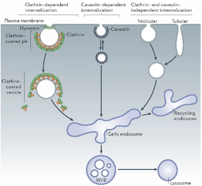
Many mechanisms of endocytosis exist and they have been divided into two major categories: clathrin-mediated endocytosis (CME) and clathrin-independent endocytosis (CIE) (Figure 1) [2]. Both these types of endocytosis are utilized to displace signaling receptors from the plasma membrane to an intracellular compartment. Endocytosis is also exploited by toxins, viruses and bacteria for penetration into the cell but this thesis will focus on the endocytosis and trafficking of receptors [3].
Endocytosis of a particular receptor can result in different outcomes depending on the endocytic route used. For example, internalization of the epidermal growth factor receptor (EGFR) with CME induces its recycling back to the plasma membrane [4, 5]. In contrast, if EGFR is internalized via CIE, the receptor is sent to the lysosome for degradation [4, 5]. In
3
addition, CME and CIE do not always produce the same effect. In Wnt/β-catenin signaling through the Frizzled receptor and LRP5/6 co-receptor, CME leads degradation of LRP5/6 while CIE leads to its recycling [6]. Therefore, the type of endocytosis alone does not determine the fate of internalized material mechanisms but rather contributes to signaling outcome, along with other factors such as the cellular context, the identity of internalized material and the trafficking machinery implicated in the process. The fate of internalized material and thus the sustainment, amplification or attenuation of signaling is intimately linked to endocytosis and subsequent sorting mechanisms. The next paragraphs provide a brief review of CME and CIE.
4
Figure 1. The different modes of endocytosis. Cells can internalize plasma membrane and receptors through a number of different routes. Clathrin-mediated and caveolin-mediated routes both require dynamin. The first destination in all routes is the early endosome, where protein sorting occurs, which directs cargo either back to the plasma membrane via the recycling endosome or into other compartments (multivesicular bodies (MVBs) or lysosomes) for degradation. [7]
5
1.2 – Clathrin-mediated endocytosis
The most studied and understood mechanism for the uptake of macromolecules is clathrin-mediated endocytosis. Clathrin is a protein that oligomerizes on the cytoplasmic side of the plasma membrane and assembles with other proteins to deform the plasma membrane, which becomes gradually invaginated, forming a clathrin-coated pit (CCP) (Figure 2). This CCP eventually buds off as a clathrin-coated vesicle, which is quickly uncoated before proceeding to their next intracellular destination. The whole process of clathrin-mediated endocytosis has been divided into five stages: nucleation, cargo selection, clathrin coat assembly, vesicle scission and uncoating [7].
1.2.1 – Nucleation
The first stage in CME is membrane invagination, triggered by the formation of a nucleation module, which is comprised of FCH domain only (FCHO) proteins, EGFR pathway substrate 15 (EPS15) and intersectins [8-10]. This nucleation module assembles at a specific site on the plasma membrane, transiently enriched with PI(4,5)P2 [11]. During nucleation, the detection
and induction of membrane deformations play important roles in the progression of endocytosis. For instance, the F-BAR domain of FCHO proteins and Snx9 bind to very low curvatures, generating further membrane bending [9, 10, 12].
6
Figure 2. Clathrin. A) First identification of clathrin by Barbara Pearse in 1976 [13]. Shown here is an electron micrograph of bovine adrenal medulla fraction containing large clathrin coated vesicles (CCV) (X67,500). B) Quick-freeze deep-etch micrography of the inner plasma membrane surface of A431 cells showing the typical clathrin lattice of growing pits [14]. Caveolae are also seen surrounded by F-actin filaments. C) An illustration of the clathrin triskelia (CHC: clathrin heavy chain; CLC: clathrin light chain). D) A model of a clathrin-coated vesicle is depicted showing assembled clathrin triskelia [15].
7
1.2.2 – Cargo selection
Once the nucleation module is assembled at the plasma membrane, the AP2 adaptor complex joins the module and together, they mediate cargo selection [9]. AP2 acts as a major hub of interactions as it can directly and simultaneously bind to PI(4,5)P2 and to motifs in the
cytoplasmic tails of transmembrane receptors [16, 17]. Indirectly, AP2 can also bind to cargo via accessory adaptor proteins. For example, during Wnt/β-catenin signaling, internalization of the Frizzled receptor depends upon the recruitment of and binding to Dishevelled, which interacts with AP2 [18]. Other proteins of the endocytosis machinery also contribute to cargo selection, such as proteins with the AP180 amino-terminal homology (ANTH) and Epsin N-terminal homology (ENTH) domains, which are membrane-binding and membrane-bending domains, respectively [19, 20].
1.2.3 – Clathrin coat assembly
Once the cargo is selected, clathrin triskelia (Figure 2C) are recruited to the site of the nucleation module and adaptor proteins. Clathrin triskelia consist of three clathrin heavy chains and three clathrin light chains that form a polyhedral lattice around the forming vesicle. As invagination of the clathrin-coated pit progresses, adaptor proteins and curvature effectors move to the edge of the forming vesicle, where they continue to promote the formation of the growing pit [21, 22].
8
1.2.4 – Vesicle scission
Near the end of CCP formation, a small portion remains attached to the plasma membrane, which must be clipped off. This is largely dependent on the large GTPase called dynamin. By assembling into a spiral around the neck of the clathrin-coated pit, dynamin mediates membrane fission and the release of clathrin-coated vesicles (Figure 3) [23]. Recent studies have also shown that BAR domain-containing proteins like sorting nexin-9 (Snx9) and actin polymerization also contribute to vesicle fission [24-26].
1.2.5 – Uncoating
Once the vesicle has detached from the plasma membrane, ATPase heat shock cognate 70 (HSC70) and auxilin disassemble the clathrin coat from its lattice arrangement back to triskelia [27, 28]. This allows the detached and uncoated vesicle to travel through the cytoplasm and fuse with the acceptor membrane of its destination, the early endosome.
Clathrin coated vesicles are also sometimes formed during vesicle formation from intracellular compartments. The stages of vesicle formation in these cases are very similar, except that some modules are interchangeable; for example, AP1 or AP3 may be substituted for AP2 during vesicle formation from endosomes and the trans-Golgi network (TGN) [29].
9
1.3 – Clathrin-independent endocytosis
Several forms of clathrin-independent endocytosis exist and these mediate primarily the intake of fluid and membrane. Clathrin-independent endocytosis accounts for about 70% of fluid uptake [30] and 60-85% of membrane uptake [31, 32]. In addition, clathrin-independent endocytosis is implicated in plasma membrane repair, cellular spreading, cellular polarization, and modulation of intercellular signaling.
1.3.1 – Caveolae
Caveolae are submicroscopic plasma membrane pits (Figure 1B) that sense and respond to plasma membrane stresses, remodel the extracellular environment, and contribute to signaling pathways as a mode of endocytosis [33]. Caveolae are irregularly distributed across tissues and individual cells, and unlike clathrin-coated pits, they have no obvious coat. For example, they are practically undetectable in kidney cells while they can represent up to 50% of plasma membrane surface on endothelial cells and adipocytes [34, 35]. The main membrane components of caveolae are the oligomeric caveolins, which drive caveolae formation with the help of cavins [33, 36]. Cholesterol and phosphatidylserine also seems to be important membrane components for caveolae formation since they are abundant in areas that are rich in caveolae and their depletion disrupts caveolae formation [37, 38]. Due to the presence of these lipids, caveolae are sometimes called lipid-rafts. Although whether and how caveolae undergo endocytosis has been a subject of controversy for many years, a
10
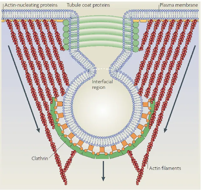
Figure 3. Membrane scission of a clathrin-coated vesicle. Contributing factors in the scission of endocytic carriers include the pinching action of dynamin as well as the force brought about by actin polymerization. Tubule coat proteins like Snx9 also contribute to membrane deformation and activation of dynamin. [39]
11
consensus has emerged that caveolae bud from the plasma membrane in a dynamin-dependent manner. In general, internalization of caveolae leads to their fusion with the early endosome, from where their components can recycle back to the surface [40].
As one of their biological functions, caveolae can flatten in response to stretching of the plasma membrane, thereby preventing damage or cell lysis. This has been shown in several cell types, including cardiomyocytes [41]. It is thus possible that caveolae and their components play important roles in mechanosensitive responses as both caveolins and cavins are released into the plasma membrane and cytoplasm, respectively, during flattening of caeolae [42]. Caveolae and caveolins have also been implicated in many signaling pathways, such as eNOS, Wnt/β-catenin and PAR-1 signaling, as well as in the regulation of lipids [33, 43, 44].
1.3.2 – RhoA, Cdc42, Arf6
In general, small GTPases like Cdc42, Arf6 and RhoA are used to differentiate between the endocytic routes employed by glycosylphosphatidylinositol-(GPI) anchored proteins, the major histocompatibility class 1 (MHCI) molecules and the interleukin 2 receptor (IL2R), respectively [45-47]. Other markers like flotillins are also involved in carrier formation and can be used to identify specific clathrin-independent routes [36]. These endocytic routes also make use of actin and actin-associated proteins [48, 49], as well as Snx9 [50, 51]. Importantly, clathrin-independent endocytosis is under differential regulation by signaling pathways and cell type [52]. While RhoA-dependent endocytosis requires dynamin and endocytosis via Cdc42 is
12
dynamin-independent, both routes are reported to be dependent on lipid rafts for vesicle formation [36, 45, 53]. Also protein toxins such as Clostridium botulinum C2 toxin make use of RhoA-mediated endocytosis [54, 55], but this is not exclusive since clathrin-dependent endocytosis can also take up a fraction of the C2 toxin [54].
The largest fraction of fluid uptake is mediated by Cdc42-dependent endocytosis [30, 53] but this pathway is also involved in the uptake of GPI-anchored proteins, and has therefore been called the GPI-enriched early endosomal compartment (GEEC) pathway [53]. The raft-associated proteins flotillin 1 and flotillin 2 play a role in both dynamin-dependent [56, 57] and -independent endocytosis [58]. Basolateral uptake of GPI-anchored proteins was found to be dependent on flotillin 2 and dynamin [56], whereas the flotillin 1-dependent uptake of GPI-linked proteins and cholera toxin B was reported to be dynamin-independent [58].
As previously mentioned, the endocytic route employed by a particular signaling molecule can have varying effects. An example of this is the endocytosis of LRP6, which is internalized in caveolar vesicles that move to early endosomes in a Rab5-dependent process, in response to Wnt3A [59]. The same study also showed that LRP6 is recycled back to the plasma membrane 4 hr after stimulation in a Rab11-dependent manner but could not exclude the possibility that a small portion of internalized LRP6 was transported to the lysosome or proteasome for degradation [59]. Interestingly, the same group later found that stimulation of LRP6 with the Wnt antagonist, Dkk1, resulted in clathrin-mediated LRP6 endocytosis, and the subsequent downregulation of this pathway [60].
This example demonstrates the intimate relationship that exists between specific signaling pathways and their underlying trafficking mechanisms. They also help appreciate the diversity
13
of responses a cell can have to an extra-cellular signal as any given endocytic pathway cannot be the sole determinant of signaling pathway outcome. Many factors need to be taken into account, including the cell-type, the extracellular signal, the receptor(s) involved, the endocytic route employed and the overall cellular context at that moment in time. The contributing factors ultimately act together to engender a cellular outcome.
1.4 – The endosomal system
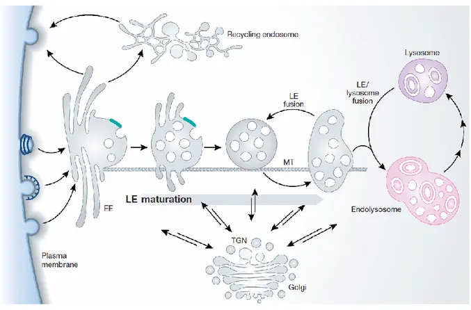
Eukaryotic cells evolved a way to perform specialized reactions within isolated and tailored micro-environments, called endosomes. Endosomes are intracellular compartments that can receive and produce fleets of transport vesicles shuttling proteins and lipids (Figure 4). Although this membranous network is very dynamic, some compartments stand out as specialized stations, occupied by specific resident proteins and lipids, where specialized functions are performed. Accordingly, the various compartments that make up the endosomal network have been labeled as early endosomes, recycling endosomes, multivesicular bodies (MVB), lysosomes, and the TGN. This simplified model is made up of a recycling pathway for plasma membrane components and their ligands, a degradative pathway for breakdown of macromolecules, and an intermediary pathway where intracellular signaling can occur and where selected components from the recycling pathway can be transported to the degradative system and vice versa. Early endosomes (EEs) are the main
14
Figure 4. The major intracellular compartments that make up the endosomal network. Internalized cargo (by CME and CIE) first arrives in the early endosome (EE) and makes its way sequentially through the endosomal network. Trafficking proteins sort incoming cargo towards their next destination, following one of two main paths, degradation or recycling. Cargo destined for degradation is sorted to the multivesicular body/late endosome (LE) which travel along microtubules (MT), eventually fusing with the lysosome where catalytic enzymes breakdown proteins into elemental components to be reused by the cell. The endolysosome that is formed matures into a classical dense lysosome. Cargo can also be recycled back to the plasma membrane from any of these endosomal compartments directly to the plasma membrane or via the TGN or recycling
15
endosome. Throughout the endosomal network, retrograde and anterograde transport shuttles proteins and lipids to and from the trans-Golgi network (TGN), respectively. [61]
sorting stations where cargo and fluid internalized from different endocytic pathways converge [62]. As cargo progresses past early endosomes, it makes its way to multivesicular bodies. MVBs are also a transient stopover for lysosomal components travelling from the trans-Golgi network (TGN) to the lysosomes. Lysosomes house hydrolases that break down proteins found in this compartment. The TGN is a major sorting station for newly synthesized proteins and lipids that have undergone serial post-translational modifications by first passing through the Golgi apparatus. Throughout the endosomal network, cargo can be shuttled to recycling endosomes and transported to the plasma membrane. Finally, the cytoplasm must also be included as it is a source for essential elements of the trafficking machinery.
How EEs arise is not completely understood, however a significant amount of membrane and volume originates from endocytic vesicles (via CME and CIE) [61]. The fate of proteins and lipids that end up in EEs is determined through sorting mechanisms. Receptors, like EGFR, are often internalized and continue to signal even when located in EEs [63]. The peripherally located EEs contain regions with tubular extensions as well as a few intralumenal vesicles (Figure 4) [64]. These morphological differences are a showcase of the dichotomy of EEs: tubular extensions of EEs are usually involved in recycling while proteins targeted for degradation cluster within multi-vesicular domains [65].
A key resident of EEs is the Rab GTPase Rab5. Rab GTPases constitute the largest family of small GTP-binding proteins and are implicated in many trafficking processes. Much of the
16
biochemical attributes of EEs are acquired through the action of Rab5 and its effectors, which include phosphatidylinositol 3-kinase (PI(3)K) (VPS34/p150), early endosomal antigen-1 (EEA1), and Rabenosyn-5 [66]. PI(3)K generates PI(3)P which is typically enriched in the membranes of EEs [66, 67] and required for trafficking from this compartment as it recruits PI(3)P-binding proteins [68]. When present on the surface of endosomal membranes, PI(3)P acts as a molecular tag recognized by proteins such as EEA1 [69, 70], Rabenosyn-5 [71] and PX domain-containing proteins like sorting nexins [72]. Accordingly, EEA1 is one of the most commonly used molecular markers for EEs due to its precise localization to this compartment through its binding to both PI(3)P and Rab5. EEA1 enables membrane fusions of incoming vesicles in coordination with members of the SNARE family [73, 74]. The FYVE domain present in EEA1 is responsible for binding PI(3)P and is also present in Rabenosyn-5 [71]. Although the role of Rabenosyn-5 is still unclear, it may mediate the recycling of cargo back to the plasma membrane by its interactions with EHD1 and Rab5 [75]. In addition, sorting nexins localized to EEs play essential roles in sorting cargo to different endosomal destinations and will be discussed later [72, 76, 77].
Multivesicular bodies, also known as multivesicular endosomes or late endosomes as defined by the time it takes for internalized tracers to reach these compartments, are distinguished from other organelles by their large number of intralumenal vesicles (ILV) [78]. This characteristic offers many options to the cell by providing a means to store material in vesicles that can be delivered to lysosomes, released by exocytosis, or stored for future use. Although many proteins passing through this endosome are en route to the lysosome, some proteins do exit the degradation pathway through “back-fusion” of intralumenal vesicles with the MVB limiting membrane [79]. Unfortunately, markers of MVBs are few, which is perhaps
17
due to the fact that these endosomes serve as trafficking hubs for proteins en route to other destinations and because the machinery involved in MVBs formation is only transiently associated to the MVB membrane. The lysosomal membrane proteins LAMP1 and LAMP2 can be found on MVB membranes but only in the presence of the cation-independent mannose-6-phosphate receptor (CI-M6PR), since this latter protein is absent from lysosomal membranes [65]. In addition, Rab5, Rab7, Rab9, Rab27 and Rab35 are sometimes associated with MVBs [80, 81]. Intralumenal vesicles are occupied by tetraspanins and lysobisphosphatidic acid (LBPA), which can be used as additional markers [82, 83].
Lysosomes are defined by their acidic pH, mediated by v-ATPase, and the presence of hydrolases. These hydrolytic enzymes originate from the trans-Golgi Network and are first transported to MVBs before being ferried to lysosomes. TGN-to-MVB transport of hydrolase receptors is the primary function of the CI-M6PR [84]. On the surface of lysosomes are structural proteins, such as LAMP1/2, ion channels, as well as trafficking and fusion machinery proteins, like Rabs and SNAREs [85].
The Golgi apparatus is a central hub for sorting and transporting proteins and lipids that travel within the secretory and degradative pathways [86]. It also houses the machinery required for the post-translational modification of proteins, which are then sorted in the trans-Golgi network. The trans-Golgi system is easily distinguishable by the shape of its structure, typically organized as stacks of flattened cisternae with dilated rims (Figure 4) [87]. This dynamic network receives cargo from the plasma membrane and endosomes, and directs transport of material coming from the endoplasmic reticulum (ER) en route to the secretory pathways [86].
18
1.5 – Receptor degradation and recycling
As cells synthesize new proteins according to their needs, proteins that are damaged, misfolded or simply no longer needed are degraded and their amino acids are reused. As such, a fine balance exists between protein synthesis, degradation and recycling. A deficiency in degradation mechanisms would be significant and could lead to catastrophic proteotoxicity within the restricted intracellular space [88, 89]. On the other hand, since degradation is irreversible, tight regulation is required to avoid reckless destruction. When possible, proteins that are still required and functional can be recycled for reuse. Rudolf Schoenheimer conducted early experiments on protein turnover in the late 1930s. In a single mass spectrometry experiment, he analyzed the fate of stable isotope-labeled amino acids that had been fed to mice, which allowed determination of the turnover rate of thousands of individual proteins [90]. For a long time, the lysosomal compartment was considered the main site of protein degradation, through the action of resident proteases. However, this view was challenged when most cellular proteins remained insensitive to alkalinization of the lysosomes. In later years, the ubiquitin-proteasome degradation system was discovered and replaced lysosomes as the major catalyst of protein degradation [91, 92].
Central to this model is the small molecule ubiquitin, which is covalently attached to lysine residues of proteins targeted for degradation, through interaction with an E3 ligase protein that recruits an E2-enzyme charged with ubiquitin [93]. Typically, proteins targeted to the proteasome are tagged with a chain of multiple ubiquitin molecules (polyubiquitination) while those destined for lysosomes are tagged with a single ubiquitin molecule (monoubiquitination). Proteasomal degradation is performed by ATP-dependent proteases, the most well-known
19
member being the 26S proteasome. ATP-dependent proteases are multi-subunit protein-wrecking machines that share the common architecture of a barrel-shaped compartmental peptidase capped by a hexameric AAA + unfoldase (ATPases associated with various cellular activities) [94]. A monoubiquitination modification induces the sorting of proteins into the internal vesicles of EEs [95]. A MVB then detaches from early endosomes and travels along microtubules to eventually fuse with lysosomes [95]. Although less common, multiple monoubiquitinations and in some cases K63-linked polyubiquitin chains have also been shown to provide a signal for MVB targeting [96, 97]. Monoubiquitination is recognized by a series of complexes called the endosomal sorting complexes required for transport (ESCRT), which then mediate the trafficking of these proteins to the lysosome [98].
In mammals, the ESCRT machinery consists of more than 20 proteins, grouped into three complexes (ESCRT-I, -II, and –III) and other associated proteins such as the ATPase vacuolar protein sorting 4 (Vps4) [99]. ESCRT is mostly known for its role in MVB formation but it is also involved in other membrane fission processes, such as the terminal stages of cytokinesis and separation of enveloped viruses from the plasma membrane [99]. In yeast, four ESCRT complexes have been identified and are numbered according to the order in which they act in the ESCRT pathway (ESCRT-0, -I, -II and -III). Studies from mammalian cells also support this model of sequential assembly and disassembly for ESCRT protein dynamics [99]. Near the end of MVB biogenesis (and other membrane fission processes), two membranes that remain connected by a thin neck are severed, a process attributed to ESCRT [99]. In mammals, ESCRT-I is recruited to sites of MVB formation by the adaptor protein Hrs [100]. This and other interactions bring about the sequential recruitment of ESCRT-II and –III, and the progression of MVB biogenesis [99].
20
Besides their roles in protein degradation, proteins found in mature MVBs can also be re-routed back to the plasma membrane or temporarily isolated from the cytoplasm. This is dependent on ESCRT-0, which promotes recycling of certain proteins, such as the β2
-adrenergic receptor and the epithelial Na+ channel [101, 102]. Another interesting example of
MVBs function, is the sequestration of cytosolic glycogen synthase kinase 3 (GSK3) into ILVs during Wnt/β-catenin signaling, which will be discussed further [103].
Sorting of receptors for recycling to the plasma membrane is achieved through extensive tubulation of the EE membranes [104]. Tubulation is an elongation of endosomal membranes where cargo accumulates and eventually buds off inside a vesicle carrier en route to an endoplasmic recycling compartment (ERC) or directly back to the plasma membrane. Accordingly, recycling directly to the plasma membrane is termed “fast recycling” while recycling via the ERC is called “slow recycling”. For example, slow recycling of the transferrin receptor takes between 30-60 minutes, while its fast recycling takes about 10 minutes [105]. Mediating these pathways are sorting proteins like Rab4, which targets receptors directly back to the plasma membrane (fast recycling), and Rab11, which regulates slow recycling [106, 107]. However, the use of these Rabs as endosomal markers should be done with caution since Rab4 has also been detected on membranes of the ERC [108].
In addition to the ESCRT machinery, other protein families, such as the sorting nexins and the small Rab GTPases, are required for sorting internalized proteins towards various endosomal destinations. These large families of proteins also contribute to protein trafficking and are thus intimately linked to protein degradation, recycling and sequestration.
21
1.6 – Sorting nexins
The first description of a mammalian sorting nexin (Snx) was published in 1996, with the identification of SNX1 in a yeast two-hybrid screen using the core tyrosine kinase domain of EGFR [109]. Mammalian Snx1 was found to contain a region homologous to a previously identified yeast protein, Mvp1p, a multicopy suppressor of Vps1p mutants deficient in carboxypeptidase Y receptor trafficking [110]. Co-localization of Mvp1p with Vps1p to Golgi membranes pointed to a potential role in membrane trafficking [110]. The relevance of the interaction between EGFR and Snx1 was uncovered by demonstrating that Snx1 could bind to the lysosomal targeting code on EGFR and contribute to its transportation to lysosomes as overexpression of Snx1 increased the rate of both constitutive and ligand-induced EGFR degradation [109]. This function was later confirmed by others and was also shown to involve the hepatocyte growth factor-regulated tyrosine kinase substrate (Hrs) [111] and enterophilin-1 (Ent-enterophilin-1) [enterophilin-1enterophilin-12]. Hrs is an ubiquitin-binding protein that sorts ubiquitylated proteins like the transferrin receptor into clathrin-coated microdomains of early endosomes, thereby targeting them to the late endosome and lysosome, and preventing recycling to the cell surface [100]. Ent-1 is an intestinal protein involved in enterocyte differentiation and the overexpression of Ent-1 reduces cell surface expression of EGFR, an effect increased by co-expression of Snx1. Shortly after the discovery of Snx1, Haft et al. (1998) introduced three new sorting nexins (Snx2, Snx3 and Snx4), which along with Snx1 as well as others proteins previously identified in Caenorhabditis elegans and Saccharomyces cerevisiae all shared a common conserved domain, the SNX-PX domain [113]. Today, mammalian sorting nexins count 33 members, all characterized by the presence of the sorting nexin PX (SNX-PX) domain (Figure 5) [72, 76, 77].
22
The roles of many sorting nexins have been investigated and members of this protein family are now recognized as important factors for endosomal protein sorting.
Figure 5. The domain structure of mammalian sorting nexins. All sorting nexins (33 members) contain a SNX-PX domain. The presence (or absence) of other conserved domains is used to divide the sorting nexins into three subfamilies: the SNXBAR contain
a C-terminal BAR domain (left), the SNXPX contain simply an isolated SNX-PX domain
(middle), and the SNXother contain another recognized domain, in addition to the SNX-PX
23
1.7 – The PX domain
The PHOX-homology domain (PX domain), as its name implies, is homologous to the PHOX domain, named according to the protein complex where it was initially identified: the phagocyte NADPH oxidase (PHOX). The PHOX motif (110 aa) is present in subunits p40phox
and p47phox of the NADPH oxidase complex of neutrophils [115]. p40phox and p47phox reside in
quiescent neutrophils, in association with a third subunit, p67phox. This tertiary complex
regulates the oxidative activity of flavocytochrome b558, composed of the catalytic heterodimer
gp91phox-p22phox, which is kept inactive in quiescent cells. In these dormant cells, p47phox has
an auto-inhibitory conformation that hides its SH3 and PHOX domains due to intramolecular interactions [116, 117]. When immune mediators activate the neutrophil, p47phox becomes
phosphorylated and exposes its SH3 domain to interact with p22phox. In addition, the PHOX
domain is also released, which allows the protein to interact with PIPs. The role of p47phox is
thus essential for the recruitment the membrane of the p47phox-p67phox-p40phox complex and for
its interaction with flavocytochrome b558. This way, the assembly of the phagocyte oxidase
complex at the membrane allows the production of reactive oxygen species that destroy invading microorganisms. Other homologs of p47phox et p67phox have since been identified, such
as the NOX organizing 1 protein (NOXO1), which contains a PHOX domain that is required for the recruitment of proteins at the plasma membrane [118-120].
In yeast, PX domain-containing proteins are implicated in processes of vesicular transport, cellular signaling, budding control and polarization [77, 121]. The evolutionary conservation of the PX domain is strongest in proteins involved in vesicular transport such as Mvp1p, Vps5p/Grd2p, Grd19p/Snx3, and Vam7p. Mvp1p, Vps5p and Grd19p are required for the
24
retrograde transport from the pre-vacuolar endosome/late endosome to the “late” Golgi (similar to the trans-Golgi in mammals) [122-125]. Snx4, Snx41 and Snx42 are other SNX-PX domain-containing proteins, which are implicated in recycling receptors from the sorting endosome (post-Golgi endosome) to the “late” Golgi [126].
In mammals, many proteins contain the PX domain, including PI3 kinases, CISK and FISH, but the majority of these proteins fall under the class of sorting nexins. Studies in yeast have shown that the PX domain is capable of interacting with PIPs, primarily PI(3)P, but also with other phosphorylated derivatives, such as PI(3,4)P2, PI(3,5)P2 and PI(4,5)P2, interactions that
should not be disregarded [77, 127-132]. A total of 27 crystal structures of the mammalian PX domain are accessible on the protein database (PDB), stemming from 14 different proteins but only two structures have been resolved in complex with PI(3)P : p40phox (PDB : 1H6H) and
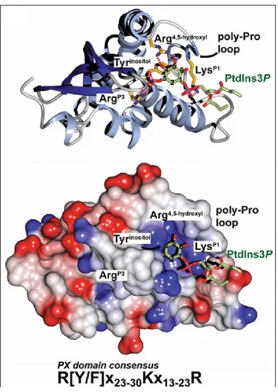
SNX9 (PDB: 2RAK). The interaction of the PX domain with PI(3)P is an electrostatic bond between the 3-phosphate of PI(3)P and a specific and conserved arginine of the PX domain. The consensus sequence of the interaction of the PX domain with PI(3)P is R[Y/F]X23-30KX 13-23R (Figure 6) [77].
25
Figure 6. The interaction between the PX domain and phosphatidylinositol-3-phosphate. Ribbon and surface structures of a representative PX domain from p40phox are shown in
complex with PI(3)P. Surface structure color codes indicate hydrophobicity, from blue (most hydrophilic) to red (most hydrophobic). Key PX-PI(3)P interactions: the arginine side chain electrostatic association with the 3-phosphate (ArgP3), stacking of the inositol
ring with the tyrosine (or phenylalanine) side chain immediately downstream from the conserved arginine residue (Tyrinositol), contact of a lysine side chain with the
1-26
phosphate (LysP1), and hydrogen bonds of the 4- and 5-hydroxy groups to a second
arginine side chain (Arg4,5-hydroxyl). [77]
The affinity of different PX domains to phospholipids is determined by two approaches: by analyzing the binding to liposomes and by “dot blots” or “strips”. The approach by PIP-strips is technically less challenging than artificially-producing liposomes, which requires delicate manipulations due to the instability of PIPs. In PIP-strips, pure phosphorylated derivatives of phosphatidylinositol are applied on nitrocellulose membranes after which proteins are incubated and then their affinities are detected by chemiluminescence. The liposome method involves the generation of liposomes composed with up to 50% of the tested phospholipid in an environment of varying proportions of phosphatidylserine, phosphatidylethanolamine or phosphatidylcholine, required for stability. Even if the PIP-strip method is easier to perform, the liposome method is considered closer to physiological conditions since it introduces a structural aspect of the protein-PIP interaction that cannot be replicated in 2-dimensional tests like PIP-strips. In fact, comparisons between both methods can produce different results for the same protein [76, 133]. Using PIP-strips, the PX domain of Snx1 had specific affinity with PI(3,4,5)P3 and weaker affinity to PI(3,5)P2 [76, 134].
However, when using the liposome method, Snx1 had comparable affinities with both PI(3)P et PI(3,5)P2 [76]. The interaction of Snx1 with PI(3)P instead of PI(3,4,5)P3 seems more
physiologically plausible since the increase of PI(3,4,5)P3 by a constitutively active PI3K did not
increase the association of Snx1 to the membrane where PI(3,4,5)P3 was enriched [76]. These
observations demonstrate the importance to use multiple methods of analysis to determine the lipid affinities of unknown proteins.
27
1.8 – Phosphatidylinositol phosphates
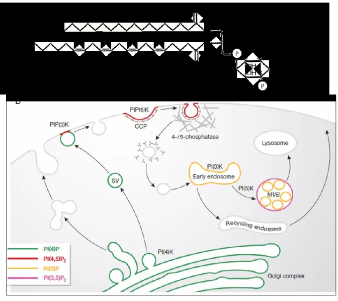
Phosphatidylinositol is used as a scaffold that can be phosphorylated at the 3-, 4- and/or 5-positions to generate phosphatidylinositol phosphates (Figure 5A). Despite their low abundance (less than 10% of total cellular phospholipids), PIPs serve as both structural and regulatory molecules in response to stimulation of certain cell surface receptors and control endosomal biology by regulating the correct timing and location of vesicular trafficking events [135, 136]. Through organelle-specific phosphatidylinositol kinases and PIP phosphatases, PIPs can undergo rapid phosphorylation/dephosphorylation cycles that lead to distinct and transient subcellular distributions of individual PI species. These species are recognized by PIP-binding modules (PIBMs), which include the FYVE, pleckstrin homology (PH), ENTH, ANTH and PX domains. The expression of proteins with these PIBMs tagged with green fluorescent protein (GFP) has permitted the construction of a map of intracellular PIP distribution (Figure 3B).
28
Figure 7. Phosphatidylinositol phosphates and their intracellular distribution. A) Phosphatidylinositol is an amphiphile lipid that can be phosphorylated on positions 3, 4 and 5 of the polar inositol headgroup. Depicted here is phosphatidylinositol-3-phosphate (PI(3)P), phosphorylated on position 3 of inositol. Other species could be generated through the action of kinases and/or phosphatases at positions 3, 4, and 5. B) Subcellular distribution of PIs and their metabolizing enzymes in exo- and endocytic membrane traffic. SV: secretory vesicle; CCP: clathrin-coated pit, MVB: multivesicular body [137].
29
Phosphatidylinositol and PI(4)P are considered as the main precursors of PIPs. However, PI(4)P has been shown to interact with cytoskeletal proteins, namely talin, suggesting a functional role [138]. PI(4)P is also enriched on the membranes of the Golgi complex, while concentrations of PI(4,5)P2 are kept low, which is likely due to the presence of PI(4)K and
PI(4,5)P2 phosphatase [136]. PI(4,5)P2 is mainly found on the inner side of the plasma
membrane [139, 140]. It is required for the invagination of clathrin-coated pits after which levels of PI(4,5)P2 drop due to 5-phosphatase activity [141]. PI(4,5)P2 is also required during the first
steps of phagocytosis but is quickly converted to PI(3,4,5)P3 by type I PI(3)K [136].
PI(3)P is mainly found on membranes of early endosomes, on intralumenal vesicles of MVBs and at the plasma membrane when it is generated during signaling processes [142]. PI(3)P is also required in Golgi-to-vacuole transport in yeast [143]. During phagocytosis, PI(3)P is generated by type III PI(3)K (Vps34) and is required for phagosomal maturation [144]. Conversion of PI(3)P to PI(3,5)P2 occurs at MVBs and is required for protein sorting at these
endosomes [136]. During the exocytic cycle, PI(4)P is generated in secretory granules [145]. PI(4)P and PI(4,5)P2 are also found in the Golgi complex, where PI(4)P promotes transport
from the Golgi and PI(4,5)P2 is important for maintaining the architecture of Golgi membranes
[146].
Generation of PIPs is also required during transportation processes along cytoskeletal routes. For example, enrichment of PI(4,5)P2 in nascent vesicles is required for actin comet tail
assembly that mediates propulsion and movement [147]. Microtubule-based motility may also be regulated by regional PIPs as the motor protein kinesin was shown to interact with PI(4,5)P2
[148]. Most phosphatidylinositol kinases and phosphatases are cytosolic and their targeting to specific regions of lipid membranes is not fully understood. It may involve small GTPases that
30
either recruit or activate phosphatidylinositol-modifying enzymes, such as Rab5, which activates PI(3)K at the early endosome [66, 136].
The interaction between the PX domain and PI(3)P (and other PIPs) is in general a weak interaction and the recruitment of sorting nexins to membranes is achieved by the contribution of other domains and molecular interactions, a concept termed “coincidence detection” [149]. For sorting nexins of the SNXBAR subfamily, this additional contribution is brought about by the
BAR domain.
1.9 – SNX-BARs
In addition to the PX domain, some sorting nexins also contain a BAR domain, referred to as SNX-BARs. The BAR (Bin/amphyphysin/Rvs) domain is an amphipathic motif exposing both a hydrophobic and hydrophilic surface. In an aqueous solution, this protein domain forms monodimers and heterodimers that form a six-helix bundle curved in a banana-shape, which bonds with the curved surface of lipid membranes [150-153]. Fifteen structures of the BAR domain have been produced, prompting the division of this domain into two sub-groups: the F-BAR (Fes/CIP4 homology-F-BAR) and the I-F-BAR (Inverse-F-BAR) [151, 154]. The association between the F-BAR dimer at the N-terminus of Toca (Transducer of Cdc42-dependent actin assembly) and lipid membranes demonstrated how this motif could associate with membranes and shape them into cylindrical tubules [155]. By integrating the atomic models of F-BAR
31
dimers [153] with cryo-EM reconstructions of membrane tubules, Frost et al. (2008) were able to show how cationic residues on the concave surface of the F-BAR motif engage the lipid bilayer and allow the rigid dimer to impose its shape onto the underlying membrane [155]. Proteins that contain the BAR domain play essential roles in the formation of tubules, which preludes vesicle budding. Tubules are localized and elongated deformations found in many endosomes.
Most of our understanding into the role of sorting nexins comes from retromer studies. The retromer is an evolutionarily conserved pentaheteromeric protein complex that is essential for the late endosome-to-TGN retrograde transport of the CI-M6PR receptor in mammals and vacuolar protein-sorting receptor-10 (Vps10p) in yeast [156]. The mammalian CI-MPR is a type I transmembrane receptor that recognizes the mannose-6-phosphate tag present on hydrolytic enzymes at the TGN and delivers these digestive enzymes to late endosomes before making its way back to the TGN [70]. The efficient retrieval of these receptors from the late endosome to the TGN is accomplished by retromer and is crucial to maintain efficient sorting and forward transport of hydrolytic enzymes to the lysosome in mammals [68, 157] or vacuole in yeast [73, 122]. Furthermore, the fundamental mechanisms for this retrograde pathway are evolutionarily conserved from lower to higher eukaryotes.
The retromer complex functions as a cytoplasmic vesicle coat that can be divided into two distinct sub complexes: a cargo recognition complex and a sorting nexin dimer. In yeast, the cargo recognition complex of retromer is composed of Vps35p, Vps26p and Vps29p, while the sorting nexin dimer is composed of Vps5p, Vps17p [158]. Vps35p associates with membranes through its interaction with Vps26p, while Vps29p stabilizes Vps35p and interacts with the second sub complex (Vps5p and Vps17p) [67, 73, 74, 159]. Vps5p and Vps17p are SNX-BARs
32
that associate with highly curved membranes and PI-enriched regions of lipid membranes [122, 160]. The yeast retromer therefore localizes to specific regions of vacuolar membranes to retrieve Vps10p and mediate tubule/vesicle formation. In mammals, the cargo recognition complex is composed of Vps26-Vps29-Vps35 [158]. However, the exact components of the sorting nexin dimer are less clear but is likely made up of a combination of Snx1 or Snx2 (Vps5 orthologs) and Snx5 or Snx6 (Vps17 orthologs) [158].
In addition to SNX1, other sorting nexins also have CI-M6PR-independent functions. For example, SNX5 has been shown to mediate both internalization and recycling of a G-protein-coupled receptor, the D1 dopamine receptor (D1R) [161]. SNX5 coexists at the plasma
membrane with D1R and GRK4 (G-protein-coupled receptor kinase 4), where SNX5 interacts
with the C-terminal of the D1R and forms a functionally cohesive complex for selective receptor
stimulation and efficient signal propagation, amplification and termination. In this role, SNX5 likely functions as a scaffold that is not only essential for endocytosis but also for receptor recycling. Accordingly, in the absence of SNX5, phosphorylation by GRK4 remains unhindered and impairs D1R endocytosis and delays recycling, leading to failure of cAMP production on
agonist stimulation [161]. This findings supports the idea that SNXs are promiscuous proteins that can associate with various other trafficking components to direct endosomal sorting of a wide variety of receptors.
33
1.10 – PX-only Sorting Nexins
In some sorting nexins, the only conserved protein domain identified to date is a N-terminal PX domain. Many of these SNXs have C-terminal regions that likely play important functional roles. Such is the case for SNX11, where a novel extended PX (PXe) domain was recently discovered by crystal structure analysis [162]. Evolutionarily, Snx11 is most closely related to Snx10, which was first shown to be important for endosome homeostasis since overexpression of Snx10 resulted in the formation of abnormally large vacuole [163]. Later studies showed that Snx10 plays a role during osteoclast formation as it is strongly up-regulated during RANKL-induced osteoclast differentiation in vitro and strongly expressed in osteoclasts in vivo [164]. In accordance, a mutation in an evolutionarily conserved residue of the PX domain of Snx10 (R51Q) was found to induce malignant osteopetrosis of infancy [165]. Osteoclasts with this mutation abnormally displayed large endosomal vacuoles and impaired resorptive function, a major factor in osteopetrosis.
34
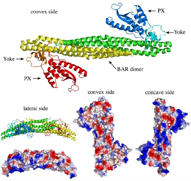
Figure 8. BAR dimers. A) Ribbon and space-fill models of the SNX9 BAR-PX dimer are shown. The two SNX9 proteins are depicted in red/orange/yellow and green/cyan/blue (PX domain: red/blue; BAR domain: yellow/green; yolk domain, which links the PX domain to the BAR domain: orange/cyan). Space-fill models showing the electrostatic distribution, red (acidic, negative residues), blue (positive, basic residues). [50]
35
Finally, SNX10 was shown to regulate the intracellular trafficking of V-ATPase, a multi-subunit complex required for ciliogenesis, acidification of osteoclasts and bone resorption [166]. Interestingly, Snx11 was found to antagonize Snx10-dependent vacuolation in vitro possibly through competitive binding with a common partner but its function remains poorly described [162].
1.11 – SNX-other
Sorting nexins can also mediate receptor recycling. For example, fast recycling of the β2 adrenergic receptor (β2AR), a seven-transmembrane signaling receptor, is a process that requires SNX27 [167, 168]. Here, VPS29 concentrates at β2AR-positive membrane tubulations. Previous to this finding, β2AR recycling was known to occur by the concentration of the receptor into endosomal tubulations, and this was dependent on a C-terminal PDZ ligand, [169]. However, initial ligand-induced endocytosis did not require retromer components. Instead, depletion of VPS35 or VPS29 significantly reduced β2AR recycling and resulted in endosomes devoid of β2AR tubules. This effect was similar to disrupting the receptor’s C-terminal PDZ ligand. Further investigation identified Rab4A as another essential component of β2AR recycling, as well as PDZ domain-containing SNX27, which acts as a cargo adaptor to retromer. In fact, SNX27 was shown to bind directly to the WASH complex, which in turn enabled its association with retromer components [168]. This indicates that retromer
36
components can mediate receptor trafficking at different endosomal compartments, by associating with various adaptor proteins.
1.12 – RABs
As previously mentioned, other important players in regulating membrane identity and vesicle traffic are the small Rab GTPases. Rab GTPases cycle between the GTP-bound state (active), catalyzed by guanine exchange factors (GEFs), and the GDP-bound state (inactive), driven by the combined action of the intrinsic GTPase activity of the Rab protein and catalyzed by GTPase-activating proteins (GAPs) [159]. In humans, over 60 members of the Rab family have been identified, which localize to distinct endosomal membranes [157]. Their association with membranes occurs through the post-translational addition of hydrophobic geranylgeranyl groups, which facilitates the attachment to target membranes and indirectly brings about interactions with coat components, motors and SNAREs. In turn, binding of Rabs to relevant membranes and to their respective effector proteins contributes to endosomal trafficking processes such as vesicle budding, vesicle uncoating, vesicle motility and vesicle fusion [157].
During vesicle budding, sorting of cargo into specific transport vesicles requires its association with cytosolic coat complexes. For example, Rab9 localizes at the multivesicular body and, like retromer, is required for retrograde transport of the CI-M6PR from this compartment to the trans-Golgi network [170, 171]. Once cargo is included into a transport
37
vesicle, coat complexes must be shed to allow membrane fusion with the acceptor membrane. For instance, Rab5, which is present on clathrin-coated vesicles, helps AP2 uncoating by promoting dephosphorylation and increasing PI(4,5)P2 turnover [172]. In addition to actin
microfilaments and microtubules, vesicle motility along these molecular cables is dependent on the kinetic force provided by various motor proteins like myosins, kinesins and dyneins. As such, certain Rab GTPases assist in proofreading the interactions between motor proteins and transport vesicles. Such is the case of Rab27a, which connects myosin Va to vesicle-like melanosomes shuttling towards the cell periphery [173]. Another example is a resident of endocytic recycling vesicles, Rab11a. Through its interaction with the Rab11 family interacting protein 2 (RAB11FIP2), Rab11a links recycling vesicles to myosin Vb [174]. As transport vesicles reach their destination, they must dock and fuse with the acceptor membrane. Here again, Rab GTPases are involved in this final step of endosomal trafficking. Evidence for this first came from studying a mutation of the yeast Rab GTPase Sec4, which causes accumulation of TGN-derived vesicles [175]. Later studies revealed that Rab GTPases contribute to vesicle fusion by recruiting elongated tethering complexes that form long distance connections between the vesicle and the acceptor membrane [157].
Another essential component of protein trafficking is the cytoskeleton, which provides the framework that guides vesicular transport and mechanical force required for reshaping the cell.
38
1.13 – The cytoskeleton
The cytoskeleton is a dynamic intracellular scaffolding system that contributes to morphology and plasticity, migration, signal transduction and intracellular trafficking. During these processes, cytoskeletal elements generate the force required for membrane deformations, create structural scaffolds and act as tracks for motor proteins. Microtubules, intermediate filaments and actin microfilaments are the three filamentous structures that make up the cytoskeleton, regulated through the fine balance between assembly and disassembly. Although a brief overview of each component will be given, the main focus will be on actin dynamics.
Microtubules are hollow tubes formed from the association of multiple alpha/beta tubulin heterodimers, which radiate from the microtubule-organizing center (MTOC) located at the centrosome in the cytoplasm [176]. Structures like the mitotic spindle of dividing cells and the core of cilia and flagella are dependent on microtubules. Microtubules are also important for structuring cell shape, maintaining organelle localization and serving as tracks for the
movement of vesicles and other cytoplasmic particles [177]. As such, both anterograde (away from cell body) and retrograde (toward cell body) movements are mediated by
microtubules [177]. To generate the force needed for transportation of particles, cells rely on motor proteins that travel along cytoskeletal tracks, using ATP for chemical energy [178]. Molecular motors travelling on microtubules are kinesins and dyneins.
Intermediate filaments (IFs) are named like this because their diameter (10nm) falls between that of microtubules (24nm) and actin microfilaments (8nm). IFs comprise a
39
heterogeneous group of structures encoded by many genes (at least 50 IF genes in humans), such as keratins, plectins, lamins, and desmins [179].
Actin microfilaments are the main driving force in cellular shape changes, which are required for processes such as cellular migration, signaling and cytokinesis [180]. Actin is an evolutionarily conserved protein found in all eukaryotic cells and it is also closely related to prokaryotic actin-like proteins, suggesting ancient evolutionary origins. Actin is an ATP-binding protein that can be found in monomeric soluble form, globular-actin (G-actin), or assembled into filamentous actin (F-actin) (Figure 6). When bound to ATP, actin monomers can be incorporated into microfilaments and shortly after, undergo ATP hydrolysis due to their ATPase activity. As the microfilament grows, actin-ATP subunits cap the growing end to prevent disassembly and promote growth. Actin microfilaments grow from their barbed (plus) end while the opposing end, where actin-ADP subunits tend to leave, is referred to as the pointed (minus) end. Since self-assembly is kinetically unfavourable, factors called actin nucleators facilitate actin polymerization.
The ARP2/3 complex is the most characterized actin nucleator, which is comprised of seven polypeptides including ARP2 and ARP3 plus five additional subunits, ARPC1-ARPC5 [180]. The ARP2/3 complex can nucleate filaments de novo and organize them into branched networks. Nascent filaments elongate at their barbed end and are capped by ARP2/3 at their pointed end. Assembly can be initiated by nucleating new filaments from monomers or by generating free barbed ends that act as templates for polymerization by uncapping or severing existing filaments. However, the activity of actin nucleation by the ARP2/3 complex alone is inefficient and requires filament binding, phosphorylation as well as the involvement of nucleation-promoting factors (NPFs). Most NPFs contain a WCA domain,
40
Figure 9. Actin. G-actin is a 42 kDa monomeric ATP-binding protein than can undergo cycles of self-assembly into filamentous actin (F-actin). ATP hydrolysis creates the ADP-bound form and subsequent depolymerization of the actin filament. Growth of the actin filament occurs primarily at the barbed end with the addition of ATP-actin and is capped by factors such as the ARP2/3 complex at its pointed end (ADP-actin) [181].
which is the minimal sequence element required for activation of ARP2/3-mediated actin nucleation, found in proteins such as the Wiskott-Aldrich syndrome protein (WASP) superfamily and the formins.
![Figure 2. Clathrin. A) First identification of clathrin by Barbara Pearse in 1976 [13]](https://thumb-eu.123doks.com/thumbv2/123doknet/2067455.6387/21.918.135.802.126.616/figure-clathrin-identification-clathrin-barbara-pearse.webp)