HAL Id: hal-03004144
https://hal-cnrs.archives-ouvertes.fr/hal-03004144
Submitted on 20 Nov 2020
HAL is a multi-disciplinary open access
archive for the deposit and dissemination of
sci-entific research documents, whether they are
pub-lished or not. The documents may come from
teaching and research institutions in France or
abroad, or from public or private research centers.
L’archive ouverte pluridisciplinaire HAL, est
destinée au dépôt et à la diffusion de documents
scientifiques de niveau recherche, publiés ou non,
émanant des établissements d’enseignement et de
recherche français ou étrangers, des laboratoires
publics ou privés.
Protein-protein interactions within the Fatty Acid
Synthase-II system of Mycobacterium tuberculosis are
essential for mycobacterial viability
Romain Veyron-Churlet, Olivier Guerrini, Lionel Mourey, Mamadou Daffé,
Didier Zerbib
To cite this version:
Romain Veyron-Churlet, Olivier Guerrini, Lionel Mourey, Mamadou Daffé, Didier Zerbib.
Protein-protein interactions within the Fatty Acid Synthase-II system of Mycobacterium tuberculosis are
essential for mycobacterial viability. Molecular Microbiology, Wiley, 2004, 54 (5), pp.1161-1172.
�10.1111/j.1365-2958.2004.04334.x�. �hal-03004144�
Molecular Microbiology (2004) 54(5), 1161–1172 doi:10.1111/j.1365-2958.2004.04334.x
© 2004 Blackwell Publishing Ltd
t3Blackwell Science, LtdOxford, UKMMIMolecular Microbiology0950-382XBlackwell Publishing Ltd, 2004? 200454511611172Original ArticleProtein-protein interactions within FAS-II of M. tuberculosisR. Veyron-Churlet et al.
Received 30 July, 2004. *For correspondence. E-mail [email protected]; Tel. (+33) 561 175 963; Fax (+33) 561 175 994.
Protein–protein interactions within the Fatty Acid
Synthase-II system of
Mycobacterium tuberculosis
are
essential for mycobacterial viability
Romain Veyron-Churlet, Olivier Guerrini,
Lionel Mourey, Mamadou Daffé and Didier Zerbib*
Département ‘Mécanismes Moléculaires des Infections Mycobactériennes’, Institut de Pharmacologie et de Biologie Structurale, Centre National de la Recherche Scientifique, 205 route de Narbonne, 31077 Toulouse Cedex 04, France.
Summary
Despite the existence of efficient chemotherapy, tuberculosis remains a leading cause of mortality worldwide. New drugs are urgently needed to reduce the potential impact of the emergence of multidrug-resistant strains of the causative agent
Mycobacte-rium tuberculosis (Mtb). The front-line antibiotic
isoniazid (INH), and several other drugs, target the biosynthesis of mycolic acids and especially the Fatty Acid Synthase-II (FAS-II) elongation system. This biosynthetic pathway is essential and specific for mycobacteria and still represents a valuable sys-tem for the search of new anti-tuberculous agents. Several data, in the literature, suggest the existence of protein–protein interactions within the FAS-II sys-tem. These interactions themselves might serve as targets for a new generation of drugs directed against
Mtb. By using an extensive in vivo yeast two-hybrid approach and in vitro co-immunoprecipitation, we have demonstrated the existence of both homotypic and heterotypic interactions between the known com-ponents of FAS-II. The condensing enzymes KasA, KasB and mtFabH interact with each other and with the reductases MabA and InhA. Furthermore, we have designed and constructed point mutations of the FAS-II reductase MabA, able to disrupt its homotypic interactions and perturb the interaction pattern of this protein within FAS-II. Finally, we showed by a transdominant genetic approach that these mutants are dominant negative in both non-pathogenic and pathogenic mycobacteria. These data allowed us to draw a dynamic model of the organization of FAS-II.
They also represent an important step towards the design of a new generation of anti-tuberculous agents, as being inhibitors of essential protein– protein interactions.
Introduction
Tuberculosis is a major infectious disease that kills 3 million people each year. According to the World Health Organization, about 40 million people will die over the next 25 years (Anonymous, 2002). Among the multiple causes for these alarming statistics, there is the growing percent-age (ª3%) of multidrug-resistant clinical isolates of the causative agent Mycobacterium tuberculosis (Mtb) in the world. In certain countries the prevalence of multidrug-resistant strains goes up to 40% (Anonymous, 2000). New drugs against Mtb are urgently needed. The most effective anti-mycobacterial drug in the last 50 years has been isoniazid (INH). INH targets the biosynthesis of mycolic acids (Banerjee et al., 1994; Dessen et al., 1995; Mdluli
et al., 1998), which represent the major and most specific lipid components of the mycobacterial cell wall (Daffe and Draper, 1998).
Mycolic acids are very long chain (C60–C90) a-branched b-hydroxylated fatty acids whose biosynthesis is under the control of at least two discrete elongation systems, namely the eukaryotic-type Fatty Acid Synthase-I (FAS-I) and the prokaryotic-like FAS-II (Kremer et al., 2000; Asselineau
et al., 2002). FAS-I consists of a multifunctional single polypeptide that performs de novo synthesis of medium length (C16–C26) acyl-coenzyme A (CoA). FAS-II catalyses the same type of reactions but it is composed of several distinct enzymes, it functions on acyl carrier protein (ACP) derivatives, and it is not capable of de novo synthesis in mycobacteria. The FAS-II initial substrates are medium length keto-acyl-ACP resulting from the condensation by the mtFabH protein of the acyl-CoA products of FAS-I with malonyl-ACP (Choi et al., 2000). After reduction by the keto-acyl-ACP reductase MabA (Banerjee et al., 1998), dehydratation by a yet unknown hydroxyl-acyl-ACP dehy-dratase, and reduction by the enoyl-ACP reductase InhA (Banerjee et al., 1994), either KasA or KasB catalyses the condensation with malonyl-ACP units in the FAS-II cycle (Schaeffer et al., 2001; Kremer et al., 2002; Slayden and Barry, 2002). Several pieces of data brought us to suspect
1162 R. Veyron-Churlet et al.
that the components of FAS-II might be tightly intercon-nected. First, a high-molecular-weight protein fraction displaying the FAS-II activity has been purified from the non-pathogenic bacterium Mycobacterium smegmatis
(Odriozola et al., 1977). Second, both InhA and MabA have been shown to be present and active in this complex fraction (Marrakchi et al., 2000; 2002). Third, the complex pattern of action of INH on FAS-II that we recently showed to fully inhibit MabA in vitro with a mechanism comparable to that of InhA (Ducasse et al., 2004) was another clue to suspect the existence of an organized FAS-II macromo-lecular complex. InhA and KasA have been proposed to be the primary target for INH (Slayden et al., 2000) and even if the mechanism of inhibition by INH is described in more details for InhA, this question is still debated (Mdluli
et al., 1996; Larsen et al., 2002; Kremer et al., 2003; Rawat et al., 2003). All these data may reflect a high degree of communication between the components of FAS-II.
An attracting hypothesis is that FAS-II would be orga-nized in such a way that the sequential enzymes of this pathway are in close contact, allowing the elongating fatty acids to be passed directly from one catalytic site to another. The elongation of fatty acids might thus be a channelled process. The channelling of metabolic inter-mediates has received a strong support in the literature over the last 20 years (Srere, 1987; Ovadi et al., 2000). It has been demonstrated in the yeast mitochondrial Krebs tricarboxylic acid cycle and it is dependent on protein– protein interactions (Velot and Srere, 2000). Protein–pro-tein interactions are essential in many cellular processes and as such, they have been used as targets for the search of potent inhibitors. For example, peptidic trans-dominant inhibitors were used to inhibit the pheromone response pathway of the budding yeast Saccharomyces cerevisiae (Caponigro et al., 1998; Geyer et al., 1999; Norman et al., 1999). These inhibitors were referred to as ‘perturbagens’ by analogy with ‘mutagens’. A pertur-bagen, as defined by Caponigro et al. (1998), instead of inducing genes mutations, acts at the level of protein, disrupting specific biochemical interactions in cells to gen-erate a mutant phenocopy.
The biosynthesis of mycolic acids is essential for myco-bacterial viability (Vilcheze et al., 2000; Portevin et al., 2004) and the fact that protein–protein interactions could exist and might be involved in the channelling of this process has prompted us to search for such interactions among the known components of the FAS-II system. Thereby, we aim at defining a perturbagen that would pave the way for the discovery of a new family of anti-tuberculous drugs. By using a yeast two-hybrid (Y2H) strategy and co-immunoprecipitation (co-IP), we showed here that both homotypic and heterotypic interactions exist between the FAS II components of Mtb. We exploited
the strongest interactions to design structural driven point mutations that were able to disrupt homotypic complexes and reveal heterotypic interactions. In vivo, these mutants are dominant negative in mycobacteria. This transdomi-nant genetic analysis allows defining a perturbagen that targets the essential FAS-II complex of Mtb.
Results and discussion
Protein–protein interactions within FAS-II revealed by Y2H genetic analysis
Yeast two-hybrid approach is a powerful technique to iden-tify protein–protein interactions. It has been shown that dissociation constants of ª70 mM were sufficient to detect protein–protein interactions in GAL4-based two-hybrid assays (Yang et al., 1995). However, the detection of false positives still represents a major risk and the analysis has to be carefully controlled. To avoid this kind of artifacts, we took several basic precautions: first, we used a three-reporter gene system (HIS3, ADE2 and MEL1) in order to dispose of a range of stringency in the Y2H analysis. It is noteworthy that numerous true protein–protein interac-tions have been identified by using only one or two reporter genes (Lei et al., 1999; Steyn et al., 2002; 2003). The second precaution was to use multiple negative con-trols. Each gene was tested against the void correspond-ing vector and each gene was also assayed against the human Lamin protein which has been shown to be very poorly interactive and represent a valuable negative con-trol (Ye and Worman, 1995). Each couple of protein was assayed in ‘both’ direction, i.e. as a fusion with binding domain (BD) and a fusion with activation domain (AD). And finally, after the selection of positive clones on selec-tive plates by streaking yeast co-transformants, the results were confirmed by plating, counting and scoring of dilutions (1.104 and 1.103) of liquid cultures of each co-transformant as indicated in Experimental procedures.
The kasA, kasB, inhA, mabA and mtfabH genes from
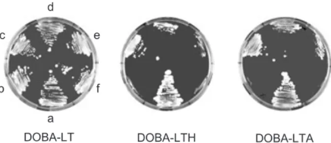
Mtb were cloned into the Y2H vectors pGAD-T7 and pGBK-T7. The resulting plasmids were used to construct all the possible AH109 co-transformants, each carrying a different couple of plasmids. As controls, AH109/pGBK-T7, AH109/pGAD-T7 and AH109/pGBK::lam were also transformed with the corresponding vectors expressing each FAS-II gene. The strains obtained were tested for their ability to grow on appropriate selective media to monitor the induction of the reporter genes HIS3 and/or ADE2. Individual co-transformants were streaked on DOBA-LT, DOBA-LTH and DOBA-LTA. An illustration of this principle is given in Fig. 1 where AH109/pGAD-T7::kasA was tested against pGBK-T7 derivatives.
The growth of each transformant was compared with the positive (AH109/pGAD::AgT/pGBK::p53) and the
neg-Protein-protein interactions within FAS-II of M. tuberculosis 1163
ative (AH109/pGAD::kasA/pGBK::lam) controls. For KasA, this first screen was interpreted as a strong inter-action with itself (quadrant c), a weaker interinter-action with KasB and InhA (quadrants d and e) and no interaction with either MabA or Lamin (quadrants f and b). After this first screen performed with each protein fused either to the AD or to the BD of GAL4, the final scoring of positives clones on four types of selective plates was performed by plating yeast cells from liquid cultures as indicated in
Experimental procedures. In this final screen, the stron-gest interactions were identified by evaluating the maxi-mum induction of the MEL1 reporter gene. A compilation of these data is presented in Table 1.
No growth was observed on any medium when each FAS-II protein was tested against either a void vector or the Lamin fusion protein indicating that these proteins did not activate any reporter gene and did not interact non-specifically with Lamin. These negative data were omitted for clarity. The Y2H analysis revealed strong homotypic interactions for each protein. Four of them (KasA, InhA, MabA and mtFabH) were positive for the three reporter genes (HIS3, ADE2 and MEL1) indicating a strong specific interactions. The homotypic interaction KasB– KasB seemed to be the weakest because, in contrary to KasA–KasA, or InhA–InhA, or MabA–MabA, or mtFabH– mtFabH, the corresponding co-transformants of AH109 were MEL1–
. These results correlate with available bio-chemical and structural data. MabA, which displayed the strongest homotypic interactions, is able to form at least homodimers in solution and has been crystallized as a tetramer (Cohen-Gonsaud et al., 2002). Similarly, InhA also displayed strong homotypic interactions and is known to form homotetramers (Dessen et al., 1995). Finally, KasA and KasB are strongly related in sequence (67% identity) and also to the Escherichia coli orthologous con-densing enzymes FabF (41% identity with KasA, 36% with KasB) and FabB (34% identity with KasB) (Schaeffer
et al., 2001) and, in this context, it is noteworthy that the FabF protein forms homodimers both in solution and dur-ing crystallization (Edwards et al., 1997; Huang et al., 1998). In the same manner, the homotypic interactions observed for mtFabH has to be correlated to the dimer-ization of the Mtb protein during crystal formation (Scars-dale et al., 2001). This coherence between our results and available data on the quaternary structures of these enzymes assess the reliability of the Y2H method used here. More novels were the heterotypic interactions revealed by this approach (Table 1). First, The KasA– KasB interaction was a ‘strong’ interaction even revealed by the MEL1 reporter gene. Second, the interaction mtFabH–KasA was seen on ‘both’ directions with two reporter genes. Finally, mtFabH–KasB interaction was seen clearly only in one direction (when mtFabH was fused with the BD of GAL4), suggesting either that this interaction is weaker than the mtFabH–KasA interaction or that the structure of the mtFabH–KasB complex is very different from the mtFabH–KasA complex and imposes to the reconstituted GAL4 activator a strong structural con-straint that prevents its activity when mtFabH is fused with the AD of GAL4.
In summary, we demonstrated that the three condens-ing enzymes of FAS-II are able to interact with each other. Another finding was given by the interaction pattern of InhA. It seemed that there is a connection between InhA and KasA which were both proposed to represent INH targets. This interaction was seen only with the ADE2 reporter gene and when InhA was fused with the BD suggesting either that KasA–InhA was a very weak inter-action or that it imposed a very strong structural constraint to the reconstituted GAL4 activator. This interaction between InhA and the ‘next’ enzyme of the FAS-II cycle, if it is confirmed, is the first clue for the existence of channelling during the biosynthesis of long-chain fatty acids. This result has also to be brought together with a recent study of the mechanism of action of INH on InhA (Rawat et al., 2003) where the possibility has been raised of a KasA–InhA interaction that might explain the
occur-Fig. 1. Y2H first screen of protein–protein interactions between KasA and the Mtb FAS-II proteins. The derivatives of AH109 containing either pGAD::AgT and pGBK::p53 (a), or pGAD::kasA and: pGBK::lam (b), or pGAD::kasA and pGBK::kasA (c), or pGAD::kasA
and pGBK::kasB (d), or pGAD::kasA and pGBK::inhA (e), or pGAD::kasA and pGBK::mabA (f) were streaked on the selective plates DOBA-LT, DOBA-LTH and DOBA-LTA and incubated 2 weeks at 30∞C. DOBA-LTH DOBA-LTA DOBA-LT e c b f a d
Table 1. Y2H analysis of protein–protein interactions within FAS-II.
AD fusions
BD fusions
kasA kasB inhA mabA mtfabH kasA ++±a ++ – – + – – – – +++ kasB + + ± + + ± – – – – – – + ± ± inhA – – – – – – + + + – – – ± – – mabA – – – – – – – – – + + + ± – – mtfabH ± ± ± ± – – – – – – – – + + +
a. Each of the three signs indicates the growth on LTH,
DOBA-LTA and DOBA-LTHA respectively. The significance of +, ± and – is given in Experimental procedures. Grey cells correspond to positive results on X-a-gal plates.
1164 R. Veyron-Churlet et al.
rence of INH-resistant mutant in KasA, even if InhA is indeed the only primary target of INH. Surprisingly, we did not observe any interaction between MabA and the other components of FAS-II. Furthermore, InhA seemed to interact only with KasA. This was striking because we shown previously that both proteins were indeed present in a FAS-II crude fraction from M. smegmatis (Marrakchi
et al., 2000; 2002). The first interpretation is that MabA do
not interact with the other FAS-II components and that the InhA interaction with them is very labile. However, these two FAS-II proteins, in contrast to the others, have been shown to crystallize as tight tetramers (Dessen et al., 1995; Cohen-Gonsaud et al., 2002) and we thought that it could be a reason that could reduce heterotypic inter-action in Y2H. To confirm the interinter-actions revealed by the Y2H approach and test the latter hypothesis of a strong inhibitory effect of the homo-multimerization of MabA in Y2H, it was necessary to develop a biochemical approach.
The co-IP approach confirms and extends the interaction pattern of the FAS-II components
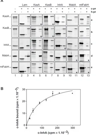
The vectors pGAD-T7 and pGBK-T7 are organized in such a way that the internal T7 promoter located just upstream from the multiple cloning site permits the in vitro transcription and translation of the gene of interest with-out including the AD or DB of GAL4. In addition, the translation products are tagged at their N-termini with either a haemagglutini (HA) or a c-Myc epitope giving, for example for KasA, proteins named h-KasA or c-KasA respectively. We evaluated the level of expression of each individual gene by loading on SDS-PAGE equivalent amounts (10 ml) of in vitro labelled transcription–transla-tion reactranscription–transla-tions performed on equivalent amounts (1 mg) of plasmid expressing either h-proteins (Fig. 2A, column 1) or c-proteins (Fig. 2A, even columns). We did not observed any striking difference from one protein to another.
Labelled or non-labelled h-proteins were used to trap c-proteins on magnetic beads coated with HA anti-bodies. When each labelled c-protein was incubated together with the coated beads without any h-protein, no significant background binding was observed (data not shown). All the combination of co-IP experiments were performed between KasA (row a), KasB (row b), InhA (row c), MabA (row d), mtFabH (row e) and the c-Lamin (Fig. 2A, column 2). We did not observe any background interactions between the five labelled h-proteins of FAS-II and the labelled c-Lamin protein (column 3) and con-cluded that the co-IP conditions were stringent enough to reveal specific interactions. When ‘cold’ h-proteins of FAS-II were used to trap labelled c-Lamin, no back-ground binding of Lamin was observed on the beads
(data not shown). The latter conditions were used to search for the homotypic interactions. In each case a clear band corresponding to the labelled c-protein was trapped by the non-labelled corresponding h-protein (row a, column 5; row b, column 7; row c, column 9; row d, column 11; and row e, column 13). The homotypic
inter-Fig. 2. Co-IP analysis of protein–protein interactions within the
FAS-II system.
A. Analysis of co-IP between the Mtb FAS-II proteins. The L-[35
S]-methionine-labelled h-proteins (HA tagged; h-prot) and c-proteins (c-Myc tagged; c-prot) from in vitro transcription/translation reactions and co-IP reactions products were fractionated by SDS-PAGE (10%) followed by phosphor-imaging analysis. In column 1, the gels con-tained the h-protein alone; h-KasA (row a), h-KasB (row b), h-InhA (row c), MabA (row d) and mtFabH (row e). The names of h-proteins are indicated in front of their position of migration. In even numbered column, the gels contained the c-protein alone. In the other columns (odd numbered except 1), the gel contained the co-IP reac-tion products performed between the c-proteins and the h-proteins. When the sizes of the proteins tested were two similar and in the case of the analysis of homotypic interactions, the co-IP reactions were performed between a labelled c-protein and a non-labelled h-protein; the corresponding panels are marked (*).
B. Co-IP analysis of KasA–InhA interaction. Increasing amounts of labelled c-InhA were incubated together with constant amount of labelled h-KasA (1.105
cpm) and the mix was subjected to standard co-IP experiment. After SDS-PAGE fractionation and quantification by phosphor-imaging, the quantities of InhA bound (in cpm) reported to 1.104 cpm of KasA bound was plotted against the amount of InhA (in
cpm) present in the reaction. The curve was a fit using a one-site-binding curve fit after the law of mass action.
c-InhA (cpm ¥ 1.10 –3) c-InhA bound (cpm ¥ 1.10 )–3 a b c d e c-prot h-prot
Protein-protein interactions within FAS-II of M. tuberculosis 1165 actions previously detected with the Y2H analysis were
confirmed for each protein of FAS-II.
All the interactions between the condensing enzymes were also detected. KasA and KasB (Fig. 2A, row a, col-umn 7 and row b, colcol-umn 5) were able to interact with each other whatever the ‘direction’ of the assay. However, the interactions observed between mtFabH (row e) and the Kas proteins (KasA, column 5, and KasB, column 7), even if it were largely above background, seemed weaker, especially when mtFabH was fused to the c-Myc epitope (column 13; rows a and b). Concerning the potential chan-nelling of intermediates between the condensing enzymes and MabA, the co-IP results did not allow concluding. MabA (row d) remained very poorly interactive and a faint co-IP band was observed in only ‘one direction’ with KasA, KasB, InhA and mtFabH (columns 5, 7, 9 and 13 respec-tively). Together with the negative Y2H results for MabA, these results were not convincing enough to prove a real interaction between MabA and the condensing enzymes. In contrast, InhA (row c) seemed to really interact with these enzymes. A new interaction was revealed between KasB (column 7) and InhA (row c) and the KasA–InhA interaction seemed to be much stronger in vitro than it was in Y2H. These interactions were seen in ‘both directions’, i.e. between c-KasA and h-InhA (row c, column 5) and between h-KasA and c-InhA (row a, column 9). To ascer-tain that the KasA–InhA interaction was specific, we performed co-IP reactions between labelled h-KasA (1.105 cpm) and labelled c-InhA using increasing amount of c-InhA. The titration curve followed a one-site binding curve suggesting the existence of a specific binding of InhA onto KasA (Fig. 2B). This was an important finding which has to be correlated with the potential channelling of intermediates between InhA and KasA. Moreover, as it has been proposed (Rawat et al., 2003), an interaction between these two proteins might represent a first expla-nation for the apparent contradiction in the literature that design both InhA and KasA as being the primary targets for INH (Mdluli et al., 1996; Larsen et al., 2002; Kremer
et al., 2003; Rawat et al., 2003).
The results obtained with the co-IP assays were in agreement with the Y2H genetic data and particularly strengthen the fact that the three condensing enzymes are able to interact with each other and with the InhA protein. The KasA–KasB interactions observed here are sup-ported by the study of the enzymatic activities of these two proteins (Slayden and Barry, 2002). In vitro, and in the presence of KasA, KasB displays a substrate prefer-ence for longer fatty acid chains than KasA suggesting a communication between these two enzymes and their successive involvement in the elongation of fatty acids by FAS-II. At this point, and because InhA interacts with the three condensing enzymes which interact with each other, we propose that three types of FAS-II complexes could
exist, each formed by a distinct condensing enzyme and at least InhA. Either these complexes might coexist or the quaternary structure of a ‘unique’ FAS-II might change from one composition to another during the time and according to the degree of elongation of the substrate. The channelling of intermediates between these specialized FAS-II complexes might be ensured either by the interac-tions between the condensing enzymes or by the tight homo-multimerization of InhA itself. The apparent absence of MabA from this complex remains striking. In view of the structure conservation of the reductases InhA and MabA (Dessen et al., 1995; Cohen-Gonsaud et al., 2002), it was indeed surprising to observe such differ-ences in the interaction pattern of the two proteins. How-ever, this behaviour seemed to be highly correlated with the strength of the homotypic interactions displayed by the proteins and that would preclude heterotypic interactions.
Structure-based design and construction of point mutations of the mabA gene
The crystallographic co-ordinates of MabA (Cohen-Gon-saud et al., 2002) were used in order to design subtle changes in the protein that would lead to the disruption of multimerization without, in theory, affecting its tertiary structure of the protein. The tertiary structure of MabA (247 residues) displays the same overall fold as the well-characterized InhA (269 residues) (Dessen et al., 1995; Rozwarski et al., 1999). This fold, which consists of a central parallel seven-stranded b-sheet flanked on both sides by a-helices, is characteristic of the short-chain dehydrogenase/reductase family of enzymes. MabA, like InhA, forms tetramers in solution and in the crystalline state (Dessen et al., 1995; Cohen-Gonsaud et al., 2002). The four subunits were labelled A, B, C and D respectively (Fig. 3A). The organization of the InhA and MabA homotetramers is also similar with a root mean square deviation of 2.6 Å for 646 matched Ca atoms. These tet-rameric structures display two significant protein–protein interfaces that occur twice, because of internal symmetry leading to the existence of two types of dimers: A–B and C–D, or A–C and B–D (Fig. 3A). Furthermore, within each interface, the relatedness of the subunits by a twofold axis implies that the interactions between subunits are dupli-cated. The first type of interface in the MabA homotet-ramer is delineated by the C-terminal polypeptidic segment comprising helix a6 and strand b7 (Fig. 3B). This interface is mainly stabilized by hydrophobic and van der Waals interactions and involves few polar interactions. A salt bridge is formed between the carboxylate group of Asp228, located between a6 and b7, and the NH1 and NH2 atoms of Arg34 on a1 of the other subunit. The mutation of the aspartic acid in an arginine (MabAD228R) might be detrimental to the stability of this interface as it
1166 R. Veyron-Churlet et al.
would lead to severe steric and electrostatic incompatibil-ities with Arg34 and other surrounding residues (Lys10, Ser222, Phe223, Ser226). If this mutation is really detri-mental, the mutant MabAD228R is expected to form only A– B or C–D dimers. The second type of interface comprises helices a4 and a5, which together with helix a3 cover one side of the central b-sheet (Fig. 3C). This interface is also mainly hydrophobic in nature with few polar contacts. In MabA, the Ca atom of Gly162 on helix a5 is at van der Waals contact with the Cb atom of Ala158 on helix a5 of the other subunit. This interaction would be impaired by any other amino acid than alanine at position 162 which would bump against Ala158 and also Ala154, and should have a profound destabilizing effect. A substitution for leucine was chosen (MabAG162L). If this mutation is effi-cient, the mutant MabAG162L is expected to form only A–C and B–D dimers.
Both single mutants and the double mutant (MabAG-D), which is expected to remain monomeric, were constructed by site-directed mutagenesis directly on the plasmids pGAD::mabA and pGBK::mabA. As we previously shown with large amounts of pure proteins, only one type of MabA dimers (A–B or C–D) involving the interface a6–b7 can be cross-linked via two lysine residues (distance Nz K173–Nz K209, 5 Å) using glutaraldehyde (Cohen-Gon-saud et al., 2002). Several other lysine residue pairs could form intramonomer cross-links (e.g. K104-K105 and K131-K173; the latter competing with the interchain cross-reaction). With this approach, there is no possibility to cross-link the A–C or B–D dimers with glutaraldehyde or to reveal the tetramerization of MabA. When this type of reactions were performed on labelled in vitro transcription/ translation product of wild-type h-mabA, we indeed
observed a dimer migrating on SDS-PAGE at the previ-ously observed (Cohen-Gonsaud et al., 2002) position of 60 kDa (Fig. 4A; D) showing that this technique can be used on proteins obtained in vitro for studying at least one type of dimerization of MabA (Fig. 4A). The mutation
D228(A)
G162(A)
G162(B)
G162(C)
G162(D)
A
B
α6
α6
β7
β7
D228(D)
D228(C)
C
α4
α5
α4
α5
G162(B)
G162(D)
D228(D)
K173(A)
K209(B)
K209(A)
K173(B)
K209(C)
K173(D)
K173(C)
K209(D)
D228(C)
(A)
(B)
(C)
(D)
Fig. 3. Quaternary structure of MabA.
A. Ribbon representation of the MabA tetramer with the different subunits coloured in magenta (A), blue (B), green (C) and yellow (D). Side-chains of residues that were subjected to mutagenesis (G162 and D228) and of lysine residues that can be cross-linked (K173 and K203) are depicted. Oxygen, nitrogen and car-bon atoms are in red, blue and grey respec-tively. Numbering is with one letter amino acid code followed by residue number and chain identifier between brackets.
B. a6–b7 interface between D and C subunits. Residues that were subjected to mutagenesis (D228) are represented.
C.a4–a5 interface between B and D subunits. Residues that were subjected to mutagenesis (G162) are represented.
Fig. 4. Cross-linking and co-IP analysis of MabA mutants.
A. In vitro cross-linking of MabA variants. Phosphor-imaging analysis of SDS-PAGE (12%) fractionation of the in vitro transcription/transla-tion-labelled products (10 ml) of pGAD-T7 derivatives of MabA (WT), MabAD228R (D228R), MabAG162L (G162L) and MabAG-D (G-D) before
(–) or after (+) glutaraldehyde cross-linking. Positions of monomer (M, 35 kDa), dimers (D, 60 kDa) are indicated. The position and size (kDa) of the molecular weight marker is indicated.
B. In vitro interactions between the MabA proteins. Co-IP analysis was as described in Fig. 2A. Labelled c-proteins derivatives of the four mabA alleles were loaded alone (left) or after co-IP with non-labelled h-MabA variants.
B -A - + - + - + - +
M
D
h-WT h-D228R h-G162L h-G-D h-D228R h-G162L h-WT c-D228R c-G162L c-G-D c-WT 97.4 66 45 31 31 31 31 31Protein-protein interactions within FAS-II of M. tuberculosis 1167 D228R, located in the a6–b7 interface of MabA and which
was designed to disrupt the A–C and B–D dimers of MabA (Fig. 3B), completely abolished the formation of glutaral-dehyde cross-linked dimers when it was present in either the single (MabAD228R) or the double (MabAG-D) mutant protein (Fig. 4A). It can be concluded that this mutation (D228R) was able to disrupt one type of dimerization and consequently tetramerization of MabA in solution. As dis-cussed above, and because of the positions of the lysine residues in MabA, it is not possible, in theory, to cross-link the A–B or C–D dimers of MabA using glutaraldehyde (Fig. 3A). Because the G162L mutation was designed to disrupt these types of MabA dimers (A–B or C–D), there was, as expected, no visible inhibition of the glutaralde-hyde cross-link of the A–C and B–D dimers (Fig. 3A) of the MabAG162L protein (Fig. 4A). However, this result did not prove that this mutation (G162L) was inefficient.
When we screened each MabA single mutant using the Y2H system, we still observed interactions with wild-type MabA (Table 2). In Y2H, a dimer of MabA seemed suffi-cient to reveal an interaction. However, in contrast to wild-type MabA–MabA interactions, those involving the mutants of MabA were all negative on X-a-gal plates (data not shown). The MabA double mutant did not interact at all with the wild-type protein. This mutant behaved like a monomer indicating that the two mutations were sufficient to abolish the multimerization of MabA. Co-IP gave essen-tially the same results (Fig. 4B). The double mutant was completely unable to interact with either the wild-type or the single mutants MabA proteins. The overall strength of interactions observed for single mutants and wild-type proteins seemed to be affected. This was particularly clear when the mutant h-proteins were tested against the wild-type c-MabA. In this case, h-MabAG162L was still able to interact with the wild-type c-MabA but this interaction seemed weaker than the interaction between the wild-type proteins. In addition, no interaction at all was observed between h-MabAD228R and the wild-type c-MabA. Taking all data together, it might be considered that each mutations designed in the present study reduced the interactivity of MabA. Each mutation has been defined to perturb one
type of dimer of the MabA tetramer. The effect of the D228R mutation which has been defined to disrupt the C– D and A–D interfaces of the MabA tetramer (Fig. 3) was clearly seen in Y2H, in cross-linking and in co-IP experi-ments. The effect of the G162L mutation which has been defined to disrupt the A–C and B–D interfaces of the MabA tetramer was seen in Y2H and co-IP and not in cross-linking because there is no possibility of glutaralde-hyde cross-link between those subunits. Only the C–D and A–D subunits can be cross-linked via the lysine K173 and K203. The effect of both mutations in the double mutant was seen by the three methods. Here, we designed precise MabA mutations perturbing its overall quaternary structure and resulting in profound effects both
in vivo and in vitro.
Mutations in MabA subunit interfaces reveal heterotypic interactions
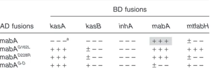
Mutants of MabA were tested in the Y2H system against the other components of FAS-II and mtFabH (Table 2). Each mutation allowed to reveal interactions with KasA and with KasB, although slightly for the latter. This sug-gests that, at least in yeast, these interactions were prob-ably masked by the high degree of homo-multimerization of MabA. In addition, only the G162L mutation allowed the detection of a significant interaction with MtFabH suggest-ing that the structure of the MabA–mtFabH complex is probably different from the ones formed with the other condensing enzymes (Table 2). These interaction patterns were clearly confirmed by co-IP. MabA as monomers (MabAG-D) or as dimers (MabAG162L and MabAD228R) in solu-tion can potentially interact with KasA, KasB, InhA and mtFabH (Fig. 5A). Quantification of the gels (Fig. 5B) obtained with the wild-type protein (Fig. 2A) and the dif-ferent variants (Fig. 5A) indicated that any defect in the multimerization of MabA increased its affinity for each condensing enzyme and InhA. We propose that each monomer of MabA can interact with each condensing enzyme and that its high propensity to multimerization might maintain the macromolecular organization of sev-eral FAS-II complexes brought together.
MabA multimerization mutants as perturbagens of the FAS-II system
In addition to contributing to unravel the mode of action of the Mtb FAS-II complex, the interaction mutants of MabA provide a valuable tool to investigate, by transdominant genetics, the viability of mycobacteria after perturbation of the FAS-II complex organization. The mabA alleles were cloned into the vector pMV261 and the resulting plasmids were used to transform M. smegmatis mc2155. The average transformation efficiencies at 37∞C were
sim-Table 2. Y2H analysis of MabA multimerization mutants.
AD fusions
BD fusions
kasA kasB inhA mabA mtfabH mabA – – –a – – – – – – + + + ± – – mabAG162L + + + ± – – – – – + + + + + + mabAD228R + + + ± – – – – – + + + ± – – mabAG-D + + + + – – – – – ± – – + – –
a. Each of the three signs indicates the growth on LTH,
DOBA-LTA and DOBA-LTHA respectively. The significance of +, ± and – is given in Experimental procedures. Grey cells correspond to positive results on X-a-gal plates.
1168 R. Veyron-Churlet et al.
ilar for pMV261, pMV261::mabAwt, pMV261::mabAD228R
and pMV261::mabAG-D (Table 3). However, upon five
inde-pendent trials, we did not observe any transformant with the vector expressing mabAG162L. Because the pHSP60
promoter is slightly thermo-inducible (Stover et al., 1991), we repeated these experiment at 30∞C and 42∞C. Except for the D228R mutation, the results were unchanged (Table 3). The mabAD228R allele seemed to express its
dominant negative effect only at 42∞C. This transdominant genetic study was extended to the vaccine strain M. bovis BCG and the pathogenic strain Mtb H37Rv (Table 3). Because these two strains do not grow at 42∞C, experi-ments were only carried out at 37∞C. The mutant
mabAG162L was also dominant negative in both strains. The
same experiments were carried out using an allele carry-ing the Y185L allele of MabA shown to be nearly inactive (28% of wild type) but capable of multimerization
(Ducasse et al., 2004). The mabAY185L allele was not
dom-inant negative in any strain. The domdom-inant negative effects observed with the G162L and D228R mutants is not likely to result from a non-productive diversion of either the substrate or the cofactor of MabA that would lead to a dead end in the biosynthesis of mycolic acids. This effect might rather be provoked by an interference of the protein brought in trans either with the resident wild-type MabA protein or with the FAS-II system itself via altered protein– protein interactions. The single mutant protein brought in
trans might form unproductive dimers with the resident
MabA wild-type protein and block the FAS-II system whereas the double mutant, which is not able to interact with MabA, has no effect. As this strong dominant nega-tive effect was revealed with both single mutations whereas no effect was seen for the wild type or the double mutant, we postulate here that these mutants act as per-turbagens of the FAS-II complex by modifying the degree of multimerization of MabA. The quaternary structure of MabA seems in turn to be determinant for the functioning of the FAS-II complex. These data point out the impor-tance and the essentiality of protein–protein interactions within the FAS-II complex of Mtb.
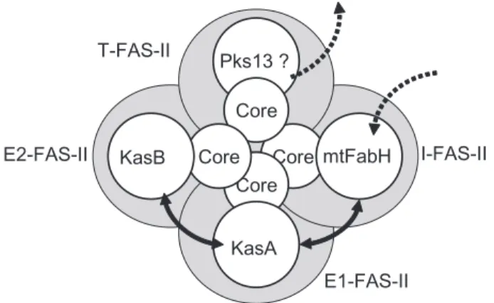
In conclusion, in the present study, a complex network of interactions between the components of the essential FAS-II system of Mtb has been revealed. Our data con-verge to a working model of the functioning of the complex where several specialized FAS-II complexes would be interconnected by protein–protein interactions between the condensing enzymes and/or the core proteins com-prising at least MabA and InhA and probably the not yet identified dehydratase (Fig. 6). If all the interactions revealed here were taken into account in a unique FAS-II complex, the resulting complex would be formed by at least a tetramer of MabA, a tetramer of InhA and three dimers of condensing enzymes (KasA, KasB and mtFabH). The molecular weight of this complex would not be in agreement with the value of 190 kDa proposed on early work on FAS-II (Odriozola et al., 1977). Further-more, it also would not be in agreement with the gel
Fig. 5. In vitro interaction between MabA mutants and the FAS-II
components.
A. Co-IP analysis of h-MabA variants with the FAS-II c-proteins. Results are presented as in Fig. 2A except that the analysis of c-proteins alone was omitted. The positions of migration of c-c-proteins (c-prot) and h-proteins (h-prot) are indicated with a black square and on the left side respectively.
B. Quantification of co-IP experiments between h-MabA derivatives and c-proteins of FAS-II. The co-IP index (in percentage) was defined as the ratio of the starting amounts of protein used in the reaction (h-protein/c-protein) reported to the final ratio of the proteins bound to the beads. The co-IP presented here was performed with h-MabA variants and c-KasA (black), c-KasB (white) or c-InhA (hatched).
B
WT G162L D228R G-D
Co-IP index (%)
A
c-KasA c-kasB c-InhAh-MabAG162L c-mtFabH h-MabAD228R h-MabAG-D
*
*
*
c-prot h-prot + + + - + + + + + +. .
. .
.
.
. .
.
.
.
.
Table 3. Transformation efficiencies of mycobacteria by
pMV261::mabA derivatives. mabA alleles M. smegmatis M. bovis BCG Mtb 30∞C 37∞C 42∞C 37∞C 37∞C None ª1.104a ª1.104 ª1.104 ª1.105 ª1.105 wild type ª1.104 ª1.104 ª1.104 ª1.105 ª1.105 mabAY185L ª1.104 ª1.104 ª1.104 Ndb Nd mabAG162L 0 0 0 0 0 mabAD228R ª1.104 ª1.104 0 ª1.105 ª1.105 mabAG-D ª1.104 ª1.104 ª1.104 Nd Nd
a. Expressed in cfu mg-1 of DNA.
Protein-protein interactions within FAS-II of M. tuberculosis 1169
filtration profiles of mycobacterial extracts used to purify active FAS-II fractions (Marrakchi et al., 2000; 2002). A more attractive model, based on the interaction network revealed here, would be the existence of several types of specialized FAS-II complexes. We propose that either these complexes might coexist or the quaternary structure of a ‘unique’ FAS-II might change from one composition to another during the time and according to the degree of elongation of the substrate. We showed that mtFabH can interact with InhA and MabA suggesting the existence of an ‘initiation-FAS-II’ containing mtFabH and a ‘FAS-II core’ formed by InhA, MabA (and probably the dehydratase) that might allow the channelling of acyl-CoA from FAS-I to FAS-II during their condensation with malonyl-ACP by mtFabH. A second FAS-II specialized complex could be the ‘elongation-1 FAS-II’ (E1-FAS-II) formed by the ‘core’ and KasA because we showed that KasA can interact with both InhA and MabA. The acyl-ACP from the ‘initiation-FAS-II’ (I-FAS-II) would be transferred to this complex maintained in close contact via either the mtFabH–KasA interactions or the multimerization of the core proteins. In the same manner, the intermediate length acyl-ACP would then ‘go’ to the ‘elongation-2 FAS-II’ (E2-FAS-II) comprising the core and KasB that could end the synthe-sis of the meromycolic chain. The channelling of interme-diates between these specialized FAS-II complexes might be insured either by the interactions between the con-densing enzymes revealed here or by the tight homo-multimerization of MabA and/or InhA. In addition, we speculated the existence of a fourth type but hypothetic FAS-II specialized complex (Termination complex; T-FAS-II) formed by the same core and the recently identified terminal condensing enzyme Pks13 (Portevin et al.,
2004). This last complex might condense the acyl-ACP coming out of E2-FAS-II and acyl-ACP (or acyl-CoA) com-ing out of I-FAS-II. Experiments will be carried out in order to confirm this model and extend it to the other partici-pants of the biosynthesis of mycolic acids like Pks13.
Another perspective of this work is the opening of a new field of investigation for the search of drugs directed against Mtb. Several human diseases (including cancer) occur as a consequence of the dissociation of particular protein–protein interactions (Sardet et al., 1997; Dyson, 1998) and peptides that bind and affect a particular target have been identified (Colas et al., 1996). The technology of selection of inhibitory peptide aptamers directed against cellular processes has been developed and opti-mized (Colas et al., 1996; Blum et al., 2000). Here, we have identified precise, specific and essential interactions whose disruption provokes the death of mycobacteria. This represents the first step towards the identification of a new generation of molecules that will be able to act as specific drugs directed against protein–protein interac-tions in the pathogen Mtb.
Experimental procedures
Strains and culture conditions
Plasmid constructions were performed in the E. coli K12 derivative Top10-F¢ (Invitrogen). The Y2H recipient strain was
S. cerevisiae AH109 from Clontech (MATa, trp1-901, leu2-3, 112, ura3-52, his3-200, gal4D, gal80D, LYS::GAL1UAS
-GAL1TATA-HIS3, GAL2UAS-GAL2TATA-ADE2, URA3::MEL1UAS
-MEL1TATA-lacZ). AH109 was cultured in YEP (BIO101) with
2% dextrose and 0.003% adenine. Selective plates were made with synthetic medium DOBA (BIO101) supplemented with the amino acids of the Complete Supplement Mixture (BIO101) lacking leucine and tryptophan (DOBA-LT). The Y2H genetic tests were performed on DOBA-LT also devoid of histidine (DOBA-LTH), or adenine (DOBA-LTA), or both (DOBA-LTHA). M. smegmatis mc2
155 was cultured in TS (Tryptic Soy; Difco) and plated on TS-Agar. M. bovis BCG Pasteur (ATCC 35 534) and Mtb H37Rv (ATCC 25 618) were grown in Middlebrook 7H9 (0.05% Tween 80) and plated on 7H11 (Difco) with 10% Middlebrook Enrichment Medium ADC or OADC (BBL) respectively. When needed media were supplemented with kanamycin (50 mg ml-1) or ampicillin
(100 mg ml-1). Electrotransformation of mycobacteria was
achieved with a Bio-Rad Gene-Pulser (2.5 kV, 25 mFD, 1000 W).
Construction of the Y2H vector derivatives
The vectors pGBK-T7 and pGAD-T7 from the Matchmaker®
Two-hybrid system 3 (Clontech) allowed the downstream cloning of genes in phase with the coding sequence of the BD or of the AD of the yeast GAL4 transcription activator. In pGBK-T7, the BD coding sequence is followed by the sequence of an internal T7 promoter and the coding sequence of an 11-amino-acid epitope tag from the proto-oncogene c-Myc, just upstream of the multiple cloning sites.
Fig. 6. Schematic representation of a model of interconnected
spe-cialized FAS-II complexes. Each putative spespe-cialized FAS-II complex is represented by a grey circle: I-FAS-II, initiation FASII; E1-FAS-II, elongation-1 FAS-II; E2-FAS-II, elongation-2 FAS-II; and T-FAS-II, terminal FAS-II. The core represents at least InhA, MabA and the non-identified dehydratase of FAS-II. Double-headed plain arrows represent observed interactions between the condensing enzymes. Stippled arrows symbolized the entry of acyl-ACP and the exit of mycolic-acids. Pks13 is the polyketide synthase 13.
Core Core Core Core Pks13 ? mtFabH KasB KasA I-FAS-II E1-FAS-II E2-FAS-II T-FAS-II
1170 R. Veyron-Churlet et al.
In addition, pGBK-T7 carries a kanamycin resistance gene and the yeast auxotrophic marker TRP1. After cloning of a gene of interest, the resulting protein will be a C-terminal fusion with the BD and the c-Myc epitope tag. The vector pGAD-T7 possesses the same genetic organization but it contains the coding sequence of the AD of GAL4, the sequence of an HA tag, an ampicillin resistance gene and the yeast LEU2 coding sequence. The five genes of Mtb (kasA, kasB, inhA, mabA and mtfabH) were amplified by polymerase chain reaction (PCR) from Mtb H37Rv chromo-somal DNA using the Pfu DNA polymerase (Promega) and specific pairs of primers (Table S1) allowing their in-phase cloning in the multiple cloning sites of both pGAD-T7 and pGBK-T7. A description of the resulting constructs together with the description of the control vectors pGAD::lam, pGAD::AgT and pGBK::p53 is presented (Table S2).
Y2H genetics analysis
AH109 has three reporter genes (HIS3, ADE2 and MEL1), under the control of three different GAL4-dependent promot-ers. The promoter driving the expression of HIS3 possesses a strong GAL4 Upstream Activating Sequences (UAS), and thus allows the detection of weak interactions. The promoter of ADE2 has a weak GAL4 UAS and thus allows only the detection of strong interactions. The third gene, MEL1, is responsible for the synthesis of a-galactosidase and is con-trolled by the natural and weak, GAL4-dependent MEL1 pro-moter region. AH109 was transformed with each couple of pGAD-T7 and pGBK-T7 derivatives as described (Clontech). Co-transformants, containing two plasmids, were selected on DOBA-LT. As a first screen for protein–protein interactions and for each couple of plasmid tested, at least five individual co-transformants were streaked on DOBA-LT with replicate on DOBA-LTH, DOBA-LTA and DOBA-LTHA. After this first screen, the validity of a given interaction was evaluated by plating dilutions (1.104
and 1.103
) of saturated liquid cultures of an individual co-transformant on the four types of selective plates. On each medium, the number of colony-forming unit (cfu) was reported to the number of cfu on DOBA-LT. After the streak test and the plating assay, the scoring of a given interaction was performed as follows: an interaction was scored as positive (+) when numerous individual colonies were visible on the streak assay and when more than 80% of the population had the appropriate phenotype on a given medium in the plating assay. It was scored as plus-minus (±) when only a few colonies were visible in the streak assay and when 50–80% of the population had the required phenotype in the plating assay. In all the other cases, the results were scored as negative (–). The induction of MEL1 was evaluated by the coloration of colonies on DOBA-LT containing 20 mg ml-1 5-bromo-4-chloro-3-indolyl
a-D-galactoside (X-a-gal; Clontech). This test is not very sensitive and we counted as MEL1+ the only colonies that displayed the same dark blue
colour as the positive control strain AH109/pGAD::AgT/ pGBK::p53.
Co-immunoprecipitation
In vitro transcription/translation of the genes of interest was
performed with supercoiled DNA (1 mg) from pGAD-T7 or
pGBK-T7 derivatives as matrix with the TnT® Quick Coupled
Transcription/Translation System (Promega). Reactions were performed in a final volume of 50 ml either in the presence of 0.4 mCi ml-1 L-[35S]-methionine (1000 Ci mmol-1; Amersham)
or with cold methionine (40 mM) to produce unlabelled h-proteins. For co-IP experiments, we used Dynabeads®
M-450 Goat mouse IgG coated with monoclonal HA anti-bodies (Sigma). The ratios of the proteins were adjusted to 1:1 by evaluating the specific activity of each protein (in cpm per ml) and by correcting their differences in the number of methionine residues. Proteins were incubated with the coated beads (2 h at 4∞C) in 20 ml of 50 mM Tris (pH 7.4), 50 mM NaCl and 0.025% Tween 20. After extensive washing of the beads with 100 mM Tris (pH 7.4), 100 mM NaCl and 0.025% Tween 20, the reactions were boiled in the SDS-PAGE loading buffer and fractionated on SDS-SDS-PAGE followed by autoradiography and Phosphor-imaging (STORM-Applied Biosystems).
Structure-based design and construction of point mutations of the mabA gene
Structural analysis was based on the crystallographic co-ordinates of MabA (Cohen-Gonsaud et al., 2002). Site-directed mutagenesis was made directly on pGAD::mabA and pGBK::mabA using inverse-PCR amplification with self-complementary primers (Table S1) carrying the desired mutation. After in vitro transcription and translation, glutaral-dehyde cross-linking of labelled h-proteins (10 ml) was per-formed as described previously (Cohen-Gonsaud et al., 2002) and analysed on SDS-PAGE and Phosphor-imaging. For in vivo analysis of mabA mutant activity in mycobacteria, the five genes, mabA, mabAY185L, mabAG162L, mabAD228R and
the double mutant mabAG-D, were cloned into the
mycobac-teria-E. coli shuttle vector pMV261 (Stover et al., 1991) under the control of the pHSP60 signals carried by the vector (Table S2).
Acknowledgements
We thank Professor Gilbert Lanéelle for communicating his invaluable knowledge of the mycolic acid biosynthesis path-way. We are grateful to P. Constant for manipulation of patho-gens, G. Labesse for providing unpublished data and O. Humbert, I. Saves and A. Quémard for fruitful discussions. R.V.-C. is a recipient of a Fellowship from the Ministère Délégué à la Recherche et aux Nouvelles Technologies (MDRNT). This work was supported by the Centre National de la Recherche Scientifique (CNRS, France) and the MDRNT (ACI ‘Molécules et cibles thérapeutiques’).
Supplementary material
The following material is available from
http://www.blackwellpublishing.com/products/journals/ suppmat/mmi/mmi4334/mmi4334sm.htm
Table S1. Oligonucleotide sequences of PCR primers and
cloning sites used for the construction of pGAD-T7 and pGBK-T7 derivatives.
Protein-protein interactions within FAS-II of M. tuberculosis 1171
References
Anonymous (2000) The WHO/IUALTD Global Project on
Anti-tuberculosis Drug Resistance Surveillance. Geneva: World
Health Organization.
Anonymous (2002) Stop TB Reports WHO/CDS/STB/
2002.17. Geneva: World Health Organization.
Asselineau, C., Asselineau, J., Laneelle, G., and Laneelle, M.A. (2002) The biosynthesis of mycolic acids by Myco-bacteria: current and alternative hypotheses. Prog Lipid
Res 41: 501–523.
Banerjee, A., Dubnau, E., Quemard, A., Balasubramanian, V., Um, K.S., Wilson, T., et al. (1994) inhA, a gene encod-ing a target for isoniazid and ethionamide in
Mycobacte-rium tuberculosis. Science 263: 227–230.
Banerjee, A., Sugantino, M., Sacchettini, J.C., and Jacobs, W.R., Jr (1998) The mabA gene from the inhA operon of
Mycobacterium tuberculosis encodes a 3-ketoacyl
reduc-tase that fails to confer isoniazid resistance. Microbiology
144: 2697–2704.
Blum, J.H., Dove, S.L., Hochschild, A., and Mekalanos, J.J. (2000) Isolation of peptide aptamers that inhibit intracellu-lar processes. Proc Natl Acad Sci USA 97: 2241–2246. Caponigro, G., Abedi, M.R., Hurlburt, A.P., Maxfield, A.,
Judd, W., and Kamb, A. (1998) Transdominant genetic analysis of a growth control pathway. Proc Natl Acad Sci
USA 95: 7508–7513.
Choi, K.H., Kremer, L., Besra, G.S., and Rock, C.O. (2000) Identification and substrate specificity of beta-ketoacyl (acyl carrier protein) synthase III (mtFabH) from
Mycobac-terium tuberculosis. J Biol Chem 275: 28201–28207.
Cohen-Gonsaud, M., Ducasse, S., Hoh, F., Zerbib, D., Labesse, G., and Quemard, A. (2002) Crystal structure of MabA from Mycobacterium tuberculosis, a reductase involved in long-chain fatty acid biosynthesis. J Mol Biol
320: 249–261.
Colas, P., Cohen, B., Jessen, T., Grishina, I., McCoy, J., and Brent, R. (1996) Genetic selection of peptide aptamers that recognize and inhibit cyclin-dependent kinase 2. Nature
380: 548–550.
Daffe, M., and Draper, P. (1998) The envelope layers of mycobacteria with reference to their pathogenicity. Adv
Microb Physiol 39: 131–203.
Dessen, A., Quemard, A., Blanchard, J.S., Jacobs, W.R., Jr, and Sacchettini, J.C. (1995) Crystal structure and function of the isoniazid target of Mycobacterium tuberculosis.
Sci-ence 267: 1638–1641.
Ducasse, S., Cohen-Gonsaud, M., Marrakchi, H., Nguyen, M., Zerbib, D., Labesse, G., and Quemard, A. (2004) In
vitro inhibition of the Mycobacterium tuberculosis b-ketoacyl-acyl carrier protein reductase MabA from M.
tuberculosis by isoniazid. Antimicrob Agents Chemother
48: 242–249.
Dyson, N. (1998) The regulation of E2F by pRB-family pro-teins. Genes Dev 12: 2245–2262.
Edwards, P., Nelsen, J.S., Metz, J.G., and Dehesh, K. (1997) Cloning of the fabF gene in an expression vector and in
vitro characterization of recombinant fabF and fabB
encoded enzymes from Escherichia coli. FEBS Lett 402: 62–66.
Geyer, C.R., Colman-Lerner, A., and Brent, R. (1999)
‘Mutagenesis’ by peptide aptamers identifies genetic net-work members and pathway connections. Proc Natl Acad
Sci USA 96: 8567–8572.
Huang, W., Jia, J., Edwards, P., Dehesh, K., Schneider, G., and Lindqvist, Y. (1998) Crystal structure of beta-ketoacyl-acyl carrier protein synthase II from E. coli reveals the molecular architecture of condensing enzymes. EMBO J
17: 1183–1191.
Kremer, L., Baulard, A., and Besra, G.S. (2000) Genetics of mycolic acid biosynthesis. In Molecular Genetics of
Myco-bacteria. Hatfull, G., and Jacobs, W.R. (eds). Washington,
DC: American Society for Microbiology Press, pp. 173– 190.
Kremer, L., Dover, L.G., Carrere, S., Nampoothiri, K.M., Lesjean, S., Brown, A.K., et al. (2002) Mycolic acid biosyn-thesis and enzymic characterization of the beta-ketoacyl-ACP synthase A-condensing enzyme from Mycobacterium
tuberculosis. Biochem J 364: 423–430.
Kremer, L., Dover, L.G., Morbidoni, H.R., Vilcheze, C., Maughan, W.N., Baulard, A., et al. (2003) Inhibition of InhA activity, but not KasA activity, induces formation of a KasA-containing complex in mycobacteria. J Biol Chem 278: 20547–20554.
Larsen, M.H., Vilcheze, C., Kremer, L., Besra, G.S., Par-sons, L., Salfinger, M., et al. (2002) Overexpression of
inhA, but not kasA, confers resistance to isoniazid and
ethionamide in Mycobacterium smegmatis, M. bovis BCG and Mycobacterium tuberculosis. Mol Microbiol 46: 453– 466.
Lei, S., Pulakat, L., and Gavini, N. (1999) Genetic analysis of nif regulatory genes by utilizing the yeast two-hybrid system detected formation of a NifL–NifA complex that is implicated in regulated expression of nif genes. J Bacteriol
181: 6535–6539.
Marrakchi, H., Laneelle, G., and Quemard, A. (2000) InhA, a target of the antituberculous drug isoniazid, is involved in a mycobacterial fatty acid elongation system, FAS-II.
Microbiology 146: 289–296.
Marrakchi, H., Ducasse, S., Labesse, G., Montrozier, H., Margeat, E., Emorine, L., et al. (2002) MabA (FabG1), a
Mycobacterium tuberculosis protein involved in the
long-chain fatty acid elongation system FAS-II. Microbiology
148: 951–960.
Mdluli, K., Sherman, D.R., Hickey, M.J., Kreiswirth, B.N., Morris, S., Stover, C.K., and Barry, C.E., III (1996) Bio-chemical and genetic data suggest that InhA is not the primary target for activated isoniazid in Mycobacterium
tuberculosis. J Infect Dis 174: 1085–1090.
Mdluli, K., Slayden, R.A., Zhu, Y., Ramaswamy, S., Pan, X., Mead, D., et al. (1998) Inhibition of a Mycobacterium
tuber-culosis beta-ketoacyl ACP synthase by isoniazid. Science
280: 1607–1610.
Norman, T.C., Smith, D.L., Sorger, P.K., Drees, B.L., O’Rourke, S.M., Hughes, T.R., et al. (1999) Genetic selec-tion of peptide inhibitors of biological pathways. Science
285: 591–595.
Odriozola, J.M., Ramos, J.A., and Bloch, K. (1977) Fatty acid synthetase activity in Mycobacterium smegmatis. Charac-terization of the acyl carrier protein-dependent elongating system. Biochim Biophys Acta 488: 207–217.
1172 R. Veyron-Churlet et al.
compartmentation and channeling. Location–location– location. Int Rev Cytol 192: 255–280.
Portevin, D., De Sousa-D’Auria, C., Houssin, C., Grimaldi, C., Chami, M., Daffe, M., and Guilhot, C. (2004) A polyketide synthase catalyzes the last condensation step of mycolic acid biosynthesis in mycobacteria and related organisms.
Proc Natl Acad Sci USA 101: 314–319.
Rawat, R., Whitty, A., and Tonge, P.J. (2003) The isoniazid-NAD adduct is a slow, tight-binding inhibitor of InhA, the
Mycobacterium tuberculosis enoyl reductase: adduct
affin-ity and drug resistance. Proc Natl Acad Sci USA 100: 13881–13886.
Rozwarski, D.A., Vilcheze, C., Sugantino, M., Bittman, R., and Sacchettini, J.C. (1999) Crystal structure of the
Myco-bacterium tuberculosis enoyl-ACP reductase, InhA, in
complex with NAD+ and a C16 fatty acyl substrate. J Biol
Chem 274: 15582–15589.
Sardet, C., LeCam, L., Fabbrizio, E., and Vidal, M. (1997) Oncogenes as transcriptional regulators. In Cell
Cycle Regulators of Chromosomal Translocation.
Ghys-dael, J., and Yaniv, M. (eds). Birkhauser Verlag, pp. 1– 63.
Scarsdale, J.N., Kazanina, G., He, X., Reynolds, K.A., and Wright, H.T. (2001) Crystal structure of the Mycobacterium
tuberculosis beta-ketoacyl-acyl carrier protein synthase III. J Biol Chem 276: 20516–20522.
Schaeffer, M.L., Agnihotri, G., Volker, C., Kallender, H., Bren-nan, P.J., and Lonsdale, J.T. (2001) Purification and biochemical characterization of the Mycobacterium
tuber-culosis beta-ketoacyl-acyl carrier protein synthases KasA
and KasB. J Biol Chem 276: 47029–47037.
Slayden, R.A., and Barry, C.E., III (2002) The role of KasA and KasB in the biosynthesis of meromycolic acids and isoniazid resistance in Mycobacterium tuberculosis.
Tuber-culosis (Edinb) 82: 149–160.
Slayden, R.A., Lee, R.E., and Barry, C.E., III (2000) Isoniazid affects multiple components of the type II fatty acid syn-thase system of Mycobacterium tuberculosis. Mol
Micro-biol 38: 514–525.
Srere, P.A. (1987) Complexes of sequential metabolic enzymes. Annu Rev Biochem 56: 89–124.
Steyn, A.J., Collins, D.M., Hondalus, M.K., Jacobs, W.R., Jr, Kawakami, R.P., and Bloom, B.R. (2002) Mycobacterium
tuberculosis WhiB3 interacts with RpoV to affect host
sur-vival but is dispensable for in vivo growth. Proc Natl Acad
Sci USA 99: 3147–3152.
Steyn, A.J., Joseph, J., and Bloom, B.R. (2003) Interaction of the sensor module of Mycobacterium tuberculosis H37Rv KdpD with members of the Lpr family. Mol Microbiol
47: 1075–1089.
Stover, C.K., de la Cruz, V.F., Fuerst, T.R., Burlein, J.E., Benson, L.A., Bennett, L.T., et al. (1991) New use of BCG for recombinant vaccines. Nature 351: 456–460.
Velot, C., and Srere, P.A. (2000) Reversible transdominant inhibition of a metabolic pathway. In vivo evidence of inter-action between two sequential tricarboxylic acid cycle enzymes in yeast. J Biol Chem 275: 12926–12933. Vilcheze, C., Morbidoni, H.R., Weisbrod, T.R., Iwamoto,
H., Kuo, M., Sacchettini, J.C., and Jacobs, W.R., Jr (2000) Inactivation of the inhA-encoded fatty acid syn-thase II (FASII) enoyl-acyl carrier protein reductase induces accumulation of the FASI end products and cell lysis of Mycobacterium smegmatis. J Bacteriol 182: 4059–4067.
Ye, Q., and Worman, H.J. (1995) Protein–protein interactions between human nuclear lamins expressed in yeast. Exp
Cell Res 219: 292–298.
Yang, M., Wu, Z., and Fields, S. (1995) Protein–peptide inter-actions analyzed with the yeast two-hybrid system. Nucleic