n
Université de Montréal
Role cf Ptfl a in the development cf endocrine and exocrine
pancreas cf
Xenopus
Iaevis embryos
par
Zeina Jarikji
Département de Biologie Moléculaire
Faculté des études supérieures
Mémoire présenté à la Faculté des études supérieures
en vue de l’obtention du grade de Maîtrise
en Biologie Moléculaire
December, 2006
de Montréal
Direction des bibhothèques
AVIS
L’auteur a autorisé l’Université de Montréal à reproduire et diffuser, en totalité ou en partie, par quelque moyen que ce soit et sur quelque support que ce soit, et exclusivement à des fins non lucratives d’enseignement et de recherche, des copies de ce mémoire ou de cette thèse.
L’auteur et les coauteurs le cas échéant conservent la propriété du droit d’auteur et des droits moraux qui protègent ce document. Ni la thèse ou le mémoire, ni des extraits substantiels de ce document, ne doivent être imprimés ou autrement reproduits sans l’autorisation de l’auteur.
Afin de se conformer à la Loi canadienne sur la protection des renseignements personnels, quelques formulaires secondaires, coordonnées ou signatures intégrées au texte ont pu être enlevés de ce document. Bien que cela ait pu affecter la pagination, il n’y a aucun contenu manquant.
NOTICE
The author cf this thesis or dissertation has granted a nonexclusive license allowing Université de Montréal to reproduce and publish the document, in part or in whole, and in any format, solely for noncommercial educational and research purposes.
The author and co-authors if applicable retain copyright ownership and moral rights in this document. Neither the whole thesis or dissertation, nor substantial extracts from it, may be printed or otherwise reproduced without the author’s permission.
In compliance with the Canadian Privacy Act some supporting forms, contact information or signatures may have been temoved from the document. While this may affect the document page count, it does flot represent any loss cf content from the document.
Faculté des études supérieures
Ce mémoire intitulé:
Role of PtfJ a in the development cf endocrine and exocrine
pancreas of
Xenopus
laevis embryos
présenté par:
Zeina Jarikji
a été évalué par un jury composé des personnes suivantes:
Dr. Muriel Aubry
président-rapporteur
Dr. Marko Horb
directeur de recherche
Dr. Mark Prentki
membre du jury
Les injections d’insuline restent le seul traitement pratique pour le diabète insulinodépendant, mais causent beaucoup de problèmes parce qu’elle ne maintient pas la glycémie tel que le fait le pancréas. Les greffes de pancréas et d’îlots sont aussi faisables mais limitées par le petit nombre de donneurs et le traitement immunosuppresseur à vie. Une solution ultime serait la production de nouvelles cellules sécrétrices d’insuline à partir de cellules souches ou différentiées provenant du receveur diabétique lui-même. Il est alors crucial d’élucider les facteurs de transcription clés qui régulent le développement endocrine du pancréas. Ptfla est un des facteurs de transcription exprimés au cours du développement précoce du pancréas et reconnu comme étant impliqué dans la détermination de la lignée pancréatique exocrine. Des résultats plus récents ont suggéré que Pifla peut aussi être impliqué dans le développement des cellules pancréatiques endocrines. Pour vérifier cette hypothèse nous avons étudié le rôle du Pifla au cours du développement du pancréas chez le Xenope. Nous avons montré que Pifla est essentiel pour le développement des deux lignées pancréatiques endocrine et exocrine. En outre, nous avons testé la suffisance du Pifla à promouvoir un sort pancréatique de façon ectopique et ceci en utilisant les techniques de surexpression par transfert de gènes et par injection d’ARN messager. Nous avons montré que Pifla est capable de promouvoir les deux sorts pancréatiques endocrine et exocrine dans une région bien définie de l’endoderme. En conclusion, nos résultats démontrent que Pifla joue un rôle potentiel au cours du développement du pancréas et éclaircissent la voie de génération de cellules insulinoproductrices in
vitro en vue d’une application thérapeutique.
Mots clés: Xenopus, Pancréas, Pifla, spécification, transdifferentiation, insuline, îlots de Langerhans, endocrine, exocrine, organogenèse
Su mm a ry
Insulin injections remain the single practical treatment for type 1 diabetes, but
stili cause many problems even under the best conditions because it cannot
reproduce the exact maintenance of euglycemia as the normal pancreas. Transplantation of whole pancreata or islets is a feasible alternative but stiil faces
problems in the lack of donors and lifetong immunosuppressive treatment. Generating new insulin producing ceils from one’s body would be the ultimate solution. Whether the sources to be used are stem ceils or differentiated celis the crucial need is to elucidate the key transcriptional regulators controlling endocrine
pancreatic development. Pifla is one of the earliest genes expressed in the pancreas and is known to be one of the key transcriptional regulators of exocrine pancreas developmerit. Recent results have suggested that the Pifla may also be
involved in endocrine cell fate specification. To address this question we studied the function of Pifla in Xenopus pancreas development. We show that Ptfla is essential
for proper development of both endocrine and exocrine cells. Furthermore, we also
tested its sufficiency to ectopically promote a pancreatic cetl fate using transgenic
and mRNA overexpression assays, and found that t is able to promote both
endocrine and exocrine celi fates within a defined region of the endoderm. In
conclusion, our resuits demonstrate that Pifla is a master regulator of pancreatic fate in Xenopus and give insight into the generation of endocrine ceils in vitro for
therapeutic application.
Key
words:
Xenopus, Pancreas, Diabetes, Pifla, specification, transdifferentiation, insulin, islet of Langerhans, endocrine, exocrine, organogenesisTable of Contents
Résumé
Summary ïi
Table of Contents iii
Listof tables viii
Listof figures viii
List of abbreviations x Dedication xii Acknowledgement xiii 1. Introduction I 1.1. Xenopus Iaevis 1 1.2. Thepancreas 2
1.2.1. Anatomy and function 2
1.2.2. Diseases cf the pancreas 5
1.2.2.1. Pancreatitis 5
1.2.2.2. Cystic Fibrosis 6
1.2.2.3. Pancreatic Cancer 6
1.2.2.4. Diabetes 8
1.2.3. Embryclogical origins 9
1 .2.4. Reg ional specification cf the pancreatic endoderm 13
1.2.4.1. Role cf the notochord 15
1.2.4.3. Role cf the pancreatic mesenchyme 18
1.2.4.4. Retinoic Acid in pancteas development 19
1 .2.4.5. Notch signaling in pancreas development 20
1.2.5. Genetic network regulating pancreas development 22
1.2.5.1. Exocrine and endocrine pancreatic transcription factors 22
Pdxl 22
HIxb9 24
Pffla/P48 25
1 .2.5.2. Exocrine specific transcription factors 30
MistI 30
1 .2.5.3. Endocrine specific transcription factors 30
Ngn3 31
NeuroD/Beta2 32
IsIl 32
Pax4 and Pax6 33
Nkx2.2 and Nkx6.1 34
1.3. Aimofthiswork 36
2. Materials and methods 37
2.1. Isolation cf XPffla 37
2.2. Embryclogical assays 38
2.2.1. Inv/trofertilization 38
2.2.3. Generation of EIas-GFP transgenics 40
2.3. Embryos fixation and in situ hybridization 40
2.3.1. Fixation 40
2.3.2. Antisense probe synthesis 41
2.3.3. In situ hybridization 41
2.3.3.1. Hybridization 42
2.3.3.2. Blocking and antibody incubation 43
2.3.3.3. Staining 43
2.4. Realtime PCR 44
3. Results 45
3.1. Summary 46
3.2. Introduction 47
3.3. Materials and Methods 52
3.3.1. Xenopus transgenics and transgene construction 52
3.3.2. Isolation ofXpffla 54
3.3.3. Embryological assays and whole mount In situ hybridization 56
3.3.4. Real time PCR 56
3.4. Results 58
3.4.1. Ptfla-VPI6 is sufficient to convert liver to pancreas 58
3.4.2. The unmodified Pifla converts duodenum and stomach to pancreas 65
3.4.3. Ptfla and Pffla-VPI6 have similar capabilities in embryonic endoderm, 69
3.4.4. Xenopus Pifla is essential for bath exocrine and endocrine pancreas 77
development
3.4.5. Mouse Pifla mRNA rescues pancreatic agenesis caused by Xptfla 83 morpholinos
3.5. Discussion 84
3.5.1. Pifla is a master regulator of pancreatic ceil fate 84
3.5.2. Twa pathways to generate endocrine celis: Pifla dependent and 87
independent
3.6. Acknowledgements 89
3.7. References 90
4. Discussïon 94
4.1. Pff la is a master reg ulatory gene of pancreatic ceH fate 94
4.2. Different protein partners promote different activities for Pifla 95 4.3. Twa pathways ta generate 3-ceIIs: Pifla dependent and independent 97 4.4. Xenopus transgenic and mRNA overexpression produce the same 99
phenotype
4.5. Different transcription factors specify pancreas, stomach and duodenum 100 4.6. The use ofXenopus transgenic for transdifferentiation studies 101
4.7. The use ofPifla and Pdxl for generating f3-cells in vitro 102
5. Conclusion 103
6. Future directions 104
6.2. Functional role of Staufen2 during pancreas development 109
List of tables
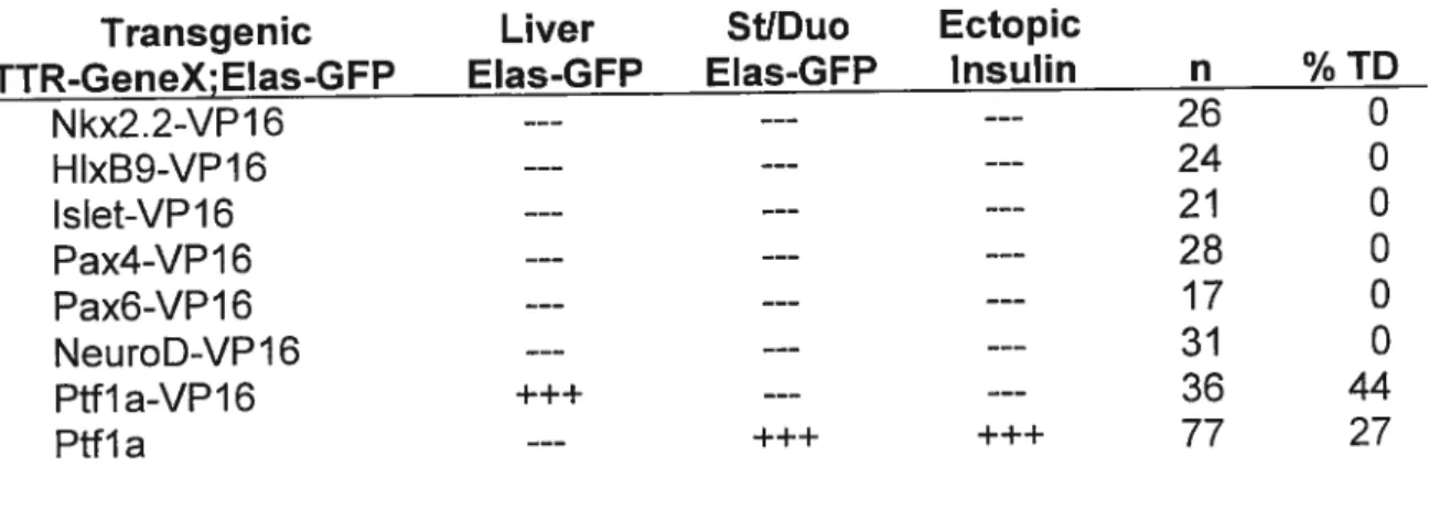
Table 3.1 Transdifferentiation ability of pancreatic transcription factors 60
List of fîgures
Figure 1.1 Anatomyofthe human pancreas 4
Figure 1.2 Pancreas development in Xenopus laevis 12
Figure 1.3 Early development 0f the dorsal pancreatic bud in mice 14
Figure 1 .4 Early development of the ventral pancreatic bud in mice 17
Figure 1 .5 Transcription factors in pancreas development 29
Figure 3.1 Transgenic overexpression of Pifla and Pifla-VPY6 promotes 63
ectopic pancreas formation
Figure 3.2 Effects of transgenic overexpression of Pifla and Pifla-VPI6 on 67 liver, liver and duodenum
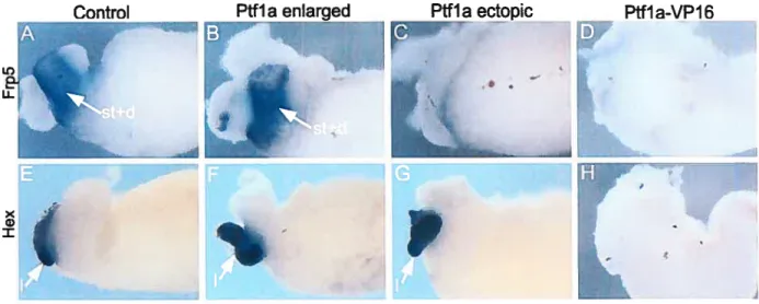
Figure 3.3 Histological analysis of Ptfla and Pifla-VPI6 transgenic whole guts 68 Figure 3.4 Overexpression of Ptfla and Ptfla-VP16 mRNA promotes ectopic 71
and enlarged pancreas formation
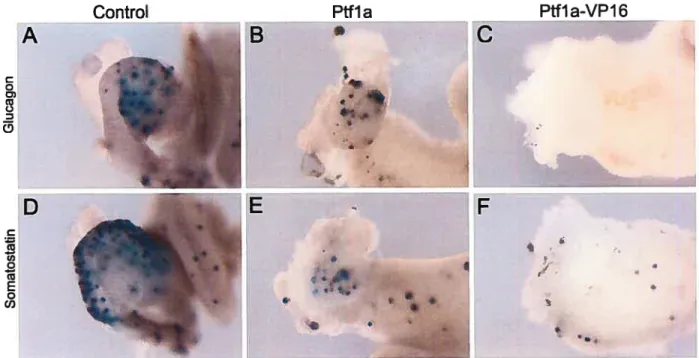
Figure 3.5 Development of glucagon and somatostatin expressing cells in 73
Figure 3.6 Effects of Pifla and Pffla-VPJ6 mRNA on organogenesis ofthe 76 liver, stomach and duodenum
Figure 3.7 Elastase expression is absent in Ptfla morphants, while insulin 81
expression is reduced at late stages, but absent at early stages
Figure 3.8 Mouse Pifla rescues the Pifla- morpholino induced phenotype 83
Figure 6.1 Outline of the microarray experiment 107
List of abbreviatïons
# Number
5’ UTR 5’ untranslated region bHLH Basic helix-Ioop-helix
BMP Bone morphogenetic protein
CMV Cytomegalovirus
U Duodenum
Dlii Delta like 1
dp Dorsal pancreas
dpc Days post-coitum
DSL Delta, Serrate, Lag2
E Embryonic day
EF1a Elongation factor 1-alpha
EGF Epidermal growth factor
Elas Elastase
ep Ectopic pancreas
FGF Fibroblast growth factor
Fig. Figure
Frp5 Frizzled related protein 5 GFP Green fluorescent protein
HepG2 Human hepatoceilular liver carcinoma ceil Une Hes Hairy I enhancer-of-split
Hex Hematopoietically expressed homeobox
Hlxb9 Homeobox 9
IDDM Insulin-dependent diabetes mellitus IFABP Intestinal fatty acid binding protein
int Intestine
lpfl lnsulin promoter factor 1
Isli Islet 1
Liver
Misti Muscle, intestine and stomach expression 1
MOl Pifla morpholino targeting the initiation codon
M02 Pff la morpholino targeting the exon-intron boundary
mPffla MousePifla
mRNA Messenger RNA
NeuroD/beta2 Neurogenic differentiation
Ngn3 Neurogenin 3
NIDDM Non-insulin-dependent diabetes mellitus
Nkx Homeobox Nk transcription factor
Notch lCD Notch intracellular domain
p Pancreas
Pax Paired box gene
PCR Polymerase chain reaction
Pdxl Pancreas duodenum homeobox 1
PP Pancreatic polypeptide
PTF Pancreatic transcription factor
Pifi a Alpha subunit of the pancreatic transcription factot 1
RACE Rapid amplification of cDNA ends
RBP-JK Recombining binding protein suppressor of hairless-J Kappa
s.t.m Septum transversum mesenchyme
Shh Sonic Hedgehog
st Stomach
St. Stage
STZ Streptozotocin
TD Transdifferentiation
TGF Transforming growth factor
TTR Transthyretin
U Unit
vp Ventral pancreas
VPI6 Herpes simplex virus regulatory protein
X.laevis Xenopus laevis
X.tropicalis Xenopus tropicalis
Xlhbox8 Xenopus laevis homeobox 8
Tb my
Tarents
Tb my Sister ancCB-rother
Tb my 3lushanc(
Acknowtedgment
I am grateful for Dr.Marko Horb for the opportunity he gave me to do my
Masters in his laboratory. I am proud of being Marko’s first student, seeing the iab growing year after year, welcoming some people and saying bye to others. I thank
Dr. Marko for the scientific formation he gave me, he taught me how to plan, perform
and discuss my experiments. His enthusiasm and interest for this project are very encouraging and motivating. I really appreciate him being so close to us,
understanding and sharing our bad and good times.
I thank ail my coileagues in the laboratory Lori, Sandeep, Farhana, Fady and
Jessica for their help. I appreciate team work with them and I wili neyer forget the good time we spent together. I also thank Mathieu Nadeau for helping me starting real time PCR experiments and Frédéric Bourque for maintenance of the frogs.
Special thanks for Mrs. Vivianne Jodoin for informing me that I should
start
my courses even before I received my acceptance due to delay in my application. Ialso owe her my actual presence in the Horb laboratory as she referred me to Dr.Marko when I was trying to find a ditector for my Masters. I appreciate her assistance and readiness to answer my questions ail the time.
1.1. Xenopus Iaevis
Xenopus Iaevis is an allotetraploid African clawed frog that has been used for many years to study early periods of embryonic development. Many teasons make Xenopus a great experimental model for studying organogenesis: 1) otgan development in frogs is much faster than in mammals; 2) the same genetic pathways operate during early stages cf celI fate specification in both species; 3) a single female can lay between 1000 and 2000 eggs per day; 4) fertilization is easily done in vitro by mixing the eggs with sperm; 5) embryos develop externally which facilitate manipulation and observation; 6) cutand-paste embryology is possible; 7) transgenesis is readily available for overexpression studies; 8) morpholino oligonucleotides permit Ioss offunction studies; 9) microarray analysis is available to compare both normal and mutant (gain or loss of function) tissues for changes in gene expression; 10) the diploid genome X. tropicalis a close relative to X. Iaevis, is completely sequenced permitting genomic analysis (Horb 2005; Heasman 2002; Amaya & Kroll 1999).
1.2.1. Anatomy and Function
Ihe mammalian pancteas is an elongated organ located actoss the back cf
the abdomen behind the stomach. It has three main sections: head, body and tau.
Ihe head, Iocated at the right extremity is the widest part of the organ and lies in the
curve of the duodenum. The body is the main portion of the pancreas Iocated in the
middle. The tau forms the Ieft part of the organ and extends toward the spleen
(Figure 1.1 A). The pancreas is a mixed exocrine and endocrine gland. The exocrine
pancreas is a Iobulated branched tissue that forms the bulk cf the pancreas.
Pancreatic exocrine ceils are arranged in grape-like clusters called acini and are
packed with numerous secretary granules containing several digestive enzymes
such as lipases, amylases, proteases and nucleases (Figure 1 .1 B). Most of these
enzymes are exocytosed as inactive precursors into the lumen of the acinus to
prevent auto degradation and auto digestion of the pancreas. From there, the
enzymes are transported via a network of larger and larger ducts which coalesce
into the main pancreatic duct that drains into the duodenum. In the duodenum, the
pancreatic enzymes are activated by gastrointestinal enteropeptidases. In the small
intestine, the digestive enzymes help break down carbohydrates, proteins, fats and
acids as part of the pancreatic juice. On the other hand, the ductal ceils secrete
buffer the acidic chyme from the stomach (Siack 1995). Endocrine ceils are grouped into spheroid structures called islets of Langerhans which are scattered arnongst the exocrine tissue. The islets are composed of five principal cell types, o, 13, 6, E and PP
that secrete respectively, glucagon, insulin, somatostatin, ghrelin and pancreatic polypeptide hormones into the bloodstream (Figure 1.1 C) (Slack 1995). Hormonal secretion of the pancreas especially insulin and glucagon is very important to regulate the level of glucose in the blood, and insulin deficiency will lead to one of the most important diseases, diabetes mellitus. Somatostatin acts by both endocrine and paracrine pathways to inhibit the secretion of other hormonal and exocrine pancreatic products (Strowski et al. 2000). Pancreatic polypeptide has a function similar to somatostatin; it suppresses the pancreatic secretion and stimulates gastric secretion. Thus, the pancreas by its exocrine and endocrine activities achieves two functions in the body that are crucial for a person’s life: food digestion and glucose
A
galI bladder bile ducttY
tait pancreatic duct duodenumB
ot pancreasn
n
C
secreteray product w pP d uctFïgure 1.1 Anatomy of the human pancreas. (A) The aduit pancreas contacts the duodenum. (B)
1.2.2. Diseases of the pancreas
As the pancreas is a mixed gland containing a wide variety of celi types that accomplish different physiological functions, malfunction of the pancreas can give rise to many health complications. Pancreatic diseases include pancreatitis, cystic fibrosis, pancreatic cancer and diabetes. I will now discuss briefly each of these diseases highlighting in particular diabetes mellitus.
1.2.2.1. Pancreatïtïs
Pancreatitis is an inflammation of the pancreas and can be acute or chronic. Gallstones and alcohol are the major causes of acute pancreatitis which will lead to back up of the exocrine secretions in the pancreas or the adjacent organs. Acute pancreatitis will improve on its own after treatment if no kidney or lung complications occur (Mergener & Baillie 1998). Chronic pancreatitis is usually associated with alcoholism and can also occur following an acute pancreatitis. The digestive enzymes attack and destroy the pancreas and the nearby tissues causing scaring and pain. Chronic pancreatitis can lead to different complications (Steer et al. 1995) such as pancreatic insufficiency, bacterial infection and type 2 diabetes and can be fatal in some cases
1.2.2.2. Cystic Fïbrosis
Cystic Fibrosis s an inherited genetic disorder that disrupts the normal function of the epithelial ceils which une passageways inside the lungs, liver, pancreas, digestive and reproductive systems. The epitheliai ceils are unable to regulate chloride transport through the celi membrane, consequently the balance of water and sait is disrupted and the mucus in the pancreas becomes thick, sticky and hard to move (Quintom 1999). The thick mucus leads to obstruction of the pancreatic ducts and impaired digestive problems.
1.2.2.3. Pancreatic Cancer
Pancreatic cancer is the fifth leading cause of cancer deaths worldwide (NCIC 2004). Most pancreatic cancers are ductal adenocarcinomas developing in the exocrine tissue (Yeo et al. 2002). Pancreatic adenocarcinoma is usually diagnosed
at a late stage because symptoms are either absent or nonspecific and only about
10% of cancers are still within the pancreas at the time of diagnosis. Survivai rate is less than six months (Warshaw & Fernandez-del Castillo 1992). Pancreatic
adenocarcinomas are characterized by an overproliferation of pancreatic ductal ceils, yet it is stili unclear whether cells responsibie for the disease originate from
pancreatic ducts or from transdifferentiation of other pancreatic ceil type such as
pancreatic cancer is in first place due to the fact that very Jittle is known about ductal
ceil development. However, in recent years several studies have attempted to
reproduce the disease by overexpressing several signaling molecuies within the pancreas or creating pancreatic-specific mutations related to human mutations
identified in the disease. For example, several studies showed that pancreatic
adenocarcinoma can be induced by overexpressing, in the developing pancreas,
signaling molecules that are normally present during pancreas organogenesis such
as TGFo (Wagner et al. 1998; Greten et al. 2001). Recently, another model of adenocarcinoma was generated by specificaily deleting in the pancreas the either
type 2 receptor of TGFI3 (TGFI3-R2) or Smad4, another downstream mediator of TGF3 (Ijichi et al. 2006; Bardeesy et al. 2006). Expression of an active form of Kras(GJ2D) in TGF3-R2 knockout mice produce pancreatic ductal adenocarcinoma
in 100% of animais with a survival rate of 59 days (Ijichi et al. 2006). Similarly,
deletion of Smad4 promotes activation cf neoplasia by Kras and accelerates
development cf the tumor (Bardeesy et al. 2006). Thus, understanding how different pancreatic lineages develop and differentiate wili help us identify the molecular mechanisms underlying pancreatic cancer development.
t22.4. Diabetes
Diabetes mellitus is a chronic, incurable disease that occurs when the body encountets some insulin insufficiencies, leading to an excess cf sugar in the blood. There are two main forms of diabetes: type 1 and type 2. Type 1 diabetes also
known as juvenile diabetes or insulin-dependent diabetes (IDDM) usually develops
in childhood or adolescence due to the autoimmune destruction of the pancreatic
3-ceils. Thus, daily insulin injections are required for proper glucose homeostasis and survival of type 1 diabetics. Type 2 diabetes also known as non-insulin-dependent
diabetes (NIDDM) can occur at any time during person’s life due to relative
insufficiency of insulin in the body caused by insulin resistance and f3 celi dysfunction. Type 2 diabetes may progress to the destruction of the insulin producing celis of the pancreas and insulin administration might be required. Type 2
diabetes is the most common form of diabetes and affects 90% of diabetic people.
Hyperglycemia associated with diabetes will slowly damage the small and large blood vessels in the body, resulting in a variety of long term complications including nephropathy, retinopathy, neuropathy and cardiovascular disease. In Canada,
diabetes is a leading cause of death by disease; two million Canadians suifer from diabetes nowadays and it is expected to rise to three million in 2010 (Health Canada
2002). The economic costs cf diabetes are also very high, it is estimated that 13.2 billion was spent in 2002 to treat people with diabetes and its complications.
The key goal of diabetes treatment is to prevent the complications associated with the disease. lnsulin injections stili cause many of problems even under the best
conditions because it cannot reproduce the exact maintenance of euglycemia as the normal pancreas. Moreover, the lack of donor tissue for whole pancreas and islet
transplantations has not been resolved yet. Replacement of the pancreatic 13-cells by
new insulin secreting ceNs represents an adequate solution (Scharfmann 2003).
One possibility is the use of pancreatic or other stem ceNs to produce 3-cells in vitro
for transplantation (Lechner & Habener 2003). Another solution will be the use of
other differentiated tissues present in the body to generate pancreatic tissue; this will overcome the two major transplantation problems which are the lack of donor tissues and the immunosuppressive treatment (Slack & Tosh 2001; Tosh & Slack
2002). To achieve such a therapeutic application, a detailed analysis of the molecular mechanisms underlying endocrine pancreatic development is necessary.
1.2.3. Embryological orïgin
In mammals, the pancreas originates as two endodermal buds developing
on the dorsal and ventral side of the foregut endoderm that will become duodenum (Edlund 2002; Kim & MacDonald 2002). At E8.5 the dorsal bud arises just below the notochord, while the ventral bud develops at E9.5 adjacent to the hepatic diverticulum (Slack 1 995). Although the cells in the pancreatic buds are committed to form a pancreas, yet they are not differentiated. Only few glucagon expressing celis
are detected in the undifferentiated ductal epithelium. At E13.5 the
two
pancreatic buds rotate, fuse and start to differentiate into endocrine and exocrine lineages.Subsequently, the differentiated pancreatic ceils proliferate extensively such that by E16 the endocrine and exocrine cells start to aggregate in islets of Langerhans and acini respectively. At E18.5 organogenesis of the pancreas is completed and the dorsal and ventral buds form a single functional organ (Figure 1 .2).
In the amphibian Xenopus Iaevis, development of the pancreas proceeds in an almost identical manner to that seen in mammals (Kelly & Melton 2000). The dorsal bud is the first to appear at stage 35/36 in the archenteron roof endoderm just below the notochord. The two ventral buds develop by stage 37/38 adjacent to the hepatic diverticulum. Subsequent morphogenesis of the endoderm and dynamic
movements of the gastrointestinal tract result in the fusion of the two ventral buds at stage 39 followed by the fusion cf the dorsal and ventral buds at stage 40 to form a
single organ (Figure 1 .2). Differentiation of exocrine and endocrine ceils occurs in a spatialiy and temporally distinct manner (KeHy & Melton 2000; Horb & Slack 2002) such that exocrine cells are initially specified in the ventral pancreas, and endocrine cells in the dorsal. The exocrine markers amylase, elastase and trypsinogen first
appear in the ventral pancreas at stage 41: expression then spreads to the dorsal pancreas at stage 42/44 and by stage 45 aIl three are detected throughout both the
ventral and dorsal pancreas (Horb & Slack 2002). Insulin is the first endocrine marker to appear in the dorsal bud at stage 32 prior to overt morphogenesis but is not detected in the ventral pancreas until stage 47 (7 days) (Horb & Slack 2002; Kelly & Melton 2000). In contrast, the other endocrine markers glucagon and
somatostatin are not detected in the pancreas until stage 45 and only in the dorsal
41 and 44. At later stages 47/48 expression spreads to the ventral pancreas. Thus,
in both mammals and amphibians, the pancreas originates from two separated and
distinct endodermal buds.
Even though the two pancreatic buds derive from the same endodermal germ layer, the corresponding developmental programs leading to their specification are
flot the same and the type of tissue specified by each bud is different, at ieast
initially. The dorsal bud gives tise to the body, taU and part of the head of the
pancreas. The ventral bud forms the remainder of the pancreatic head and the uncinate process. Interestingiy, the distribution of endocrine celis within the islets in each reg ion of the pancreas differs: islets in the tau are rich in beta and alpha celis, but contain few PP celis, while islets in the head are rich in PP celis with few beta or alpha ceils. Those differences are due to the fact that the endodermai domains that specify the pancreatic buds are independent and receive different signais from their surrounding tissues.
A B_vp C D
sth
L
Stage In I 39 E F G H4 t st Stage $h.
in
40«
- Stage 41Figure 1.2 Pancreas development in Xenopus Iaevis. (A) Stage 39 tadpole. (B,C) Stage 39 dissected guts showing ventral pancreatic bud (dark blue) and dorsal pancreatic bud (light blue). (B) Left view. (C) Right view. (D) Transverse section of stage 39 gut showing the separated pancreatic buds, anterior view. (E) Stage 40 tadpole. (F,G) Stage 39 dissected guts. (F) Left view. (G) Right view. (H) Transverse section of stage 40 gut showing the pancreatic buds starting to fuse, anterior view. (I) Pancreas at stage 41. Stomach (st), intestine (int), duodenum (d), ventral pancreas (vp), dorsal pancreas (dp) and pancreas (p). (Adapted from Kelly and Melton 2000 with modifications)
1.2.4. Regional Specification of the pancreatic endoderm
During embryogenesis the prepancreatic endoderm contacts several mesodermal tissues that play a fundamentai role in specifying the different pancreatic lineages, including the notochord and the heart (Figure 1.3). Those mesodermal tissues has been shown to secrete several different growth factors that have a direct influence on the prepancreatic endoderm and play a fundamental role
in pancreas development including the Notch, Hedgehog, EGF, FGF, and TGF-!3
pathways. Early during embryogenesis, the dorsal prepancreatic endoderm in the posterior foregut contacts the notochord while the ventral prepancreatic endoderm is adjacent to the splanchnic mesoderm and to the aortic endothelial ceils (Figure 1.3
A). (Kim & Hebrok 2001 Slack 1995). Later on between 9 and 9.5 dpc the fusion of
the paired dorsal aorta separates the notochord from the endoderm (Figure 1 .3 B),
and by 10 dpc mesenchyme surrounds the whole gut (Figure 1.3 C). Thus signais between the endoderm and the mesoderm direct ail stages of pancreatic development. Early during development the notochord is the signaling centre that permits dorsal budding and pancreatic gene expression. Later on signais from the mesenchyme are necessary for dorsal and ventral bud development (Kim et al. 1997a; Kïm et al. 1997b). During pancreas organogenesis the notochord emits the
A
B
C
Figure 1.3 Early development of the dorsal pancreatic bud in mice. (A) At 15 somite stage the notochord contacts the gut endoderm. (B) At 20 somite stage the dorsal aorta separates the notochotd from the gut endoderm. (C) By the 28 somite stage the mesenchyme surrounds the gut and the dorsal pancreatic bud has formed.
Signais from the notochord are requïred for early induction cf pancreatic endoderm. Experiments in chick embryos showed that no pancreatic rnarkers are
detected when the notochord is separated from the endoderm. Yet this expression is
restored in the anterior endoderm when endodermal epithelium is co-cultured with isoiated notochord (Kim et ai. 199fb). Eariy removal cf the notochord resuits in ectopic expression of Sonic Hedgehog Shh, in the pancreatic endoderm disrupting both pancreatic morphogenesis and gene expression (Hebrok et ai. 1998). The notochord secretes components cf the TGF-13 signaiing pathways, activin-/3 and fibrobiast growth factor FGF2 which tepress Shh expression in the pancreatic
endoderm region. Purified FGF2 and activin-f3 mimic the activity cf isolated
notochord in the pancreatic endoderm assay and induce PUxi and insulin
expression (Hebrok et ai. 1998). Activin-f3 and TGF-f32, another mem ber cf the TGF
3 famiiy, have been shown to disrupt branching and acinar morphogenesis when
overexpressed in embryonic mouse pancreas (Ritvos et aI. 1995). Yet the notochord
is net the oniy signaling centre that represses Shh; it is aise repressed in the iateral
endodermal region which does not contact the notochord that gives rise to the ventral pancreas (Kim et al. 1997a).
1.2.4.2. Role of the heart mesenchyme
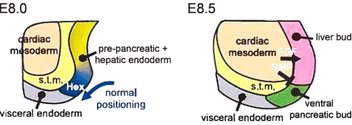
It is unclear whether repression of Shh in the ventral prepancreatic endoderm is similar to its repression from the dorsal region. But it is clear that ventral pancreatic development differs from dorsal pancreatic development because the notochord does not contact the endoderm and because several gene knockouts have been shown to differentially affect the ventral versus the dorsal buds. The ventral pancreatic bud arises adjacent to the liver and organogenesis of both organs is closely related. The region of the foregut endoderm receiving high levels of FGFs, FGF2 from the surrounding cardiac mesoderm initiates a hepatic program. While the other region, which is not in direct contact with the cardiac mesenchyme maintains the default pancreatic fate (Figure 1.4) (Deutsch et al. 2001; Duncan 2001). In addition, it appears that BMPs signaling from the septum transversum mesenchyme are required with FGEs to initiate hepatic development within the ventral endoderm (Rossi et al. 2001). In fact, BMP4 and FGF2 converge to change the pancreatic fate of ventral endodermal cells, with the latter forming liver instead. On the other hand it was shown that the homeobox gene Hex controls the specification of the ventral pancreatic bud (Bort et al. 2004). In the mouse embryo Hex is expressed at E7.0 in the ventral-lateral foregut that gives rise to the ventral pancreas and the liver (Bogue et al. 2000). In Hex nuil embryos, the ventral definitive endodermal cells do flot proliferate and therefore cannot be positioned beyond the influence of the cardiac mesoderm, resulting in a complete failure of ventral pancreatic bud specification. Moreover, Hex -I- endoderm explants cultured in the absence of cardiac mesoderm
are able to activate early pancreatic genes (Bort et aI. 2004). Thus, Hex controls the
growth of the ventral endodermal ceils and their positioning beyond the cardiac
mesoderm, allowing them to escape the hepatic induction and form the ventral
pancreatic bud.
E8.O
pre-pancreatic+ hepatic endoderm normal positioningE8.5
liver budFigure 1.4 Early development of the ventral pancreatic bud in mice. At E8.0, Hex maintains the definitive endodermal ceils in a proliferation state which Iead to positioning them beyond the catdiac mesoderm. At E8.5, FGF and BMP signaling from the cardiac mesoderm induce hepatic fate (arrow), and prevent pancreatic fate (bar) in the adjacent endoderm. The region of the endoderm that does flot contact the heart forms the ventral pancreatic bud. s.t.m: septum transversum mesenchyme (Adapted from Bort et al. 2004 with modifications)
vsceraI endoderrn
ventral visceral endoderm pancreatic bud
1.2.4.3. Role ofthe pancreatic mesenchyme
Epithelial-mesenchymal signaling represents another interaction that
governs later proliferation and differentiation of the pancreatic celis. The first
classical studies showed that when the pancreatic mesenchyme is separated from
the epithelium the latter faits to grow and to develop a mature pancreas, and concluded that the mesenchyme is indispensable for the pancreatic epithelium to
proliferate and differentiate producing ail pancreatic celi types (Wessels 1967).
Recent studies show that the mesenchyme controls proliferation of the pancreatic
epithelium via members cf the fibrobtast growth factor family (EGEs). It was shown that in EgflO-/- mouse embryos the two pancreatic buds form normally but the celis
fait to proliferate and subsequent differentiation is arrested (Bhushan et al. 2001). Implication of the mesenchyme in pancreatic epithelium differentiation is more
understood and in vitro experiments demonstrated that the epithelium has different
responses when exposed to different environments and that the default fate of the pancreatic epithelium is to form islets (Debas 1997). However, the mesenchyme has
specific signaIs that permit the pancreatic epithelium to form acinar pancreatic
tissue. The mesenchyme promotes exocrine pancreas development by repressing development of the endocrine iineage. Growth factors from the mesenchyme have been shown te induce celI division in the developing pancreas promoting exocrine
differentiatïon and inhibiting endocrine differentiation (Horb & Slack 2000). The
mesenchyme secretes Follistatin a potential inhibitor of Activin is expressed in the pancreatic mesenchyme at E12.5 and is able to mimic the effect of the mesenchyme
(Miralles 1998). Similarly in Xenopus, specification of the endoderm is controfled by
the adjacent mesoderm (Horb & Slack 2001). Cuftured explants from neurula and
tailbud stages containing both endoderm and mesoderm, form specific morphological structures that express regiona markers. Endodermal explants,
lacking mesoderm, do flot develop any recognizable morphological structures, white
recombination between mesoderm and endoderm Iead to the expression of
endodermal markers with mesodermal characteristics (Horb & Sfack 2001). Results
from mouse, chick and fly have also shown that the gut mesoderm can specify the
underlying endoderm (Kedinger et aI. 1986; Yasugi 1993; Roberts 1998). Those experiments demonstrate that during gut development mesodermal signaIs are key players for proper patterning of the endoderm into distinct differentiated tissues.
1.2.4.4. Retinoic acid in pancreas development
It is weB known that in vertebrate, retinoic acid (RA) signaling s essential for
patterning ectoderm and mesoderm (Cavalas & Krumlauf 2000; Maden 1999). Studying the role of RA in patterning the endoderm has emerged recently. First studies done on zebrafish embryos demonstrated that RA is essential for
regionalization of the endoderm (Stafford & Prince 2002). No pancreatic and liver markers are detected in RA nuli zebrafish embryos. In Xenopus embryos, inhibition
of RA at gastrula stage bIocks development of both exocrine and endocrine
concentration of RA disrupts the antero-posterior patterning of zebrafish endoderm and ectopic pancreatic and hepatic tissue are detected in the anterior endoderm (Stafford & Prince 2002). In Xenopus embryos, overexpression of RA Ieads to an
expansion of endocrine development and blocks exocrine deveiopment in the dorsal pancreatic endoderm. In the ventral endoderm, RA promotes exocrine pancreatic differentiation and inhibits hepatic development (Chen et ai. 2004; Stafford & Prince 2002). Chen et al. showed that RA promotes pancreatic development by repressing Shh from the dorsal prepancreatic endoderm. They also demonstrated that expansion of endocrine celI population on the expense of the exocrine population after RA induction is due to inhibition of Notch signaling activity (Chen et al. 2004). In mice, retinoids have been shown to inhibit acinar differentiation and promote ductal differentiation through epitheliai-mesenchymal interaction (Kobayashi et al. 2002). In conclusion we can say that RA signaling is essential at the gastruiation stage of
pancreas, development to regulate exocrine pancreas iineage.
1.2.45. Notch sîgnaling in pancreas development
Notch signaling in pancreas development is responsible for maintaining the undifferentiated celi population. The Notch signaling cascade was first described in the neural system, where lateral specification restricts neuronal fate; later, Notch has been shown to regulate celi fates and paffern formation in most tissues. Notch s a receptor that is able to receive extraceilular signais and to regulate gene expression
in the nijcleus. Upon activation via DSL (Delta, Serrate, Lag-2) ligand interaction,
Notch receptor is activated and its intracellular domain (lCD) is released and
translocated into the nucleus (Mumm & Kopan 2000). Notch lCD wotk in concert
with another DNA binding protein RBP-JK to express hairy/enhancer-of-split (Hes)
transcription factors which are known to repress key regulator genes such as neurogenin (Beatus & Lendahl 1998). In the pancreas, Notch signaling modulates the differentiation of progenitor ceNs. Loss cf function experiments, affecting different components of the Notch pathway (Hesi, Dlii, and RBP-JK), showed that Notch signaling inhibits premature and excessive differentiation cf pancreatic progenitors
into endocrine cells (Apelqvist et al. 1999; Jensen et al. 2000). Directed
misexpression cf Notch lCD to mouse dorsal and ventral pancreatic buds using the Pdxi promoter, inhibits differentiation of both endocrine and exocrine cells and traps
the pancreatic progenitots in an undifferentiated state (Murtaugh et al. 2003). Moreover, activation of Notch in normal mouse pancreas causes loss of acinat celI differentiation and expansion of the ductal cell population thus inducing acinar-to ductal metaplasia - characteristic of pancreatic adenocarcinoma - (Miyamoto et al.
2003). More recently, it was shown that endogenous Notch signaling activates the initial commitment of the exocrine lineage but blocks terminal acinar differentiation (Esni et al. 2004). Thus, early during development, Notch signaling activates initial commitment to the exocrine fate and inhibits endocrine celI differentiation to maintain
the undifferentiated pancreatic precursors. In the mature pancreas, Notch is silenced
while misexpression of Notch in mature 13-ceil does flot alter their differentiation (Murtaugh et al. 2003; Lardon et al. 2004; Rooman et al. 2006).
1.2.5. Genetic network regulatïng pancreas development
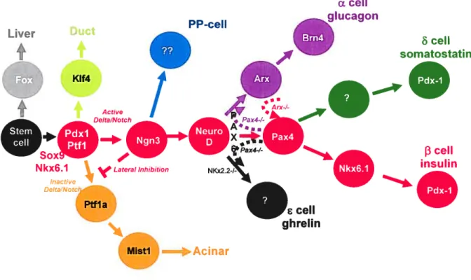
1.2.51. Exocrine and endocrine pancreatic transcription factors
Pdxl
Several classes of transcription tactors are involved in the specifïcation and differentiation of endocrine and exocrine lineages from a common precursor population which is first established in dorsal and ventral pancreatic buds (Figure
1.5). Lineage tracing analysis show that pancreatic progenitors expressing the Para
hox transcription factor Pdxl give rise to aIl pancreatic cell types: endocrine, exocrine and ductal cells (Gu et al. 2002; Gu et al. 2003). Interestingly, theXenopus Xlhbox8 was the first Pdxl homologue to be isolated. Xlhbox8 is first expressed in the dorsal and ventral pancreatic buds as well as in the duodenum (Wright et al. 1989). In mice, Pdxl has the same expression pattern. It is expressed in the pancreatic buds at E8.5. Later on ai E9.5 and E11.5 expression is detected in the duodenum and stomach (Guz et al. 1995; Offield et al. 1996). In the aduit pancreas
PUxI expression is restricted to the f3-cells (Jonsson et al. 1995). Loss and gain of function experiments have shed light on the role of PUxI in specifying the various pancreatic lineages and established Pdxl as the first master regulatory gene in
pancreas development.
Mice lacking Pdxl do not form a pancreas, however a small dorsal pancreatic bud forms but without any expression of insulin or amylase (Jonsson et
al. 1994; Offield et al. 1996). The primitive bud formed does contain few ci and
f3-cells. These results suggest that initial budding of the endocrine celis can occut n the absence of Pdxl while the subsequent steps to reach a mature and functional
stage are PUxI dependent. Conditional inactivation of Pdxl in the differentiated
f3-celis shows that Pdxl is indeed required for maintaining the hormonal secretion characteristic of the f3-cell (Ahlgren et al. 1998). Moreover, targeted depletion cf
Pdxl in the whole pancreas at late gestational stage Ieads to agenesis of the acinar
tissue, instead immature duct-like ceils are formed (Hale et al. 2005). Mutations in the human homologue, lpfl, are also associated with pancreatic agenesis (Stoffers et al. 1997). In conclusion, these loss-of-function studies showed that Pdxl is
essential for initial pancreas development, but they have not addressed the
sufflciency of Pdxl in specifying pancreatic fates.
On the other hand, gain of function experiments prove that ectopic expression of Pdxl is able to generate functional pancreatic celis. The first misexpression experiment of PUxI in the stomach and hindgut region did flot show any ectopic pancreatic tissue even though agenesis of the cecum was promoted (Heller et al. 1998). Later, Ferber et al. showed that expression cf PUxi in mouse
livers activates endogenous expressÏon cf insulin and they also showed that mature hepatic insulin teduces hyperglycemia in diabetic mice (Ferber et al. 2000; Ber et al. 2003; Meivar-Levy & Ferber 2003). We have shown previously that ectopic expression of Pdxl-VP76 in Xenopus liver is able to convert Uver to pancreas containing both exocrine and endocrine markers (Horb et al. 2003). Similarly, Pdxl VP16 transdifferentiates rat hepatic celis into pancreatic endocrine precursor celis that rescue diabetes when exposed to high glucose levels (Cao et aI. 2004). More recently, it was shown that overexpression of both Pdxl and Pifla was sufficient to promote a pancreatic fates in nonpancreatic endodermal celis (Afelik et aI. 2006). These results suggest that transdifferentiation of hepatic and other endodermal ceNs into functional insulin producing ceils wilI serve as a new therapy for insulin dependent diabetes.
Both Ioss and gain of function experiments demonstrate that Pdxl plays a fundamental role in the specification of ail pancreatic lineages. In concert with specific protein partners, Pdxl has the abiiity first to switch the fate of endodermal progenitor celis into a pancreatic fate and second to convert differentiated cells into functional pancreatic cells.
Hlxb9
HIxb9 is a homeodomain transcription factor with different expression patterns during pancreas development. HIxb9 expression is first detected around E8
in dorsal and ventral pancreatic buds even before initiation of Pdxl expression, yet
HIxb9 expression is transient and by E10.5 very Iow levels are detected ony in the ventral pancreas (Li et al. 1999). Later during development, HIxb9 becomes restricted to the differentiated endocrine ceils. Homozygous mutant embryos fail to develop a dorsal pancreatic bud. The ventral pancreatic bud develops with aberrant islet structure and a reduction in 13-ceil number (Harrison et al. 1999; Li et al. 1999).
These loss of function studies demonstrate that HIxb9 is required in a specific period
of time in the dorsal pancreas for pancreatic specification and in the ventral
pancreas for endocrine differentiation. In agreement with this, overexpression of
HIxb9 from the PUxI promoter results in continuous expression of HIxb9 in early
pancreas development resulting in aberrant development of the pancreas. It seems that the pancreatic epithelium and its neighboring mesenchyme adopt an intestinal
fate (Li & Edlund 2001). This gain of function experiment confirm furiher that only
transient expression of HIxb9 is required for pancreatic development, and that in extended expression HIxb9 may function as a repressor in the developing pancreas.
Pifi aIP48
Ptfla first identified as P48 is the celi specific component of the pancreatic
transcription factor one Pifi (Krapp et al. 1996). PTF is a heterooligomer that contains three different subunits p75, p48 and p64. p75 is required to transport the complex into the nucleus and does flot contact the DNA (Sommer et al. 1991). p48
and p64 are the DNA binding subunits, they contact the DNA as a heterodimer recognizing two different motifs. p48 binds to consensus sequence CANNTG, whiie p64 recognizes the TGGGA motif (Cockeli et ai. 1989). The tissue specific component of Ptfl isp48 and wiiI be referred to as Pifla throughout the thesis. Ptfla
is a basic helix loop helix (bHLH) transcription factor that was initiaiiy identified as a key transcriptional regulator of exocrine pancreas development that binds to the 5’ promoter regions of ail acinar digestive enzyme genes and activates them (Cockef I et al. 1989; Krapp et al. 1996). Recent resuits have suggested that Ptfla may aiso
be invoived in endocrine ceil fate specification (Krapp et ai. 1998; Kawaguchi et al. 2002).
In mammais, Pifla is expressed in eariy pancreatic progenitors (dorsal and
ventral buds) (Kawaguchi et ai. 2002), but in aduits it is oniy expressed in acinar celis (Krapp et ai. 1996). In Xenopus endoderm, Pifla is expressed in the dorsal and
ventral pancreatic anlagen at stage 32 prior to overt morphogenesis and differentiation. Later Pffla expression is maintained in the entire pancreas ((Afeiik et
ai. 2006). Aortal endotheliai ceils induce Ptfla specificaily in the dorsal pancreatic
endoderm (Yoshitomi & Zaret 2004), while FgflO is necessary to maintain this dorsal
expression (Jacquemin et ai. 2006). Ventral induction of Ptfla does not require the
viteliin veins (Yoshitomi & Zaret 2004).
Loss-of-function studies in mice have demonstrated that Pifla is essential
for acinar ceil deveiopment and piays an important role in endocrine ceil deveiopment as weli. Mice homozygous for a nuli mutation of Pifla lack exocrine
E18 however, pancreatic endocrine celi markers are detected in the spleen, but they are not organized into islets (Krapp et ai. 1998). In humans, PIFJA gene mutations are associated with pancreatic and cerebeliar agenesis (Seiiick et aI. 2004). In zebrafish, Pifla is only expressed in a subset of pancreatic progenitors in the Ieft ventrolateral endoderm, and not in the dorsal posterior endoderm (Lin et ai. 2004; Zecchin et al. 2004). Morpholino knockdown studies in zebrafish have shown that
Pifla is required for development of ail acinar ceils and a subset of endocrine celis
(Lin et al. 2004). In Pifla morphants, acinar development is inhibited while the early
endocrine celis are unaffected (Zecchin et al. 2004). Another study showed that late endocrine ceils that normaNy develop from ventral anterior endoderm are absent in Pifla morphants (Lin et al. 2004). Several other studies have suggested that Ptfla may function as a master regulator of pancreatic celi fate. Lineage tracing analysis
based on Cre-mediated recombination showed that pancreatic ceils iackïng Pifla fail
to initialize a pancreatic program and acquire a duodenal fate instead (Kawaguchi et
ai. 2002). Knockdown studies in Xenopus (Afeiik et ai. and cutrent study) are
concordant with what have been shown previousiy in mice and zebrafish; implicating
Pifla oniy in exocrine pancreas deveiopment and establishing the eariy insulin ceils as Ptfla independent (Lin et al. 2004; Krapp et al. 1998). Thus, ioss of function
experiments demonstrate that Pifla s essential for exocrine pancreas deveiopment but they do not ask whether Ptfla is sufficient to initiate a pancreatic program.
Gain cf function experiments have reveaied the importance cf Ptfla in specifying the pancreas in the endodermai progenitors. Loss of Hesi, a downstream
stomach, duodenum and bile duct regions of mice embryos. Endodermai celis ectopicaiiy expressing Pifla switch their fate into pancreatic progenitors that wiiI later differentiate into endocrine, exocrine and ductai celis (Fukuda et al. 2006). In Xenopus, direct overexpression cf Pifla in the anterior gut endoderm leads ta the Ioss of stomach and duodenum at the expense of exocrine pancreatic celis (Afelik et
ai. 2006). In the same study, they showed that overexpression of Pifla and Pdxl
promotes ectopic pancreas differentiation in the posterior endoderm. Altogether, work from Afeiik et ai. with our current study demonstrate that Pifla is impiicated in specifying ail pancreatic lineages and suggest that Pifla piays a central roie in the decïsion to become stomach, duodenum, bile duct or pancreas.
c ceil 6 ceIl somatostatin
—‘Q
f3 celi insulinFigure 1.5 Transcription factors in pancreas development. Hierarchy cf transcription factors in the developing pancreas showing the relationships among the various transcription factors. Lineage relationships are based on gene expression patterns and phenotypes from mouse knockout studies. Different colors represent different pancreatic lineages.
PP-ceII Liver Duct K1f4 Nkx6.1
4
LateraI Inhibition Inactive ci —+Acinar s ceil g h relïnMistl
Most of the studies on pancreatic development focus on identifying regulatory genes involved in f3-ceIl formation. Very littie is known about exocrine
pancreas development and function. Only few transcription factors specific to the
exocrine tissue have been identified, including Pifla and Misti. Misti is the only
pancreatic gene restricted to exocrine ceils. Misti is a bHLH transcription factor expressed in a variety of exocrine tissues including the salivary gland, pancreas,
stomach and prostate (Pin et al. 2000). Ihis type of expression pattern suggests that Misti may be involved in regulating the process of exocytosis. Inhibition of Misti disrupts first the organization of the acinar cells; later on exocrine tissue is dramatically injured with loss of acinar cells at the expense of ductal cells (Pin et al. 2000). These resuits indicate that Misti is required to maintain exocrine function and identity.
1.2.5.3. Endocrine specific transcription factors
Understanding the transcriptional regulatory network underlying endocrine ceil development IS of great interest for generating -ceIls either from stem celis or
from differentiated tissues. Some transcription factors are required ta differentiate the endocrine lineage from the pancreatic progenitor celis, others permit ta specify the different endocrine celi types.
Ngn3
The bHLH transcription factor Ngn3 is the most important endocrine
transcription factor. Ngn3 is transiently expressed between E9.5 and E15.5 in the pancreatic epithelïum prior ta endocrine differentiation. Epithelial pancreatic ceils expressing bath PUxi and Ngn3 reptesent the endocrine precursot population
(Schwitzgebel et aI. 2000; Jensen 2004). Inactivation of Ngn3 by hamalogaus recombination resuits in diabetes. Islets of Langerhans are missing in the mutant
pancreas and ail faut major hormones (insulin, glucagon, somatastatin and PP) are undetectable (Gradwahl et aI. 2000). Hawever, averexpressian of Ngn3 undet the PUxi pramater in pancreatic progenitor ceils promates an endocrine fate,
characterized by an accelerated differentiation, 0f glucagan producing celis (Apelqvist et al. 1999). Although Ngn3 is necessary for the deveiopment cf the
endocrine precursors before their differentiation it seems that other factors are required far their subsequent specification.
NeuroDlbeta2
NeuroD is another member cf the bHLH class cf transcription factor which plays a fundamental role in pancreas deveiopment. NeuroD was first isolated from the pancreas and the brain. in the pancreas, the activity cf the insulïn and giucagon promotets depend on the presence cf an E-box that binds to the ceil specific bHLH NeuroD/Beta2 (Naya et ai. 1995; Dumonteii et ai. 1998). NeuroD is expressed in the endocrine ceils, the intestine and the brain (Naya et ai. 1995; Lee et ai. 1995). Mice lacking NeuroD die from severe diabetes. NeuroD-deficient pancreases form ail types of endocrine ceiis, yet morphogenesis of the isiets is disrupted and the number of 3-ceiis is reduced due to prenatai apoptosis (Naya et ai. 1997). n the endocrine transcriptionai cascade, expression cf Ngn3 precedes and overiaps with expression cf NeuroD (Naya et ai. 1997), which precedes that cf cther endocrine differentiated markers such as Pax genes. Thus, Ngn3 seems te be the upstream activator cf NeuroD, which in turn activates other transcription factors such as Pax6 (Huang et ai. 2000; Marsich et ai. 2003).
is M
Isli is a LIM homeodcmain transcription factor expressed eariy during development in the dorsal pancreatic mesenchyme and in the aduit pancreas Isli expression is detected in the isiet ceiis. inhibition cf Isil Ieads te dorsal agenesis,
confirming the role of the mesenchyme in the development 0f the dorsal pancreatic bud (Ahlgren et al. 1997). In addition, Isli embryos lack any differentiated endocrine islets, which reveals the requirement of endodermal Isli for endocrine celi differentiation in the pancreas.
Pax4 and Pax6
Pax4 and Pax6 are members of a subclass 0f the Pax gene family,
containing both a paired domain and a homeodomain (Dahl et al. 1997). Pax4
expression is detected as early as E9.5 in the dorsal pancreatic; one day after Pax4
is present in dorsal and ventral pancreatic buds. Expression peaks at the secondary
transition (E13.5-E15.5) and then diminishes to very low levels at birth (Sosa-Pineda et aI. 1997). Pax4 null embryos fail to develop any 13- and & cells; instead an expansion 0f ci- and E- cell lines is observed (Sosa-Pineda 2004; Prado et al. 2004).
Pax4 colocalizes with other endocrine specifÏc transcription factors such as Ngn3,
Isil, Nkx2.2 and Pax6. Moreover, it has been shown that Pax4 is a target of Ngn3. Thus, the role of Pax4 in pancreas development is to control f3- and ci- ceil specification after initiation by Ngn3 (Sosa-Pineda et al. 1997; Sosa-Pineda 2004).
Based on the knockout phenotype, one can tell that early during endocrine development Pax4 promotes
13-
and cx- ceil differentiation by repressing ci- and Eceils. In tact, Peterson et al. (2002) have demonstrated in vitro that endogenous glucagon expressed in rat cells 15 inhibited by Pax4 expression.
In the same study the authors showed that Pax4 competes with Pax6 to repress insulin transcription mediated by Pax6. Expression 0f Pax6 is detected in ail endocrine ceils at earty stage of development and in the mature pancreas. First gene inactivation analysis ieads to a total loss of glucagon producing a-ceils (St Onge et al. 1997), which indicates that Pax6 is tequired for a-cells development but not necessary for that of 3- and 6- cells. Interestingly, in Pax4-Pax6 double mutant
ail endocrine islet cells are missing. Thus, Pax4 and Pax6 seem to have reciprocal
and non-redundant functions during endocrine pancreatic development; Pax4 is
responsible for the differentiation of Ç3- and 6- cells while Pax6 is responsible for differentiating the o-celIs.
Nkx2.2 and NkxG.J
Nkx2.2 and Nkx6. I are members of the homeodomain class of transcription factors. Nkx2.2 is detected very early in pancreatic precursor epithelium, and
becomes restricted to endocrine f3, o, and PP cells when differentiation is initiated (Sussel 1998). In Nkx2.2 mutants, endocrine celis do not show any obvious phenotype and a large population of islet celis is still present. However, these celis do not express insulin and expression of glucagon and pancreatic polypeptide s dramatically reduced. Ceils in the mutant islet clusters were thought to be immature
that 3-cefls in the Nkx2.2 mutant mice are replaced by ghrelin producing E-cells (as we have described before for Pax4 mutants) (Prado et aI. 2004).
Early expression of Nkx6. 7 is similar to that of Nkx2.2 but later expression is limited to 13-ceils only (Jensen J et al. 1996; Oster et al. 1998). Deletion of Nkx6.1
does not affect endocrine precursor celis, however after the secondary transition
there is a great decrease in islet size due to a reduction in the number of -cells. The other islet cells are present with normal hormonal expression. Unlike Nkx2.2, Nkx6. I mutant pancreases do not show any evidence of ghrelin cells expansion (Sander et al. 2000; Prado et al. 2004), and expression of Nkx2.2 is normal. lnterestingly Nkx6.1 is flot detected in Nkx6.I mutants and double deletion of both Nkx2.2 and Nkx6.1 is similar to Nkx2.2 mutant phenotype. Taken together, aIl these data place Nkx6.I downstream of Nkx2.2 in the endocrine transcriptional network and suggest
that the phenotype observed in Nkx2.2 mutant pancreas may be due to the loss of other transcription factors such as Nkx6.7 (Sussel 1998; Sander et aI. 2000). In
conclusion, it is important to know that Nkx2.2 is essential for the specification 0f the
mature 3-celI phenotype and Nkx6.1 is essential to stabilize the 3-ceJl phenotype
1.3. Aim ofthis work
The rote of Ptfla in pancreas developrnent rernains controversial since it
appears to have different rotes in different organisms. It is stili unclear if Pifla is
involved in endocrine pancreas developrnent. We decided to investigate its rote in embryonic pancreatic cell specification using the frog Xenopus Iaevis as our
experimental model. Xenopus Iaevis has been widely used to study early
embryogenesis. We have also shown in our previous work that Xenopus Iaevis is a
good system to study pancreatic growth and differentiation since the development of the exocrine and endocrine ceils occurs in a spatially and temporally distinct manner, such that the exocrine celis appear first in the ventral pancreas, while the
endocrine cells corne from the dorsal pancreas. In addition the fate map of early Xenopus embryos is well deterrnined and by targeting specific blastomeres we can target specifically the ventral or the dorsal pancreas. We have cloned the full length Xenopus Iaevis Pifla cDNA and studied its expression pattern during embryos
development. We carried out gain and loss of function experiments to study the functional role of Pifla in pancreas developrnent. Overexpression cf Pifla and
Pffla-VPI6 has different ability to convert endoderm derived organ to pancreas. On
the other hand, inhibition of Pifla using morpholino antisense oligonucteotides affects the initial specification cf both endocrine and exocrine lineages. Taken together, our results dernonstrate that Ptfla is necessary and sufficient for endocrine and exocrine pancreatic celi fate in Xenopus.
2.1. Isolation ofXPffla
Degenerate PCR primers used to amplify a partial fragment cf the bHLH region of Xptfla were based on the following peptides: PTLPYEKR,
TCCCCA000TGCCCtaygaraarmg-3’ for the forward primer and ENEPPFEFV, 5’-CACGAACTCGAAAGGGggytcrttytc-3’ for the reverse primer. The PCR product was cloned into the pCR-Script vector, and the sequence was used to identify a Pifla
open reading frame from the X. tropicalis genome assembly. Based on the X.
tropicalis Pifla sequence the following primers were designed to amplify the 5’end cf
X. Iaevis Pifla from stage 42 whole gut cDNA: forward
5’-CCGGCACCATGGAAACGGT-3’ and reverse primer
5’-ATCCTCAGGAGTCCACACT-3’. The PCR product was cloned into the pCR-Script vector and ligated to the bHLH region of Xptfla previously isolated by cloning into the Notl-Bsu361 sites. The 5’ UTR of Xpifla was cloned using 5’ RACE (BD Biosciences). 5’ ready cDNA was prepared from stage 42 Xenopus Iaevis whole tadpoles (gift of G. Andelfinger). Two different reverse primers were designed: the
first primer was positioned 720 bp from the start site
ATCCTCAGGAGTCCACACT-3’ and the second 500 bp from the start site 5’-TGAGGAAGTTMTGTAGC-3’. The PCR product was cloned into the pCR11 vector
For cloning the X. Iaevïs intron, we designed the foflowing primers 60 bp
upstream and downstream cf the predicted site of the intron: forward
5’-GTACAGTCCGATCTGCCGCT-3’ and reverse 5’-CTCAGTTGCTTCTCATCAGT-3’. We expected the X. Iaevis intron to be approximately 500bp in size since the X.
tropicalis and mouse introns are 477 bp and 330 bp in length, respectively. We
amplified the X. Iaevis intron from stage 42 whole gut cDNA; this can be accomplished since X. Iaevis cDNA frequently contains intronic sequence. A single
band of 832 bp was amplified and cloned into the pCRII vector and sequenced; the
intron being 712 bp long. lnterestingly, when compared to the X. tropicalis intron only
one region of 43 bp was similar in sequence, showing 84% nucleotide identity (data
flot shown). The accession number for the complete cDNA sequence including
5’UTR is AY372268.
2.2. Embryological assays
2.2.1. In vitro fertilizatïon
Pigmented Xenopus Iaevis females were injected in the dorsal lymph sacs
with 500-600 U of human chorionic gonadotrophin (HCG) 10 to 12 hours before
eggs collection. Freshly squeezed eggs were fertilized in vitro with minced testes in
pH7.8, 0.1 mM EDTA). Fertilized eggs were then dejellied with 3 % cysteine hydrochioride (pH 7-8) and cultured in 0.1XMMR at different temperatures between 14 and 23°C. Embryos were staged accotding to Niewkoop and Faber (Nieuwkoop
& Faber 1967).
2.2.2. Microïnjections
Embryonic injections were preformed in 2 ¾ Ficoll, JXMMR. Antisense
morpholino oligonucleotides were designed by Gene Tools, LLC. MOl
5’-CAACTGCTCCAGGACCGTTTCCATG-3’ tatgets the initiation codon ofXpffla. M02 5’-ACGUGGACUACTTGTGCCCCGG-3’ targets the exon-intron boundary. Forty ng (which corresponds to 4.5 pM) of each morpholino were injected individually into the four vegetal blastomers of 8 ceIl stage Xenopus embryos. In case of double morpholino 20 ng of MOl were injected along with 20 ng of M02. As control for
Pifla morpholinos we injected K1f4 (K1f4 is present but not specific to the pancreas)
morpholinos in the vegetal blastomeres of Xenopus embryos and we did not get any phenotype. For Pifla mRNA, 800 pg were injected along with 400 pg CMV-GFP
mRNA ta ttack our injections. For Pffla-VPI6 mRNA only 300 pg with 400 pg of the CMV-GFP were injected into the dorsal vegetal blastomers of 8 celI stage Xenopus laevis embryos. Synthetic mRNA transcripts were transctibed with the SP6 in vitro
transcription kit mMessage mMachine (Ambion). Injected embryos were cultures in 2
kept at 18°C overnight. The following day embryos were changed to 0.1XMMR at different temperatures.
2.23. Generatïon of EIas-GFP transgenics
F0 EJas-GFP transgenics were generated using the EIas-GFP transgene as
described by Beck and Slack (Beck & Sack 1999). F1 offspring were generated by
crossing F0 a female EIas-GFP aduit wïth a wild type male, whereas F2 offspring were generated by fertilizing Fi female transgenic eggs with Fi transgenic sperm in vitro. Germiine transmission cf the Elas-GFP transgene from these Fi adults s found in 75% cf the offspring when ttansgenic eggs are fertilized with Fi transgenic male sperm (n>300). When transgenic sperm is used te fertilize wild type female
eggs, only 45% cf the embryos are transgenic.
2.3. Embryos Fixation and In Situ Hybridization
2.3.1. Fixation
Embryos were collected at different stages cf development as whole
temperature, then washed with pute ethanol for 15 minutes and stored in fresh ethanol at -20°C until subsequent hybridization.
2.3.2. Antisense probe synthesis
Antisense digoxigen probes for amylase, elastase, and insulin were
prepared as previously described (Horb & Slack 2002). Antisense digoxigenin probe for XPffla was synthesized from XPffla in pCR-Script linearized with Sacil and transcribed with T7 RNA polymerase. Probe for XHex was prepared from XHex in Bluescript Iinearized with Noti and transcribed with T7 RNA polymerase. Ail the
probes were purified using sephadex G50 columns. The Dig-Iabeed antisense RNA probes were then used for in situ hybridization.
2.3.3. In Situ Hybridization
Whole mount in situ hybridization were done as described using BM purpie (Hariand 1991). Embryos are subjected to three major steps: hybridization, anti-body incubation and staining each ofwhich is completed in one day.
2.3.31. Hybridization
Fixed embryos must be rehydrated, treated with proteinase and
prehybridized before adding the probe for hybridization.
Rehydration and Proteinase K treatment
5 min sequential washes at room temperature with:
o Methanol (MeOH)
o 75 ¾ MeOH + 25 ¾ H20
o 5O¾MeOH+50%H20
o 25 % MeOH + 75 % PTw (1XPBS, 0.1% Tween20)
o 100% PTw(4 times)
o 5 to 15 minutes with proteinase K
o 0.1 M Triethanolamine (TEA) (2 times)
o 0.1 M TEA + 12.5 I acetic anhydride (2 times)
o 100% PTw(2 times)
o 20 minutes with 4 ¾ Paraformaldehyde
o 100 ¾ PTw (5 times)
Preh ybrïd ïzation
o 18 to 20 hours in hybridization buffer with 1 qglml antisense probe at 60°C
2.3.3.2. Blocking and Antibody incubation
o 10 minutes wash with hybridization buffer at 60°C
o 20 minutes wash with 2XSSC at 60°C (2 times)
o 30 minutes wash with 0.2XSSC at 60°C (2 times)
o 15 minutes wash with Maleic acid buffer (MAS) at room temperature (2 times)
o 1 hour incubation with MAB + 2 % Blocking buffer (BMB) at room temperature
o 1 hour incubaiton with MAS + 2 ¾ Blocking buffer (BMB) + 20 ¾ goat serum
at room temperature
o Overnight incubation with MAB + 2 % Blocking buffer (BMB) + 20 % goat
serum + anti-digoxigenin antibody (1:2000 dilution) at 4°C
2.3.3.3. Stainïng
Prestaining washes and staining are done at room temperature
o 1 hour wash with MAS (5 times)