against a Neuroinvasive Human Respiratory Virus
Elodie Brison, Hélène Jacomy, Marc Desforges, Pierre J. TalbotLaboratory of Neuroimmunovirology, INRS-Institut Armand-Frappier, Laval, Québec, Canada ABSTRACT
Human coronaviruses (HCoVs) are recognized respiratory pathogens with neuroinvasive and neurotropic properties in mice
and humans. HCoV strain OC43 (HCoV-OC43) can infect and persist in human neural cells and activate neuroinflammatory
and neurodegenerative mechanisms, suggesting that it could be involved in neurological disease of unknown etiology in
hu-mans. Moreover, we have shown that HCoV-OC43 is neurovirulent in susceptible mice, causing encephalitis, and that a viral
mutant with a single point mutation in the viral surface spike (S) protein induces a paralytic disease that involves glutamate
exci-totoxicity in susceptible mice. Herein, we show that glutamate recycling via the glial transporter 1 protein transporter and
glu-tamine synthetase are central to the dysregulation of glutamate homeostasis and development of motor dysfunctions and
para-lytic disease in HCoV-OC43-infected mice. Moreover, memantine, an N-methyl-
D-aspartate receptor antagonist widely used in
the treatment of neurological diseases in humans, improved clinical scores related to paralytic disease and motor disabilities by
partially restoring the physiological neurofilament phosphorylation state in virus-infected mice. Interestingly, memantine
atten-uated mortality rates and body weight loss and reduced HCoV-OC43 replication in the central nervous system in a
dose-depen-dent manner. This novel action of memantine on viral replication strongly suggests that it could be used as an antiviral agent to
directly limit viral replication while improving neurological symptoms in various neurological diseases with a viral involvement.
IMPORTANCEMutations in the surface spike (S) protein of human respiratory coronavirus OC43 appear after persistent infection of human
cells of the central nervous system, a possible viral adaptation to this environment. Furthermore, a single amino acid change in
the viral S protein modulated virus-induced neuropathology in mice from an encephalitis to a neuropathology characterized by
flaccid paralysis, which involves glutamate excitotoxicity. We now show that memantine, a drug that is used for alleviating
symptoms associated with neuropathology, such as Alzheimer’s disease, can partially restore the physiological state of infected
mice by limiting both neurodegeneration and viral replication. This suggests that memantine could be used as an antiviral agent
while improving neurological symptoms in various neurological diseases with a viral involvement.
C
oronaviruses (CoVs) are enveloped RNA viruses recognized
to induce respiratory, enteric, and neurological diseases in
several animal species (
1
). Human coronaviruses (HCoVs) are
pathogens responsible for upper and lower respiratory tract
infec-tions (
2
) and for the severe acute respiratory syndrome (SARS)
epidemic (
3
). Over the years, HCoVs have also been associated
with other serious human pathologies, such as development of
pneumonia, myocarditis, and meningitis (
4
,
5
), as well as,
occa-sionally, acute disseminated encephalitis (
6
).
We have previously shown that HCoVs can infect and persist in
human neural (neuronal and glial) cells and can activate glial cells
to produce proinflammatory mediators (
7–10
). We have also
shown that, like its murine counterpart, the mouse hepatitis virus
(MHV), which causes a neurodegenerative and
neuroinflamma-tory disease in susceptible mice and rats (
1
) and which is used to
establish a viral animal model of multiple sclerosis (MS), HCoV
strain OC43 (HCoV-OC43) has neuroinvasive properties in mice
(
11
). Furthermore, as we have demonstrated that this strain of
human coronavirus is neuroinvasive in humans (
12
), we
hypoth-esized that HCoV-OC43 might be associated with
neuroinflam-matory and/or neurodegenerative human diseases.
Glutamate is a major excitatory neurotransmitter of the central
nervous system (CNS) involved in several neurophysiological
functions. A disruption of its homeostasis can lead to
excitotoxic-ity, a pathological process by which neural cells may be damaged
following an excessive stimulation of glutamate on its specific
iono-tropic receptors [the
2-amino-3-(3-hydroxy-5-methyl-isoxazol-4-yl)propanoic acid (AMPA) and N-methyl-
D-aspartate (NMDA)
re-ceptors] (
13
,
14
). Glutamate is mainly synthesized from glutamine
by two different isoforms of glutaminase, designated kidney-type
glutaminase (KGA; also known as GLS1) and liver-type
glutami-nase (LGA; also known as GLS2) (
15
), and is stored in vesicles
before being released into the synaptic cleft. In some pathologies,
astrocytes and microglial cells can also synthesize and release
glu-tamate by expressing glutaminase (
16–18
).
Overactivation of the AMPA and NMDA receptors may result
in neural Ca
2⫹overload, which can mediate excitotoxicity by
means of a cascade of events involving free radical production,
mitochondrial dysfunction, and the activation of several enzymes,
such as phospholipase A2, resulting in damage to cell membranes,
the cytoskeleton, and DNA (
19
). Glutamate reuptake is necessary
Received 11 October 2013 Accepted 10 November 2013 Published ahead of print 13 November 2013
Address correspondence to Pierre J. Talbot, pierre.talbot@iaf.inrs.ca, or Marc Desforges, marc.desforges@iaf.inrs.ca.
Copyright © 2014, American Society for Microbiology. All Rights Reserved.
for the regulation of physiological extracellular glutamate
concen-trations and because it can potentially damage nerve cells.
Gluta-mate homeostasis is mainly mediated by high-affinity
sodium-dependent transporters, such as the glial transporter 1 (GLT-1)
protein. In neurological diseases, disruption of GLT-1 expression
was associated with an alteration in glutamate uptake (
20
). After
reuptake, glutamate is converted to glutamine by the glial enzyme
glutamine synthetase (GS) (
21
,
22
), and glutamine is transported
in nerve terminals and reconverted to glutamate (
23
).
NMDA receptors have been shown to play a significant
physi-ological role by controlling synaptic plasticity and memory
func-tions, and their overactivation by glutamate may lead to
excito-toxicity because of their high permeability to calcium ions (
24
).
Some high-affinity NMDA antagonists, such as dizocilpine (also
designated (
⫹)-MK-801 maleate), were shown to block
excito-toxicity and cell death but also to induce severe side effects in
humans, due to the blockade of normal neuronal function (
25
,
26
). Memantine, a low-affinity voltage-dependent uncompetitive
antagonist which blocks NMDA receptors only when they are
overstimulated by glutamate (
27
), acts only under pathological
conditions during prolonged activation of NMDA receptors
with-out affecting normal neuronal NMDA receptor functions (
28
).
Interestingly, glutamate excitotoxicity was reported to be involved
in several human neurological diseases, such as Alzheimer’s
dis-ease, MS, epilepsy, Huntington’s disdis-ease, and Parkinson’s disease
(
29
,
30
), and memantine was reported to have a beneficial effect in
several of these human neuropathologies (
31–34
). Moreover,
glu-tamate excitotoxicity could be involved after CNS infections by
West Nile Virus (
35
), human immunodeficiency virus (HIV) (
36
),
human herpesvirus 6 (HHV-6) (
37
), human T-lymphotropic
vi-rus type 1 (HTLV-1) (
38
), bornavirus (
39
), and Sindbis virus (
40
).
We have recently reported that mice infected with an
HCoV-OC43 variant harboring one point mutation in its surface spike
(S) glycoprotein (Y241H) (
41
,
42
) suffered from glutamate
exci-totoxicity that induced a neuropathology characterized by an
MS-like flaccid paralysis where motor dysfunctions and hind-limb
pa-ralysis were associated with dysregulation at the level of the AMPA
receptors (
42
).
In the present study, we now demonstrate that NMDA
recep-tors are also involved, further characterize the process that leads to
a dysregulation of glutamate homeostasis during infection, and
show that memantine improved clinical scores (CSs) related to
paralytic disease and motor disabilities and reduced viral
replica-tion in the CNS of HCoV-OC43-infected mice in a
dose-depen-dent manner.
MATERIALS AND METHODS
Ethics statement. All animal experiments were approved by the
Institu-tional Animal Care and Use Ethics Committee (IACUC) of the Institut National de la Recherche Scientifique (INRS) and conformed to the Ca-nadian Council on Animal Care (CCAC). Animal care and use protocol number 0902-01 was issued by the IACUC of INRS for the animal exper-iments described herein.
Viruses and cell lines. The wild-type reference virus HCoV-OC43
(VR-759) was obtained in the 1980s from the American Type Culture Collection (ATCC). The recombinant virus HCoV-OC43 (rOC/ATCC) was generated using the full-length cDNA clone pBAC-OC43FLand dis-played the same phenotypic properties as the wild-type virus, as previ-ously described (43). This recombinant virus was used as the reference control virus for all experiments. Recombinant virus designated rOC/ US241results from the introduction of a single point mutation in the spike
glycoprotein of HCoV-OC43, as previously described (42). The HRT-18 cell line (a gift from David Brian, University of Tennessee) was cultured in minimal essential medium alpha (MEM-alpha; Life Technologies) sup-plemented with 10% (vol/vol) fetal bovine serum (FBS; PAA GE Health-care) and was used to produce viral stocks and in experiments with NMDA antagonists.
Primary cultures of mouse CNS cells. Embryos at 11 to 13 days of
gestation were removed from pregnant anesthetized BALB/c mice. The cortex and hippocampus of the embryonic pup brains were dissected and placed in Hanks balanced salt solution (HBSS) medium, without Ca2⫹ and Mg2⫹, supplemented with 1.0 mM sodium pyruvate and 10 mM HEPES buffer. The tissues were incubated in 5 ml of trypsin-EDTA (0.25%; Life Technologies) supplemented with 20g/ml of DNase I (0.1%; Sigma) for 20 min at 37°C. After digestion, the trypsin-DNase mix was removed from the tissue and enzyme was inactivated with HBSS me-dium supplemented with 10% (vol/vol) FBS for 2 min, and this meme-dium was removed and replaced by fresh HBSS medium (without Ca2⫹and Mg2⫹, supplemented with 1.0 mM sodium pyruvate and 10 mM HEPES buffer). Samples were then briefly centrifuged at 250⫻ g, and the super-natant was removed and replaced by 5 ml of HBSS. Tissues were gently pipetted up and down with a pipette with a 1-ml tip to dissociate the cells. Supernatants were then transferred into a 15-ml tube and centrifuged for 5 min at 1,000⫻ g. The pellet was resuspended in neurobasal medium (Invitrogen) supplemented with 0.5 mM GlutaMAX-I (Life Technolo-gies) and B27 supplement (Life TechnoloTechnolo-gies). Cells were then seeded at approximately 1⫻ 106/cm2and grown on poly-D-lysine-treated glass cov-erslips in the same medium, which was replaced by fresh neurobasal me-dium with B27-GlutaMAX-I after 4 days in culture. The cultures were ready for infection after 10 days in culture.
Infection of human cell lines and primary murine CNS cell cultures.
HRT-18 cells and primary mouse CNS cell cultures were infected at a defined multiplicity of infection (MOI) or mock infected and then incu-bated at 33°C or 37°C, respectively, for 2 h (for virus adsorption), washed with phosphate-buffered saline (PBS), and incubated at 33°C with fresh MEM-alpha supplemented with 1% (vol/vol) FBS (for HRT-18 cells) or at 37°C with fresh neurobasal medium with B27-GlutaMAX-I (for primary murine CNS cell cultures) for different periods of time before processing at the indicated time points.
Endpoint reverse transcription-PCR (RT-PCR) for expression of NMDA-NR1 receptor subunit gene expression. Total RNA was extracted
from HRT-18 cells and primary mouse CNS cell cultures using an RNeasy minikit (Qiagen) according to the manufacturer’s instructions. The cDNA was synthesized by use of a SuperScript III first-strand synthesis Supermix kit (Life Technologies) according to the manufacturer’s instructions and was subsequently quantified using an ND1000 spectrophotometer (NanoDrop). The primers for the NMDA-NR1 subunit of the NMDA receptor were ana-lyzed via the BLAST database to ensure the specificity of binding. The forward primer used was 5=-CAAGTATGCGGATGGGGTGA-3=, and the reverse primer used was 5=-CAGTCTGGTGGACATCTGGTA-3= (NMDA-NR1; GenBank accession numberNM_000832.6). Two micrograms of cDNA sample was added to a PCR mixture of Accuprime Pfx reaction mix, MgSO4, and deoxynucleoside triphosphates (Life Technologies). PCR was subse-quently run on samples for 35 cycles under the following conditions: dena-turation at 95°C for 35 s, annealing at 55°C for 45 s, and extension at 68°C for 40 s.
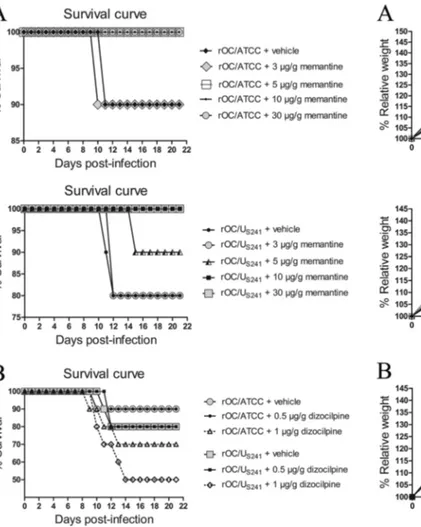
Mice, survival curves, body weight variations, and clinical scores.
Female BALB/c mice (Jackson Laboratories) with a postnatal age of 22 days were inoculated by the intracerebral route with 102.550% tissue culture infective doses of recombinant virus, as previously described (42). Groups of 10 mice infected with each recombinant virus were observed, and survival, body weight variations, and clinical scores re-lated to motor dysfunctions were evaluated on a daily basis over a period of 21 days postinfection (dpi). Mice infected with rOC/US241 were evaluated and scored according to a scale based on experimental allergic encephalitis (EAE) clinical score (CS) evaluation (0 to 1,
nor-mal with no clinical signs; 1.5 to 2, partial hind-limb paralysis with a walk close to ground level; 2.5 to 3.5, complete hind-limb paralysis; 4 to 5, moribund state and death).
NMDA receptor antagonists. The specific uncompetitive NMDA
re-ceptor antagonists memantine [3,5-dimethyl-tricyclo(3.3.1.13,7)decan-1-amine hydrochloride] and dizocilpine [chemical name, (⫹)-MK-801 maleate; (5S,10R)-(⫹)-5-methyl-10,11-dihydro-5H-dibenzo(a,d)cyclo-hepten-5,10-imine maleate] were obtained from R&D Systems-Tocris.
For animal treatments, memantine and dizocilpine were dissolved in PBS. To investigate the effect of treatment with memantine or dizocilpine, groups of 10 BALB/c mice infected with rOC/ATCC or rOC/US241were treated intraperitoneally with defined doses (g/g of body weight) of me-mantine or dizocilpine at 12 h postinfection and then daily for 3 weeks, except when stated otherwise, or only with PBS (vehicle) to normalize experimental stress conditions. To verify the noncytotoxic effect of differ-ent doses of memantine and dizocilpine, sham-infected mice received the same doses of these NMDA receptor antagonists.
For treatment of cell cultures after virus adsorption, HRT-18 cells or primary murine CNS cell cultures were washed with sterile PBS and incu-bated with fresh MEM-alpha supplemented with 1% (vol/vol) FBS (for HRT-18 cells) or at 37°C with fresh neurobasal medium with B27-GlutaMAX-I (for primary mouse CNS cell cultures) containing defined concentrations (M) of memantine or dizocilpine for different periods of time before processing at the time points indicated below.
Infectious virus assay using an IPA. Mouse brain and spinal cord
tissues or cell culture supernatants were processed for the presence and quantification of infectious virus by an indirect immunoperoxidase assay (IPA) on HRT-18 cells, as previously described (44). Briefly, cells were incubated with a mouse primary antibody used to detect the S protein of HCoV-OC43. After three PBS washes, cells were incubated with a second-ary horseradish peroxidase-conjugated goat anti-mouse immunoglobulin antibody (Kirkegaard & Perry Laboratories). Finally, immune complexes were detected by incubation with 0.025% (wt/vol) 3,3=-diaminobenzidine tetrahydrochloride (Bio-Rad) and 0.01% (vol/vol) hydrogen peroxide in PBS, and infectious virus titers were calculated by the Karber method, as previously described (44). Statistical analyses were conducted by one-way analysis of variance (ANOVA), followed by Tukey’s post hoc test.
Extracellular glutamate concentration assay. Extracellular glutamate
concentrations were measured using a colorimetric (optical density at 450 nm [OD450]) glutamate assay kit (ab83389; Abcam) according to the manufacturer’s instructions. Statistical analyses were conducted by one-way ANOVA, followed by Tukey’s post hoc test.
Cell viability and LDH release assay. The viability of the cell cultures
was assayed by use of a lactate dehydrogenase (LDH) cytotoxicity assay kit (Roche) according to the manufacturer’s instructions. It consisted of a colorimetric assay (OD490) for quantification of cell death and cell lysis, based on the measurement of the lactate dehydrogenase activity released from the cytosol of damaged cells into the supernatant. Sta-tistical analyses were conducted by one-way ANOVA, followed by Tukey’s post hoc test.
Immunohistochemistry/immunofluorescence. For
immunohisto-chemistry, groups of three BALB/c mice either sham infected or infected with recombinant virus and treated with memantine, dizocilpine, or ve-hicle were randomly selected and perfused with a solution of 4% (wt/vol) paraformaldehyde at 10 dpi, which corresponds to the time of the peak of viral replication in the spinal cord and the outcome of clinical scores related to paralytic disease. Lumbar segments from spinal cords were cryoprotected in 30% (wt/vol) sucrose, frozen at⫺20°C, and processed with a cryostat (Microm HM 525) for sets with section sizes of 15m. Spinal cord sections were first blocked with horse serum in 1⫻ PBS for 1 h at room temperature. Axonal damage was investigated by assessing the heavy neurofilament (NF-H) phosphorylation state. Tissue sections were incubated with a mouse antinonphosphorylated neurofilament monoclo-nal antibody (MAb; SMI 311; 1/1,000; Covance) for 2 h at room temper-ature. Activation of the glutaminase enzyme was investigated with a
poly-clonal goat anti-phosphate-activated glutaminase antibody (KGA; 1/500; Santa Cruz Biotechnology) or with a polyclonal rabbit anti-phosphate-activated glutaminase GLS2 (GLS2; 1/250; ab113509; Abcam) overnight at 4°C. Recycling of glutamate by the GS enzyme was investigated with a monoclonal mouse anti-GS antibody (clone GS-6; 1/500; MAB302; Mil-lipore) overnight at 4°C. Tissue sections were then washed with 1⫻ PBS and incubated with a biotinylated secondary antimouse, antigoat, or an-tirabbit antibody before revealing with stain from an ABC Vectastain kit (Vector Laboratories), as previously described (45).
For immunofluorescence staining, primary murine CNS cell cultures were washed with sterile PBS and then fixed with 4% (wt/vol) paraformal-dehyde for 30 min at room temperature. After washing, cells were perme-abilized with 100% methanol at⫺20°C for 5 min and washed with PBS. Before staining, cells were preincubated with a blocking solution contain-ing horse serum in PBS for 1 h at room temperature and then incubated with primary antibodies: a polyclonal rabbit anti-glial fibrillary acidic pro-tein (GFAP) antibody (1/1,000; Dako), a polyclonal goat anti-phosphate-activated glutaminase antibody (isoform KGA; 1/500; Santa Cruz Bio-technology), a polyclonal rabbit anti-phosphate-activated glutaminase antibody (isoform GLS2; 1/250; ab113509; Abcam), or a monoclonal mouse anti-glutamine synthetase antibody (clone GS-6; 1/500; MAB302; Millipore) overnight at 4°C. After several washes with PBS, cells were incubated in the dark for 2 h at room temperature with the secondary fluorescent antibodies Alexa Fluor 568 antigoat, antimouse, or antirabbit (1/1,000; Life Technologies) or Alexa Fluor 488 antirabbit or antimouse (1/1,000; Life Technologies). After final PBS washes, tissue sections were incubated for 5 min at room temperature with 4=,6-diamidino-2-phe-nylindole (DAPI; 1g/ml; Life Technologies) and then mounted with Immuno-Mount mounting medium (Fisher Scientific). Immunohisto-chemical and fluorescent staining were observed under a Nikon Eclipse E800 microscope with a QImaging Retiga-EXi Fast 1394 digital camera using Procapture system software.
Protein extraction and Western blot analysis. Spinal cords from
groups of three mice selected randomly were homogenized in radioim-munoprecipitation assay buffer (150 mM NaCl, 50 mM Tris, pH 7.4, 1% [vol/vol] NP-40, 0.25% [wt/vol] sodium deoxycholate, 1 mM EDTA) supplemented with a protease cocktail inhibitor (P8340; Sigma). Lysates were cleared by centrifugation for 5 min at 4°C and 17,000⫻ g, and supernatants were aliquoted and stored at⫺80°C until further analysis. A bicinchoninic acid protein assay kit (Novagen) was used to determine protein concentrations, according to the manufacturer’s protocol. Pro-teins (20g per sample) were separated on a 4 to 12% gradient gel (Novex NuPage; Life Technologies) and transferred to a polyvinylidene difluoride membrane (Immobilon-P transfer membrane; Millipore) with a Bio-Rad Trans-Blot semidry transfer cell apparatus. Membranes were blocked with Tris-buffered saline (TBS) buffer containing 1% (vol/vol) Tween (TBS-T) and 5% (wt/vol) nonfat milk overnight at 4°C, and then the membranes were incubated with a polyclonal rabbit anti-GFAP antibody (1/1,000; Dako), a monoclonal guinea pig anti-glutamate transporter an-tibody (GLT-1; 1/1,000; AB1783; Millipore), a monoclonal rat anti-Mac-2 antibody (1/100; ATCC; Cedarlane), a polyclonal goat anti-phosphate-activated glutaminase antibody (KGA; 1/1,000; Santa Cruz Biotechnol-ogy), a polyclonal rabbit anti-phosphate-activated glutaminase antibody (GLS2; 1/1,000; ab113509; Abcam), a monoclonal mouse GS anti-body (clone GS-6; 1/1,000; MAB302; Millipore), or a polyclonal rabbit anti-GAPDH (glyceraldehyde-3-phosphate dehydrogenase) antibody (1/ 1,000; Santa Cruz Biotechnology) for 1 h at room temperature.
Membranes were washed three times with TBS-T and then incubated with antirabbit (1/1,000; GE Healthcare Lifesciences), antimouse (1/ 1,000; GE Healthcare Lifesciences), anti-guinea pig (1/1,000; Millipore), antirat (1/1,000; Kirkegaard & Perry Laboratories), or antigoat (1/1,000; R&D Systems) secondary antibodies coupled to horseradish peroxidase for 1 h at room temperature, and antibodies were detected by chemilumi-nescence using a Bio-Rad Immun-Star horseradish peroxidase substrate kit. Band detection and semiquantification were done using GeneSnap
software from a Chemi Genius Syngene apparatus. ANOVA tests followed by Tukey’s post hoc analysis were performed to determine the statistical significances in the differences in protein expression between different groups of mice using SPSS software (version 16.0).
RESULTS
Daily treatment of mice with NMDA receptor antagonists
(me-mantine and dizocilpine) attenuates the motor dysfunctions
and severe paralysis induced following infection rOC/US241. We
have recently reported that a viral variant with a single point
mu-tation (Y241H) in the surface spike (S) glycoprotein of
HCoV-OC43 (rOC/U
S241), acquired during viral persistence in human
neural cells (
41
), led to the induction of motor dysfunctions and a
paralytic disease in infected BALB/c mice (
42
). In order to
char-acterize the involvement of NMDA receptors in rOC/U
S241-in-duced hind-limb paralysis, mice infected with rOC/U
S241were
treated with various doses of the NMDA receptor antagonist
me-mantine or dizocilpine or with vehicle (PBS). The highest dose of
dizocilpine that could be administered was 0.5
g/g; above that
dose, the drug was toxic to mice (catatonia, weight loss; data not
shown). Both dizocilpine and memantine treatments attenuated
clinical scores related to the motor dysfunctions and paralytic
dis-ease induced following rOC/U
S241infection, with a more effective
effect for memantine treatment than dizocilpine treatment (
Fig.
1A
and
B
). Indeed, fewer mice treated with memantine presented
mild paralysis (CSs, 1.5 to 2), and they recovered more rapidly
than dizocilpine-treated mice or control vehicle-treated mice.
Moreover, daily memantine treatment attenuated (dose, up to 10
g/g) and even abolished (dose, 30 g/g) clinical scores related to
the paralytic disease of mice infected with rOC/U
S241in a
dose-dependent manner (
Fig. 1B
).
Daily memantine treatment attenuates mortality rates and
body weight loss of infected mice compared to those achieved by
dizocilpine and vehicle (control) treatment. To investigate the
effect of treatment with memantine or dizocilpine on survival
rates, groups of 10 BALB/c mice infected with reference wild-type
OC43 coronavirus (rOC/ATCC) or S-mutant OC43 virus (rOC/
U
S241) were treated intraperitoneally daily with defined doses of
memantine or dizocilpine or with PBS (vehicle). Mice infected
with rOC/ATCC or rOC/U
S241and treated with PBS showed,
re-spectively, 10% and 20% mortality. Daily memantine treatment
attenuated (doses, up to 5
g/g) and totally suppressed (doses, 10
g/g and over) the mortality of mice infected with either virus in a
dose-dependent manner (
Fig. 2A
) compared to that achieved with
dizocilpine treatment, which did not attenuate mortality
com-pared to that achieved with PBS (
Fig. 2B
). Moreover, a higher dose
of dizocilpine (1
g/g) by itself increased mortality in noninfected
mice (
Fig. 2B
). We had verified beforehand that the treatment of
sham-infected mice with various doses of memantine (3 to 30
g/g) or dizocilpine (0.5 g/g) was not toxic and did not affect the
survival rate of mice (data not shown).
Mice were also investigated for weight variations because, as
previously described (
45
), weight loss is a specific symptom of
mice infected with HCoV-OC43. Mice infected with rOC/ATCC
or rOC/U
S241and treated with PBS showed a loss of body weight of
about 10 to 15% at 10 days postinfection and recovered at between
12 and 22 days postinfection. Daily memantine treatment
signif-icantly attenuated (dose, 5
g/g; P ⬍ 0.05 and P ⬍ 0.01) and even
abolished (doses, 10
g/g and greater) the weight loss symptoms
of mice infected with either virus in a dose-dependent manner
(
Fig. 3A
) compared to the effects of dizocilpine treatment, which
FIG 1 Daily memantine and dizocilpine treatment attenuates the motor dysfunctions and severe paralysis induced following infection of mice by
S-mutant human coronavirus OC43. Groups of 10 BALB/c mice infected with rOC/US241were treated daily by intraperitoneal injection of a defined dose
of memantine or dizocilpine or of PBS (vehicle). Motor dysfunctions were evaluated and scored according to a scale based on experimental allergic encephalitis (EAE) clinical score (CS) evaluation (0 to 1, normal with no clinical signs; 1.5 to 2, partial hind-limb paralysis with a walk close to ground level; 2.5 to 3.5, complete hind-limb paralysis; 4 to 5, moribund state and death). (A) Daily treatment with 0.5g/g dizocilpine attenuated the clinical scores in mice infected with rOC/US241. Higher doses of dizocilpine had strong side effects on mice and increased the mortality rate, as illustrated inFig.
2. (B) Daily memantine treatment attenuated (doses, up to 10g/g) and even abolished (dose, 30 g/g) the clinical scores in a dose-dependent manner. Results are representative of three independent experiments.
did not attenuate the weight loss of mice infected with either virus
(
Fig. 3B
). As mentioned above, we made sure that the treatment of
sham-infected mice with various doses of memantine (3 to 30
g/g) or dizocilpine (0.5 g/g) had no effect and that it did not
affect the relative weight of the mice (data not shown).
Increase in extracellular glutamate levels and LDH release
following infection of mouse primary CNS cell cultures. In order
to investigate whether HCoV-OC43 causes a dysregulation of
glu-tamate homeostasis that may lead to neuronal loss, primary
mu-rine CNS cell cultures were infected with rOC/ATCC or rOC/
U
S241at various multiplicities of infection (MOIs) and treated
with memantine (20
M) or vehicle (medium). At 24 h
postinfec-tion, cell culture supernatants were assessed for extracellular
glu-tamate and lactate dehydrogenase (LDH) release. Increasing
MOIs of each virus led to a significant increase in extracellular
glutamate levels following infection with rOC/ATCC (P
⬍ 0.05
for MOIs of 1 and 3) or rOC/U
S241(P
⬍ 0.05 for MOIs of 0.1 and
1; P
⬍ 0.01 for an MOI of 3) compared to the level for
mock-infected cells (
Fig. 4A
). Moreover, infection with rOC/U
S241usu-ally led to a more significant extracellular glutamate release (P
⬍
0.05) than rOC/ATCC infection (
Fig. 4A
). Memantine treatment
led to a significant decrease in the extracellular glutamate
concen-tration following infection of primary cultures of mouse CNS cells
with rOC/ATCC (P
⬍ 0.05 for an MOI of 1) and rOC/U
S241(P
⬍
0.05 for MOIs of 0.1 and 1) (
Fig. 4A
).
The viability of cell cultures was assayed via the LDH release
assay. Increasing MOIs of each virus led to a significant increase in
LDH release following infection with rOC/ATCC (P
⬍ 0.05 for an
MOI of 1 and 3) or rOC/U
S241(P
⬍ 0.05 for MOIs of 0.1, 1, and 3)
compared to that for mock-infected cells (
Fig. 4B
). Infection of
primary cultures of mouse CNS cells with rOC/U
S241usually led to
FIG 2 Daily memantine treatment attenuates the mortality rates of
in-fected mice compared to those achieved with dizocilpine and vehicle (con-trol) treatment. Groups of 10 BALB/c mice infected with reference wild-type virus (rOC/ATCC) or S-mutant virus (rOC/US241) were treated daily
with an intraperitoneal injection of a defined dose of memantine or dizo-cilpine or of PBS (vehicle). (A) Daily memantine treatment attenuated (doses, up to 5g/g) and totally suppressed (doses, 10 g/g and above) the mortality rates for mice infected with either virus in a dose-dependent manner. (B) Daily dizocilpine treatment did not attenuate the mortality rates for mice infected with either virus. A higher dose of dizocilpine (1 g/g) by itself increased the mortality rate. Results are representative of three independent experiments.
FIG 3 Daily memantine treatment attenuates/abolishes the weight loss of
infected mice compared to that achieved with dizocilpine and vehicle (control) treatments. Groups of 10 BALB/c mice infected with reference wild-type virus (rOC/ATCC) or S-mutant virus (rOC/US241) were intraperitoneally treated
daily with a defined dose of memantine or dizocilpine or with PBS (vehicle). (A) Daily memantine treatment attenuated (doses, up to 5g/g) and abolished (dose, 10g/g and above) weight loss in mice infected with either virus in a dose-dependent manner. (B) Daily dizocilpine treatment did not attenuate weight loss in mice infected with either virus. *, P⬍ 0.05 (Dukey’s test) for comparison with infected mice treated with vehicle for a defined day postin-fection; **, P⬍ 0.01 (Dukey’s test) for comparison with infected mice treated with vehicle for a defined day postinfection. Results are representative of three independent experiments.
a more significant LDH release (P
⬍ 0.05) compared to that
achieved with infection with rOC/ATCC (
Fig. 4B
). Memantine
treatment led to a significant decrease in LDH release following
infection of primary cultures of mouse CNS cells by both rOC/
ATCC (P
⬍ 0.05 for an MOI of 1) and rOC/U
S241(P
⬍ 0.05 for
MOIs of 0.1 and 1) (
Fig. 4B
).
The increased extracellular glutamate level is not a result of
an increase in expression of phosphate-activated glutaminase.
Given that an increase in the extracellular glutamate
concentra-tion could result from an increase in glutamate synthesis, groups
of three BALB/c mice infected with rOC/ATCC or rOC/U
S241and
treated with memantine or vehicle were sacrificed and evaluated
for phosphate-activated kidney-type glutaminase (KGA) or
liver-type glutaminase (GLS2) in lumbar spinal cord grey matter (GM)
segments at 10 dpi (the time of the peak of viral replication in the
spinal cord and the outcome of clinical scores related to paralytic
disease). Infection of mice with rOC/ATCC or rOC/U
S241did not
lead to changes in KGA or GLS2 expression in lumbar spinal cord
GM (
Fig. 5A
and
B
). Western blot analysis of spinal cord proteins
confirmed the histological findings. Infection of mice with rOC/
ATCC or rOC/U
S241did not lead to changes in expression of
phos-phate-activated KGA or GLS2, and memantine treatments did not
affect glutaminase expression (
Fig. 5C
). To confirm these in vivo
results, primary murine CNS cell cultures were infected with rOC/
ATCC or rOC/U
S241and treated with memantine or vehicle.
Ex-pression of KGA and GLS2 by immunofluorescence and
immu-nohistochemistry revealed that it was restricted to neurons, with
no changes in the level of expression of either enzyme at 24 h
postinfection for both viruses (
Fig. 5A
and
B
). We had made sure
beforehand that the treatment of sham-infected mice with 3
g/g
of memantine or primary murine CNS cell cultures with 20
M
memantine did not affect the basal level of expression of KGA and
GLS2 compared to the level obtained after vehicle (control)
treat-ment (data not shown).
Astrocytes and microglial cells may contribute to an excess of
glutamate release by expressing glutaminase. Mice infected with
either virus showed significant GFAP expression (P
⬍ 0.001)
compared to that for sham-infected mice, as previously shown
(
42
), and memantine treatment did not attenuate this astrocytic
activation (
Fig. 5C
). However, mice infected with either virus
showed significant (P
⬍ 0.01 and P ⬍ 0.05) microglial cell
activa-tion (as monitored by Mac-2 expression) compared to that for
sham-infected mice, which was increased significantly (P
⬍ 0.05)
more following infection with rOC/U
S241than following infection
with rOC/ATCC, as previously shown (
42
). Memantine
signifi-cantly (P
⬍ 0.05) attenuated this microglial cell activation in mice
infected with rOC/U
S241(
Fig. 5C
).
Expression of GS and GLT-1 is downregulated in mice
in-fected with rOC/US241
and partially restored following
treat-ment with memantine. Groups of three BALB/c mice infected
with rOC/ATCC or rOC/U
S241treated with memantine or vehicle
were sacrificed and evaluated by immunohistochemistry for GS
and glutamate transporter 1 (GLT-1) of lumbar spinal cord GM
segments at 10 dpi. GS staining was downregulated in the GM of
mice infected with rOC/U
S241(
Fig. 6A
). To confirm these in vivo
results, primary cultures of murine CNS cells were infected with
rOC/ATCC or rOC/U
S241(MOI
⫽ 1), treated with memantine
(20
M) or vehicle (medium), and assessed for GS expression by
immunofluorescence at 24 h postinfection. GS was exclusively
expressed in astrocytes (GFAP marker) and was dramatically
downregulated in GM and in infected murine CNS cell cultures
infected with rOC/U
S241, and memantine treatment led to an
in-crease in GS expression (
Fig. 6A
). Western blot analysis of spinal
cord proteins confirmed the histological findings. Infection of
mice with rOC/U
S241led to a significant (P
⬍ 0.01 and P ⬍ 0.01)
downregulation of GS expression compared to that in
sham-in-fected mice or rOC/ATCC-insham-in-fected mice treated with vehicle.
Me-mantine treatment led to a significant (P
⬍ 0.01) increase in GS
expression (
Fig. 6B
). Moreover, infection of mice with rOC/U
S241led to a significant (P
⬍ 0.01 and P ⬍ 0.01) downregulation of
GLT-1 expression compared to that in sham-infected mice or
rOC/ATCC-infected mice treated with vehicle, as previously
shown (
42
). Memantine treatment led to a significant (P
⬍ 0.01)
increase in GLT-1 expression (
Fig. 6B
). We had made sure
before-hand that the treatment of sham-infected mice with 3
g/g of
FIG 4 Increase in extracellular glutamate levels and LDH release following
infection of primary cultures of mouse CNS. Primary cultures of mouse CNS cells were infected with reference wild-type virus (rOC/ATCC) or S-mutant virus (rOC/US241) at different MOIs and treated with memantine (20M) or
vehicle (medium). (A) Increasing MOIs of both viruses led to an increase in the extracellular glutamate level that was significantly decreased by memantine (MOIs, 0.1 and 1). (B) Increasing MOIs of both viruses also led to an increase in LDH release that was also significantly reduced by memantine (MOIs, 0.1 and 1). #, P⬍ 0.05 (Dukey’s test) for comparison to mock-infected cells; ##,
P⬍ 0.01 (Dukey’s test) for comparison to mock-infected cells; &, P ⬍ 0.05
(Dukey’s test) for comparison of rOC/ATCC- or rOC/US241-infected cells at a
defined MOI; *, P⬍ 0.05 (Dukey’s test) for comparison of memantine- and vehicle-treated infected cells.
memantine or primary murine CNS cell cultures with 20
M
memantine did not affect the basal level of expression of GS and
GLT-1 compared to the level obtained after the control treatment
(data not shown).
Daily treatment with memantine reduces neuronal
dysfunc-tion in mice infected with rOC/ATCC or rOC/U
S241. Spinal cords
from mice infected with rOC/U
S241or rOC/ATCC and treated with
memantine or vehicle were harvested at 10 dpi (the time of peak of
viral replication in the spinal cord and the outcome of motor
dys-functions and severe paralysis) to evaluate whether a neuronal
alter-ation associated with excitotoxicity was under way in infected mice.
Neuronal dysregulation was investigated by evaluating the axonal
neurofilament phosphorylation state. Infection of mice with rOC/
ATCC or rOC/U
S241resulted in an abnormal loss of soma
nonphos-phorylated NF-H immunoreactivity in the spinal cord GM compared
to that in sham-infected mice. This modification in the
phosphory-lation state was more pronounced following infection with rOC/
U
S2410than following infection with rOC/ATCC (
Fig. 7
; SMI 311
GM, white arrows). On the other hand, the spinal cord white matter
(WM) of mice infected with rOC/ATCC and rOC/U
S241showed an
abnormal presence of nonphosphorylated NF-H immunoreactivity,
and axonal swelling in WM was more evident following rOC/U
S241FIG 5 The increase in extracellular glutamate levels is not a result of an increase in expression of phosphate-activated glutaminase. Phosphate-activated kidney-type
glutaminase (KGA) or liver-type glutaminase (GLS2) was detected in lumbar spinal cord GM segments in infected mice treated with memantine or vehicle at 10 dpi (black arrows). Primary cultures of murine CNS cells were infected with reference wild-type virus (rOC/ATCC) or S-mutant virus (rOC/US241) and treated with
memantine (20M) or vehicle (medium). Immunofluorescence revealed no changes in KGA (A) or GLS2 (B) activation at 24 h postinfection in lumbar spinal cord GM in primary murine CNS cell cultures following infection by either virus. MAP2, microtubule-associated protein 2. Magnifications,⫻400. (C) Semiquantitative Western blot analysis of spinal cord proteins confirmed that activation of KGA and GLS2 was not modulated and that the levels of astrogliosis (a significant increase in GFAP expression) and microglial cell activation (an increase in Mac-2 expression) compared to those in sham-infected mice occurred. Memantine treatment significantly reduced Mac-2 expression in mice infected with rOC/US241compared to that in rOC/ATCC-treated mice. Data are represented as the mean⫾ SEM (n ⫽ 3). ###, P ⬍
0.001 (Dukey’s test) for comparison to sham-infected mice; ##, P⬍ 0.01 (Dukey’s test) for comparison to sham-infected mice; #, P ⬍ 0.05 (Dukey’s test) for comparison to sham-infected mice; *, P⬍ 0.05 (Dukey’s test) for comparison of groups of memantine treated- or nontreated infected mice. 1, sham infection plus vehicle treatment; 2, rOC/ATCC infection plus vehicle treatment; 3, rOC/ATCC infection plus treatment with 3g/g memantine; 4, rOC/US241infection plus vehicle treatment; 5,
infection than following rOC/ATCC infection (
Fig. 7
; SMI 311 WM,
black arrows).
Higher doses of memantine treatment attenuate viral
rep-lication in the CNS of infected mice. Because memantine
treatment had a dose-response effect on various parameters,
including mortality rates and body weight loss, and because
memantine is a derivative of adamantane (an antiviral drug),
we wished to evaluate whether the treatment may at some point
affect viral replication in the CNS. Daily treatment over a
pe-riod of 21 days with higher doses of memantine (10
g/g and
above) significantly reduced, in a dose-dependent manner, the
replication of rOC/ATCC (P
⬍ 0.05 and P ⬍ 0.01) and rOC/
U
S241(P
⬍ 0.05, P ⬍ 0.01, and P ⬍ 0.001) in the brains and
spinal cords of infected mice (
Fig. 8A
), indicative of an antiviral
effect. However, dizocilpine treatment did not affect the
repli-cation of either virus (
Fig. 8B
).
Prophylactic transient treatment with memantine
attenu-ates viral replication in the CNS of infected mice. Having shown
that the long-term use of high doses of memantine (daily
treat-ment for 21 days) altered viral replication, we wished to evaluate
whether a preventive treatment for a short period of time would
also have the same effect. Groups of mice were treated with
me-mantine (5
g/g) 1 day prior to viral infection and daily for only 4
days postinfection; viral replication in brains and spinal cords was
evaluated every 2 days. This preventive use of memantine for a
shorter period of time significantly (P
⬍ 0.05) reduced viral
rep-lication in the brain and spinal cord of mice infected with rOC/
ATCC or rOC/U
S241(
Fig. 9
).
Memantine reduces viral replication in an NMDA
blockade-independent manner. In order to investigate the effect of
me-mantine on HCoV-OC43 replication, primary cultures of
mouse CNS cells (which express NMDA receptors, as verified
FIG 6 Expression of GS and GLT-1 is downregulated in mice infected with S-mutant human coronavirus OC43 and partially restored following treatment with
memantine. GS was evaluated by immunohistochemistry in lumbar spinal cord GM segments of mice infected with rOC/ATCC or rOC/US241and treated with
memantine or vehicle at 10 dpi (black arrows) and by immunofluorescence at 24 h postinfection in primary cultures of murine CNS cells infected and treated with memantine (20M) or vehicle (medium). (A) GS expression was dramatically downregulated in GM and in primary cultures of murine CNS cells infected with the S-mutant virus (rOC/US241), and memantine treatment partially restored GS expression. GFAP, glial fibrillary acidic protein. Magnifications,⫻400. (B)
Semiquanti-tative Western blot analysis of spinal cord proteins confirmed the histological findings. Infection of mice with rOC/US241led to a significant downregulation of GS and
GLT-1 expression that was significantly restored by memantine. Data are represented as the mean⫾ SEM (n ⫽ 3). ##, P ⬍ 0.01 (Dukey’s test) for comparison to sham-infected mice; **, P⬍ 0.01 (Dukey’s test) for comparison of groups of memantine-treated or nontreated infected mice. 1, sham infection plus vehicle treatment; 2, rOC/ATCC infection plus vehicle treatment; 3, rOC/ATCC infection plus treatment with 3g/g memantine; 4, rOC/US241infection plus vehicle treatment; 5,
by RT-PCR for the expression of the NMDA-NR1 receptor
subunit;
Fig. 10D
) or cells of the HRT-18 cell line (which do not
express NMDA receptors;
Fig. 10D
) were infected with rOC/
ATCC or rOC/U
S241and treated with increasing noncytotoxic
doses (data not shown) of memantine or dizocilpine.
Meman-tine treatment of cells significantly reduced the production of
intra- and extracellular infectious virus in primary cultures of
mouse CNS cells (P
⬍ 0.05 for 30 M and P ⬍ 0.01 for 70 M;
Fig. 10A
) and HRT-18 cells (P
⬍ 0.05 for 30 M and P ⬍ 0.01
for 70
M;
Fig. 10B
) compared to the levels of production by
dizocilpine- or PBS-treated cells. To investigate whether
me-mantine affected virus entry into cells, it was added to the cell
medium either before, during, or after infection. Memantine
treatment (70
M) significantly reduced the intra- and
extra-cellular production of infectious rOC/ATCC and rOC/U
S241,
when administered after infection (P
⬍ 0.05), compared to that
in the control (PBS-treated infected cells) or when memantine
treatment was added before or during infection (
Fig. 10C
).
DISCUSSION
We have recently reported that a single point mutation in the
surface spike (S) glycoprotein of human coronavirus OC43
(Y241H) modulates virus-induced neuropathology in a mouse
model from an encephalitis to an MS-like paralytic disease related
to glutamate excitotoxicity, with involvement of AMPA receptors
(
42
). In the current study, we made use of two uncompetitive
antagonists of NMDA receptors and demonstrate that these
glu-tamate receptors are also involved in the neuropathology induced
following infection of mice with HCoV-OC43. Moreover, we
show that memantine protected mice from infection by having a
dose-dependent effect on both neurological symptoms and viral
replication.
Our results show that HCoV-OC43 infection causes a
dysregu-lation of glutamate homeostasis that may lead to neuronal loss
(neurodegeneration). The significant increase in the extracellular
glutamate level and LDH release shown in
Fig. 4
underlines the
importance of glutamate dysregulation after infection, as shown
in other virus infections (
46–48
). The fact that this increase is
more significant following infection with rOC/U
S241than
follow-ing infection with rOC/ATCC strongly suggests that the viral S
glycoprotein is an important factor in this dysregulation. The
ob-servation that memantine treatment led to a significant decrease
in the extracellular release of glutamate and LDH at 24 h
postin-fection shows that glutamate homeostasis is altered during
infec-tion and that it appears to involve NMDA receptors, leading to
excitotoxicity.
Glutamate homeostasis relies on two different mechanisms in
order to prevent excitotoxicity. In order to decipher the possible
mechanism leading to an increase in extracellular glutamate
fol-lowing infection by HCoV-OC43, we first investigated the
synthe-sis of glutamate (
Fig. 5
). HCoV-OC43 infects neurons as primary
targets of infection in the murine CNS (
11
) and in human neuron/
astrocyte cocultures (
49
) and induces neuronal degeneration and
eventual cell death. This could be associated with a stress that can
disturb the phosphate-activated glutaminase enzyme expression
responsible for glutamate synthesis, as was shown in
HIV-1-in-fected microglia (
18
). Our results (
Fig. 5
) show that expression of
the two glutaminase isoforms found in the CNS (
50
) is not
mod-ified in the spinal cord of mice or in primary cultures of mouse
CNS cells infected with HCoV-OC43. Moreover, we show that
expression of GFAP (an astrocyte marker) and Mac-2 (a marker of
microglial cell activation) is upregulated following infection with
HCoV-OC43 and that microglial activation is significantly
in-creased following infection by the S mutant (rOC/U
S241)
com-pared to that in the reference wild-type virus (rOC/ATCC). Some
studies have shown that, in some circumstances, astrocytes and
microglial cells may express glutaminase and release glutamate
(
16–18
,
51
,
52
). However, our results clearly show that neither
astrocytes nor microglial cells expressed any detectable
phos-phate-activated glutaminase (
Fig. 5
). This strongly suggests that
the increase in extracellular glutamate release following infection
by HCoV-OC43 is not a result of an increased expression of
phos-phate-activated glutaminase enzyme.
As previously reported, recycling of glutamate is also an
im-portant key factor for the physiological regulation of glutamate
homeostasis to prevent excitotoxicity (
53
). Under physiological
conditions, glutamate homeostasis is in large part regulated by
glial glutamate transporter 1 (GLT-1), which is mainly expressed
FIG 7 Daily treatment with a low dose of memantine reduces neuronal
dys-functions in infected mice. Groups of three BALB/c mice infected with refer-ence wild-type virus (rOC/ATCC) or S-mutant virus (rOC/US241) and treated
with vehicle or memantine (3g/g) were sacrificed and evaluated by immu-nohistochemistry for the levels of neurofilament phosphorylation in the lum-bar spinal cord GM and WM segments at 10 dpi compared to those in control mice. Following infection, GM showed a loss of normal nonphosphorylated NF immunoreactivity compared to the GM of sham-infected mice. This loss was more pronounced following rOC/US241infection than following rOC/
ATCC infection (white arrows). The spinal cord WM of infected mice showed a higher level of abnormal axonal nonphosphorylated NF-H with abnormal axonal swelling, which was more pronounced following infection with rOC/ US241than following sham infection (black arrows) and infection with rOC/
ATCC. Results are representative of two independent experiments with three mice per group. Magnification,⫻400.
(A) Memantine treatment reduced viral replication in the brains and spinal cords of infected mice in a dose-dependent manner (between 10g/g and 30 g/g). (B) Dizocilpine treatment did not affect viral replication in the brains and spinal cords of infected mice. Data are represented as medians with ranges (n⫽ 3). *, P⬍ 0.05 (Dukey’s test) for comparison to control mice (infected mice treated only with vehicle) for a defined day postinfection; **, P ⬍ 0.01 (Dukey’s test) for comparison to control mice (infected mice treated only with vehicle) for a defined day postinfection; ***, P⬍ 0.001 (Dukey’s test) for comparison to control mice (infected mice treated only with vehicle) for a defined day postinfection. Each symbol on the graph represents a single mouse. L.O.D., limit of detection; TCID50,
on astrocytes and which is responsible for up to 90% of the total
glutamate clearance in adult CNS (
54
). As seen in
Fig. 5
, astrocytes
are strongly activated following infection with either recombinant
virus. However, expression of GLT-1 did not correlate with this
activation associated with an increased number of astrocytes
ex-pressing high levels of GFAP, as previously shown (
42
). Indeed,
the results presented in
Fig. 6
show that GLT-1 expression was
significantly downregulated following rOC/U
S241infection
com-pared to that in mice infected with rOC/ATCC or sham-infected
mice. Interestingly, decreased expression of this transporter was
reported in several neurological diseases, as well as in other viral
models (
35
,
37
,
55
,
56
). Of note, memantine treatment led to a
major restoration of GLT-1 expression.
Glutamate homeostasis is also regulated by glutamine
synthe-tase. Indeed, after reuptake, glutamate is converted to glutamine
by the astroglial enzyme GS (
21
,
22
). Glutamine is then
trans-ported into nerve terminals and reconverted to glutamate for an
eventual new glutamate-glutamine cycle (
23
). As shown in
Fig. 6
,
GS expression was significantly downregulated following rOC/
U
S241infection compared to that in mice infected with rOC/
ATCC or sham-infected mice, and memantine treatment led to a
major restoration of GS expression.
We have previously reported that infection of mice with
HCoV-OC43 led to the release of proinflammatory cytokines
(tu-mor necrosis factor alpha [TNF-
␣], interleukin-1 [IL-1], and
IL-6) in the spinal cord of infected mice, with a significant increase
in IL-6 in mice infected with an S mutant virus compared to that in
mice infected with the reference wild-type virus (
45
).
Proinflam-matory cytokines such as IL-6 are known to downregulate
gluta-mate transporter GLT-1 expression (
56–58
) and GS expression
(
59
,
60
). In the present study, we show that infection with the
S-mutant virus rOC/U
S241led to significant
microglia/macro-phage activation compared to what was observed with infection
with the wild-type reference virus (rOC/ATCC) and that
treat-ment with memantine significantly reduced microglial cell
activa-tion, as previously reported (
61
,
62
).
We show that the increased microglial cell/macrophage
activa-tion correlated with the downregulaactiva-tion of expression of GLT-1
and GS (
Fig. 5C
and
6B
). Our results suggest that memantine may
prevent downregulation of GLT-1 and GS expression by
down-regulating microglial cell activation, which is responsible for the
major release of some proinflammatory molecules, like IL-1, IL-6,
and TNF-
␣, that could act on disruption of glutamate
homeosta-sis by downregulating glutamate recycling (
53
,
63
,
64
).
Glutamate excitotoxicity may damage the cytoskeleton of
ax-ons and affect the axonal transport rate by disrupting the
neuro-filament phosphorylation state (
65
,
66
). Signs of neuronal injury
are monitored by evaluation for a change in the NF
phosphoryla-tion state, which is characterized by a loss of nonphosphorylated
NF in soma and an increase in nonphosphorylated NF in axons
(
67
). Mice infected with rOC/U
S241or rOC/ATCC also showed an
abnormal loss of nonphosphorylated NF-H in soma in the grey
matter (GM), and this was more evident following rOC/U
S241in-fection. Moreover, the spinal cord white matter (WM) of mice
infected with rOC/U
S241or rOC/ATCC presented abnormal
axonal nonphosphorylated NF-H, which was also more evident
FIG 9 Prophylactic transient treatment with memantine attenuates viral replication in the CNS of infected mice. Groups of three BALB/c mice were treated with
memantine (5g/g) 1 day before viral infection and daily for 4 days postinfection. Mice infected with reference wild-type virus (rOC/ATCC) or S-mutant virus (rOC/US241) were sacrificed and evaluated for viral replication in brains and spinal cords every 2 days. The preventive memantine treatment reduced viral
replication in the brains and spinal cords of infected mice. Data are represented as medians with ranges (n⫽ 3). *, P ⬍ 0.05 (Dukey’s test) for comparison to control mice (infected mice treated only with vehicle) for a defined day postinfection. Each symbol on the graph represents a single mouse. L.O.D., limit of detection; TCID50, 50% tissue culture infective dose. Results are representative of those from two independent experiments.
Memantine treatment significantly reduced intra- and extracellular infectious virus titers in primary cultures of mouse CNS cells (A) and in cells of the human epithelial HRT-18 cell line (B) compared to those achieved with dizocilpine and control (medium alone) treatment. h.p.i., hours postinfection. *, P⬍ 0.05 (Dukey’s test) for comparison of memantine- or dizocilpine-treated infected cells with PBS-treated infected cells; **, P⬍ 0.01 (Dukey’s test) for comparison of memantine- or dizocilpine-treated infected cells with PBS-treated infected cells. (C) Memantine treatment (70M) significantly reduced intra- and extracellular infectious virus titers when added after virus adsorption. B, before infection; D, during infection; A, after infection. *, P⬍ 0.05 (Dukey’s test) for comparison of memantine-treated infected cells and control (PBS-treated infected) cells. Results are representative of three independent experiments. (D) Expression of the NR1 subunit of the NMDA receptor was confirmed by RT-PCR in primary cultures of mouse CNS cells (lane 1; the 211-bp fragment size is indicated). No expression was detected in the HRT-18 cell line (lane 2). Lane MM, molecular size marker; lane⫺, negative control for PCR.
following rOC/U
S241infection, even leading to axonal beading
and swelling (
Fig. 7
). It has already been reported that
modifica-tions in the phosphorylation state of neurofilaments contribute to
motor neuron disease, as observed in amyotrophic lateral sclerosis
(ALS) and multiple sclerosis (MS), as well as following infection
with Theiler’s murine encephalomyelitis virus (TMEV) in an
an-imal model of MS (
68
,
69
,
87
). This abnormal disruption of the
NF-H phosphorylation state in axons and cell bodies observed
following infection could lead to neuronal dysfunction and
dis-ruption of axonal transport with perturbations in neuronal
trans-mission, which was demonstrated to account for motor
disabili-ties (
70
). Daily treatment with memantine (3
g/g) significantly
reduced the imbalance in NF-H phosphorylation and also
im-proved motor dysfunctions, probably through both a direct effect
on protection of neurons from glutamate overactivation on
NMDA receptors (
71
) and an indirect effect by downregulating
microglial cell activation, that result in the release of
proinflam-matory mediators or toxic molecules that may damage neurons
(
61
,
62
).
Memantine is an adamantane derivative. Some of these
deriv-atives are used as drugs in human medicine and have antiviral
properties. These include amantadine and rimantadine (with
ac-tivity against influenza A virus [
72
]), tromantadine (with activity
against herpes simplex virus [
73
]), and bananin (with activity
against the SARS coronavirus [
74
]).
In order to explore the mode of action of memantine as an
antiviral molecule, we made use of mouse primary CNS cell
cul-tures (which express NMDA receptors) and of a human epithelial
cell line (commonly used in the laboratory to amplify
HCoV-OC43, which does not express NMDA receptors). The
observa-tion that memantine affects viral replicaobserva-tion in both cell types
strongly suggests that memantine acts as an antiviral drug without
the involvement of NMDA receptors (
Fig. 10A
and
B
). Moreover,
our observation that memantine shows a highly significant
anti-viral activity after primary infection of susceptible cells (
Fig. 10C
)
indicates that it inhibits viral replication after virus attachment to
the cell receptor, as was also shown for other adamantane
deriva-tives. Indeed, amantadine and rimantadine inhibit the early steps
of uncoating of influenza virus in endosomes (
75
,
76
),
tromanta-dine inhibits early steps in herpes simplex virus replication before
macromolecular synthesis and a late event, such as assembly or
release of virus (
77
), and bananin inhibits the helicase activities of
SARS coronavirus (
74
). Memantine could act by inhibition of the
ion channel activity of the HCoV-OC43 E protein, as amantadine
and rimantadine do by binding the influenza virus M2 protein
proton channel (
75
,
76
), or it could inhibit the ATPase activity of
the HCoV-OC43 helicase, as bananin does against the SARS
coro-navirus (
74
).
To our knowledge, our study shows the first evidence that
me-mantine, widely used in humans to treat neurological diseases,
shows antiviral properties against a human neuroinvasive and
neurotropic virus. This novel action of memantine strongly
sug-gests that it could be used as an antiviral agent in various
neuro-logical diseases with a viral involvement, such as herpes
encepha-litis or meningitis, or neurological diseases for which the etiology
could involve viruses, such as multiple sclerosis (
78
), Alzheimer’s
disease (
79
), and Parkinson’s disease (
80
).
Moreover, as shown in
Fig. 9
, transient treatment with
me-mantine (5
g/g) significantly (P ⬍ 0.05) attenuated viral
replica-tion in the CNS of infected mice, which strongly suggests that
me-mantine could be used as a prophylactic agent for a short treatment in
an attempt to control some viral outbreaks, as previously shown for
other antiviral agents effective against influenza virus (
81
).
In summary, we show that memantine improved clinical
scores related to paralytic disease and motor disabilities by
par-tially restoring the physiological NF phosphorylation state in
S-mutant-virus-infected mice. Moreover, memantine attenuated, in
a dose-dependent manner, mortality rates and body weight loss
and reduced viral replication in the central nervous system of
HCoV-OC43-infected mice. Furthermore, glutamate recycling
via the GLT-1 transporter and the GS enzyme, rather than
gluta-mate synthesis, appears to be the key factor in glutagluta-mate
homeo-stasis dysregulation that may lead to the motor dysfunctions and
paralytic disease induced in mice following infection with
HCoV-OC43. Even though we are aware of the potential limitations of
studying a human virus in a mouse model and we are interpreting
our results accordingly, we believe that this approach is relevant
and important for predicting how a virus could interact with
hu-mans, as shown for several other human viruses used in mouse
models (
82–86
). Our results also strongly suggest that memantine
could be used as both a prophylactic and a therapeutic antiviral
agent against neuroinvasive and neurotropic human viruses that
cause viral encephalitis or meningitis or neurological diseases in
which the etiology could involve viruses. Further studies are under
way to investigate both the role of inflammation on the
down-regulation of glutamate recycling and the precise mechanism that
triggers NMDA receptor-associated excitotoxicity, to identify the
viral and cellular factors involved in the process following
infec-tion of mice by the human coronavirus OC43, and to understand
how memantine plays such an important role in controlling both
neurodegeneration and viral infection itself.
ACKNOWLEDGMENTS
This work was supported by grant no. MT-9203 from the Institute of Infection and Immunity (III) of the Canadian Institutes of Health Re-search (CIHR) to Pierre J. Talbot, who is the holder of the Tier-1 (Senior) Canada Research Chair in Neuroimmunovirology award. Elodie Brison gratefully acknowledges a doctoral studentship from the Multiple Sclero-sis Society of Canada.
REFERENCES
1. Buchmeier MJ, Lane TE. 1999. Viral-induced neurodegenerative dis-ease. Curr. Opin. Microbiol. 2:398 – 402. http://dx.doi.org/10.1016 /S1369-5274(99)80070-8.
2. Talbot PJ, Jacomy H, Desforges M. 2008. Pathogenesis of human coro-naviruses other than severe acute respiratory syndrome coronavirus, p 313–324. In Perlman S, Gallagher T, Snilder EJ (ed), The nidoviruses. ASM Press, Washington, DC.
3. Rota PA, Oberste MS, Monroe SS, Nix WA, Campagnoli R, Icenogle JP,
Penaranda S, Bankamp B, Maher K, Chen MH, Tong S, Tamin A, Lowe L, Frace M, DeRisi JL, Chen Q, Wang D, Erdman DD, Peret TC, Burns C, Ksiazek TG, Rollin PE, Sanchez A, Liffick S, Holloway B, Limor J, Mc-Caustland K, Olsen-Rasmussen M, Fouchier R, Gunther S, Osterhaus AD, Drosten C, Pallansch MA, Anderson LJ, Bellini WJ. 2003. Characterization
of a novel coronavirus associated with severe acute respiratory syndrome. Science 300:1394 –1399.http://dx.doi.org/10.1126/science.1085952. 4. Riski H, Hovi T. 1980. Coronavirus infections of man associated with
diseases other than the common cold. J. Med. Virol. 6:259 –265.http://dx .doi.org/10.1002/jmv.1890060309.
5. Forgie S, Marrie TJ. 2009. Healthcare-associated atypical pneumonia. Semin. Resp. Crit. Care 30:67–85.http://dx.doi.org/10.1055/s-0028-1119811. 6. Yeh EA, Collins A, Cohen ME, Duffner PK, Faden H. 2004. Detection
of coronavirus in the central nervous system of a child with acute dissem-inated encephalomyelitis. Pediatrics 113:e73– e76.