Estrogen withdrawal and liver fat accumulation:
contribution of hepatic VLDL-TG production and effect
of exercise training
par Razieh Barsalani
Département de kinésiologie
Thèse présentée à la Faculté des études supérieures en vue de l’obtention du grade de Philosophiae Doctor (Ph.D.)
en Sciences de l’activité physique
Avril, 2010
Faculté des études supérieures et postdoctorales
Cette thèse intitulée:
Estrogen withdrawal and liver fat accumulation: contribution of hepatic VLDL-TG production and effect of exercise training
Présentée par: Razieh Barsalani
a été évaluée par un jury composé des personnes suivantes:
Daniel Curnier, président-rapporteur Jean-Marc Lavoie, directeur de recherche
Raynald Bergeron, membre du jury Denis Prud’homme, examinateur externe Daniel Curnier, représentant du doyen de la FES
Résumé
L’accumulation de triglycérides (TG) dans les hépatocytes est caractéristique de la stéatose hépatique non-alcoolique (SHNA). Cette dernière se produit dans diverses conditions dont le facteur commun est le métabolisme anormal des lipides. Le processus conduisant à l'accumulation des lipides dans le foie n’a pas encore été totalement élucidé. Toutefois, des lipides s'accumulent dans le foie lorsque les mécanismes qui favorisent leur exportation (oxydation et sécrétion) sont insuffisants par rapport aux mécanismes qui favorisent leur importation ou leur biosynthèse. De nos jours il est admis que la carence en œstrogènes est associée au développement de la stéatose hépatique. Bien que les résultats des études récentes révèlent l'implication des hormones ovariennes dans l'accumulation de lipides dans le foie, les mécanismes qui sous-tendent ce phénomène doivent encore être étudiés. En conséquence, les trois études présentées dans cette thèse ont été menées sur des rates ovariectomizées (Ovx), comme modèle animal de femmes post-ménopausées, pour étudier les effets du retrait des œstrogènes sur le métabolisme des lipides dans le foie, en considérant l'entraînement physique comme étant un élément positif pouvant contrecarrer ces effets. Il a été démontré que l'entraînement physique peut réduire l'accumulation de graisses dans le foie chez les rates Ovx.
Dans la première étude, nous avons montré que chez les rates Ovx nourries à la diète riche en lipides (HF), les contenus de TG hépatiques étaient élevées (P < 0.01) comparativement aux rates Sham, 5 semaines après la chirurgie. Le changement de la diète HF par la diète standard (SD) chez les rates Sham a diminué l’accumulation de lipides dans le foie. Toutefois, chez les rates Ovx, 8 semaines après le changement de la HF par la SD le niveau de TG dans le foie était maintenu aussi élevé que chez les rates nourries continuellement avec la diète HF. Lorsque les TG hépatiques mesurés à la 13e semaine ont été comparés aux valeurs correspondant au retrait initial de la diète HF effectué à la 5e semaine, les niveaux de TG hépatiques chez les animaux Ovx ont été maintenus, indépendamment du changement du régime alimentaire; tandis que chez les rats Sham le passage à la SD a réduit (P < 0.05) les TG dans le foie. Les mêmes comparaisons avec la concentration des
TG plasmatiques ont révélé une relation inverse. Ces résultats suggèrent que la résorption des lipides au foie est contrée par l'absence des œstrogènes. Dans cette continuité, nous avons utilisé une approche physiologique dans notre seconde étude pour investiguer la façon dont la carence en œstrogènes entraîne l’accumulation de graisses dans le foie, en nous focalisant sur la voie de l'exportation des lipides du foie. Les résultats de cette étude ont révélé que le retrait des œstrogènes a entraîné une augmentation (P < 0.01) de l’accumulation de lipides dans le foie en concomitance avec la baisse (P < 0.01) de production de VLDL-TG et une réduction l'ARNm et de la teneur en protéines microsomales de transfert des triglycérides (MTP). Tous ces effets ont été corrigés par la supplémentation en œstrogènes chez les rates Ovx. En outre, l'entraînement physique chez les rates Ovx a entraîné une réduction (P < 0.01) de l’accumulation de lipides dans le foie ainsi qu’une diminution (P < 0.01) de production de VLDL-TG accompagnée de celle de l'expression des gènes MTP et DGAT-2 (diacylglycérol acyltransférase-2). Des études récentes suggèrent que le peptide natriurétique auriculaire (ANP) devrait être au centre des intérêts des recherches sur les métabolismes énergétiques et lipidiques. Le ANP est relâché dans le plasma par les cellules cardiaques lorsque stimulée par l’oxytocine et exerce ses fonctions en se liant à son récepteur, le guanylyl cyclase-A (GC-A). En conséquence, dans la troisième étude, nous avons étudié les effets du blocage du système ocytocine-peptide natriurétique auriculaire (OT-ANP) en utilisant un antagoniste de l’ocytocine (OTA), sur l'expression des gènes guanylyl cyclase-A et certains marqueurs de l’inflammation dans le foie derates Ovx. Nous avons observé une diminution (P < 0.05) de l’ARNm de la GC-A chez les rates Ovx et Sham sédentaires traitées avec l’OTA, tandis qu’une augmentation (P < 0.05) de l'expression de l’ARNm de la protéine C-réactive (CRP) hépatique a été notée chez ces animaux. L’exercice physique n'a apporté aucun changement sur l'expression hépatique de ces gènes que ce soit chez les rates Ovx ou Sham traitées avec l’OTA.
En résumé, pour expliquer l’observation selon laquelle l’accumulation et la résorption de lipides dans le foie dépendent des mécanismes associés à des niveaux d’œstrogènes, nos résultats suggèrent que la diminution de production de VLDL-TG induite par une déficience en œstrogènes, pourrait être un des mecanismes responsables de
l’accumulation de lipides dans le foie. L’exercice physique quant à lui diminue l'infiltration de lipides dans le foie ainsi que la production de VLDL-TG indépendamment des niveaux d'œstrogènes. En outre, l'expression des récepteurs de l’ANP a diminué par l'OTA chez les rates Ovx et Sham suggérant une action indirecte de l’ocytocine (OT) au niveau du foie indépendamment de la présence ou non des estrogènes. L’axe ocytocine-peptide natriurétique auriculaire, dans des conditions physiologiques normales, protègerait le foie contre l'inflammation à travers la modulation de l’expression de la GC-A.
Mots-clés: Stéatose hépatique, ovariectomie, rat, hormones ovariennes, diète riche en lipides, protéine microsomale de transfert des triglycérides (MTP), diacylglycérol acyltransférase-1 et -2 (DGAT-1 et -2), entraînement en endurance, récepteur hépatique de GC-A, antagoniste de l’ocytocine (OTA).
Abstract
Excessive accumulation of triglycerides (TGs) in hepatocytes is the characteristic of non-alcoholic hepatic steatosis (NAHS). NAHS occurs in various conditions in which abnormal fat metabolism is a common factor. The primary processes leading to lipid accumulation in the liver are not well understood. However, lipid in the form of TG accumulates within liver cells when mechanisms that promote their removal (by oxidation or secretion) cannot keep pace with mechanisms that promote lipid import or biosynthesis. Today, it is well accepted that estrogen deficiency is associated with the development of a state of hepatic steatosis. Although recent findings indicated the implication of ovarian hormones in liver lipid accumulation, mechanisms underlying this phenomenon need to be further investigated. Therefore, the three studies presented in this thesis have been conducted in ovariectomized (Ovx) rats, as animal model of post-menopausal women, to investigate the effects of estrogen withdrawal on liver fat metabolism and considering the effects of exercise training as a positive counteractive factor. It has been shown that exercise training can reduce liver fat accumulation in Ovx rats.
In the first study, we showed that in high fat (HF) fed animals, liver TG content was higher (P < 0.01) in Ovx compared to Sham rats as soon as 5-week after the surgery. Switching from the HF to a standard (SD) diet resulted in a decrease in liver fat accumulation in Sham animals. However, 8 weeks after the diet switch, liver fat accumulation was as high in Ovx rats as those maintained on the HF diet. When liver TG content measured at week 13 was compared to initial pre-switching values (week 5), liver TG levels in Ovx animals were maintained at the same level independently of the diet switch, while in Sham rats switching to a SD diet reduced liver TG accumulation (P < 0.05). The same comparisons with plasma TG levels revealed an opposite relationship. These results may be taken as evidence that indeed liver fat resorption is hampered in the absence of estrogens. To go one step further, we used a physiological approach in our second study to investigate how estrogen deficiency affects liver fat accumulation putting
an emphasis on the pathway of lipid exportation from the liver. Results of this study showed that estrogen withdrawal resulted in higher (P < 0.01) liver fat accumulation concomitantly with lower (P < 0.01) very low density lipoprotein-triglyceride (VLDL-TG) production and lower mRNA and protein content of hepatic microsomal triglyceride transfer protein (MTP). All of these effects in Ovx rats were corrected with estrogen supplementation. Moreover, exercise training in Ovx rats reduced (P < 0.01) liver fat accumulation and further reduced (P < 0.01) hepatic VLDL-TG production along with gene expression of MTP and diacylglycerol acyltransferase-2 (DGAT-2). A recent growing body of literature suggests that atrial natriuretic peptide (ANP) hormone should be the interest of new investigations in the field of energy and lipid metabolism. ANP is released from the heart into plasma by oxytocin (OT) stimulation and exerts its biological action by binding to its receptor, guanylyl cyclase-A (GC-A: ANP receptor). Therefore, in the third study, we investigated the effects of blocking the oxytocin-atrial natriuretic peptide (OT-ANP) system, using an OT antagonist (OTA), on the gene expression of hepatic guanylyl cyclase-A and some inflammatory markers in the liver of Ovx rats. Hepatic GC-cyclase-A mRNcyclase-As were decreased (P < 0.05) in Ovx and Sham OTA-treated rats in the sedentary state, contrary to hepatic C-reactive protein (CRP) mRNA expression that increased in these animals (P < 0.05). Exercise training had no effect on hepatic expression of these genes in both Sham and Ovx rats receiving OTA.
Overall, our results point to the interpretation that hepatic fat accumulation and resorption are dependent on mechanisms associated with a normal estrogenic status; indicating that a decrease in VLDL-TG production might be a contributing factor responsible for the hepatic fat accumulation induced by estrogen deficiency. Exercise training lowers liver fat accretion and VLDL-TG production independently of the estrogen levels. Moreover, hepatic expression of ANP receptors is decreased by OTA in both Sham and Ovx rats suggesting an indirect action of the OT system on the liver independently of the estrogenic status of the animal. Oxytocin-atrial natriuretic peptide axis may contribute to the protection of hepatic tissue under normal physiological conditions such as reducing
inflammatory markers within the hepatocytes by exerting its role through guanylyl cyclase-A expression.
Keywords: Hepatic steatosis, Ovariectomy, Rat, Ovarian hormones, High-fat diet, Microsomal triglyceride transfer protein (MTP), Diacylglycerol acyltransferase-1 and -2 (DGAT-1 and -2), Endurance training, Hepatic GC-A receptor, Oxytocin antagonis (OTA).
Table of contents
Résumé ... i
Abstract ... iv
Table of contents ... viii
List of figures ... ix
Abbreviations ... x
Acknowledgements ... xiii
Introduction ... 1
Chapter 1: Review of literature ... 4
1.1 Non-alcoholic hepatic steatosis (NAHS) ... 4
1.1.1 Free fatty acid (FFA) influx into the liver ... 5
1.1.2 Metabolic pathways contributing in the development of NAHS ... 9
1.1.2.1 Fatty acid uptake pathway ... 11
1.1.2.2 De novo lipogenesis pathway ... 13
1.1.2.3 Hepatic lipid oxidation ... 16
1.1.3 Hepatic VLDL-TG production ... 17
1.1.3.1 Mechanism of hepatic VLDL-TG production ... 18
1.1.3.2 Molecular mediators of hepatic VLDL-TG production ... 22
1.1.3.2.1 Microsomal triglyceride transfer protein (MTP) ... 22
1.1.3.2.2 Apolipoprotein B (ApoB) ... 23
1.1.3.2.3 Diacylglycerol acyltransferase (DGAT)-1 and-2 ... 24
1.1.3.3 Regulation of hepatic VLDL-TG production ... 25
1.1.3.3.1 Liver lipid availability ... 25
1.1.3.3.2 Insulin and hepatic VLDL-TG production ... 27
1.1.3.4 Exercise and hepatic VLDL-TG production ... 28
1.2 Implication of estrogen withdrawal in the development of hepatic steatosis ... 30
1.2.1 Sources of estrogens ... 30
1.2.3 Estrogen deficiency and hepatic steatosis ... 34
1.2.4 Estrogen withdrawal and hepatic VLDL-TG production ... 38
1.2.5 Exercise training and estrogen withdrawal-induced hepatic steatosis ... 39
1.3 Oxytocin-Atrial natriuretic peptide (OT-ANP) system ... 41
1.3.1 Oxytocin ... 41
1.3.2 ANP: synthesis and secretion ... 42
1.3.3 GC-A/NPRA: signal transduction ... 45
1.3.4 Metabolic functions of ANP/GC-A in the liver ... 48
1.3.5 OT-ANP system: effects of estrogen and exercise training ... 52
1.4 General objective of the thesis and presentation of the manuscripts ... 54
Chapter 2: Original research articles ... 57
2.1 Article 1 ... 57
2.2 Article 2 ... 82
2.3 Article 3 ... 112
Chapter 3: General discussion and conclusion ... 143
3.1 General discussion ... 143
3.2 Conclusion ... 151
List of figures
Figure 1. Sources of fatty acids for liver fat. ... 7
Figure 2. Overview of the four main pathways involved in the development of NAHS and their regulatory factors. ... 10
Figure 3. A model for cellular fatty acid uptake. ... 12
Figure 4. SREBP-1c and ChREBP act in synergy to regulate lipogenic gene expression... 15
Figure 5. Role of the cytosolic and microsomal pools of liver TG in the assembly of VLDL in ER. ... 19
Figure 6. Targets for the regulation of the TG-rich particle precursor of VLDL. ... 21
Figure 7. Relationship between liver fat and basal hepatic VLDL-TG secretion. ... 26
Figure 8. The multifaceted mechanisms of E2 and ER signaling. ... 33
Figure 9. A schematic representation of the natriuretic peptides to specifically activate the natriuretic peptide receptors; NPRA, NPRB, and NPRC, is indicated. ... 44
Figure 10. Schematic diagram of proposed mechanism of OT induced ANP release in the right atrium. ... 46
Figure 11. Changes over time from initial release of ANP from perfused heart with buffer alone or with oxytocin (10-6 mol/liter) in the presence or absence of OTA, compound V1 (10-7 and 10-6 mol/liter). ... 47
Figure 12. Schematic representation of ANP-dependent activation and post-binding events of GC-A/NPRA. ... 49
Figure 13. Proposed role of ANP and BNP in energy metabolism under conditions of normal diet and activity levels vs. HF diet and inactivity. ... 51
Figure 14. Effect of energy restriction in Ovx rats equivalent to food intake of intact rats on the hepatic TG accumulation. ... 150
Abbreviations
ACC : Acetyl-CoA carboxylase AMPK : AMP-activated protein kinase
ANP : Atrial natriuretic peptide ApoB : Apolipoprotein B
ChREBP : Carbohydrate response element-binding protein cGMP : Cyclic guanosine monophosphate
CM : Chylomicron CRP : C-reactive protein
DGAT-1: Diacylglycerol acyl transferase-1 DGAT-2 : Diacylglycerol acyl transferase-2
DNL : de novo lipogenesis E2 : 17β-estradiol ERs : Estrogen receptors
ER : Endoplasmic reticulum FFA : Free fatty acid
FAS : Fatty acid synthase GC-A : Guanylyl cyclase-A
HF : High fat
HSL : Hormone sensitive lipase I/R : Ischemia/reperfusion LPL : Lipoprotein lipase
mRNA : Messenger ribonucleic acid MAPK : Mitogen activated protein kinase
MTP : Microsomal triglyceride transfer protein NAFLD : Non-alcoholic fatty liver disease
NAHS : Non-alcoholic hepatic steatosis NF-κB : Nuclear factor-kappa B
NPs : Natriuretic peptides
NPR : Natriuretic peptide receptor
PPAR-α : Peroxisome proliferator-activated receptor alpha PPAR-γ : Peroxisome proliferator-activated receptor gamma
OT : Oxytocin
OTA : Oxytocin antagonist Ovx : Ovariectomy
SCD-1 Stearoyl-Coenzyme A desaturase-1 SD : Standard
SREBP-1c : Sterol regulatory element-binding protein-1c TG : Triglyceride
Acknowledgements
I wish to express my gratitude to my supervisor, Professor Jean-Marc Lavoie for his continued encouragement and invaluable suggestions throughout my Ph.D. study. I would also like to acknowledge the help of the laboratory personnel, professional staff, and administration staff of the Department of Kinesiology at Université de Montréal for their contribution to my formation and academic development. I am grateful to all my fellow graduate students and my "helpers" who have given me valuable assistance during my research projects. I would also like to thank Dr. Jolanta Gutkowska and her laboratory team at Centre Hospitalier de l'Université de Montréal (CHUM) Hôtel-Dieu.
I am also grateful to all of my family members abroad (my sisters, brothers, nephews and niece) along with my friends for their endless moral support.
Finally and most importantly, I am forever indebted to my husband Naser (Abdolnaser Pighon) for his understanding, patience and encouragement when it was most required.
Introduction
Obesity is now recognized as a major public health problem that constitutes a risk factor for life-threatening diseases such as type 2 diabetes and cardiovascular disease. These disorders represent major causes of morbidity and mortality throughout the world. Excess caloric intake and/or dietary fat intake along with sedentarity play a central role in inducing obesity which translates not only on peripheral fat accumulation but additionally impacts on fat accumulation in the liver (Liu, Bengmark et al. 2010). In fact, the majority of non-alcoholic fatty liver disease (NAFLD) patients are obese (Marchesini, Brizi et al. 2001). NAFLD represents a spectrum of liver diseases that range from simple non-alcoholic hepatic steatosis (NAHS), to a more severe stage termed non-non-alcoholic steatohepatitis which may in turn progress to hepatic fibrosis, cirrhosis, and liver failure (Patrick-Melin, Kalinski et al. 2009). Although it is a very common disorder, NAFLD has only recently gained broader interest among physicians and scientists (Duvnjak, Lerotic et al. 2007). Excessive fat accumulation within hepatocytes has been reported to play an important role in the development of insulin resistance and is even considered as a hepatic component of the metabolic syndrome (Kadowaki, Hara et al. 2003; Samuel, Liu et al. 2004; Marchesini, Marzocchi et al. 2005). The importance of the phenomenon is highlighted by recent data suggesting that ectopic fat in liver may be even more important than visceral fat in characterization of metabolically benign obesity in humans (Stefan, Kantartzis et al. 2008).
There is accumulating evidence that estrogen deficiency increases the risk of hepatic steatosis in post-menopausal women (Park, Jeon et al. 2006; Suzuki and Abdelmalek 2009) as well as in different animal models (Deshaies, Dagnault et al. 1997; Picard, Deshaies et al. 2000). Hepatic steatosis is twice as common in post-menopausal women as in pre-menopausal women (Hagymasi, Reismann et al. 2009). It thus seems that menopause plays a central role in the pathogenesis of NAHS in women. However, strong valuable evidence on the protective effects of estrogen on the liver is lacking (Volzke, Schwarz et al. 2007; Frith and Newton 2010). The increased risk of liver lipid infiltration in post-menopausal women is, therefore, a concern that needs to be well characterized, especially in relation to
the high dietary fat intake of Western societies. Over the last decade, the laboratory of Prof. Jean-Marc Lavoie has been using ovariectomized (Ovx) rat model as an experimental model of human post-menopausal obesity. The studies conducted in his laboratory convincingly indicated the importance of estrogen withdrawal on hepatic fat accumulation (Latour, Shinoda et al. 2001; Shinoda, Latour et al. 2002; Paquette, Shinoda et al. 2007). However, it was not until recently that our group started to investigate the underlying mechanisms of the relationship between estrogen deficiency and liver lipid infiltration. Therefore, the purpose of the studies presented in this thesis was to extend our understanding regarding the development of NAHS associated with estrogen deficiency state using an Ovx rat model.
In the first study, we designed an experiment to test the hypothesis that the livers of Ovx rats are resistant to reversal of liver lipid infiltration. To verify our hypothesis, we stimulated fat accretion in livers of Ovx and Sham rats by submitting them to a high fat (HF) diet (containing 43% of its energy from lipids) and evaluated reversal of liver fat accumulation by switching the feeding to a standard (SD) diet (containing 12.5% of its energy from lipids). Results of this study supported our hypothesis. Also, the results suggest that liver fat infiltration in Ovx rats is not solely related to an increased hepatic lipid uptake, but also facilitated by an intra-hepatic mechanism related to the absence of estrogens. Results of this study led us to test a mechanism that might explain liver fat accumulation in Ovx rats. In line with this, we used a physiological approach in the second study to determine if hepatic very low density lipoprotein-triglyceride (VLDL-TG) production is altered following a 3-h infusion of lipids in Ovx rats. We also measured the hepatic gene expression and the protein content of microsomal triglyceride transfer protein (MTP), a molecule that exerts a central role in the synthesis and secretion of hepatic VLDL (Gibbons, Wiggins et al. 2004).
The protective effects of exercise training on some of the mechanisms involved in hepatic lipid accumulation in this hormonal context, such as liver lipogenesis and lipid oxidation, have been recently investigated (Pighon, Gutkowska et al. 2010). To
complement these studies, we also examined the effects of exercise training on hepatic VLDL-TG production and gene expression of related makers in Ovx rats and compared these responses with the effects of 17β-estradiol supplementation. Finally, it has been suggested that cardiac oxytocin-atrial natriuretic peptide (OT-ANP) system is under estrogenic control and it is positively influenced by exercise training (Gutkowska, Paquette et al. 2007). In the last study, we investigated the OT-ANP system in Ovx rats with a hepatico-metabolic approach. Many studies have shown an organ-protective effect of ANP against serious liver damages such as ischemia/reperfusion (I/R) injury (Bilzer, Witthaut et al. 1994; Gerbes, Vollmar et al. 1998; Kiemer, Vollmar et al. 2000; Kiemer, Gerbes et al. 2002; Carini, De Cesaris et al. 2003; Gerwig, Meissner et al. 2003; Kulhanek-Heinze, Gerbes et al. 2004). In addition, OT-ANP axis has been shown to have an effect on lipid metabolism via increased lipolysis (Moro, Crampes et al. 2004). This triggers our interest in investigating if this axis influence lipid metabolism in liver. Therefore, the aim of this study was to investigate the metabolic actions of OT antagonist (OTA) in liver of Ovx rats and if exercise training plays a role in these actions.
This thesis consists of three chapters. Chapter 1 is devoted to the review of the literature which is divided into three sections: the objective of the first section is to provide the reader with an overview of NAHS, with an emphasis on major metabolic pathways associated with this phenomenon; in the second section, we review the role of estrogen in the development of hepatic steatosis with a special attention given to hepatic lipid elimination via the VLDL-TG production pathway; lastly, we review the OT-ANP system focusing on the ANP receptor; guanylyl cyclase-A (GC-A) and its physiological and metabolic functions. Chapter 2 introduces the original research articles of this thesis that are presented according to the format required by the journals to which they are published or submitted. Finally, chapter 3 provides a general discussion and conclusion on the findings of the thesis.
Chapter 1: Review of literature
1.1 Non-alcoholic hepatic steatosis (NAHS)
As the worldwide obesity epidemic continues to increase, the prevalence of NAHS, will become increasingly prominent (Pillai and Rinella 2009). NAHS has been identified as an independent risk factor of insulin resistance, metabolic syndrome, and cardiovascular disease (Johnson, Sachinwalla et al. 2009). In recent years, high dietary fat intake has been considered as a main contributing factor for obesity (Satia-Abouta, Patterson et al. 2002) inducing several obesity related metabolic deteriorations including liver lipid infiltration (Marchesini, Marzocchi et al. 2005; Kotronen, Westerbacka et al. 2007). Both alcoholic and non-alcoholic fatty liver are characterized by lipid deposition in hepatocytes. NAFLD is defined as accumulation of fat in the liver which exceeds 5% to 10% of liver weight in individuals who do not use significant amounts of alcohol (Neuschwander-Tetri and Caldwell 2003). Almost one quarter of adults in many industrialized countries have excessive hepatic fat accumulation (Lazo and Clark 2008). There is currently no accepted treatment for NAHS. To date, the most effective treatments for this disease are lifestyle changes like diets inducing weight reduction and exercise (Postic and Girard 2008). Importantly, post-menopausal women are sub-group of the population that is particularly at an increased risk of hepatic fat accumulation. There are several evidences indicating that, indeed, menopause is associated with an increased risk of hepatic steatosis development (Clark 2006; Volzke, Schwarz et al. 2007). Increased risk of liver lipid infiltration in post-menopausal women is, therefore, a concern that needs to be characterized especially in relation to the high dietary fat intake of Western societies. The origin of the fat (mainly TG) that accumulates in the liver is complex and only partially understood (Pessayre and Fromenty 2005; Postic and Girard 2008). On the other hand, it is a condition usually associated with obesity, diabetes, and insulin resistance. The overall mechanism of liver fat accumulation involves an imbalance between lipid availability (from circulating lipid uptake or de novo lipogenesis) and lipid disposal (through fat oxidation or triglyceride-rich lipoprotein secretion). In the following sections we will review the metabolic pathways
implicated in the metabolism of lipids in liver that may have an incidence on hepatic fat accumulation.
1.1.1 Free fatty acid (FFA) influx into the liver
The mammalian body greatly relies on fatty acids as suppliers of chemically stored energy, building blocks of cellular membranes and signal transducers (van der Vusse 2009). The main source of fatty acids is dietary lipid, digested in the gastro-intestinal tract by the catalytic action of pancreatic hydrolytic enzymes. Part of fatty acid production also originates from synthesis in the liver using carbohydrates as substrate. Large amounts of fatty acids are stored in fat cells in adipose tissue. Fatty acids are transported in the body via the lymphatic and vascular systems. Basically, two transport forms are used: fatty acids are transported as TG, the main lipid component of circulating lipoproteins such as chylomicrons (CM) and VLDL, or FFA (van der Vusse 2009). FFA whether obtained through dietary sources or produced via de novo lipogenesis, may generally undergo three fates: stored intracellularly in adipose tissue in the form of TG (Yu and Ginsberg 2005), exported from the liver into the plasma in the form of VLDL-TG, or used as an energy source via FFA oxidation. FFA may also be stored in non-adipocyte cells as TG which is usually a source of lipotoxicity (Manco, Calvani et al. 2004). Non-adipose TG storage usually happens in situations where available FFA exceeds the catabolic capacity of peripheral tissues, or when adipose tissue storage is impaired (Yu and Ginsberg 2005). It has been shown that mammalian liver is normally capable of storing considerable amounts of TG. The liver accommodates TG in storage droplets in the cytosol of hepatocytes (Gibbons, Islam et al. 2000).
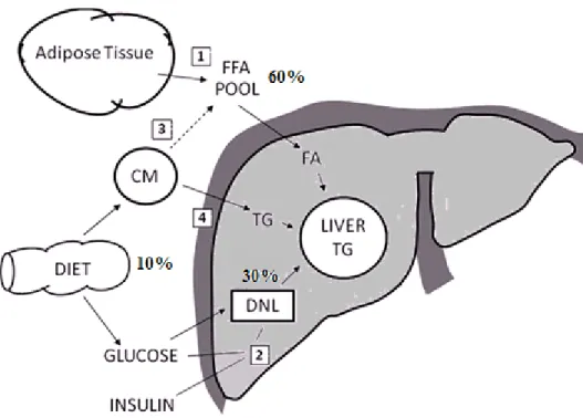
It is now proposed that the development of NAHS is closely linked to an excess flow of FFA toward the liver (Cusi 2009). Potential sources of FFA for liver fat consist of: (1) peripheral fats stored in adipose tissue that flow to the liver by the plasma FFA pool; (2) FFAs synthesized within the liver through de novo lipogenesis; (3) dietary fatty acids that are transported through CM from the intestine to the FFA pool and then to the liver; and (4)
uptake of CM remnants by the liver (Fig. 1) (Adiels, Olofsson et al. 2008; Postic and Girard 2008). Using a multiple stable isotope approach in humans, Donnelly et al. (Donnelly, Smith et al. 2005) estimated that while 60% of the accumulated TG in the hepatic steatosis conditions originates from plasma FFA pool, approximately 10% comes from the diet and about 30% from de novo lipogenesis.
Catecholamines (e.g. epinephrine and norepinephrine) and insulin are the major hormones that control lipolytic activity. On the adipocyte surface catecholamines bind to its receptor and stimulate lipolysis though β-adrenergic receptors (β-ARs) coupled to stimulatory GTP-binding protein (Gs). Activation of Gs proteins stimulates the adenylate cyclase (AC), the enzyme that catalyzes the formation of cyclic adenosine monophosphate (cAMP) (Duncan, Ahmadian et al. 2007). Increasing concentrations of cAMP in the cell activates protein kinase A (PKA) (Belfrage, Fredrikson et al. 1982), which catalyzes the phosphorylation and subsequent activation of hormone sensitive lipase (HSL) (Holm 2003; Duncan, Ahmadian et al. 2007; Granneman and Moore 2008). Activated HSL is able to break down TGs to fatty acids and glycerol (Arner and Langin 2007). The cAMP/PKA-HSL pathway has long been considered to be the only regulator of adipocyte lipolytic cascade (Langin 2006). However, a novel lipolytic pathway in human adipocytes which does not involve cAMP has been observed, this pathway acts through natriuretic peptides (NPs) (Lafontan, Moro et al. 2005). NPs bind to their specific receptors (this action will be explained in details in the section of metabolic effects of NPs) on human fat cells and subsequently activate protein kinase G (PKG), which causes phosphorylation and activation of HSL. It seems that these NPs stimulate lipolysis to the same extent as catecholamines (Arner and Langin 2007). On the other hand, insulin acts as a potent inhibitor of lipolysis via binding to its receptor. Binding of insulin to its receptor stimulates the degradation of cAMP using the enzyme phosphodiesterase 3B (PDE 3B), leading to a decreased activity of PKA and inhibition of HSL activity (Arner and Langin 2007).
It appears that insulin resistance in adipose tissue plays an important role in the pathogenesis of NAHS. In the insulin resistance state, the inhibitory action of insulin on
Figure 1. Sources of fatty acids for liver fat. Fatty acids (FA) may enter the liver via 4 different pathways: (1) FFA derived from adipose tissue, (2) hepatic de novo lipogenesis (DNL), (3) spillover of FA from lipolysis of chylomicron (CM)-triglycerides (TG) into the FFA pool, (4) and uptake of TG from CM remnants. Adapted from Adiels et al. (Adiels, Olofsson et al. 2008).
adipocyte lipolysis is impaired, thus resulting in an increase in the rate of adipocyte lipolysis and an increased influx of FFA into the liver (Browning and Horton 2004; Zak and Slaby 2008). Additionally, fatty acid flux into the plasma FFA pool is more facilitated in insulin resistance by decreasing glucose uptake in adipocytes which reduces glycerol-3-phosphate levels, thus reducing the reutilization of fatty acids for TG synthesis (Tamura and Shimomura 2005). Moreover, it has been shown that the severity of NAHS is positively related to the visceral fat accumulation independently of body mass index (Eguchi, Eguchi et al. 2006). In this regard, in vitro evidence indicates that lipolytic sensitivity to catecholamines is higher in fat cells from intra-abdominal adipose tissue than from subcutaneous fat of the gluteal/femoral region (Richelsen, Pedersen et al. 1991). Furthermore, this study shows that the antilipolytic effect of insulin is greater in fat cells from subcutaneous than from intra-abdominal adipose tissue suggesting the enhanced lipolytic activity in intra-abdominal adipose tissue. Higher fatty acid flux from this depot into the portal circulation is primarily taken up by the liver (Horowitz 2001) providing an important source of substrate for hepatocellular TG synthesis (Bradbury and Berk 2004). Nevertheless, it has been reported that the major source of FFA delivered to the liver might be derived from FFA released from subcutaneous adipose tissue during postabsorptive conditions through the systemic circulation (Fabbrini, Sullivan et al. 2010). As a whole, Adiels et al. suggest that the increased release of fatty acids to the liver from adipose tissue is the most important factor in liver fat accumulation (Adiels, Olofsson et al. 2008).
On the other hand, dietary fatty acids after entering the circulation through CM from the intestine can be taken up by the liver as CM remnants. Alternatively, lipoprotein lipase (LPL) catalyzes the release of fatty acids from the CMs at a rate that exceeds tissue uptake, resulting in a spillover of these fatty acids into the plasma FFA pool (Barrows and Parks 2006). The contribution of dietary fatty acids to liver TGs, therefore, depends on the fat content of the diet (Adiels, Olofsson et al. 2008). It has been shown that NAHS is linked to obesity for which caloric overconsumption is considered as a main factor (Festi, Colecchia et al. 2004). Since dietary fat is the most energy-dense macronutrient, with about 38 kJ/g
(in comparison, carbohydrate and protein only provide about 17 kJ/g), an increase in dietary fat intake can easily promote an increase in energy intake (Schrauwen and Westerterp 2000). It is well established that HF diets induce deleterious metabolic effects in both rodents and humans (Kraegen, Clark et al. 1991; Ghibaudi, Cook et al. 2002; Satia-Abouta, Patterson et al. 2002). Animal studies clearly indicate that the ingestion of a HF diet in sedentary rats results in obesity which is accompanied by liver lipid infiltration (Collin, Chapados et al. 2006; Gauthier, Favier et al. 2006). There are also studies in human identifying high dietary fat intake as a main contributing factor to obesity (Satia-Abouta, Patterson et al. 2002) that induced several obesity related metabolic deteriorations including liver lipid infiltration (Marchesini, Marzocchi et al. 2005; Kotronen, Westerbacka et al. 2007). Taken together, the main contributing factor for excess TG accumulation in NAHS seems to be the increased release of fatty acids from adipose tissue and dietary fatty acids, which flow to the liver via the FFA pool.
1.1.2 Metabolic pathways contributing in the development of NAHS
NAHS occurs when there is an imbalance between pathways of lipid accumulation and lipid elimination. TGs consisting of a glycerol and three long chain fatty acids (LCFA) are the main lipid component in the liver (Choi and Diehl 2008). The four pathways leading to hepatic TG accumulation include: (1) increased lipid uptake by liver (2) increased hepatic de novo lipogenesis (3) reduced hepatic oxidation of fatty acids and (4) diminished export of lipids from the liver (Fig. 2). Imbalance between these metabolic steps will increase TG accumulation within the cytoplasm of hepatocytes (Wei, Rector et al. 2008). In the next section we will explain the intra-hepatic-related metabolic pathways leading to NAHS.
Figure 2. Overview of the four main pathways involved in the development of NAHS and their regulatory factors. NAHS is characterized by (1) an increase in the uptake of lipids by the liver, (2) an increase in hepatic de novo lipogenesis (DNL), and an insufficient elimination of excess liver triacylglycerol (TAG) by means of (3) hepatic lipid oxidation and (4) very low density lipoprotein (VLDL) assembly and secretion. HSL, hormone sensitive lipase; LPL, lipoprotein lipase; FAT/CD36, fatty acid translocase/cluster of differentiation 36; SREBP-1c, sterol regulatory element-binding protein-1c; ChREBP, carbohydrate response element-binding protein; LXR, liver X receptors; PPAR, peroxisomal proliferator-activated receptors; AMPK, AMP-activated protein kinase; PGC-1α, peroxisome proliferator-activated receptor gamma coactivator-1 alpha; MTP, microsomal triglyceride transfer protein; DGAT, diacyglycerol acyltransferase; ARF-1, ADP-ribosylation factor 1; ApoB, apolipoprotein B. Taken from Lavoie et al. (Lavoie and Gauthier 2006).
1.1.2.1 Fatty acid uptake pathway
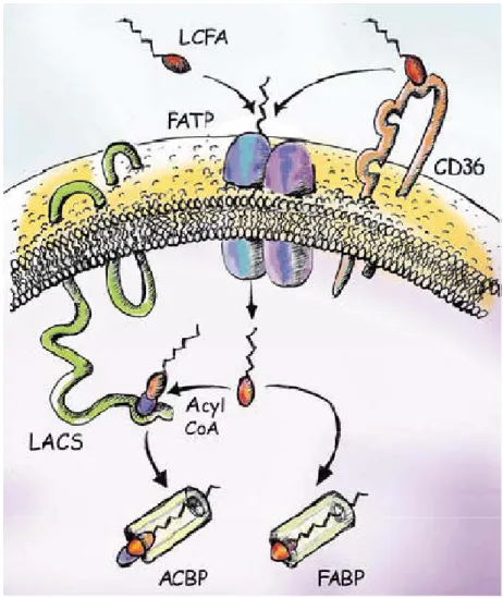
Circulating FFAs, either of dietary or endogenous origin (adipose tissue lipolysis), provide most of the hepatic lipid content in the development of NAHS (Musso, Gambino et al. 2009). It seems that the rate of hepatic FFA uptake depends on the delivery of FFA to the liver and the liver’s ability for FFA transport (Fabbrini, Sullivan et al. 2010). LCFA cross the mammalian cells either through the diffusion or the facilitated protein-mediated mechanism (Bradbury and Berk 2004). Over 90% of the LCFA uptake into tissues including hepatocytes is mediated by proteins through a facilitated mechanism (Stump, Fan et al. 2001). Several membrane proteins that are involved in the uptake of LCFA have been identified. The most important of these proteins are: fatty acid translocase (FAT, also known as cluster of differentiation 36 (CD36)), long chain fatty acyl-coenzyme A synthetases (LACS) and fatty acid transport protein (FATP). Stahl (Stahl 2004) suggested a model for LCFA uptake in which, LCFA are either transported directly by FATP complexes across the plasma membrane or alternatively, are first accumulated on the plasma membrane by binding to CD36, which subsequently gives the fatty acids to the FATP (Fig. 3). After uptake, LCFA is activated quickly by LACS to prevent efflux. In addition, binding of intracellular LCFA and acyl-CoA to fatty acid binding protein (FABP) and acyl-CoA binding proteins (ACBP) facilitate the unloading of transporters and act as an intracellular fatty acid buffer (Fig. 3) (Stahl 2004). FFA and fatty acyl-CoA bounded to FABP and ACBP transport them to intracellular compartments for metabolism or the nucleus to interact with transcription factors (Nguyen, Leray et al. 2008).
Any condition that constantly raises plasma FFA concentrations (obesity, metabolic syndrome and type 2 diabetes) will lead to increased hepatic FFA uptake (Bradbury 2006). For instance, HF diet in rats resulted in significantly higher plasma FFA leading to hepatic steatosis (Gauthier, Couturier et al. 2003). In contrast, in mice lacking HSL, plasma FFA
Figure 3. A model for cellular fatty acid uptake. Extracellular long chain fatty acids (LCFA) might directly bind to fatty acid transport protein (FATP) complexes (blue) and be transported into cells. Alternatively, LCFA could bind first to cluster of differentiation 36 (CD36) (yellow), which hands on the LCFA to FATP dimmers. Intracellular LCFA are coupled to coenzyme A (CoA) by long chain fatty acyl-CoA synthetase (LACS, green), preventing their efflux, while fatty acid binding proteins (FABP) act as a cytoplasmic buffer for incorporated LCFA (ACBP acyl-CoA binding protein). Taken from Stahl et al. (Stahl 2004).
concentration is low and as a consequence no steatosis is observed (Voshol, Haemmerle et al. 2003). In this regard, it has been shown that following a HF diet or fasting, the liver of LFABP (liver type FABP) knockout mice (LFABP-/-) were protected from steatosis while wild type mice developed fatty liver in both situations (Martin, Danneberg et al. 2003; Newberry, Xie et al. 2006). Newberry et al. (Newberry, Xie et al. 2003) also showed that in response to 48-h fasting, wild type mice demonstrated a 10-fold increase in hepatic TG content while LFABP-/- mice demonstrated only a 2-fold increase. In this last study the lower hepatic TG content observed in LFABP-/- mice was due to a reduction in fatty acids uptake by the liver in a situation of increased mobilization from adipocytes TG stores and FFA availability due to the fasted state, and not by increased hepatic TG secretion or fatty acid oxidation, since both of them were reduced in LFABP-/- mice. In another study it was reported that gene expression and/or protein content of FAT/CD36 were increased in liver of obese subjects with NAFLD compared with those who have normal intra-hepatic TG content (Greco, Kotronen et al. 2008; Fabbrini, Magkos et al. 2009).
1.1.2.2 De novo lipogenesis pathway
Fatty acid synthesis is expressed in two major tissues, liver and adipose tissues, but the relative contribution of these sites to de novo lipogenesis is variable among species. In human, it appears that the liver is the major site of de novo lipogenesis (Patel, Owen et al. 1975), while in rodents, both liver and adipose tissue are important (Pullen, Liesman et al. 1990). In adipose tissue, de novo synthesis of fatty acids contributes to fat deposition and long term energy reservoir while in the liver, synthesized fatty acids are exported via lipoprotein production, and thus provide an energy source for the body and structural component for membrane building (Nguyen, Leray et al. 2008). The de novo lipogenic pathway is highly dependent upon nutritional and hormonal conditions as it is now clearly established that insulin and glucose are required for lipogenic enzyme transcription (Foufelle and Ferre 2002). Conditions associated with high rates of lipogenesis, such as a low fat/high carbohydrate diet, hyperglycemia and hyperinsulinemia, are associated with a
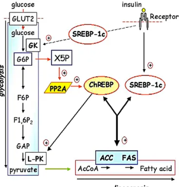
shift in cellular metabolism from lipid oxidation to TG synthesis, thereby increasing the availability of liver TG (Postic and Girard 2008). The two key transcriptional regulators in hepatic de novo lipogenesis, sterol regulatory element-binding protein-1c (SREBP-1c) and carbohydrate responsive element-binding protein (ChREBP), are respectively activated in response to insulin and glucose, and control lipogenic gene expressions such as acetyl-CoA carboxylase (ACC) and fatty acid synthase (FAS) (Fig. 4) (Dentin, Girard et al. 2005). It appears that abnormal transcription of one or two of these regulators can result in accumulation of TG in the liver. The role of SREBP-1c in the pathogenesis of fatty liver has been explored in different animal models. SREBP-1c levels are elevated in fatty livers of obese, insulin resistant and hyperinsulinaemic ob/ob mice (Shimomura, Bashmakov et al. 1999; Shimomura, Matsuda et al. 2000). Conversely, expression of lipogenic enzymes and liver fatty acid infiltration are dramatically reduced by SREBP-1c suppression (Sekiya, Yahagi et al. 2003; Teran-Garcia, Adamson et al. 2007). On the other hand, studies using ChREBP-/- rodents have indicated that hepatic ChREBP is required for the normal lipogenic response to a carbohydrate load (Iizuka, Bruick et al. 2004). Dentin et al. (Dentin, Pegorier et al. 2004) demonstrated that glycolytic and lipogeneic gene expression is synergistically regulated by SREBP-1c and glucose acting through ChREBP and showed that decreased hepatic ChREBP gene expression resulted in reducing lipogenic gene expressions of FAS and ACC. It seems like liver de novo lipogenesis is a highly regulated metabolic pathway in which transcription factors such as liver X receptor (LXR), SREBP-1c, and ChREBP play an important role over the enzymes involved in de novo synthesis of fatty acids including ACC, FAS, and stearoyl-coenzyme A desaturase-1(SCD-1) (Strable and Ntambi 2010). In humans, it has been reported by Schwarz et al. (Schwarz, Linfoot et al. 2003) that the contribution of FFA synthesized from hepatic de novo lipogenesis in liver TG formation was 4 times higher in hyperinsulinemic patients with NAFLD compared to healthy subjects. Hyperinsulinemia and hyperglycemia, often found in a context of NAFLD, and their effect on SREBP-1c and ChREBP respectively, (Girard, Perdereau et al. 1994) are therefore likely to induce an abnormal increase in lipogenic activity and
Figure 4. SREBP-1c and ChREBP act in synergy to regulate lipogenic gene expression. The phosphorylation of glucose in glucose 6-phosphate, by hepatic glucokinase, is an essential step for glucose metabolism as well as for the induction of glycolytic and lipogenic genes. The recent identification of ChREBP has shed light on the possible mechanism whereby glucose affects gene transcription. The activity of ChREBP requires a mechanism of phosphorylation/dephosphorylation which is determined by the relative activity of protein phosphatase 2A (PP2A), regulated by X5P concentrations. SREBP-1c, which is induced by insulin, also plays an important role in mediating insulin signaling on lipogenic gene expression. These two transcription factors work synergistically to induce transcription of the lipogenic genes in the presence of glucose and insulin. Adapted from Dentin et al. (Dentin, Girard et al. 2005).
contribute to the development of NAFLD. After converting fatty acids into TG, TG can then be stored as lipid droplets within hepatocytes or secreted into the blood as VLDL, but they can also be hydrolyzed and the fatty acids channeled towards the β-oxidation pathway (Postic and Girard 2008).
1.1.2.3 Hepatic lipid oxidation
Lipid oxidation is one of the lipid elimination pathways in the liver. Fatty acid oxidation is the main source of energy for skeletal muscle and the heart, while the liver oxidizes fatty acids mostly under the conditions of prolonged fasting, during illness and increased physical activity (Wei, Rector et al. 2008). Fatty acid oxidation in the liver takes place in three sub-cellular organelles: the β-oxidation occurs in mitochondria and peroxisomes, whereas ω(omega)-oxidation occurs in the smooth endoplasmic reticulum (Tessari, Coracina et al. 2009). It seems that peroxisome proliferator–activated receptor α (PPARα) plays a key role in these oxidation systems by transcriptionally controlling their important enzymes. Although mitochondria and peroxisomes have similar function (β-oxidation), there is a difference between these two pathways for fatty acid oxidation. Peroxisomal β-oxidation is responsible for the metabolism of very LCFA while mitochondrial β-oxidation is responsible for the oxidation of short, medium, and long chain fatty acids (Nguyen, Leray et al. 2008). Short and medium chain fatty acids (12 carbon or less) freely enter the mitochondria and via intra-mitochondrial oxidation results in the formation of acetyl-CoA (Wei, Rector et al. 2008). LCFA (14 carbon or more) entry into the mitochondria is regulated by the activity of the enzyme carnitine palmitoyl transferase-1 (CPT-1) (Kerner and Hoppel 2000). Therefore this enzyme is considered as a rate limiting step in the oxidation of fatty acids (Horowitz 2001). Once inside the mitochondria, the fatty acids proceed through a sequence of metabolic processes to synthesize adenosine triphosphate (ATP) for energy. Destruction in any of these processes could reduce fat oxidation resulting in liver lipid accumulation (Horowitz 2001). For instance, it has been shown that the activity of mitochondrial respiratory chain complex is decreased in the liver
of patients and animal models with NAFLD (Perez-Carreras, Del Hoyo et al. 2003; Garcia-Ruiz, Rodriguez-Juan et al. 2006). Moreover, many enzymes are implicated in mitochondrial β-oxidation and deficiency of these enzymes can lead to the development of hepatic steatosis. For instance, mice with disrupted medium chain and very long chain acyl-CoA dehydrogenase genes manifest defects in fatty acid oxidation that likely lead to the observered micro and macrovascular hepatic steatosis found in these mice (Wei, Rector et al. 2008). As we mentioned earlier, peroxisomal β-oxidation metabolizes very LCFA (> C20) (Musso, Gambino et al. 2009). Deficiency in peroxisomal β-oxidation enzymes has been recognized as an important cause of microvesicular steatosis and steatohepatitis (Fan, Pan et al. 1998). Long chain and very long chain fatty acids are also metabolized by the cytochrome P450 CYP4A ω-oxidation system to dicarboxylic acids that provide as substrates for peroxisomal β-oxidation (Reddy and Hashimoto 2001). Dicarboxylic acids are toxic for mitochondria, since they inhibit fatty acid oxidation system (Macdonald and Prins 2004). An effective peroxisomal β-oxidation system is needed to minimize the deleterious effects of dicarboxylic and other toxic fatty acids to prevent hepatic steatosis (Musso, Gambino et al. 2009).
1.1.3 Hepatic VLDL-TG production
This section on VLDL-TG production will be presented as a separate one in regards to its importance for the work presented in this thesis. Lipoproteins are particles that contribute to overall metabolic homeostasis by transporting hydrophobic lipids including TG and cholesterol ester (CE) (Dixon 1970) in the circulation to and from different tissues in the body (Mason 1998). Transport of TG throughout the body is crucial for the maintenance of whole body energy balance. A particular lipoprotein class termed VLDL is the primary vehicle synthesized by the liver for the transport of endogenous TG (Mason 1998). In the body, two types of lipoproteins mainly transport TG to peripheral tissues such as muscle and adipocytes: CM and hepatic VLDL. CMs produced by enterocytes transport
the majority of TG from nutrition while VLDL synthesized and secreted by hepatocytes transport TG from the liver (Gibbons, Wiggins et al. 2004).
For many years it has been thought that the major role of VLDL was the effective control of the plasma glucose concentration by transporting de novo fatty acids synthesized in liver toward adipose tissue for storage as TG (Schwarz, Linfoot et al. 2003). This view has been challenged by the fact that, even under conditions in which hepatic de novo lipogenesis is high, the hepatic VLDL-TG is not derived from this source but from pre-formed fatty acids entering the liver from adipose tissue (Gibbons 1990; Hellerstein, Schwarz et al. 1996). VLDL-TG is hydrolyzed by LPL to fatty acids which are stored in adipose tissue. Therefore, liver plays a very important protective role in efficiently modulating plasma FFA concentrations. Liver, as a valuable buffer, removes fatty acids from the circulation, temporarily stores them as a benign derivative (TG) and secretes them at a later time as VLDL when the period of maximum danger is passed (Gibbons, Wiggins et al. 2004). However, although fat may accumulate in liver substantially, the capacity of liver tissue for TG storage is limited (Berk and Stump 1999). It seems that higher liver lipid concentrations resulting from enhanced entrance of fatty acids, high rate of de novo lipogenesis, down-regulated fatty acid oxidation (or their combinatory effect) increases the secretion of VLDL-TG from the liver (McGarry, Mannaerts et al. 1977; Schwarz, Linfoot et al. 2003). The importance of hepatic TG for the assembly and secretion of VLDL is supported by in vivo observations that the secretion of VLDL increases with increasing hepatic concentrations of lipids (Adiels, Taskinen et al. 2006).
1.1.3.1 Mechanism of hepatic VLDL-TG production
The hepatic VLDL-TG production is a complex mechanism that involves several regulatory molecules and enzymes, which takes place in the membrane and lumen of endoplasmic reticulum (ER: the secretory apparatus) (Gibbons, Wiggins et al. 2004) (Fig. 5). In the process of VLDL assembly, the newly formed cytosolic TG pool from extracellular fatty acids or de novo synthesized endogenous fatty acids, is not directly
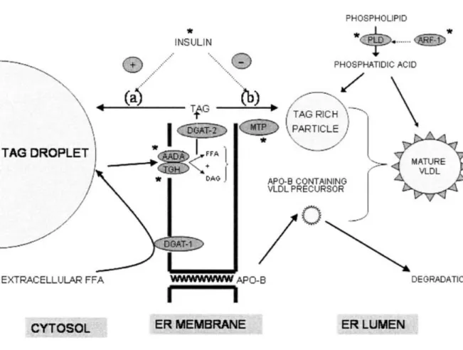
Figure 5. Role of the cytosolic and microsomal pools of liver TG in the assembly of VLDL in ER. For explanation refer to text and see list of abbreviations for meaning of acronyms. Taken from Lavoie et al. (Lavoie and Gauthier 2006).
incorporated into the VLDLs. Extracellular fatty acids from the plasma come into the hepatocyte and are esterified by diacylglycerol acyltransferase-1 enzyme (DGAT-1, also called overt DGAT) producing TG which is stored in the cytosol (cytosolic TG pool). Most of the TG utilized for the assembly of VLDL in the ER of the hepatocyte, is mobilized by lipolysis of the cytosolic TG pool (through the lipolytic action of arylacetamide deacyclase (AADA) and TG hydrolase (TGH)). The products of lipolysis are re-esterified by diacylglycerol acyltransferase-2 enzyme (DGAT-2, also called latent DGAT) producing the microsomal TG pool in ER membrane (Fig. 5). Some of these microsomal TGs are recycled to the cytosol and some are channeled into a TG-rich VLDL precursor. The formation of this precursor is positively regulated by MTP and negatively regulated by insulin (Gibbons, Wiggins et al. 2004).
A useful working model of VLDL assembly originates from the data of Alexander et al. in 1976 (Alexander, Hamilton et al. 1976). This model proposes that TG becomes associated with apolipoprotein B (apoB) in at least two distinct stages of the assembly process (Shelness and Sellers 2001). In the first stage, a small quantity of TG is transferred to apoB in the ER forming small apoB-containing VLDL precursors which is dependent upon MTP (Rustaeus, Stillemark et al. 1998). The second stage of VLDL formation, referred as the maturation phase, is characterized by the fusion of the apoB-containing VLDL precursor with a larger droplet of TG resulting in a ready for secretion mature VLDL (Rava, Ojakian et al. 2006). The details of this process remain unclear, but it seems that the formation of TG-rich particle and fusion of this molecule with the VLDL precursor is dependent on the activity of the ADP-ribosylation factor-1 protein (ARF-1) that activates phospholipase D (PLD) to form phosphatidic acid (Gordon 1997; Asp, Claesson et al. 2000) (Fig. 6). It has been shown that inhibition of ARF-1 slows down the maturation phase of VLDL assembly without affecting the formation of the apoB-containing precursor (Rustaeus, Lindberg et al. 1995). VLDL particles are mostly composed of TG (60%), phospholipids (15%), cholesterol (15%) and proteins (10%) (Gordon, Wetterau et al. 1995). Secretion of hepatic VLDL is an effective way for eliminating hepatic TG, thus preventing
Figure 6. Targets for the regulation of the TG-rich particle precursor of VLDL. Phosphatidic acid formed by the ARF-1-mediated activation of phospholipase D (PLD) contributes TG either to the TG-rich VLDL precursor particle or to the mature VLDL. Taken from Gibbons et al. (Gibbons, Wiggins et al. 2004).
potential liver TG accumulation. Accordingly, any alteration in the regulation of this mechanism could contribute to the development of NAHS.
1.1.3.2 Molecular mediators of hepatic VLDL-TG production 1.1.3.2.1 Microsomal triglyceride transfer protein (MTP)
MTP is a heterodimer protein complex primarily present in the lumen of the endoplasmic reticulum in hepatocytes and enterocytes. It consists of a unique large (97-kDa) subunit and a smaller multifunctional subunit (58-(97-kDa) which is identical to protein disulfide-isomerase (PDI) (White, Bennett et al. 1998; Mohler, Zhu et al. 2007). The large subunit possesses lipid transfer activity on the complex (Jamil, Dickson et al. 1995; Mohler, Zhu et al. 2007) and PDI mediates the folding of the large apoB protein during translation (van Greevenbroek, van Meer et al. 1996). The complex is responsible for the transport of neutral lipid (TG and CE) between the phospholipid surfaces of the ER (Gordon, Jamil et al. 1994; Benoist, Nicodeme et al. 1996). It is essential for the assembly of VLDLs by liver and CMs by small intestine (Gordon and Jamil 2000; Hussain, Shi et al. 2003). Synthesis of TG and CE is beneficial in avoiding toxicities associated with excess FFA and free cholesterol (Hussain and Bakillah 2008). MTP is expressed primarily in tissues that synthesize apoB containing lipoproteins such as liver and small intestine (Shoulders, Brett et al. 1993; Nakamuta, Chang et al. 1996). Its expression in other tissues including myocardium, ovary, testis, kidney, and retina as well as mouse brown and white adipose tissue has also been reported (Shoulders, Brett et al. 1993; Nielsen, Perko et al. 2002; Li, Presley et al. 2005; Swift, Kakkad et al. 2005). Since some of these tissues do not express apoB, MTP might be implicated in other aspects of lipid trafficking or storage (Mohler, Zhu et al. 2007).
MTP lipid transfer activity is involved in importing TGs into the lumen of the ER. In addition to its lipid transfer activity, MTP physically interacts with apoB (Hussain, Iqbal et al. 2003). The absolute requirement for the role of MTP in VLDL-TG assembly is shown by the clinical condition of abetalipoproteinaemia, a recessive genetic disease in humans
characterized by a mutation of the MTP gene (Gregg and Wetterau 1994), and from experimental studies in liver-specific MTP knockout mice (Raabe, Veniant et al. 1999). The absence of MTP in abetalipoproteinemia patients results in concentrations of plasma apoB undetectable in these patients (Wetterau, Aggerbeck et al. 1992; Shoulders, Brett et al. 1993). Similarly, the deletion of the MTP gene in mice liver resulted in impaired secretion of VLDL (Raabe, Flynn et al. 1998; Raabe, Veniant et al. 1999). Moreover, a polymorphism of this gene in human has been associated with the development of NAFLD (Gambino, Cassader et al. 2007).
1.1.3.2.2 Apolipoprotein B (ApoB)
Circulating plasma TG is a mixture of lipoprotein which is derived either from intestine (CMs) or the liver (VLDL). Lipoproteins are characterized by different densities and apoprotein compositions. A critical element of VLDL is a large protein of apoB which seems to preserve the structural integrity of the lipoprotein by associating both with the outer hydrophilic shell and with the hydrophobic core (Dixon and Ginsberg 1993; Gruffat, Durand et al. 1996). ApoB can be found in two types: apoB100 and apoB48. The name of apoB48 comes from the fact that its molecular weight is approximately 48% of apoB100. In human, apoB100 is associated with VLDL and is synthesized exclusively in the liver, while apoB48 is synthesized exclusively by enterocytes (intestinal absorptive cells) and is associated with CMs (Krishnaiah, Walker et al. 1980; Chen, Habib et al. 1987; Yao and McLeod 1994). ApoB is synthesized in the ER of hepatocytes where it is combined with lipids stored in liver (predominantly with TG) to form VLDL particles as it passes through the secretory pathway and is secreted into plasma as a lipid-rich lipoprotein particle (Mason 1998). It appears that lipids contained in VLDL cannot be secreted in the absence of apoB (Cartwright and Higgins 1996). When the availability of TG is not sufficient for the formation of VLDL, apoB is degraded and VLDL particle formation is reduced (Mason 1998). A genetic defect in apoB100 has been shown to be associated with impaired
VLDL-TG production resulting in the excess deposition of VLDL-TG in the liver (Badaloo, Reid et al. 2005; Schonfeld, Yue et al. 2008).
1.1.3.2.3 Diacylglycerol acyltransferase (DGAT)-1 and-2
DGAT is an endoplasmic reticulum membrane-associated enzyme that catalyzes the final step of TG synthesis by facilitating the linkage of 1, 2 diacylglycerol (DAG) to a long chain fatty acyl-CoA. There are two isoforms of the enzyme: the overt (visible) type (on the cytosolic side of ER membrane; DGAT-1) that catalyzes the synthesis of TG destined to cytoplasmic droplets, and latent (invisible) type (on the lumen side of ER membrane; DGAT-2) that catalyzes the TG synthesis for VLDL formation (Owen, Corstorphine et al. 1997). DGAT-2 is expressed primarily in the liver, intestine, and white adipose tissue, whereas DGAT-1 is expressed in all tissues (Cases, Smith et al. 1998; Cases, Stone et al. 2001). It has been shown that greater activities of both DGATs are implicated in the increased rate of hepatic TG secretion and intracellular accumulation of TG in ob/ob, suggesting causal importance of both DGATs for the steatosis and hypertriglyceridemia observed in the ob/ob genotype (Waterman and Zammit 2002).
However, the precise role of each enzyme in hepatic TG synthesis and VLDL secretion is unclear. Since it appears that cytosolic droplet TG cannot be incorporated directly into VLDL formation, the relative activities of these two functions of DGAT may have a significant impact on the level of triglyceridemia as well as on the development of hepatic steatosis (Yamazaki, Sasaki et al. 2005). DGAT-1 lacking mice have normal liver and plasma TG levels but are resistant to HF diet induced obesity through a mechanism involving increased energy expenditure as well as enhanced sensitivity to insulin and leptin (Smith, Cases et al. 2000). In contrast, DGAT-2 deficient mice which die shortly after birth because of lipopenia (an abnormally small amount or a deficiency of lipids in the body) and severe skin abnormality have large reductions in carcass, liver, and plasma TG as well as plasma FFA and glucose (Stone, Myers et al. 2004). Recently, Liu et al. showed that inhibition of DGAT-2 in wild type mouse liver resulted in decreased VLDL secretion in a
dose dependent manner and reduced plasma TG, total cholesterol, and apoB (Liu, Millar et al. 2008). In this last study, inhibition of DGAT-2 in DGAT-1 knockout mice produced the same effect. This indicates that DGAT-2 is the isoform responsible for synthesizing TG targeted for secretion and that presence and absence of DGAT-1 does not affect the process.
1.1.3.3 Regulation of hepatic VLDL-TG production
The regulation of hepatic VLDL-TG production depends mainly on lipid availability, activity of molecular mediators, mostly key MTP enzyme, and insulin (Julius 2003). The importance of the VLDL-TG production’s molecular mediators including MTP has been reviewed earlier. In the next section the two other important regulators, lipid availability and insulin, are discussed.
1.1.3.3.1 Liver lipid availability
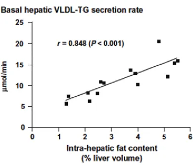
For lipoprotein synthesis, four sources of fatty acids are used: de novo lipogenesis, cytoplasmic TG stores, fatty acids derived from lipoproteins taken up directly by the liver, and plasma FFA (Julius 2003). It appears that de novo lipogenesis plays a minor role in regulating VLDL synthesis. However, it is clearly elevated under conditions of high carbohydrate intake. On the other hand, plasma FFAs which seem to play an important role in hepatic TG storage (Diraison and Beylot 1998) also stimulate hepatic VLDL production. It has been shown that an increased delivery of fatty acids increases the secretion of VLDL-TG from the liver tissue (Lewis, Uffelman et al. 1995; Lewis 1997). Moreover, the importance of liver fat content for the assembly and secretion of VLDL has been demonstrated by in vivo turnover studies (Adiels, Taskinen et al. 2006). Secretion of VLDL increases with increasing concentrations of liver lipids and cytoplasmic TG stores appear to fundamentally contribute to VLDL-TG (Adiels, Olofsson et al. 2006; Fabbrini, Mohammed et al. 2008) (Fig. 7). In fact, the relationship between oxidation and esterification of fatty acids in hepatocytes appears to be important in regulating the VLDL synthesis: an enhanced esterification is accompanied by increased VLDL
Figure 7. Relationship between liver fat and basal hepatic VLDL-TG secretion. Basal VLDL-TG secretion rate increases linearly with increasing amount of intra-hepatic fat content within the normal range of liver fatness. Taken from Magkos (Magkos 2009).
secretion (Julius 2003). In addition, it has been suggested that hepatic TG concentration may positively regulate hepatic MTP activity and gene expression (Taguchi, Omachi et al. 2002).
1.1.3.3.2 Insulin and hepatic VLDL-TG production
Studies that investigated the acute effect of insulin on VLDL kinetics indicate a decreased secretion of VLDL-TG (Lewis, Uffelman et al. 1993). Insulin seems to stimulate the suppression of some factors that are responsible for the normal transfer of the newly mobilized TG pool into the TG-rich VLDL precursor (Wiggins and Gibbons 1992; Gibbons, Wiggins et al. 2004) (Fig. 5 and 6). In general, insulin decreases VLDL formation by two mechanisms: (A) indirectly by regulating the amount of fatty acids in the circulation, and (B) by direct suppression of the production of VLDL in the liver, independently of the availability of fatty acids (Malmstrom, Packard et al. 1998). Since the VLDL production is regulated by the availability of intra-hepatic substrates, insulin may indirectly interfere with the production of hepatic VLDL by its anti-lipolytic effect on adipose tissue (Coppack, Jensen et al. 1994). Acute hyperinsulinemia in humans suppressed plasma FFA, inhibiting VLDL-TG production (Lewis, Uffelman et al. 1993). On the other hand, several mechanisms have been proposed for the molecular mechanisms involved in the direct suppression of VLDL by insulin. For instance, it has been shown that insulin can directly reduce the MTP gene expression via negatively regulating promoter region of MTP (Lin, Gordon et al. 1995), thus decreasing the rate of synthesis and secretion of VLDL. It seems that insulin down-regulates MTP expression through activation of the mitogen activated protein kinase (MAPK) pathway (Allister, Borradaile et al. 2005). Insulin also suppresses hepatic VLDL secretion by directly interfering with the maturation phase of VLDL assembly by the activation of phosphoinositide 3-kinase (PI3-K) in rat hepatocytes (Sparks and Sparks 1994; Sparks, Phung et al. 1996; Phung, Roncone et al. 1997). In agreement with these observations, results reported by Brown and Gibbons using labeled method in cultured rat hepatocytes suggest that insulin signaling via PI3-K inhibited the
maturation phase of VLDL assembly by preventing bulk lipid transfer to a VLDL precursor, thus enhancing the degradation of apoB (Brown and Gibbons 2001). Moreover, Lin et al. showed that the mRNA levels and secretion rate of apoB were decreased by 31% and 43% respectively, when cultured hepatocytes were incubated with insulin (Lin, Gordon et al. 1995).
1.1.3.4 Exercise and hepatic VLDL-TG production
Regular exercise has broad beneficial effects on the lipoprotein profile (Kraus, Houmard et al. 2002). It is well known that exercise training results in lowering plasma TG concentration in obese/overweight (Kelley, Kelley et al. 2005) and also in healthy subjects (Kelley, Kelley et al. 2004; Kelley and Kelley 2006). Since hepatic VLDL-TG production is a component of plasma TG concentration, it is reasonable to think that hepatic VLDL production is reduced following exercise training, thus resulting in improved plasma TG. In animals, it has been shown that exercise training is associated with reduced rate of hepatic VLDL-TG secretion (Simonelli and Eaton 1978; Lira, Tavares et al. 2008). In support of these results, the effects of interval aerobic training on in vivo hepatic VLDL production in human has been recently reported (Tsekouras, Magkos et al. 2008). After two months of exercise training, a 35% decrease in VLDL-TG secretion rate in the exercise group compared to the non-exercise control group was observed. The effects of exercise training on decreased intra-abdominal adipose tissue and liver fat along with increased insulin sensitivity are probably the primary mechanisms whereby exercise training could bring about a decrease in hepatic VLDL secretion. Decreased intra-abdominal fat is expected to limit the delivery of FFA to the liver through the portal vein (Nielsen, Guo et al. 2004) thus in turn leading to lower VLDL-TG secretion (Bjorntorp 1990; Chan, Barrett et al. 2004). However, the reduction in VLDL-TG secretion after exercise training occurs even in the absence of changes in body weight and body composition (Tsekouras, Magkos et al. 2008). Furthermore, high levels of physical activity are inversely associated with liver fat accumulation (Perseghin, Lattuada et al. 2007; Spassiani and Kuk 2008; Zelber-Sagi,
Nitzan-Kaluski et al. 2008) the latter being directly associated with basal VLDL-TG secretion rate (Fabbrini, Mohammed et al. 2008). In this regard, it has been suggested that stimulated lipid oxidation and reduced lipid synthesis in liver by exercise via possible mechanisms such as activation of AMP-activated kinase pathway, might play a role in VLDL-TG production (Lavoie and Gauthier 2006). Nevertheless, the effects of exercise training on liver TG and VLDL-TG production require further investigation especially in light of their important role in metabolic deregulation.
To our knowledge, little is known on the effects of exercise training on the intra-hepatic regulation of VLDL production and enzymes involved such as MTP. In two recent studies in animals, a 60% reduction in hepatic MTP gene expression (Lira, Tavares et al. 2008) and a 25% reduction in hepatic MTP protein content with exercise training have been reported (Chapados, Seelaender et al. 2009). Moreover, reduced VLDL-apoB100 secretion rate and total apoB100 after a single, prolonged bout of moderate-intensity endurance exercise has been reported (Magkos, Wright et al. 2006). The exact mechanisms underlying these observations are not clear, however, it is speculated a possible role of insulin sensitivity following exercise training. Since whole-body as well as liver insulin sensitivity is increased after either a single bout or chronic exercise training (Devlin, Hirshman et al. 1987; Mikines, Sonne et al. 1988; Perseghin, Price et al. 1996; Magkos, Tsekouras et al. 2008), it is possible that insulin-sensitization after exercise reduces plasma FFA availability (Magkos, Mohammed et al. 2009) and brings temporal decrease in MTP protein content in the liver (Kamagate and Dong 2008). Therefore, this would be consistent with reduced hepatic VLDL-TG secretion (Tsekouras, Magkos et al. 2008) and VLDL-apoB100 (Alam, Stolinski et al. 2004) observed 2-3 days after exercise training in human. However, the counteracting effect of increased post-exercise FFA availability should be considered in the hepatic VLDL-TG secretion process (Magkos, Mohammed et al. 2009). It seems that most of the suppressing action of insulin on hepatic VLDL-TG secretion is mediated by the diminution in plasma FFA availability (Lewis, Uffelman et al. 1995). In this regard, it has been shown that greater fatty acid availability after exercise does not stimulate VLDL-TG