Université de Montréal
DETECTION AND QUANTIFICATION OF STAPHYLOCOCCUS AUREUS ENTEROTOXIN B IN FOOD PRODUCT USING ISOTOPIC DILUTION
TECHNIQUES AND MASS SPECTROMETRY
par
KHANH DANG BAO
Département de biomédecine vétérinaire
Faculté de médecine vétérinaire
Mémoire présenté à la Faculté de médecine vétérinaire
en vue de l’obtention du grade de
Maître ès Sciences (M.Sc.)
en sciences vétérinaires
option pharmacologie
Mai, 2012
RÉSUMÉ
L’entérotoxine B staphylococcique (SEB) est une toxine entérique hautement résistante à la chaleur et est responsable de plus de 50 % des cas d’intoxication d’origine alimentaire par une entérotoxine. L’objectif principal de ce projet de maîtrise est de développer et valider une méthode basée sur des nouvelles stratégies analytiques permettant la détection et la quantification de SEB dans les matrices alimentaires. Une carte de peptides tryptiques a été produite et 3 peptides tryptiques spécifiques ont été sélectionnés pour servir de peptides témoins à partir des 9 fragments protéolytiques identifiés (couverture de 35 % de la séquence). L’anhydride acétique et la forme deutérée furent utilisés afin de synthétiser des peptides standards marqués avec un isotope léger et lourd. La combinaison de mélanges des deux isotopes à des concentrations molaires différentes fut utilisée afin d’établir la linéarité et les résultats ont démontré que les mesures faites par dilution isotopique combinée au CL-SM/SM respectaient les critères généralement reconnus d’épreuves biologiques avec des valeurs de pente près de 1, des valeurs de R2 supérieure à 0,98 et des coefficients de variation (CV%) inférieurs à 8 %. La précision et l’exactitude de la méthode ont été évaluées à l’aide d’échantillons d’homogénat de viande de poulet dans lesquels SEB a été introduite. SEB a été enrichie à 0,2, 1 et 2 pmol/g. Les résultats analytiques révèlent que la méthode procure une plage d’exactitude de 84,9 à 91,1 %. Dans l’ensemble, les résultats présentés dans ce mémoire démontrent que les méthodes protéomiques peuvent être utilisées efficacement pour détecter et quantifier SEB dans les matrices alimentaires.
Mots clés : spectrométrie de masse; marquage isotopique; protéomique quantitative;
ABSTRACT
Staphylococcal enterotoxin B is a highly heat-resistant enteric toxin and it is responsible for over 50% of enterotoxin food poisoning. It represents a particular challenge during food processing since, even if the bacteria have been destroyed, the biological activity of the toxin remains unchanged. The objective of this study was to develop and validate a new method based on a novel proteomic strategy to detect and quantify SEB in food matrices. Tryptic peptide map was generated and 3 specific tryptic peptides were selected and used as surrogate peptides from 9 identified proteolytic fragments (sequence coverage of 35%). Peptides were label with light and heavy form of acetic anhydride to create an isobaric tag that will allow quantification. The linearity was tested using mixtures of different molar ratios and the results showed that measurements by LC-MS/MS were within generally accepted criteria for bioassays with slope values near to 1, values of R2 above 0.98 and less than 8% coefficient of variation (%CV). The precision and accuracy of the method were assessed using chicken meat homogenate samples spiked with SEB at 0.2, 1 and 2 pmol/g. The results indicated that the method can provide accuracy within 84.9 – 91.1% range. Overall, the results presented in this thesis show that proteomics-based methods can be effectively used to detect, confirm and quantify SEB in food matrices.
Keywords: mass spectrometry; stable isotope labeling; quantitative proteomics;
TABLE OF CONTENTS
RÉSUMÉ ... iii
ABSTRACT ... iv
TABLE OF CONTENTS ... v
LIST OF FIGURES ... viii
LIST OF ABBREVIATIONS ... ix
ACKNOWLEDGMENTS ... xii
Chapter 1. INTRODUCTION ... 1
Chapter 2. LITERATURE REVIEW ... 4
2.1. Microbiology of staphylococci ... 5
2.1.1. Historical background ... 5
2.1.2. Taxonomy ... 5
2.1.3. Biochemical and metabolic characteristics ... 6
2.2. Epidemiology ... 7
2.3. Virulence factors of staphylococci ... 8
2.3.1. Overview of staphylococcal virulence factors ... 8
2.3.2. Staphylococcal toxins ... 8
2.3.2.1. Hemolysins ... 9
2.3.2.2. Leukocidin ... 10
2.3.2.3. Exfoliative toxins A and B ... 11
2.3.2.4. Toxic shock syndrome toxin-1 ... 12
2.3.3. Staphylococcal enterotoxins ... 12
2.3.3.1. Structure and Properties ... 12
2.3.3.2. Enterotoxin gene location ... 17
2.3.3.3. Environmental factors that affect staphylococcal enterotoxin production ... 18
2.4. Food poisoning an overview ... 19
2.4.1. Food safety activities ... 19
2.4.1.2. Food safety activities in Vietnam ... 20
2.4.2. Staphylococcal food poisoning ... 23
2.5. Analytical method for the detection of S. aureus related toxins ... 24
2.5.1. Bioassays ... 24
2.5.2. Molecular tools ... 25
2.5.3. Immunological tools ... 26
2.5.3.1. Gel diffusion ... 26
2.5.3.2. Radioimmunoassay ... 27
2.5.3.3. Enzyme linked immunosorbent assay ... 28
2.5.3.4. Reversed passive latex agglutination assay ... 30
2.5.4. Mass-spectrometry based methods ... 31
2.6. Analytical strategies ... 32
2.6.1. Proteomic methods used in mass spectrometry ... 32
2.6.2. Liquid chromatography- Mass spectrometry ... 33
2.6.2.1. General overview ... 33 2.6.2.2. Electrospray ionisation ... 34 2.6.2.3. Mass analyzers ... 36 2.6.3. Peptide separation ... 38 2.6.4. Peptide identification ... 38 2.6.5. Peptide quantification ... 40 OBJECTIVES ... 42
Chapter 3. Analysis of Staphylococcus Enterotoxin B using Differential Isotopic Tags and Liquid Chromatography Quadrupole Ion Trap Mass Spectrometry ... 43
Chapter 4. GENERAL DISCUSSION ... 77
Chapter 5. CONCLUSIONS ... 87
LIST OF TABLES
Literature review
Table 1. Genus Staphylococcus: coagulase-positive species………...6 Table 2. Grouping of SEs and SEls based on amino acid sequence comparisons...…...13 Table 3. General properties of SEs and SEls………...………...15 Table 4. Reported foodborne disease in Vietnam from 2006-2010………...….21 Table 5. Distribution of food poisoning outbreaks in Vietnam 2009-2010………...….22
Article
Table 1. The mass transitions for quantitation in MRM mode………...65 Table 2. Summary of peptides obtained following the digestion of SEB with Trypsin………66 Table 3. Summary of precision and accuracy data for SEB determination in chicken meat……….67
LIST OF FIGURES
Literature review
Figure 1. 3D structure of SEB………..16 Figure 2. Staphylococcal enterotoxin B sequence………16 Figure 3. Nomenclature for the product ions generated in the fragmentation of peptide molecules by tandem mass spectrometry……….40 Figure 4. Generic reaction of tryptic peptide N-terminal modifications with acetic anhydride (Ac2O) and introduction of differential isotopic tag………41
Article
Figure 1. Generic reaction of tryptic peptide N-terminal Modifications with Acetic Anhydride (Ac2O) and Introduction of Differential Isotopic tag……….…...69 Figure 2. Full Scan LC-MS Chromatogram of SEB Tryptic Peptides……….70 Figure 3. Representative chromatograph of Acetylated SEB Tryptic Peptides Mixed at a
molar 1/1 (1H/2H) ...……….71
Figure 4. Product ion spectra of targeted Ac2O-derivatized tryptic peptides…………..72 Figure 5. Representative blank and LOQ chromatogram for (A) LGNYDNVR, (B)
IEVYLTTK and (C) FTGLMENMK………...……...73
Figure 6. LC-MS/MS Quantitative Analysis of Selected SEB Tryptic Peptides Labeled with 1H6-Ac2O or 2H6-Ac2O using a Reference Internal Standard Strategy………76
LIST OF ABBREVIATIONS
agr Accessory genome regulator
APC Antigen-Presenting cell
CA-MRSA Community-associated methicillin resistant S. aureus
CID Collision-induced dissociation
CPL-SM/SM Chromatographie en phase liquide- Spectrométrie de masse/Spectrométrie de masse
CPS Coagulase Positive Staphylococci
Da Dalton
ELISA Enzyme-linked immunosorbent assay
ESI Electrospray ionization
ETs Exfoliative toxins
FAO Food and Agricultural Organization
FBD Foodborne disease
FTICR Fourier transform ion cyclotron resonance
GC Gas chromatography
HPLC High performance liquid chromatography
LC Liquid chromatography
LC-MRM-MS Liquid chromatography-Multiple reaction monitoring-Mass spectrometry
LOD Limit of detection
LOQ Limit of quantitation
MHC class II Major histocompatibility complex class II
MODS Multiple organ dysfunction syndrome
MRM Multiple Reaction Monitoring
MRSA Methicillin Resistant Staphylococcus aureus
MS Mass spectrometry
m/z Mass to charge ratio
ng nanogram
PCR Polymerase Chain Reaction
pmol picomole
PVL Panton Valentine Leukocidin
RIA Radioimmunoassay
RPLA Reversed passive latex agglutination assay
SAgs Superantigens
SaPIs Staphylococcus aureus pathogenicity islands S. aureus Staphylococcus aureus
SCC Staphylococcal cassette chromosome
SE Staphylococcal enterotoxin
se Enterotoxin genes
SEB Staphylococcal enterotoxin B
SEl Staphylococcal enterotoxin like proteins
SFP Staphylococcal food poisoning
SIS Synthetic internal standard
SSSS Staphylococcal scalded skin syndrome
TCR T cell Receptor
TOF Time of flight
TSS Toxic Shock Syndrome
VFA Vietnam Food Administration
VISA Vancomycin-intermediate S. aureus
VRSA Vancomycin resistant S. aureus
WHO World Health Organization
%CV Coefficient of variation (precision)
ACKNOWLEDGMENTS
First of all, I would like to express my special thanks to my director Dr Francis Beaudry for his important guidance, encouragement, patience and invaluable advices which made this thesis a reality. I do not know what I would have done without you. Thank you for all.
I would also like to give my sincere appreciation to my Co-director Dr Ann Letellier for providing supports and valuable comments for my study and qualified research works. My acknowledgement also goes to Dr Sylvain Quessy, the program director for organizing this project which not only gives me a chance of working this thesis but also give me to better understand the cultures and people of Canada.
I want to thanks all the staff of FAPQDCP-CIDA project especially Mrs Hélène Bergeron, my second mother who helped me a lot. Your care, supports have made an unforgettable moment and made my stay so nice during the past two years.
I would like to thanks to Floriane Pailleux, Mohamed Rhouma, my best friends for all the wonderful times spent both inside and outside the laboratory. Appreciation also goes to Vietnamese students in Canada for providing help and encouragements during the entire period of completing thesis.
I would like to send my deepest thanks to my beloved family, my parents, without them I would not be the person I am today.
Finally, I owe special gratitude to my wife, whom without her full love and supports, I would never achieved fruitful results. I wish to dedicate my result to my beloved family members.
In recent years, foodborne diseases (FBD) have been a widespread and growing public health problem and a financial burden for the public and the food industry (Sockett and Todd, 2000). More than 250 known diseases can be transmitted through food and bacteria are the most common reasons of FBD outbreaks (Le Loir et al., 2003). Among these, Staphylococcus aureus (S. aureus) is a one of the major pathogenic bacteria which causes gastroenteritis resulting from the consumption of staphylococcal enterotoxins (SEs) produced in contaminated food (Hennekinne et al., 2009; Chiang et al., 2008). Consumption of food contaminated with SEs of S. aureus results in the onset of acute gastroenteritis within 2-6h (Seo and Bohach, 2007; Murray, 2005). The symptoms associated with staphylococcal food poisoning are characterized by nausea, vomiting, abdominal cramps and headache (Hennekinne et al., 2010; Pinchuk et al., 2010; Balaban and Rasooly, 2000). The illness usually resolves within 24h. Improper food handling is the most common reason of contamination and consequently, S. aureus enters the food chain during preparation and handling (Pinchuk et al., 2010). Additionally, SEs are also responsible for toxic shock syndromes and other conditions frequently involved in allergic and autoimmune diseases (Vasconcelos and Cunha, 2010; Balaban and Rasooly, 2000). In order to better recognize food poisoning related to SEs, the identification and quantification of SEs in food is important and therefore analytical assays need to be specific, sensitive, accurate and precise.
Staphylococcal enterotoxin B (SEB) is an exotoxin produce by S. aureus (Arvidson and Tegmark, 2001). SEB is a single polypeptide chain containing a total of 239 amino acid residues with one disulfide bond (Spero et al., 1973). The molecular weight of SEB is 28,336 Da (Hennekinne et al., 2010). It belongs to a family of microbial proteins called ‘‘pyrogenic toxin superantigens’’ (Chiang et al., 2008). SEB is a highly heat resistant
enteric toxin (Pinchuk et al., 2010; Balaban and Rasooly, 2000). Although there are more than 20 different SEs only a few of them have been clearly understood and SEB is one of the most common toxins associated with food poisoning (Pinchuk et al., 2010). Moreover, SEB is also considered as agents of biological warfare (Ahanotu et al., 2006). During the 1960s, the USA deployed an offensive biological warfare program and SEB was one of the agents studied. SEB was an attractive agent due to low quantities requires to trigger acute poisoning (Ulrich et al., 1997). Extensive researches have been conducted in the area of detection of enterotoxins in food resulting in the development of radioimmunoassay and enzyme linked immunosorbernt assay methods (ELISA) (Bennett, 2005; Candlish, 1991). However, these methods are not used for the quantitative determination of enterotoxins but rather as a detection tool. Consequently, the development of a rapid, sensitive, selective, accurate and precise method for the direct detection and quantification of enterotoxins in food is needed (Vasconcelos and Cunha, 2010).
The purpose of this research project is to demonstrate that proven targeted quantitative proteomic strategies can be applied for the quantification of S. aureus enterotoxin B in food products. The method will use the principles of an isotope dilution technique using surrogate tryptic peptides to quantify SEB by liquid chromatography tandem mass spectrometry. Specific tryptic peptides of SEB will be labeled with a non-isobaric amine labeling reagent to create internal standards used to develop a robust and reproducible Liquid Chromatography-Multiple Reaction Monitoring-Mass Spectrometry (LC-MRM-MS) assay. The project includes SEB tryptic mapping and molecular characterization of selected tryptic peptide based on MS/MS analysis. Moreover, method linearity, precision and accuracy will be assessed and compared with generally accepted criteria.
2.1. Microbiology of staphylococci
2.1.1. Historical background
The genus Staphylococcus belongs to the bacterial family of Staphylococcaceae; they are gram-positive and catalase-positive bacteria, nonsporulating, nonmotile and forming grape-like clusters when observed under the microscope (Vasconcelos and Cunha, 2010; Bennett and Monday, 2003; Le Loir et al., 2003). The organism was first described in 1881 by Alexander Ogston in purulent infections (Bergdoll and Wong, 2006). After microscopic analysis, Ogston discovered grape-like clusters of round, golden cells (Ogston, 1881). In 1884, Rosenbach described staphylococci according to the colony types: the pigmented type of the cocci which produced yellow colonies, called Staphylococcus aureus (S. aureus) and the other produced non-pigmented or white colonies, named Staphylococcus albus (Cowan et al., 1954). The latter species is now known as Staphylococcus epidermidis forming relatively small white colonies (Bergdoll and Wong, 2006).
2.1.2. Taxonomy
Coagulase has been used to distinguish between different types of Staphylococcus isolates and allowed the classification of 50 species and subspecies (Hennekinne et al., 2010, Vasconcelos and Cunha, 2010). Coagulase producing staphylococci strains are divided into two groups: coagulase-positive staphylococci (CPS) (Table 1) and coagulase-negative staphylococci with more than different 30 species (Cunha, 2009). However, only CPS are clearly involved in food poisoning incidents. Among CPS
group, S. aureus sp. aureus is the main causative agent described in staphylococcal food poisoning (Hennekinne et al., 2010; Cunha, 2009; Murray et al., 2005).
Table 1. Staphylococcus Genus: coagulase-positive species (Hennekinne et al., 2010)
Species Main sources
S. aureus sp. aureus Humans, animals S. aureus sp. anaerobius Sheep
S. intermedius Dog, horse, mink, pigeon
S. hyicus Pig, chicken
S. delphini Dolphin
S. schleiferi sp. coagulans Dog (external ear)
S. lustrate Otter
2.1.3. Biochemical and metabolic characteristics
Staphylococci are non-motile, facultative anaerobes and they can grow by aerobic respiration (facultative anaerobes) or by fermentation producing lactic acid (Bennett and Monday, 2003). The ability to grow in high saline concentrations is a special characteristic of the organism and most of them are able to grow in media with 10% NaCl. The organism is able to grow at a wide temperature ranging from of 7°C to 48.5°C with an optimum of 30°C to 37°C, a pH ranging from 4.2 to 9.3 (with an optimum of 7 to 7.5) (Le Loir et al., 2003; Stewart et al., 2002). S. aureus has positive
reactions when testing for coagulase, heat stable nuclease, alkaline phosphatase and mannitol fermentation (Todar, 2009).
2.2. Epidemiology
Staphylococci are ubiquitous bacteria and they are common inhabitants of the nasal passage, skin and others anatomical sites on human and other warm blooded mammals such as on mucous membranes of the upper respiratory tract, lower urogenital tract and gastrointestinal tract (Tolan et al., 2010; Bergdoll and Wong, 2006). Approximately 20% of healthy individuals are carriers the organism, 60% of individuals who carry the organism intermittently and 20% are non-carriers (VandenBergh et al., 1999; Kluytmans et al., 1997). The common S. aureus infections are superficial infections like styes. Initially, these infections start locally and then are spread into bloodstream and may result in life threatening condition like bacteremia, endocarditis, meningitis and pneumonia (Lee, 1998; Lowy, 1998). The clinical manifestations of some staphylococcal diseases are such as impetigo, staphylococcal scalded skin syndrome (SSSS), toxic shock syndrome (TSS) and staphylococcal food poisoning. S. aureus is also notorious for its resistance to antibiotics such as penicillin resistance, methicillin resistant S. aureus (MRSA), vancomycin resistant S. aureus (VRSA) and vancomycin-intermediate S. aureus (VISA).
2.3. Virulence factors of staphylococci
2.3.1. Overview of staphylococcal virulence factors
Virulence factors are often involved in direct interactions with the host tissues or in concealing the bacterial surface from the host’s defense mechanisms (Wu et al., 2008). Staphylococcal virulence factors can be divided into several groups based on the mechanism of virulence and the function: (i) Surface proteins that promote colonization of host tissues; (ii) immune-avoidance (Protein A, coagulase, capsule, leukocidin, biofilm formation ability); (iii) invasion that promote bacterial spread in tissues (leukocidin, kinase, hyaluronidase, staphylokinase, ADNase, fatty acid modifying enzyme); (iv) biochemical properties that enhance their survival in phagocytes (carotenoids, catalase production); (v) damage to cell membranes (hemolysins, leukocidin); (vi) damage to host tissues (SEs, Toxic shock syndrome toxin, Exfoliative toxins) and (vii) antimicrobial resistance factors (Todar, 2009; Wu et al., 2008; Haghkhah, 2003, Nilsson et al., 1999).
2.3.2. Staphylococcal toxins
S. aureus is notorious not only for its ability to develop antibiotic resistance quickly but also for the wide variety of virulence factors which contribute to its ability to invade and colonize tissues. S. aureus can produce several molecules associated to virulence factors including cell surface-associated proteins, capsular polysaccharides, exoenzymes and exotoxins (Nilsson et al., 1999). S. aureus can produce five different membrane damaging toxins and four hemolysins (alpha-, beta-, gamma-, and delta hemolysin) (Nilsson et al., 1999).
2.3.2.1. Hemolysins
Alpha toxin (α haemolysin) is encoded in the bacterial chromosome and plays an important role in pathogenesis (Haghkhah, 2003; O’Callaghan et al., 1997). S. aureus ability to adhere to plasma and extracellular matrix proteins is a significant factor in the pathogenesis of infections (Harris et al., 2002). Several specific adhesins are expressed on the surface of S. aureus, which interact with a number of host proteins, such as fibronectin, fibrinogen, collagen, vitronectin and laminin (Foster and McDevitt, 1994). S. aureus is able to penetrate host cell by producing a number of membrane damaging toxins. Specific integration in the hydrophobic regions of the host cell membrane can lead to pore formations. Αlpha toxin can produce cytolysis, due to an osmotic imbalance that has caused excess water to move into the cell (Krull et al., 1996; Harshman et al., 1989).
Beta (β) toxin is an important cause of the reduction of the macrophage activity induced by most strains of S. aureus. The toxin, Mg2+-dependentsphingomyelinase C degrades sphingomylin in the outer phospholipid layer of the erythrocytes (Nilsson et al., 1999). The toxin lyses a variety of cells such as erythrocytes, leukocytes, macrophages and fibroblasts, known to scavenge nutrients (Huseby et al., 2007). Beta toxin is responsible for tissue destruction and abscess formation characteristic of staphylococcal disease (Murray et al., 2005; Haghkhah, 2003).
The gamma (γ) toxin locus occurs in 99% of S. aureus (Frinck-Barbancon, 1991). The γ toxin locus expresses three proteins, two class S components (HlgA and HlgC) and one
class F component (HlgB) (Nilsson et al., 1999). The gamma toxin is able to lyses human erythrocytes as well as human lymphoblastic cells (Haghkhah, 2003). The gamma toxin is believed to be responsible for the pathogenesis of Toxic shock syndrome (TSS) together with TSS toxin 1 (TSST-1) (Nilsson et al., 1999).
Delta (δ) toxin is surface active protein and can readily insert itself into hydrophobic membrane structures and form ion channels (Schmitz et al., 1997; Colacicco et al., 1977). Delta toxin is responsible for various pathological effects during an infection. Differentiation between delta toxins from other hemolysins is done by determining heat stability and the pattern of its activity on erythrocytes of various species (Bohach et al., 1997). The toxin is able to lyse erythrocytes as well as mammalian cells by formation of pores in the membrane (Haghkhah, 2003) and has different affinities for different cells such as neutrophils, monocytes and erythrocytes (Alouf and Freer, 1999).
2.3.2.2. Leukocidin
Panton-Valentine Leukocidin (PVL) is a staphylococcal exotoxin belonging to the pore forming toxin family that induces lysis of some immune system cells such as polymorphonuclear neutrophils, monocytes and macrophages (Genestier et al., 2005). Recently, PVL have been strongly associated with human primary necrotizing infection including community-associated methicillin resistant S. aureus (CA-MRSA) and the often lethal necrotizing pneumonia (Labandeira-Rey et al., 2007; Gillet et al., 2002). PVL plays a significant role in leukocyte destruction and tissue necrosis (Genestier et al., 2005; Lina et al., 1999). Pore formation of PVL requires the assembly of two components of the toxin, LukS-PV and LukF-PV which alter phospholipid metabolism
and cause disruption of normal cellular activities (Haghkhah, 2003). The toxin damages membranes of host defense cells and erythrocytes by the synergistic action of those components (Lina et al., 1999).
2.3.2.3. Exfoliative toxins A and B
Staphylococcal scalded skin syndrome (SSSS) is a blistering skin disorder caused by S. aureus strains which produce exfoliative toxins (ETs) (Mockenhaupt et al., 2005). The illness usually begins abruptly with a fever and redness of the skin, often near the mouth and spreading over the entire body in the course of a few days. When the skin is lightly rubbed, the top layer of skin may be begins to peel off the epidermal layer wrinkles. Among 4 different serotypes of ETs (named ETA, ETB, ETC and ETD), ETA and ETB are the major causes of SSSS (Prévost et al., 2003). ETD was associated with epidermal blister (Hanakawa et al., 2002). ETA is heat resistant protein and retains its exfoliative activity after being heated whereas ETB is heat labile. ETA is encoded on a chromosome whereas the gene encoding ETB is located on a large plasmid. Similar to the other virulence factors of S. aureus, the regulation of the ETs is under control of the agr locus (Novick, 2003). Both ETA and ETB have significant amino acid identity (share 40% identical to each other) (Amagai et al., 2000). ETs have proteolytic activity manifested only under specific, still undermined, conditions (Bukowski et al., 2010). Their proteolytic activity seems directly responsible for skin exfoliation while mitogenic activity, despite being physiologically relevant or observed under particular experimental conditions, was probably not directly associated with the primary manifestations of SSSS (Bukowski et al., 2010).
2.3.2.4. Toxic shock syndrome toxin-1
Toxic shock syndrome (TSS) is an acute systemic disease caused by TSS toxin 1 (TSST-1) which is secreted by S.aureus (Vasconcelos and Cunha, 2010). It is characterized by a rapid fever, arterial hypotension, diffuse cutaneous rash, myalgias, vomiting, diarrhea, multiple organ failure (multiple organ dysfunction syndrome – MODS) and desquamation of hands and feet (Chesney, 1989). A fatal shock may be developed 24 h after the onset of symptom if it is not treated promptly. The disease was associated with young women in their menstrual period (Bergdoll et al., 1981). More than 45% of TSS cases are associated with menstruation and most cases caused by TSST-1. In non menstrual TSS, 50% is due to TSST-1 and another is also attributed to staphylococcal enterotoxin B and C (Vasconcelos and Cunha, 2010; Bohach et al., 1990). Non menstrual TSS may occur in any individuals with other infections such as post-surgery TSS, influenza associated TSS skin infections, erythematous syndrome and TSS associated with the use of diaphragms such as contraceptive methods (McCormick et al., 2001).
2.3.3. Staphylococcal enterotoxins 2.3.3.1. Structure and Properties
Staphylococcal enterotoxins are proteins produced by certain of Staphylococcus strains. SEs belongs to a family of various types of heat stable enterotoxins that are a leading cause of gastroenteritis resulting from consumption of contaminated food (Balaban and Rasooly, 2000). In addition, SEs are powerful superantigens that stimulate non-specific T-cell proliferation. SEs share close phylogenic relationship with similar structures and
activities. Twenty two different types of enterotoxins have been described including staphylococcal enterotoxins (SEs: SEA to SEE, SEG to SEI, SER to SET) which are characterized by emetic activity and staphylococcal enterotoxin–like (SEl) proteins, which are not emetic in a primate model (SElL and SElQ) or have yet to be tested (SElJ, SElK, SElM to SElP, SElU, SElU2 and SElV) (Argudín et al., 2010; Vasconcelos and Cunha, 2010).
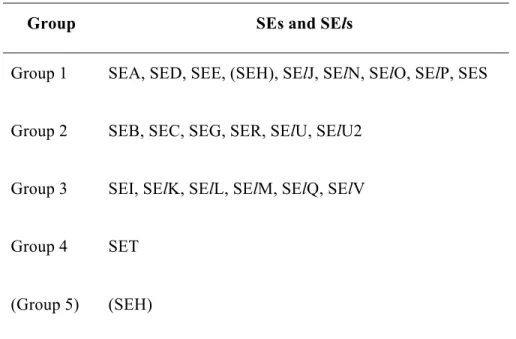
Based on amino acid sequence comparisons, they can be divided into five major groups (Table 2). SEH has been placed within Groups 1 or 5 (Vasconcelos and Cunha, 2010; Thomas et al, 2007; Uchiyama et al., 2006). The percentage of amino acid in the primary sequence was used for classification and comparison purposes. SEA, SED and SEE share 70-90% of homology in the amino acid sequence, while only 40-60% with SEB, SEC and TSST-1 (Pinchuk et al., 2010; Balaban and Rasooly, 2000).
Table 2. Grouping of SEs and SEls based on amino acid sequence comparisons *.
Group SEs and SEls
Group 1 SEA, SED, SEE, (SEH), SElJ, SElN, SElO, SElP, SES Group 2 SEB, SEC, SEG, SER, SElU, SElU2
Group 3 SEI, SElK, SElL, SElM, SElQ, SElV Group 4 SET
(Group 5) (SEH)
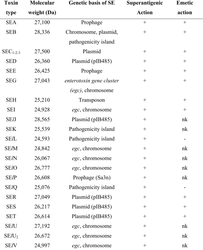
These toxins are globular single chain proteins with molecular weights ranging from 22-29kDa (Table 3) (Argudín et al., 2010) and their mature length is approximately 220-240 amino acids (Pinchuk et al., 2010). The three dimensional structure of several SEs and TSST-1 have been determined by crystallography. They are compact ellipsoidal proteins with two major unequal domains with a β strand and a few α helices, separated by a shallow cavity. The larger of the two domains contains both the amino and carboxyl termini (Pinchuk et al., 2010; Balaban and Rasooly, 2000). The two domains are highly conserved among all SEs.
Table 3. General properties of SEs and SEls (Hennekinne et al., 2010)
Toxin type
Molecular weight (Da)
Genetic basis of SE Superantigenic Action
Emetic action
SEA 27,100 Prophage + +
SEB 28,336 Chromosome, plasmid,
pathogenicity island
+ +
SEC1-2-3 27,500 Plasmid + +
SED 26,360 Plasmid (pIB485) + +
SEE 26,425 Prophage + +
SEG 27,043 enterotoxin gene cluster (egc), chromosome
+ +
SEH 25,210 Transposon + +
SEI 24,928 egc, chromosome + +
SElJ 28,565 Plasmid (pIB485) + nk
SEK 25,539 Pathogenicity island + nk
SElL 24,593 Pathogenicity island + -
SElM 24,842 egc, chromosome + nk
SElN 26,067 egc, chromosome + nk
SElO 26,777 egc, chromosome + nk
SElP 26,608 Prophage (Sa3n) + nk
SElQ 25,076 Pathogenicity island + -
SER 27,049 Plasmid (pIB485) + +
SES 26,217 Plasmid (pIB485) + +
SET 26,614 Plasmid (pIB485) + +
SElU 27,192 egc, chromosome + nk
SElU2 26,672 egc, chromosome + nk
SElV 24,997 egc, chromosome + nk
* +: positive reaction; -: negative reaction; nk: not known
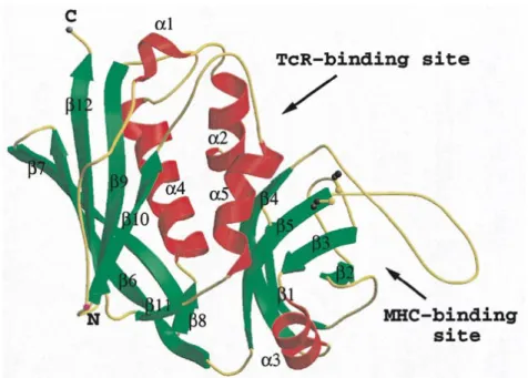
SEB is a single polypeptide chain containing total 239 amino acid residues with one disulfide and no free group (Spero et al., 1973). The molecular weight of SEB is 28,336 Da.
Figure 1. 3D structure of SEB (Papageorgiou et al., 1998)
Figure 2. Staphylococcal enterotoxin B sequence (Nema et al., 2007)
ESQPDPKPDE LHKASKFTGL MENMKVLYDD NHVSAINVKS IDQFLYFDLI YSIKDTKLGN YDNVRVEFKN KDLADKYKDK YVDVFGANYY YQCYFSKKTN DINSHQTDKR KTCMYGGVTE HNGNHLDKYR SITVRVFEDG KNLLSFDVQT NKKKVTAQEL DYLTRHYLVK NKKLYEFNNS PYETGYIKFI ESENSFWYDM MPAPGDKFDQ SKYLMMYNDN KMVDSKDVKI EVYLTTKKK
One of the most important properties of SEs is thermal stability. Generally, heat treatments commonly used in food processing are not effective for complete inactivation of enterotoxins. SEs are also partially resistant to proteolytic enzymes (e.g. pepsin, trypsin, rennin and papain) retaining some activities in the digestive tract after ingestion (Balaban and Rasooly, 2000).
They are pyrogenic and share some other important functions in the ability to induce emesis and gastroenteritis as well as superantigenicity (Pinchuk et al., 2010). SEs belong to the broad family of pyrogenic toxin superantigens (Sags). Unlike conventional antigens, Sags do not need to be processed by antigen-presenting cell (APC) before being presented to T cells. Balaban and Rassoly (2000) suggested that the enterotoxin activity may facilitate transcytosis, enabling the toxin to enter the bloodstream, that allow the interaction with T cells and leading to superantigenic activity. Superantigens bind directly to class II MHC complex (Major histocompatibility complex class II) on the surface of APC. Interaction typically occurs to the variable region of TCR β chain (Vβ). Thus, a large number of T cells are stimulated and proinflammatory cytokines are released in large amounts causing systematic toxicity and suppression of the adaptive immune response (Pinchuk et al., 2010; Vasconcelos and Cunha, 2010; Balaban and Rasooly, 2000). Although the superantigenic activity of SEs is well understood, the mechanisms leading to the emetic activity are not clearly define (Balaban and Rasooly, 2000). The biological strength of the Sags is determined by its affinity for the TCR. Sags with the highest affinity for the TCR elicit the strongest response.
2.3.3.2. Enterotoxin gene location
Genes encoding SEs have different genetic supports. All se and sel genes are located on accessory genetic elements, including plasmids, prophages, S. aureus pathogenicity islands (SaPIs), genomic island vSa, or next to the staphylococcal cassette chromosome (SCC) elements (Argudín et al., 2010). Most of these are mobile genetic elements and their spread among S. aureus isolates can modify their ability to cause disease and contribute to the evolution of this important pathogen. For instance, sea gene is carried
by a family of temperate phages (Coleman et al., 1989). SEB is encoded by seb gene and is chromosomally located in some clinical isolates (Shafer and Iandolo, 1978). Some of the SE genes are controlled by the accessory gene regulator (arg) which is the main regulatory system controlling the gene expression of virulence factors in S. aureus (Kornblum et al., 1990).
2.3.3.3. Environmental factors that affect staphylococcal enterotoxin production
Staphylococcal food poisoning (SFP) is often associated with growth in protein rich food such as meat and dairy products. These products are highly complex matrices. Many studies have been carried out in laboratory media and in diverse foodstuff to investigate the conditions for producing SEs of S. aureus. Some amino acids are essential elements: valine is necessary for growth, cystein and arginine are necessary for both growth and SE production strains of S. aureus producing specifically SEA, SEB or SEC (Le Loir et al., 2003). The same factors that affect growth of the organism in general also affect the production of enterotoxin (Bergdoll and Wong, 2006). Moreover, growth ability of S. aureus is influenced by a variety of microorganisms and S. aureus is quite sensitive to microbial competition. Lactic organisms may inhibit the production of proteases and enterotoxins associated to S. aureus (Haines and Harmon, 1973).
2.4. Food poisoning an overview
2.4.1. Food safety activities
2.4.1.1. Food safety activities in the world
Based on report of World Health Organization (WHO), up to one third of the population in developed countries acquire foodborne illnesses each year (WHO, 2006). A great proportion of these cases can be attributed not only by food contamination of food but also by contaminated drinking water (WHO, 2007; Mead et al., 1999). Many incidents of food poisoning were not reported because symptoms are mild and can be resolved quickly. In addition, the evaluation of FBD incidences is difficult to monitor in many countries due mainly to poor monitoring and health systems (Le Loir et al., 2003). To date, more than 250 known FBDs were identified (Le Loir et al., 2003). Food poisoning can be due to known or unknown causes. The known causes of food poisoning are infectious agents and toxic agents. Mainly, the infectious agents are bacteria, viruses, prions, parasites and the toxic agents are toxins as well as inorganic and organic chemicals. They can be found and detected in food with appropriate analytical methods (Le Loir et al., 2003; Mead et al., 1999). Food poisoning caused by infectious agents can be classified into two groups: (i) foodborne infections and (ii) foodborne intoxications. Foodborne infections occur when pathogenic bacteria present in food are consumed. Among these, bacteria have accounted for more than 70% of deaths related to foodborne diseases (Hughes et al., 2007; Lynch et al., 2006; Le Loir et al., 2003; Mead et al., 1999). The symptoms vary widely depending on the etiological agent (Le Loir et al., 2003). They include abdominal pain, vomiting, diarrhea, and headache (Kass and Riemann, 2006). More serious cases can result in life-threatening neurologic, hepatic, and renal syndromes leading to permanent disability or death particularly in susceptible
groups such as the elderly, people with diminished immunity or infants and young children (Kennedy et al., 2004; Le Loir et al., 2003).
The WHO identified five factors associated with these illnesses: (i) improper cooking procedures, (ii) incorrect temperature during storage, (iii) lack of hygiene and sanitation by food handler, (iv) cross-contamination between raw and fresh ready-to-eat foods and (v) acquiring food from unsafe source (WHO, 2007). Among these factors mentioned above, four of five factors are related directly to food handler behaviours (acquiring foods from unsafe sources is the exception) (Chapman et al., 2010). According to the CDC, the majority of food poisoning occurrence is related to improper food handling (97%). Among the food poisoning cases, 79% of cases are associated with food prepared in commercial or institutional establishment and 21% of cases are associated with preparation of food at home (Gamarra et al., 2009) and meals prepared outside of the home have been implicated in up to 70% of traced outbreaks(Klein and DeWaal, 2008; Lee and Middleton, 2003). Cross-contamination is often a cause of food poisoning that is overlooked. It occurs when harmful bacteria are spread between food and contaminated surfaces or equipment. Thus food can become contaminated at any stage of food chain and contamination can occur at anytime from farm to folk.
2.4.1.2. Food safety activities in Vietnam
Vietnam has been facing enormous challenges in improving its food safety and safety regulations. A fundamental problem is directly related to the lack of trained resources, including management, leading inevitably to poor implementation of a surveillance system. Moreover, the current legal framework is inadequate and ambiguous (ASIA
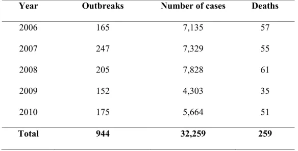
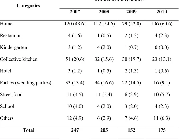
Invest Program 2006-2007; Van, 2007). With a population of more than 86 million and 75% of the population living in rural areas (General Statistic Office of Vietnam, data in 2009), the country is facing a lot of challenges related to food safety. Based on report of Vietnam Food Administration (VFA, 2011), the incidence of food poisoning have slightly decreased from 2006 to 2010 (on average 7.8/100,000 person-year) with 944 cases of FBDs which resulted in 32,259 victims and 259 people died (Table 4). Almost all food poisoning outbreaks were associated with preparation of food at home followed by collective kitchen and street vended food (Table 5) (VFA, 2011). In fact, the true incidence of diarrheal disease (includes food borne and waterborne etiologies) could be significantly higher than the official figures due to poor implementation food surveillance program, lack of trained resources, lack of appropriately laboratory equipments as well as the poor organization and inaccessibility of the health system (Khan, 2009; Van, 2007; Kim, 2002).
Table 4. Reported foodborne disease in Vietnam from 2006-2010 (VFA, 2011)
Year Outbreaks Number of cases Deaths
2006 165 7,135 57 2007 247 7,329 55 2008 205 7,828 61 2009 152 4,303 35 2010 175 5,664 51 Total 944 32,259 259
Table 5. Distribution of food poisoning outbreaks in Vietnam 2009-2010 Categories Results of surveillance 2007 2008 2009 2010 Home 120 (48.6) 112 (54.6) 79 (52.0) 106 (60.6) Restaurant 4 (1.6) 1 (0.5) 2 (1.3) 4 (2.3) Kindergarten 3 (1.2) 4 (2.0) 1 (0.7) 0 (0.0) Collective kitchen 51 (20.6) 32 (15.6) 30 (19.7) 23 (13.1) Hotel 3 (1.2) 1 (0.5) 2 (1.3) 1 (0.6)
Parties (wedding parties) 33 (13.4) 34 (16.6) 22 (14.5) 16 (9.1)
Street food 11 (4.5) 11 (5.4) 6 (3.9) 10 (5.7)
School 10 (4.0) 4 (2.0) 3 (2.0) 4 (2.3)
Others 12 (4.9) 6 (2.9) 7 (4.6) 11 (6.3)
Total 247 205 152 175
• In parenthesis: percentage of cases (based on statistics of VFA, 2011)
Currently, the Vietnamese government is making great efforts to improve the food safety and management system in order to reduce risks (Kim, 2002). The Food Law has been approved and promulgated by the Vietnamese Congress in 2010. Moreover, Vietnam has applied international standards for food hygiene and safety in the entire production chain following the warning of WHO and the United Nations Food and Agricultural Organisation (FAO). Food safety must be controlled from “farm to fork”, meaning from the growing, harvesting to processing, distribution and consumption phases (FAO, 2002).
2.4.2. Staphylococcal food poisoning
SFP is caused by the consumption of SEs produced in contaminated foods. SFP are also the second most common cause of reported foodborne illnesses (Argudín et al., 2010; Hennekinne et al., 2010; Pinchuk et al., 2010; Le Loir et al., 2003; Balaban and Rasooly, 2000). S. aureus is an ubiquitous bacteria and is found in variety of domestic animals as well as humans and transfer to food through two main sources: human carriage and dairy animals in cases of mastitis (Hennekinne et al., 2010; Pinchuk et al., 2010). The amount of toxin ingested from contaminated food needed to cause disease is less than 1.0 µg, comparable to 106 CFU/g (Pinchuk et al., 2010; Bergdoll and Wong, 2006). Onset of the illness can occur rapidly (2 to 6h) with symptoms such as nausea followed by vomiting and diarrhea, abdominal cramps, dizziness and shivering (Seo and Bohach, 2007; Balaban and Rasooly, 2000). In more severe cases, headache, muscle cramping, and transient changes in blood pressure and pulse may occur. The disease resolves within 24-48h without specific treatment (Bennett, 2005; Murray, 2005; Dinges et al., 2000). Occasionally, it can be severe enough to require hospitalization, particularly when infants, elderly or debilitated people are concerned (Murray, 2005). Death is rare (Balaban and Rasooly, 2000; Mead et al., 1999).
Amongst the SEs family, only a few have been the focus of specific studies (Pinchuk et al., 2010; Vasconcelos and Cunha, 2010). SEA is the most common toxin in staphylococcus- related food poisoning (80%) with relatively mild symptoms while SEB is a toxin associated with severe symptoms. SEB has very low toxic and lethal doses and was studied for potential use as an inhaled bio-weapon (10%) (Ahanotu et al., 2006; Ler et al., 2006; Casman, 1965). There are other identified SEs. SED is one of the most
common staphylococcal toxin associated to food poisoning with relatively low toxic dose but it is associated to mild symptoms (Pinchuk et al., 2010). SEE was identified in rare cases, while SEF was presumed to be implicated with the toxic shock syndrome (Vasconcelos and Cunha, 2010). SEG, SEH and SEI were not studied in depth, however SEH was identified as one of the cause for a massive outbreak associated with the reconstituted milk consumption in Osaka (Japan) in 2000 (Ikeda et al., 2005).
Foods that require hand preparation and kept at slightly elevated temperatures after preparation are frequently involved in staphylococcal food poisoning due to the insufficient pasteurization/decontamination of raw material or its contamination during preparation and handling by food handlers. Foods that are incriminated in SFP include meat, poultry products, eggs products, canned meat, salads, cooked meals (especially pasta based products), sandwich fillings and other dairy products. Milk and milk products are also related to staphylococcal food poisoning. Although, bacteria can be killed by heating, the SEs are heat resistant, they will not degrade extensively and consequently contaminated food products will remain toxic even after cooking (Evenson et al., 1988).
2.5. Analytical method for the detection of S. aureus related toxins
2.5.1. Bioassays
Detection and identification of SEs in food were initially performed using biological methods. Surgalla et al (1953) successfully identify the enterotoxins. Biological assays are based on the capacity of an extract of the suspected food to induce symptoms such as vomiting, gastrointestinal symptoms in animals or superantigenic action in cell cultures.
Initially, animal studies were conducted in order to find out a link between the enterotoxicity of foods and different organisms isolated from foods. SEs have been identified based on their emetic activity in monkey feeding test and kitten-intraperitoneal test and more recently, house musk shrews Suncus murinus are used as animal models (Ono et al., 2008). Monkey feeding tests were not sufficiently sensitive to detect the amount of toxin in food during outbreaks and the procedure of injecting cats or kittens was rapidly considered non-specific (Casman and Bennett, 1965). Therefore, the biological assays utilizing animals have been replaced by immunoassays, and molecular biological methods (Normanno et al., 2006; Martin et al. 2004; Nakano et al., 2004).
2.5.2. Molecular tools
Polymerase chain reaction (PCR) is a noteworthy technique used in molecular biology. This method usually detects genes encoding enterotoxins in strains of S. aureus isolated from contaminated foods (Vasconcelos and Cunha, 2010). However, these molecular methods have two major limitations: (i) staphylococcal strains must be isolated from food and (ii) the results inform on the presence or absence of genes encoding SEs but do not provide any information on the concentration of the toxins in food. This method therefore cannot be the sole method to detect SEs in food (Hennekinne et al., 2010). Other related techniques such as reverse transcription polymerase chain reaction (RT-PCR) detect and quantify mRNA. The method includes the reverse transcription of RNA strand into its DNA complement. The resulting cDNA is amplified using PCR (Vasconcelos and Cunha, 2010). This method offers more specificity but still does not allow direct detection and/or quantification of the toxins (Hennekinne et al., 2010). Recently, several PCR-based methods have been used for staphylococcal enterotoxin
genotyping but these methods are time-consuming and laborious because many separate reactions are required to identify subsets of different enterotoxin genes (se). The advantage of this method is its sensitivity for the detection of enterotoxin genes. In contrast, results of these methods may show false positive due to the presence of different copy numbers of the genes resulted (including new and unexpected toxin genes) in varying signal intensities in the array (Vasconcelos and Cunha, 2010).
2.5.3. Immunological tools
Several immunological methods have been used for the detection of staphylococcal enterotoxins in food: (i) immunodiffusion assays (ii) radioimmunoassays (RIA) and (iii) enzyme-linked immunosorbent assays (ELISA) (Thompson et al., 1986).
2.5.3.1. Gel diffusion
There are two kinds of gel diffusion: (i) single gel diffusion tube assay and (ii) double gel diffusion tube assay. A single gel diffusion assay was described for the detection of SEA and SEB by Hall and colleagues (Hall et al., 1965). In single and double gel diffusion tube assay, when enterotoxin antigen reacts with its corresponding antibody, a visible precipitate may occur, called precipitin reaction. Melted agar containing antiserum is poured into test tubes and is overlaid with a solution containing enterotoxins. The enterotoxin diffuses downward into the antiserum agar layer and forms a precipitin band at the interface in the test tube. The diameter of the precipitin ring is plotted against the concentration of enterotoxin resulting in a straight line. The double gel diffusion tube assays are modified from the single gel system. A layer of
plain agar separates the antibody containing agar layer and the enterotoxin solution. The limit of detection of double gel diffusion is approximately 1µg/ml. The tube diffusion assays has been supplanted by the micro slide and plate assays. In 1969, Casman et al (1969) developed the micro slide gel double diffusion assays and now it is used as the current standard for evaluation new methods. In micro slide gel double diffusion assay, small wells are cut in agar coated micro slides, antiserum added to the center well and enterotoxin placed in the wells surrounding the antiserum well.
2.5.3.2. Radioimmunoassay
Radioimmunoassay (RIA) is a highly sensitive technique used for measurement of primary antigen and antibody interactions and for the determination of the amount of substances present in samples. First discovered by Rosalyn Yalow and Solomon Berson, RIA was used to measure blood volume, iodine metabolism and hormones like insulin (Yalow and Berson, 1960). Since its development, RIA has been considered a revolution in bioanalysis because of its rapidity, precision, sensitivity and simplicity. The principle of this method is based on the reaction of the antigen with specific antibody resulting in a competitive binding assay using an antigen as a ligand and an antibody as the binding protein (‘‘carrier’’). To perform a radioimmunoassay, a known quantity of an antigen is made radioactive (i.e. labeled with gamma-radioactive isotopes of iodine attached to tyrosine). Radiolabeled antigens are then mixed with a known amount of the antibody for that antigen, and both will bind to one another (Smith and Bencivengo, 1985). The unknown concentration of the antigenic substance in a sample is obtained by comparing its inhibitory effect on the binding of radiolabeled antigen to a specific amount of
specific antibody (Smith and Bencivengo, 1985). A standard curve is established and the amount of antigen in the unknown samples can be calculated based samples with increasing concentrations of unlabeled ligand. The analysis of food samples by RIA require minimal preparation allowing fast and sensitive detection of staphylococcal enterotoxins from foods with sensitivity near 1 ng/g (Thompson et al., 1986; Smith and Bencivengo, 1985). Several methods were developed, validated and used for the detection of SEA, SEB, SEC: (i) solid-phase RIA with polystyrene tubes; (ii) RIA with bromoacetyl-cellulose as an immunoadsorbent; (iii) the double-antibody technique with anti-rabbit gamma globulin as co-precipitant and RIA with cells containing protein A as coprecipitant (Miller et al., 1978). The assay involved labeling of the enterotoxins with radioactive 125I or 131I-chloramine-T, lactoperoxidase and gaseous iodine (Miller et al., 1978). Although, RIA has been widely used in research and routine analysis, there are many limitations. Limitations include the lack of specificity and linearity leading to accuracy problems. Moreover, the handling and disposal of radioactive waste are a concern and represent an additional cost (Smith and Bencivengo, 1985).
2.5.3.3. Enzyme linked immunosorbent assay
ELISA can also be referred as enzyme immunoassay but does not have the same limitation compared to RIA regarding the handling and disposal of radioactive chemicals and wastes (Freed et al., 1982; Kauffman, 1980). ELISA is an immunoassay technique involving an enzymatic reaction to detect the presence of a specific antigen-antibody reaction (Candlish, 1991; Clark and Engvall, 1980). The enzyme converts a colorless substrate to a colored product that allows the detection of antigen-antibody binding. An ELISA can be used to detect either the presence of antigens or antibodies in a sample,
depending on how the test is designed. In 1971, Engvall and Perlmann published their first paper on ELISA and demonstrated quantitative measurement of IgG in rabbit serum with alkaline phosphatase as the reporter label (Engvall and Perlmann, 1971). Saunders and Bartlett (1977) described a double antibody solid-phase enzyme immune assay for detection of SEA from foods. Presently, many ELISA methods are available for the detection of staphylococcal enterotoxins in food products. Among ELISA techniques, there are three types which are frequently for the detection of SEs: (i) the single sandwich ELISA; (ii) the double sandwich ELISA; and (iii) competitive methods. In the single sandwich ELISA technique, a solid phase is coated with antibody and enterotoxin is added and allowed to react. This technique could be carry out in microtiter plate tubes or spheres, or polystyrene tubes. The assay uses peroxidase or alkaline phosphatase antibody conjugates (Smith and Bencivengo, 1985; Saunders and Bartlett, 1977). In the double sandwich method, the enzyme is coupled to the specific antibody. A solid phase anti-enterotoxin complex with enterotoxin reacted with a second anti enterotoxin produced in an animal species different from that of the first. An anti-IgG-enzyme conjugate is used in the assay (Smith and Bencivengo, 1985; Saunders and Bartlett, 1977). With the competitive method, the enzyme is conjugated with the toxin molecule (Kauffman, 1980). There are many advantages including speed, simplicity and sensitivity. However, there are also many limitations. ELISA methods are prone to false positive and false negative results due to the cross reactivity of the antibodies with antigens. ELISA methods provide information on the presence of an analyte but no information on its chemical properties (i.e. chemical structure) and consequently, the specificity of the technique can be questioned. The method accuracy strongly depends on the specificity and ELISA methods are rather used as a detection tool that should be
used in combination with more sophisticated analytical techniques for confirmation and accurate quantification.
2.5.3.4. Reversed passive latex agglutination assay
For rapid detection of enterotoxin, a reversed passive latex agglutination assay was developed (Shingaki et al., 1981; Oda et al., 1979). This method allows the detection of soluble antigens such as enterotoxins. The antibody is coated to particles such as latex beads and reacts with the soluble antigen which is visibly agglutinated in the presence of the corresponding enterotoxin. The latex particles are sensitized with rabbit globulins and these latex particles agglutinate in the presence of staphylococcal enterotoxins. This is now commercially available in SET-RPLA kit from Basingstoke (Hampshire, UK). The kit is more convenient, simple and rapid to use and is more sensitive than the immune diffusion assay with a limit of detection of 1 ng/g (Bankes and Rose, 1989). Fujikawa and Igarashi (1988) modified the RPLA which using high density latex particles for the detection of SEA, SEB, SEC, SED and SEE. The assay can be performed using the SET-PPLA commercial kit (Denka Seiken Co.Ltd, Tokyo Japan). The uses of this method provides some advantages including a simpler procedure and a shorter incubation time (reduce from 20-24h to 4h). Moreover, there is no need for expensive equipment but similar limitations are seen compared to traditional ELISA assays. Therefore, SET-RPLA kits could be used for the rapid detection of toxins in variety of foods (Park and Szabo, 1986) but will require more sophisticated analytical techniques to adequately identify and quantify the toxins.
2.5.4. Mass-spectrometry based methods
The limitations outlined in previous sections, more specifically the fact that all methods described above do not provide any information on the toxin chemical properties, the development of new analytical alternatives to detect, identify and quantify SEs in food matrices are needed. Moreover, the lack of available antibodies against the newly describes SEs has lead bioanalytical scientist to develop direct detection methods base on specific physicochemical properties. Mass spectrometry (MS) has become an indispensable technique for the identification, characterization and quantification of proteins. The method provides unparalleled specificity, rapidity and reliable analytical results (Hennekinne et al., 2010; Chaerkady and Pandey, 2008; Griffiths et al., 2001). The development and implementation of electrospray ionization (ESI) and matrix-assisted laser desorption/ionization (MALDI) has allowed structural characterization of biomolecules especially proteins and peptides (Griffiths et al., 2001). However, single MS cannot be used for all proteins and all purposes (Hennekinne et al., 2010). Therefore, the MS method requires the development of a series of techniques and liquid chromatography coupled to tandem MS is applied for the sensitive identification of complex protein mixtures (Chaerkady and Pandey, 2008). Recent studies illustrated that LC-MS proteomic strategies can be applied to develop more selective, accurate and precise assays for the characterization and quantification of SEs (Brun et al., 2007; Callahan et al., 2006; Bernardo et al., 2002). Isotopically labeled internal standard is one of the popular strategies in which the relative concentration of proteins can be measured by isotopic dilution. Various labeling methods have been developed and the mass tags can be introduced into peptides by chemical or metabolic labeling techniques (Kito and Ito, 2008). Accordingly, known amounts of isotope labeled synthetic peptide standards
are combined with the analyte and then two peptide ions of isotope pairs can be simultaneously analysed by LC-MS/MS. The mass difference between the labeled and unlabeled peptide can be distinguished and the absolute amount of the analyte is calculated based on ratio of peak intensity between isotope pair ions (Kito and Ito, 2008).
2.6. Analytical strategies
2.6.1. Proteomic methods used in mass spectrometry
Proteomic is a promising tool for studying global gene expression profiles at the protein levels (Yan and Chen, 2005). In general, proteomics involve the profiling of protein component, identifying their modifications and measurement of protein abundance through the use of purification and characterization techniques (Kito and Ito, 2008). There are many proteomic methods that play an important role for understanding the alterations of biological systems especially protein structures, activities and interactions (Cravatt et al., 2007). Among proteomic techniques, mass spectrometry has become the most powerful tool to generate information on the structure and mass of the peptide due to its high sensitivity and specificity. Furthermore, MS-based analyses can provide accurate and precise concentrations (Kito and Ito, 2008; Brun et al., 2007; Cravatt et al., 2007). Currently, there are several widely used methods to generate global quantitative protein profiles including top down and bottom up approaches but most of these methods include stable isotope labeling for quantitation (Yan and Chen, 2005).
2.6.2. Liquid chromatography- Mass spectrometry 2.6.2.1. General overview
Since the first introduction of chromatography in the early 20th century, chromatography has become the preferred technique in most bioanalytical laboratories. Chromatography is a physical method of separation in which the components to be separated are distributed between two phases, one of which is stationary (the stationary phase), while the other (the mobile phase) moves in a definite direction (IUPAC, 1993). Chromatographic methods include two categories depending on the nature of the mobile phase such as gas chromatography (GC) and liquid chromatography (LC). A mobile phase is described as ‘‘a fluid which percolates through or along the stationary bed in a definite direction’’. It may be a liquid, a gas or a supercritical fluid, while the stationary phase may be a solid, a gel or a liquid. If a liquid, it may be distributed on a solid, which may or may not contribute to the separation process (IUPAC, 1993).
GC is suitable for gaseous or volatile substances that are heat-stable (Manz et al., 2004) and is not adequate for peptide and protein analysis. Unlike GC, liquid chromatography (LC) is more versatile and can be applied to safely separate a very wide range of organic compounds from small molecules, such as drugs and metabolites up to larger molecules such as peptides and proteins (Manz et al., 2004). The majority of LC separations can be classified as normal and reversed phase chromatography. In normal phase chromatography, the stationary phase consists of hydrophilic material for instance silica particles and the mobile phase is a hydrophobic organic solvent such as hexane. In reversed phase chromatography, the stationary phase is hydrophobic and the mobile phase is a mixture of polar solvents such as water and acetonitrile. Chromatography can
separate components from a complex mixture by differential adsorption between a stationary phase and a mobile phase. The separation is based on the chemical properties of the analytes, the stationary phase and the mobile phase which requires optimization to obtain adequate selectivity. Traditional LC methods use ultraviolet-visible, fluorescence, electrochemical and refractive index detectors, but more recently LC was couple to single or multistage MS to significantly enhance the selectivity and sensitivity as well as to obtain structural information on targeted analytes.
Nowadays, MS is a widely used in a number of fields such as chemistry, biochemistry (Siuzdak, 1994), pharmacology (Fenselau, 1992), microbiology (Easterling et al., 1998) and the proteomics (Pandey and Mann, 2000). The development of MS is a direct consequence of the improvement of soft ionization techniques like electrospray (ESI) and matrix-assisted laser desorption ionization (MALDI) (Karas and Hillenkamp, 1988) which allowed the direct analysis of polar, thermally labile biomolecules without fragmentation (Lane, 2005; Griffiths et al., 2001). MS accurately measures the mass to charge (m/z) ratios of ionizable compounds. Generally, ESI-MS analysis requires the samples to be injected using a chromatographic system to avoid problems associated with ionization suppression when analytes are introduced into the ion source (Griffiths et al., 2001; Jonsson, 2001).
2.6.2.2. Electrospray ionisation
ESI was first described by Malcolm Dole when transferred large molecules to the gas phase in the late 1960s (Dole et al., 1968). In 1984, ESI was used for the first time to create gas phase ions for MS analysis (Yamashita and Fenn, 1984). This achievement
was recognized in 2002. The Nobel Prize in Chemistry 2002 was awarded to John B. Fenn specifically for his contribution into the development of ESI-MS. Electrospray ionization is the most common atmospheric pressure ion source currently employed to couple LC to MS (Jonsson, 2001; Yamashita and Fenn, 1984). The ion source is necessary to evaporate the liquid, produce ions and generate an electric field to transport the ions into the orifice of the MS. ESI-MS was introduced by Yamashita and Fenn in 1984 (Yamashita and Fenn, 1984) and has made a significant commercial impact since 1990 (Balogh, 1997). In an ESI source, the liquid from the HPLC is directed through the free end of an electrode (capillary) set at 3 to 5 kV. In the case of pure ESI, the high electric field at the tip of the capillary pulls the liquid emanating from the electrode into a fine jet that breaks up, typically a millimeter from the tip of the electrode, into a fine spray of electrified droplets. The fine droplets in the spray evaporate in about one millisecond to liberate charged molecules from the droplets as ions, which the electric field of the electrode tip then transports toward the entrance of the MS. Currently, the ESI process is one of the softest ionization techniques available and has the strong advantage of generating molecular ions ([M+nH]n+ or [M-nH]n-). Several other techniques were derived for this general concept such as ionspray (Bruins et al., 1987), microspray (Covey, 1995) and nanospray (Wilm and Mann, 1996). The latter techniques are mainly used for proteomic analysis and the ionspray or pneumatic assisted ESI has a higher nebulization capacity and can accommodate significantly higher flow rates. Ionspray is the most often used version of ESI currently in bioanalysis. ESI is considered one of the mildest desorption techniques available since little or no extra internal energy is imparted to the ions and, therefore, little fragmentation occurs (Bruins et al, 1987). Furthermore, one of the most widely accepted features of ESI-MS is that ions observed in a given mass spectrum are preformed in solution. This implies that unless the species