Université de Montréal
Développement du squelette du crinoïde Florometra serratissima et évolution des protéines de la matrice de spicules chez les ambulacraires
par Ariane Comeau
Sciences biologiques, Université de Montréal, Faculté des arts et des sciences
Mémoire présenté à la Faculté des arts et des sciences en vue de l’obtention du grade de M.Sc. en sciences biologiques
Août, 2014
i
Résumé
Les crinoïdes sont bien connus pour leurs fossiles, mais la biominéralisation de leurs stades larvaires n’est que peu documentée. La première partie du projet présente le développement des ossicules des trois stades larvaires du comatule Florometra serratissima : doliolaria, cystidienne et pentacrinoïde. Les ossicules du crinoïde démontraient de la plasticité phénotypique et de la désynchronisation dans leur développement. Les crinoïdes étant la classe basale des échinodermes modernes, ceci porte à croire que ces traits étaient aussi caractéristiques des échinodermes ancestraux et auraient joué un rôle dans la radiation hâtive et la grande disparité des échinodermes. Pour notre deuxième étude, comme les patrons de morphologie des crinoïdes et des autres échinodermes sont nombreux et sont régulés par des protéines spécifiques, nous avons vérifié la présence de quatre familles de protéines de la matrice de spicules (SMAP) connues chez les oursins dans les transcriptomes des autres échinodermes et d’autres deutérostomes. La famille des spicules matrix (SM) et l’anhydrase carbonique CARA7LA étaient absentes chez tout autre organisme que les oursins, les protéines spécifiques au mésenchyme (MSP130) étaient présentes en nombres différents chez tous les ambulacraires suggérant de multiples duplications et pertes, et les métalloprotéases étaient nombreuses chez chacun. Le développement des ossicules chez les échinodermes est un sujet qui a gagné en popularité au cours des dernières décennies, spécialement chez les oursins, et inclure les crinoïdes dans ce type d’étude permettra de nous renseigner sur l’origine et l’évolution des échinodermes modernes.
Mots clés : biominéralisation, comatule, crinoïde, échinoderme, ambulacraire, transcriptome, algue coralline
ii
Abstract
While the fossil record of crinoids is widespread and largely known, biomineralization of their larval stages is poorly documented. The first part of the project focuses on the ossicle development of the three larval stages of the feather star Florometra serratissima: doliolaria, cystidean and pentacrinoid. The ossicles of the crinoid showed phenotypic plasticity and asynchronous development. Crinoids form the basal class of living echinoderms; this prompts one to believe that these traits were also characteristic of the ancestral echinoderms and would have played a role in the early radiation and large disparity of the modern echinoderms. For the second study, as patterns of morphology of crinoids and of other echinoderms are numerous and are regulated by specific proteins, we verified the presence of four families of spicule matrix associated proteins (SMAPs) known among sea urchins in transcriptomes of the other echinoderms and deuterostomes. The family of spicule matrix (SMs) proteins and the carbonic anhydrase CARA7LA were absent in any other organism aside from sea urchins, mesenchyme specific proteins (MSP130s) were present in varying numbers in all ambulacrarians suggesting multiple duplications and losses and matrix metalloproteases were numerous in every organisms. The development of ossicles in echinoderms is a topic that has gained popularity in the last decades, especially in sea urchins, and including crinoids in this type of study will inform us about the origin and evolution of the modern echinoderms.
Keywords: biomineralization, feather star, crinoid, echinoderm, ambulacrarian, transcriptome, coralline algae
iii
Table des matières
Résumé ... i
Abstract ... ii
Table des matières ... iii
Liste des tableaux ... vi
Chapitre 2 ... vi
Chapitre 3 ... vi
Liste des figures ... vii
Chapitre 1 ... vii
Chapitre 2 ... vii
Chapitre 3 ... x
Liste des sigles et abréviations ... xii
Remerciements ... xv
Chapitre 1. Introduction ... 1
1.1 Phylogénie ... 3
1.2 Reproduction et stades larvaires ... 4
1.3 Biominéralisation ... 6
1.4 Protéines impliquées dans la biominéralisation ... 7
1.5 Objectifs ... 8
1.6 Méthodologie ... 8
1.7 Contribution des auteurs ... 10
Chapitre 2. Skeletal development in larval stages of the feather star Florometra serratissima 11 2.1 Abstract ... 12
iv
2.2.1 Larval ossicle development ... 13
2.2.2 Crystal polymorphs and composition of calcium carbonate in invertebrates ... 16
2.2.3 Larval settlement ... 16
2.3. Materials & Methods ... 17
2.3.1 Ossicle isolation and observation ... 18
2.3.2 Settlement observations ... 20
2.3.3 Scanning electron microscopy (SEM) and electron-dispersive spectroscopy (EDS) ... 21
2.3.4 Confocal Raman spectroscopy ... 21
2.4. Results ... 21
2.4.1 Ossicles in larvae of F. serratissima ... 21
2.4.2 Elemental composition and crystallization of pentacrinoid ossicles ... 24
2.4.3 Settlement ... 24
2.5. Discussion ... 25
2.5.1 Comparison of F. serratissima ossicles with those of other crinoids and echinoderm larvae ... 25
2.5.2 Elemental composition and crystallization of pentacrinoid ossicles ... 31
2.5.3 Settlement ... 32
2.7 Acknowledgements ... 33
2.8 References ... 34
2.9 Figures ... 41
Chapitre 3. Among Ambulacrarians, sea urchins have a complex, derived protein skeletal matrix ... 60
3.1 Abstract ... 61
v
3.3 Materials & Methods ... 63
3.4 Results ... 65
3.5 Discussion ... 69
3.7 Acknowledgements ... 71
3.8 References ... 72
3.9 Figures ... 76
Chapitre 4. Discussion générale ... 82
Chapitre 5. Conclusion ... 88
5.1 Perspectives pour des études futures ... 89
vi
Liste des tableaux
Chapitre 2
Table 2.1. Timetable of developmental events of F. serratissima, from fertilization to pentacrinoid, between 9.5 and 11.5 oC. All descriptions are taken from Mladenov and Chia (1983). F. serratissima development begins by free spawning of gametes into the water where fertilization takes place.
Table 2.2. EDS analysis of 22 days old pentacrinoid ossicles (plates, columnar ossicles and attachment disk). nscans = 26, on 10 ossicles. Quantities are reported as mass percentage.
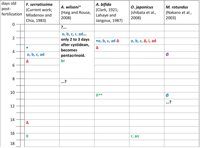
Table 2.3. Timeline of skeletal elements appearance from fertilization to pre-juvenile stage in four feather stars and one sea lily. Information is taken from the works listed under the respective species. The color code indicates the larval stage at which the first skeletal element was observed (purple for auricularia (sea lily only), blue for doliolaria, red for cystidean and green for pentacrinoid). The geometric symbols indicate the beginning of the larval stages if known (blue circle for doliolaria, red triangle for cystidean and green diamond for pentacrinoid). Two of the same larvae symbols on one line indicate its possible delay before metamorphosis into the larvae symbolized. The circle with a bar (Ø) indicates that no biominerals were observed. A question mark followed by an ellipsis (?...) indicates that the skeletal formation events up to this time are unknown and an ellipsis followed by a question mark (...?) indicates that later skeletal formation events are unknown. an: 1 anal (radianal) plate; ad: 1 attachment disk; ax: axillary ossicles; b: 5 basal plates; br: brachial ossicles; c: columnar ossicles; ci: cirri; i: infrabasal plates; cos: costal ossicles; o: 5 oral plates; p: 1 proximal; r: 5 radial plates.
Chapitre 3
Table 3.1 Homologous SMAPs of echinoid S. purpuratus found (ID and or Accession numbers) or not found (None) in transcriptome and genome of seven invertebrate deuterostomes. Echinoid and hemichordate S. kowalevskii data are taken from two other works, Mann et al. (2010) and Cameron and Bishop (2012) respectively and are shown here in italics. E-values are shown following each sequences between brackets. E-values for the MSP130 sequences are based on comparison with Sp-Msp130r2 except for Ssp-Msp130_2 that is based on comparison with Sp-Msp130r3 as it was not retrieved with Sp-Msp130r2. Selected sequences of MMPs and their e-values are the most similar sequence retrieved in transcriptomes and genome for each four sea urchin MMPs. There are less than four sequences in each organism because some retrieved sequences were identical for different sea urchin MMP proteins.
vii
Liste des figures
Chapitre 1
Figure 1.1. Arbres phylogénétiques A : des deutérostomes (modifié de Röttinger and Lowe, 2012) et B : des ambulacraires (modifié de Telford et al., 2014). Les échinodermes et les hémichordés forment le clade des ambulacraires. Les classes d’échinodermes vivant aujourd’hui sont les crinoïdes, les astéroïdes (étoiles de mer), les ophiuroïdes (ophiures), les holothuroïdes (concombres de mer) et les échinoïdes (oursins).
Figure 1.2. Stades larvaires du comatule F. serratissima : A : Doliolaria, B : cystidienne et C : pentacrinoïde.
Chapitre 2
Figure 2.1. Cladogram of ambulacrarians phylogeny including example species of crinoids. 1: Ambulacrarians; 2: Echinoderms; 3: Eleutherozoa (sister group of crinoids); 4: Crinoids (Articulata are the extant crinoids); 5: Sea lilies; 6: Comatulids (feather stars). Phylogeny of Eleutherozoa and of crinoids are in debate. This cladogram was constructed following the works of Telford et al. (2014) and Hemery et al. (2013), two recent studies using molecular data to construct trees.
Figure 2.2. A: Crinoids were kept in the dark in tables with flow through seawater. B: Rocks and bottles taken from their habitat were added to the tables because the animals like to settle on raised objects from the bottom. C: Mature eggs are perfectly spherical. D: Immature eggs are irregularly shaped and aggregated into masses. E: An individual pinnule that has been dissected from an arm is showing immature eggs. F: In vivo, eggs and sperm are released by the gonopore (shown by the arrow). Scales = 800 µm
Figure 2.3. Development of plate ossicles from doliolaria to cystidean. A: 5d21h old doliolaria tri-radiate early plate; B: 4d5h old doliolaria plate; arrows point early spicules becoming plates that did not have a tri-radiate form; C: 7d old doliolaria plate; D: 10d19h old doliolaria plate; E: 7d7h old cystidean plate; F: 11d old doliolaria, the square encompasses the area where the five oral and five basal plates in the larvae are located; G: Photo montage of 11d2h old cystidean calyx with some visible plates. H to Q: Set of the ten plate ossicles from an individual 8d7h doliolaria. No plate is identical to another, basal and oral plates are not distinguished here. ant: anterior; bp: basal plate(s); op: oral plate(s); post: posterior
Figure 2.4. Skeletal parts of a cystidean. Some of the 5 oral and 5 basal plates are visible. The stalk is divided into three regions, apical, median and basal regions. Around 10 columnar ossicles and the attachment disk form the xenomorphic stalk of this cystidean. The
viii
attachments between them are not visible here. The line marks are bulge of the middle of each ossicle. ad: attachment disk; bp.1 to 3: basal plates; c.1 to 10; columnar ossicles of the stalk starting at the calyx; op.1 to 3: oral plates
Figure 2.5. A-B: Spicule growth in two oral plate ossicles of 22 day old pentacrinoids. Arrows show the direction of the growth of ossicles when the spicules elongated to form stroma of the stereom. They also grew in width (illustrated by double arrow in A).
Figure 2.6. The columnar ossicles from ten larvae from different developmental time periods. A : 5d5h old doliolaria columnar spicules, at earliest ellipsoidal shape (1), semicircle shapes (2-3) ; B, C, D: 6d1h, 8d7h and 9d17h old doliolaria columnar ossicle, respectively; E : 9d17h old portion of a doliolaria stalk; F : 11d3h old doliolaria; G: 9d old cystidean columnar ossicles; H: Photo montage of 10d2h old recently settled cystidean; I: The calyx has rotated into a nearly upright position in this 7d7h old cystidean; J: 13d old cystidean stalk with xenomorphic columnar ossicles. Rectangular dash areas demarcate the stalk position in F, H and I and the circle dash area demarcates calyx position in H. ad: attachment disk; ant: anterior; dor: dorsal region; post: posterior; ven: ventral region
Figure 2.7. New columnar ossicles of cystidean seems to appear under the calyx and they are shorter than the ossicles in the middle of the stalk.
Figure 2.8. The attachment disks from four larvae from at developmental time periods. A: Ossicle of an 8d1h old doliolaria is an early form of the attachment disk; B: Attachment disk and a part of the stalk of an 11d4h old cystidean that had settled five days earlier; C: Photo montage of a 15d old cystidean that had settled on a flattened substrate with its attachment disk having taken a flattened enlarged disk-shape; D: The attachment disk of this 45d old pentacrinoid has an irregular cylindrical shape compared to the flattened shape illustrated in B and C and it is a consequence of attaching on a non-flat substrate.
Figure 2.9. Ossicles of pentacrinoid of 22 days old. A: Calyx; B: Oral plate; C: Basal plate; D: Immediately under the calyx with newer columnar ossicle. bp: basal plate; c.1 to c.5: first five columnar ossicles of the stalk starting at the calyx; cr : concave region; op: oral plate
Figure 2.10. Pinnules at the tip of an arm in regeneration of an adult crinoid F. serratissima possessed ossicles that are reminiscent to cystideans and pentacrinoids columnar ossicles. A: The tip of an adult arm in regeneration possessed two new pinnules in formation (pi3 and pi4). The arrow shows the beginning of the part in regeneration. B: Ossicles were isolated from a pinnule in formation and like the cystideans and pentacrinoids, the middle of the columnar ossicles are marked by a bulge (indicated by the arrow). C: The ossicles of a newly regenerated pinnule (see A: pi1 and pi2) were aligned like the stalk of the pentacrinoids. D: Two isolated ossicles of the newly regenerated pinnule. ap: apical blastema; pi: pinnule; pi o: pinnule ossicles; tf o: tube feet ossicles
ix
Figure 2.11. Complete skeleton of a 9d7h old doliolaria consisting of 10 plates (A to J) and 10 columnar ossicles (K to P) shows the disparity of their shapes and sizes within a single larva. The basal and oral plates were not identified here. At that stage, some plates were composed of numerous stereom holes (E to H) while other lacked of them (B). K: Two columnar ossicles were still imbricated together (1-2). At that stage, most of the columnar ossicles had also spicules developed perpendicularly on both sides of the circle to assure the imbrications of the stalk in addition to the basic circular shape (K to O). Other columnar ossicles lacked of the perpendicularly spicules and were probably positioned in the apical region of the stalk under the pre-calyx (P). This individual did not possessed a recognizable attachment disk.
Figure 2.12. Selected ossicles of five sister doliolariae of 9d7h old show the shape disparity between and among larvae. Each row (larvae 1 to 5) represents two plates (columns A and B), two columnar ossicles (columns C and D) and the attachment disk (column E) of one larva. At that stage, two patterns were retrieved several times among columnar ossicles. In a same larva, some columnar ossicles fully formed the middle stereom (C) while others were not completely closed (D). These latter were found in the apical region of the stalk under the pre-calyx. A rare pattern of columnar ossicles consists of a more oval shape with smaller sterom holes (2C-2D). The attachment disk (E) was also circular but was recognized by the absence of the middle stereom and by the dense perpendicular spicules on the top of one side. The attachment disk were not retrieved in all doliolariae of the study. The plate shapes, the developmental rates, and the diameter of the ossicles differed between larvae. The development of larva 1 seems the slowest compared to the other larvae as its ossicles are smaller. The individual shown in Figure 2.11 was from the same culture of these larvae.
Figure 2.13. Maximum dimension of oral and basal calyx plate ossicles according to the age of doliolaria (nlarvae = 16; nossicles =115), cystideans (nlarvae = 15; nossicles = 124) and
pentacrinoids (nlarvae = 7; nossicles = 52) defined as the time after fertilization. Average sizes of
ossicles in each larva are shown in black. Some were only spicules (biominerals without stereom holes). Some larvae grew in the presence of algae which may affected the speed of ossicle growth.
Figure 2.14. A: Oral plates are mobile in pentacrinoids to allow the tube feet to get out out and become a suspension feeder. B: Additional small plate (pointed by the arrow) was found in a 9d7h old doliolaria close to a columnar ossicle. C: Additional small plate (pointed by the arrow) was found in a 15d old cystidean close to the basal plates. The plates shown in B and C might be homologous to the infrabasals seen in other species. D: A sixth thinner basal plate was occasionally found in a 45 days old pentacrinoid. E: The plate was aligned with the normal basal plates (photo montage). The abnormal plate is shown within the dashed area. F: EDS scan with five points on a plate ossicle of a 22 days old pentacrinoid. op : oral plate; tf : tube feet
x
Figure 2.15. Crystal spectrum of ossicles in pentacrinoids using confocal Raman spectroscopy is characteristic of calcite.
Figure 2.16. Proportion of larvae settled in the different substrate treatment groups. Blue bars illustrate proportions at day 7 post fertilization while red bars show proportions at day 9 post fertilization. Groups with corresponding letters (lower case letters correspond to day 7 and upper case letters correspond to day 9) are not significantly different from each other according to a Tukey post-hoc test.
Figure 2.17. Aggregation of advanced cystideans. A: Three cystideans aggregated on a piece of a branched coralline alga; B: Many aggregated cystideans of a culture settled on the glass bowl, which was found more rarely than on algae. Scales = 1 mm
Figure 2.18. Effect of coralline algae addition on the abundance of settled larvae. A culture of 47 sister larvae was made and separated in two bowls after fertilization, 25 larvae were transferred to a first bowl and 22 larvae were transferred to a second bowl. On day 3 fertilization, coralline algae was added to the bowl containing the 22 larvae. On day 5 post-fertilization, only 4% of the larvae without algae was settled (one larva) while 82% of the larvae with algae was settled.
Figure 2.19. Effect of coralline algae on ossicle growth of doliolariae and cystideans. Some were only spicules (biominerals without a stereom hole). Error bars show the standard deviation. The average ossicle diameter per larva was significantly larger in cystideans with algae (red linear regression line) than the doliolariae without algae (blue linear regression line) (F = 8.540, p = 0.011)
Chapitre 3
Figure 3.1. Alignment of the MSP130 proteins in the echinoid S. purpuratus (Sp), the holothuroid P. parvimensis (Pp), the ophiuroid A. filiformis (Af), the asteroid P. miniata (Pm), the crinoid F. serratissima (Fs) and the hemichordates Schizocardium sp (Ssp) and
S. kowalevskii (Sk). If a gap was present in the same position in six or more of the twenty one
sequences, it was removed from the alignment.
Figure 3.2. Phylogenetic relationships and the presentation of the MSP130 proteins in the echinoid S. purpuratus (Sp), the holothuroid P. parvimensis (Pp), the ophiuroid A. filiformis (Af), the asteroid P. miniata (Pm), the crinoid F. serratissima (Fs) and the hemichordates
Schizocardium sp (Ssp) and S. kowalevskii (Sk) shown as phylogram (A) and as radial (B).
The number at each node represents the bootstrap of 1000 replicates as a percentage.
Figure 3.3. Alignment of the MMPs proteins in the echinoid S. purpuratus (Sp), the holothuroid P. parvimensis (Pp), the ophiuroid A. filiformis (Af), the asteroid P. miniata (Pm),
xi
the crinoid F. serratissima (Fs), the hemichordates Schizocardium sp (Ssp) and S. kowalevskii (Sk). and the tunicate O. dioica (Od). Only the first sequence obtained with each four echinoid MMP proteins in every transcriptome are part of this alignment, which were sometimes the same for different queries. If a gap was present in the same position in six or more of the sixteen sequences, it was removed from the alignment.
Figure 3.4. Phylogenetic relationships and the presentation of the MMPs proteins in the echinoid S. purpuratus (Sp), the holothuroid P. parvimensis (Pp), the ophiuroid A. filiformis (Af), the asteroid P. miniata (Pm), the crinoid F. serratissima (Fs), the hemichordates
Schizocardium sp (Ssp) and S. kowalevskii (Sk). and the tunicate O. dioica (Od). shown as
phylogram (A) and as radial (B). Only the first sequence obtained with each four echinoid MMP proteins in every transcriptome are part of these trees. The number at each node represents the bootstrap of 1000 replicates as a percentage.
xii
Liste des sigles et abréviations
a.a. : amino acid
A. bifida : Antedon bifida
A. filiformis : Amphiura filiformis
A. mediterranea : Antedon mediterranea
A. wilsoni : Aporometra wilsoni
ab : apical blastema ad : attachment disk an : anal plate ant : anterior ax : axillary ossicle As : arsenic
BLAST : basic local alignment search tool
blastp : protein databases searched using a protein query BMSC : Bamfield Marine Sciences Centre
b : basal plate bp : basal plate br : brachial ossicle c : columnar ossicle C : carbon Ca : calcium
CaCO3 : calcium carbonate
ci : cirri
cm : centimeter
cos : costal ossicle
cr : concave region
CTL : C-type lectin
CTLLD : C-type lectin-like domain
xiii
dor : dorsal region
EDS : energy dispersive spectroscopy EST : expressed sequence tag
F. serratissima : Florometra serratissima
g : gram
GRN : gene regulatory network
h : hour
i : infrabasal plate
kV : kilovolt
L : liter
m : meter
M. rotundus : Metacrinus rotundus
Ma : million years ago
Mg : magnesium
MgCO3 : magnesium carbonate
mm : millimeter
mmol : millimole
MMP : matrix metalloprotease
mol : mole
mRNA : messenger ribonucleic acid MSP130 : mesenchyme specific protein 130
mW : milliwatt
n : number
Na : nitrogen
NCBI : National Center for Biotechnology Information
o : oral plate
O : oxygen
O. dioica : Oikopleura dioica
O. japonicus : Oxycomanthus japonicus
op : oral plate
xiv
P. parvimensis : Parastichopus parvimensis
pi : pinnule
pi o : pinnule ossicle
PMC : primary mesenchyme cell
post : posterior
pr : proximal
psu : practical salinity unit
r : radial plate
S. kowalevskii : Saccoglossus kowalevskii
S. purpuratus : Strongylocentrotus purpuratus
SCUBA : self contained underwater breathing apparatus SEM : scanning electron microscopy
SM : spicule matrix
SMAP : spicule matrix associated protein
Ssp : Schizocardium sp.
tblastn : translated nucleotide databases searched using a protein query
tf : tube feet
tf o : tube feet ossicle
ven : ventral region
wt% : weight percent
% : percent
°C : Celsius degree
xv
Remerciements
Je tiens à remercier plus que tout mon directeur de recherche, le professeur Christopher Cameron, pour m’avoir donné la chance de réaliser ce projet et d’explorer la recherche du monde marin. Merci pour son soutien, pour sa confiance et pour m’avoir généreusement transmis sa passion pour la biologie développementale et l’évolution. Merci au professeur Cory Bishop, St. Francis-Xavier University pour avoir accepté de me diriger pour un stage dans son laboratoire, professeure Paola Oliveri et David Dylus, University College London, pour le travail collaboratif, Professeur Chris Lowe, Judith Levine et Paul Gonzalez, Standford University pour la réalisation et le partage des transcriptomes, Kevin Learning, Simon Fraser University pour sa contribution aux tests de fixation des larves aux différents substrats, Siobhan Gray, officière de plongée et de la sécurité au Bamfield Marine Sciences Centre (BMSC) et Ross Whippo, son assistant pour les collectes de crinoïdes et pour m’avoir initiée à la plongée scientifique, docteur Eric Clelland, coordinateur de recherche du BMSC et George Robertson, technicien de recherche à St. Francis-Xavier University pour leur assistance en laboratoire, Marie-Line Fiola pour avoir contribué aux manipulations en laboratoire au BMSC, Maureen Vo, Karma Nanglu et Lina Kabbadj pour avoir été de plaisants camarades de laboratoire, ma famille et mes amis pour leur support perpétuel et sans oublier, merci aux merveilleux crinoïdes pour être aussi adorables et remplis de mystères.
2
La capacité de biominéralisation d’innombrables organismes distincts leur ont permis d’être fossilisés depuis plus de 800 millions d’années (Cohen et al., 2011; Macdonald et al., 2010). L’étude des fossiles nous permet de retracer l'histoire de l'évolution, entre et parmi les différents taxons des êtres vivants. Les crinoïdes sont célèbres pour leur grande quantité de spécimens fossilisés (Mooi, 2001) et leur phylogénie est principalement construit par rapport à la configuration de leurs plaques thécales (Ausich, 1996; Moore and Teichert, 1978; Simms, 1993; Ubaghs, 1969). Par contre, les stades larvaires des crinoïdes ne sont que très peu documentés comparativement à leur représentants fossilisés et aux autres échinodermes vivants (Clark, 1921; Lahaye and Jangoux, 1987; Mortensen, 1920b; Shibata et al., 2008). Les oursins représentent un modèle important en biologie développementale et moléculaire, essentiellement parce que les gamètes adultes sont matures en tout temps, leur développement est régulier et leur larve planctonique pluteus est transparente, ce qui est commode pour l’observation. L’oursin pourpre Strongylocentrotus purpuratus a son génome complet séquencé et il est souvent utilisé pour représenter les échinodermes en général. Les avancées moléculaires sur l’espèce sont rapides. Le réseau de régulation des gènes qui dirige la formation du squelette dans l’embryon est même déchiffré (Oliveri et al., 2008). Par contre, les échinodermes forment un phylum comportant des organismes qui diffèrent grandement les uns des autres, principalement par leur morphologie générale. On connaît très peu la génétique des concombres de mer, des ophiures, des étoiles de mer et des crinoïdes, les quatre autres classes d’échinodermes. Puisqu’ils constituent la classe basale des échinodermes modernes, l’étude du développement des crinoïdes a le potentiel de complémenter les données paléontologiques et moléculaires et de mettre en lumière l’évolution du squelette chez les échinodermes. Le développement des crinoïdes modernes est peu étudié parce qu’il pose plusieurs difficultés. Contrairement aux oursins, le cycle de reproduction de la plupart des crinoïdes est mal compris et probablement irrégulier, et leur larve planctonique est opaque, ce qui rend l’observation des structures plus ardue. Dans le bras de mer vis-à-vis le Bamfield Marine Sciences Centre (BMSC), Barkley Sound, CB, une population de crinoïdes de l’espèce
Florometra serratissima se trouve à 30 mètres de profondeur. Nous avons donc rapporté
quelques adultes dans le laboratoire du BMSC afin de cultiver des larves et étudier le développement du squelette aux trois stades larvaires de F. serratissima. Des embryons et des larves ont aussi été rapportés précédemment au Hopkins Marine Lab, Université de Stanford,
3
afin que les membres du laboratoire en extraient les transcriptomes. Ils nous ont partagé les transcriptomes des crinoïdes, des autres échinodermes et d’un hémichordé, ce qui nous a permis de chercher des séquences homologues aux gènes impliquées dans la biominéralisation des oursins, chez les autres ambulacraires.
L’espèce F. serratissima (Clark, 1907) est un crinoïde sessile qui ne vit que sur la côte ouest de l’Amérique de Nord, entre l’Alaska et la Californie, à des profondeurs variant entre 11 et 1 252 m (Clark and Clark, 1967). Plusieurs études sur F. serratissima ont été entreprises à partir de la population du bras de mer du Bamfield Marine Sciences Centre (BMSC), Barkley Sound, CB, Canada, depuis les années 80 (Byrne and Fontaine, 1981, 1983; Chia et al., 1986; Holland and Grimmer, 1981a; McEdward et al., 1988; Mladenov, 1983, 1986; Mladenov and Chia, 1983; Shaw and Fontaine, 1990; Telford et al., 2014). L’animal a aussi été recueilli à d’autres endroits dans la région de l’île de Vancouver, Canada (Bickell et al., 1980; Byrne and Fontaine, 1981; Mortensen, 1920b; Scouras and Smith, 2001), en Oregon, États-Unis (Puniwai, 2002) et en Californie, États-Unis (Holland and Grimmer, 1981b; Webster, 1975). Tout de même, peu de recherches ont été entreprises sur le crinoïde F. serratissima relativement aux autres échinodermes et une variété d’aspects de l’espèce est encore à clarifier. Jusqu’en 2001, 55% des articles scientifiques ayant comme sujet les échinodermes excluant les fossiles portaient sur les oursins et moins de 7% sur les crinoïdes (Mooi, 2001).
1.1 Phylogénie
Les échinodermes et les hémichordés sont des embranchements frères et forment ensemble le taxon des ambulacraires (Swalla and Smith, 2008) (Figure 1.1). Les échinodermes ont divergé des hémichordés durant l’édiacarien, il y a environ 570 millions d’années (Erwin et al., 2011). Les plus vieux ossicules fossiles d’échinodermes dateraient de plus de 520 millions d’années (Mooi, 2001; Sprinkle and Wilbur, 2005) et ceux des hémichordés de 525 millions d’années, au cambrien inférieur (Hou et al., 2011). Les crinoïdes sont apparus lors du début de l’ordovicien il y a environ 485 millions d’années (Guensburg and Sprinkle, 2001; Hess et al., 1999; Pisani et al., 2012; Ubaghs, 1969). Les crinoïdes vivant aujourd’hui sont soit sessiles suite à la perte du pédoncule au stade juvénile (comatules) ou pédonculés à l’âge adulte (lys de
4
mer) (Cohen et al., 2004) et forment la classe basale des échinodermes modernes (Ruppert et al., 2004; Smith, 1997) (Figure 1.1).
Figure 1.1. Arbres phylogénétiques A : des deutérostomes (modifié de Röttinger and Lowe, 2012) et B : des ambulacraires (modifié de Telford et al., 2014). Les échinodermes et les hémichordés forment le clade des ambulacraires. Les classes d’échinodermes vivant aujourd’hui sont les crinoïdes, les astéroïdes (étoiles de mer), les ophiuroïdes (ophiures), les holothuroïdes (concombres de mer) et les échinoïdes (oursins).
1.2 Reproduction et stades larvaires
Alors que certains crinoïdes vivent 1) en eaux peu profondes, 2) couvent leurs larves et / ou 3) possèdent un cycle de vie relativement court (Haig et al., 2012), F. serratissima est un comatule 1) qui est principalement retrouvé en mer profonde, 2) dont les femelles et les mâles relâchent leurs gamètes dans la colonne d’eau pour que la fertilisation y ait lieu et 3) dont le cycle de vie est relativement long et mal compris (Clark and Clark, 1967). De plus, les animaux vivant en eaux profondes sont rarement observés à relâcher leurs gamètes in situ, tel
5
est le cas de F. serratissima. Ces aspects rendent le développement de cette espèce une étude à défis. En fait, seulement Mladenov (1986) a publié sur le cycle de reproduction de
F. serratissima et aucune autre révision n’a été publiée par la suite. Chez d’autres espèces de
crinoïdes telles que Comanthus japonica et Lamprometra klunsingeri (Dan and Dan, 1941; Fishelson, 1968; Mladenov, 1986), les mâles relâchent leurs gamètes en premier. La présence des spermatozoïdes pourrait être nécessaire pour induire la ponte d’œufs des femelles chez
F. serratissima aussi (Mladenov, 1986). Trois stades larvaires surviennent lors du
développement des comatules : doliolaria, cystidienne et pentacrinoïde (Figure 1.2). La doliolaria (Figure 1.2.A) possède des cils et nage afin de trouver un substrat adéquat, s’y attacher et se métamorphoser en larve cystidienne (Figure 1.2.B). Cette dernière développera un pédoncule sur lequel le calice est soutenu. La cystidienne deviendra pentacrinoïde (Figure 1.2.C) lorsque le calice s’ouvrira pour laisser les ambulacres sortir et permettre à la larve de se nourrir comme suspensivore (Mladenov and Chia, 1983). La larve doliolaria de
F. serratissima peut nager entre 4,6 et 14 jours avant de s’installer au substrat (Mladenov and
Chia, 1983). Chez un grand nombre d’invertébrés marins, il y a un stade larvaire planctonique et certains nécessitent un signal spécifique pour s’installer (Bishop et al., 2006). Certaines espèces de crinoïdes s’attachent à une grande variété de substrats tels que des algues et des hydroïdes, alors que d’autres s’installent seulement sur leurs conspécifiques (Clark, 1921). En nature, le peu de pentacrinoïdes de F. serratissima qui ont été retrouvés étaient attachés aux cirres des adultes de la même espèce (Mortensen, 1920b), alors qu’en laboratoire, ils s’attachent à de multiples substrats (Clark, 1921).
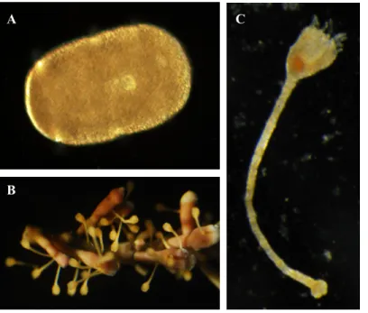
Figure 1.2. Stades larvaires du comatule F. serratissima : A : Doliolaria, B : cystidienne et C : pentacrinoïde. A C B 100 µm
6
1.3 Biominéralisation
Un squelette de carbonate de calcium (CaCO3) est sécrété chez de nombreux groupes
d’invertébrés à partir des éléments nécessaires qui sont disponibles dans leur environnement (Lowenstam and Weiner, 1989). Le CaCO3 est retrouvé chez ces animaux sous trois différents
polymorphes de cristallisation : aragonite, vatérite et calcite (Lowenstam, 1954). Le mécanisme influençant la production d’un squelette calcaire en aragonite, en vatérite ou en calcite n’est pas tout à fait clarifié. L’aragonite a été récemment découvert dans les biominéraux réduits des hémichordés (Cameron and Bishop, 2012). Les échinodermes forment le seul embranchement possédant un endosquelette complètement cristallisé en calcite (Lowenstam and Weiner, 1989), et ce datant au moins depuis l’hélicoplacoïde, l’un des plus vieux représentants fossiles d’échinodermes (Durham, 1993). Lors du développement larvaire de l’oursin, le CaCO3 des spicules passe par un stade amorphe avant de devenir calcite,
c’est-à-dire qu’il ne possède aucune cristallisation pendant ce stade (Beniash et al., 1997). Les premiers minéraux de la larve bipinnaria des astéroïdes est fait de calcite (Koga et al., 2014). Le polymorphe de cristallisation chez les larves de crinoïdes n’est pas connu et pourrait se modifier lors du développement des ossicules, ou encore ressembler à celui des hémichordés et être en aragonite.
Dans la composition chimique du calcite des échinodermes, le magnésium se trouve en concentration relativement élevée (entre 3 et 43,5 moles % du MgCO3), et peut varier en
fonction de facteurs génétiques, physiologiques et environnementaux, notamment la température, la salinité et le rapport de Mg à Ca dans l'eau de mer (résumé par Gorzelak et al., 2013). Lors des différentes ères géologiques, il y a eu des fluctuations de ratio Mg/Ca des océans. Les périodes avec des océans à ratio élevé en Mg/Ca sont appelées aragonites et les périodes à ratio Mg/Ca bas sont calcites. Ainsi, il est suggéré que les espèces ayant évolué récemment formeraient la plupart du temps un squelette calcaire de même minéralogie que leur ère d’apparition. Toutefois, certains passages de la calcite vers l’aragonite et vice versa sont survenues chez certaines espèces lors des changements du ratio Mg/Ca des océans. Jusqu’à maintenant, la manière dont la concentration de Mg est contrôlée par ces facteurs est incertaine (résumé par Gorzelak et al., 2013). Au cours de la formation de la nacre de la coquille des abalones, il y a une transition de la phase calcite à la phase plus stable, aragonite.
7
Il y a transfert entre des protéines solubles et glycoprotéines spécifiques au calcite à celles de l’aragonite pour que cette transition entre les deux cristaux se produise (Fritz et al., 1994; Michenfelder et al., 2003). Dans ce cas-ci, la transition entre les deux cristaux ne dépend donc pas de son environnement ou du ratio Mg/Ca dans l’eau de mer, mais de changements au niveau génétique.
1.4 Protéines impliquées dans la biominéralisation
L’endosquelette des échinodermes est nommé stétérome, il est caractérisé par une structure fenestrale (Lowenstam and Weiner, 1989) et par des cavités appelées stroma (Ruppert et al., 2004). Dans cette structure de carbonate de calcium, des protéines sont présentes afin de permettre la formation et le maintien de la structure (Benson et al., 1986; Wilt, 2005). La minéralisation des oursins est régulée par des protéines associées à la matrice de spicules (SMAP). Quatre familles de protéines sont particulièrement abondantes et se trouvent dans la carapace, la dent, les épines et les spicules de l’oursin (Mann et al., 2010). i) Les protéines
spicule matrix (SM) et d'autres protéines à domaine lectine de type C (CTLLC) sont de loin les
plus abondantes et omniprésentes des SMAP d'oursins et sont principalement impliquées dans la cristallisation. ii) Les protéines spécifiques au mésenchyme (MSP130) sont le deuxième groupe de protéines les plus abondantes et sont nécessaires à la croissance des spicules. iii) Les anhydrases carboniques sont des enzymes qui catalysent la conversion réversible du dioxyde de carbone en protons et en bicarbonate. iv) Les métalloprotéases sont impliquées dans la maturation protéolytique des précurseurs de matrice. Le rôle de chacun des membres de ces familles de gènes pour la biominéralisation a été expérimentalement vérifié chez l’oursin (Mann et al., 2010). Par contre, leur présence dans les autres classes d'échinodermes n'est pas connu, excepté quelques unes chez l’ophiure : Les SM et les MSP130 n’ont pas été identifiés dans le transcriptome du gastrula de l’espèce Ophiocoma wendtii (Vaughn, 2012). Chez l’hémichordé Saccoglossus kowalevskii, les protéines SM sont absentes, trois MSP130 sont présentes, l’anhydrase carbonique CARA7LA ainsi que des homologues aux métalloprotéases de la matrice sont présents (Cameron and Bishop, 2012).
8
1.5 Objectifs
L'objectif général du projet est de mettre en lumière deux aspects majeurs reliés aux crinoïdes : le développement des ossicules présents aux stades larvaires et les protéines liées à la biominéralisation des crinoïdes et autres ambulacraires. La biologie développementale et comparative sont les deux principales disciplines qui ont été utilisées lors de ce projet. Nous mettons en parallèle ces deux aspects étant donné que la formation et la morphologie des ossicules dépendent des protéines qui y sont présentes et qui régulent leur formation. Les échinodermes fossiles et modernes diffèrent grandement les uns des autres par la structure de leur squelette. Jusqu’à maintenant, les protéines qui synthétisent cette structure sont inconnues chez presque tous les échinodermes sauf chez les oursins modernes. La combinaison de différentes disciplines telles que l’ontogénie, la paléontologie et la biologie moléculaire est nécessaire afin de construire des arbres phylogénétiques robustes et comprendre l’évolution rapide au sein du phylum (Roux et al., 2013).
Le deuxième chapitre présente le premier article portant sur le développement du squelette aux stades larvaires du crinoïde F. serratissima. Le but est de caractériser les premières formes d’ossicules dans les larves, de déterminer leur composition chimique et leur cristallisation à des fins de comparaison avec d’autres espèces de crinoïdes et de compréhension du cycle de vie du comatule.
Le troisième chapitre présente le deuxième article et porte sur les protéines associées à la matrice de spicules (SMAP) chez les ambulacraires. L’objectif de cette étude est d’identifier des familles de protéines du SMAP de l’oursin chez les autres ambulacraires et qui par conséquent pourrait aussi avoir dans ces organismes un rôle pour la biominéralisation.
1.6 Méthodologie
Afin d’étudier le développement des ossicules aux stades larvaires de F. serratissima, pendant l’été 2013, nous avons recueilli des adultes crinoïdes de la population du bras de mer du Bamfield Marine Sciences Centre, Île de Vancouver, Colombie-Britannique. Cette population se trouvant à environ 30 mètres de profondeur, nous avons eu recours à la plongée
sous-9
marine pour recueillir les crinoïdes adultes. Les mâles et les femelles récoltés étaient gardés dans des réservoirs d’eau salée courante. Leurs gamètes étaient utilisés pour fertilisation en laboratoire afin d’élever des larves et en observer les ossicules à différents stades de développement, sous microscope optique équipé de filtres polarisées. Des larves ont été fixées afin de conserver leur squelette à un stade précis pour observation ultérieure. Le squelette de certaines larves fixées a été observé et analysé sous spectroscopie à l’Université St. Francis-Xavier, Antigonish, Nouvelle-Écosse, afin de déterminer le polymorphe de cristallisation et la composition élémentaire constituant les ossicules. Pour ce faire, l’analyse dispersive en énergie (EDS) et un microscope électronique à balayage (SEM) ont été utilisés.
La formation de ces ossicules larvaires dépend de protéines spécifiques qui jusqu’à maintenant sont inconnues chez les crinoïdes et chez la plupart des autres échinodermes. Ces protéines sont connues chez l’oursin S. purpuratus et font partie du squelette de l’animal (Mann et al., 2010). Elles s’appellent les protéines associées à la matrice de spicules (SMAP). Quatre familles de protéines se retrouvent à l’intérieur de toutes les parties du squelette de l’oursin (carapace, dent, épines et spicules) et elles ont une fonction dans la régulation du squelette qui a été expérimentalement vérifiée (Mann et al., 2010). Nous avons cherché ces quatre familles de protéines dans le transcriptome de chacune des autres classes d’échinodermes (crinoïde, astéroïde, ophiuroïde et holothuroïde), d’un hémichordé et dans le génome d’un tunicier avec l’outil tblastn. Les séquences de protéines de l’oursin ont été récupérées sur le SpBASE portal (http://www.spbase.org/SpBase/). Pour être considérées homologues, les séquences retrouvées dans les transcriptomes devaient atteindre le seuil de ressemblance établi, qui était de minimalement 25% identique sur la longueur d’au moins 100 acides aminés, avec un e-value inférieur à 10-10, et ce en dehors des domaines conservés (Cameron and Bishop, 2012). Les
alignements de séquences et les arbres phylogénétiques étaient construits à l’aide des logiciels CLC Main Workbench 7 (http://www.clcbio.com/products/clc-main-workbench/), Clustal Omega (http://www.ebi.ac.uk/Tools/msa/clustalo/) et Jalview 2.8 (http://www.jalview.org/).
10
1.7 Contribution des auteurs
L’article présenté au Chapitre 2 s’intitule Skeletal development in larval stages of the feather
star Florometra serratissima. Les coauteurs sont Cory Bishop et Christopher Cameron. J’ai
contribué à cet article en le rédigeant et en exécutant la plupart des manipulations. L’expérimentation comparant l’installation sur algue, coquille de moule, biofilm, tube de ver serpulide et rouille ont été effectuées par l’étudiant Kevin Learning de l’Université Simon Fraser pendant l’été 2011 et j’ai réalisé les manipulations avec l’algue coralline ramifiée. Christopher Cameron m’a dirigé tout au long de mes expérimentations au Bamfield Marine Sciences Centre et à l’Université de Montréal. Cory Bishop m’a dirigé lors des manipulations à l’Université de St. Francis-Xavier.
Le deuxième article est présenté au Chapitre 3 et s’intitule Among Ambulacrarians, sea
urchins have a complex, derived protein skeletal matrix. Les coauteurs sont David Dylus,
Paola Oliveri et Christopher Cameron. J’ai contribué à cet article en le rédigeant et en effectuant la partie bioinformatique, c’est-à-dire ce qui fait suite à l’obtention des transcriptomes. J’ai effectué la recherche de protéines dans les transcriptomes de chacun des organismes, à l’exception de celui de l’ophiuroïde, effectuée par David Dylus. La préparation des transcriptomes a été performée par Chris J. Lowe, Judith Levine, Paul Gonzalez (Hopkins Marine Lab, Université de Stanford), Paola Oliveri, David Dylus (University College London) et Christopher Cameron (Université de Montréal). Christopher Cameron m’a dirigé lors de mes manipulations et Paola Oliveri a dirigé David Dylus.
Chapitre 2. Skeletal development in larval stages of the
feather star Florometra serratissima
Authors: A. Comeau1, C. Bishop2, C. B. Cameron1
1 Département de Sciences Biologiques, Université de Montréal, C.P. 6128, Succ.
Centre-ville, Montréal, Québec, Canada H3C 3J7
2Department of Biology, St Francis-Xavier University, 2320 Notre Dame Avenue, Antigonish,
Nova Scotia, Canada B2G 2W5 Manuscript in preparation for submission
12
2.1 Abstract
Crinoids constitute the basal group of living echinoderms. They are well known for their fossils, and their adult stage skeleton has been much studied, whereas biomineralization in early larval stages is not well documented. In this work we describe ossicle development of the feather star Florometra serratissima at its three larval stages doliolaria, cystidean and early pentacrinoid. The earliest form of plate ossicles were tri-radiate spicules and appeared at the doliolaria stage. By the late doliolaria, three types of ossicles developed: calyx plates (5 basals and 5 orals), columnar ossicles and an attachment disk. The columnar ossicles began development as ellipsoidal spicules. We document the elaboration of these plates through the cystidean to the 56 day old early pentacrinoid stage. The oldest pentacrinoids had no radial plates. The crystal polymorph of the pentacrinoid ossicles is calcite, as found in adult crinoids and all other echinoderms. Doliolaria larvae grown in the presence of coralline algae settled in greater numbers and the development of the ossicles was accelerated. Contrary to what is reported for sea urchins, crinoid larval development is asynchronous, and highly phenotypically plastic, two evolvable traits that may have contributed to the early radiation and phenotypic disparity of echinoderms.
Keywords: feather star, biomineralization, calcite, ossicle, larva, doliolaria, cystidean, pentacrinoid.
2.2 Introduction
Florometra serratissima (Clark 1907) is an unstalked crinoid with a range from Alaska to
South California, occurring at depths of 11 to 1 252 m (Clark and Clark, 1967). Among extant echinoderms, crinoids are the sister group to the remaining echinoderm classes, the Eleutherozoa (Smith, 1997) (Figure 2.1). The subclass Articulata are the only living crinoids and include the stalked sea lily forms and the comatulids (Cohen et al., 2004). The pentacrinoid stage of comatulids is stalked, reminiscent of the sea lilies, but the stalk is abandoned in the transition to the mobile juvenile stage. Morphological phylogenies have placed feather stars and sea lilies as monophyletic sister taxa. Molecular studies on the other
13
hand suggest that some sea lilies have evolved from a paedomorphic pentacrinoid stage of a feather star, which makes the sea lilies a polyphyletic group (Rouse et al., 2012). Relationships among the comatulids are also in contest as members of the family Antedonidae, including
Florometra and Antedon, may be para- or polyphyletic (Hemery et al., 2013). Ontogenetic
investigations on crinoids are rare but they certainly should provide useful information about phylogeny. Moreover, the fossils of adult crinoids are numerous but their early stages are rarely found. Of what is observed, Paleozoic crinoids seem to follow similar developmental trajectories than to the modern crinoids (Brower, 1974). Thereby, the study of the skeleton in early stages of modern crinoids is required for a better understanding of the fossil crinoid ontogeny.
During post-embryonic development, feather stars pass through three larval stages: doliolaria, cystidean and pentacrinoid. Doliolaria larvae swim with their cilia to find a substrate on which to settle and attach with an adhesive pit. F. serratissima doliolariae begin to settle at around 4.6 days after fertilization and settlement can be delayed up to 14 days after fertilization. During the first hour after settlement, attachment to the substrate is temporary and the larva can resettle if removed. This capacity to resettle is lost after about one hour post settlement. At this point the larva metamorphoses into the sessile non-feeding cystidean stage, it loses its cilia, the calyx rotates to an upward position and the stalk begins to elongate. About one month after settlement, the sessile cystidean becomes a pentacrinoid when it extends its tube feet from the calyx and begins to feed. Pentacrinoids of F. serratissima abandon the stalk to become a free moving juvenile months later (Mladenov and Chia, 1983).
2.2.1 Larval ossicle development
The endoskeletal material of adult echinoderms is called stereom. It is characterized by a fenestrated structure (Lowenstam and Weiner, 1989) and by labyrinthine cavities called stroma (Ruppert et al., 2004). Contrary to feather stars, the auricularia and doliolaria stages of sea lilies and sea cucumbers lack ossicles (Massin et al., 2000; Nakano et al., 2003). Instead, sea cucumbers possess one or more hyaline spheres also referred to as elastic balls (Hamel et al., 2001; Ito and Kitamura, 1998; Massin et al., 2000; Mortensen, 1937). The only stalked crinoid larvae observed so far are Metacrinus rotundus auriculariae and doliolariae and they both lack ossicles (Nakano et al., 2003). The auricularia larval stage is not present in feather
14
stars, only being seen in stalked crinoids, asteroids and holothuroids. The earliest spicules in larval sea urchins (echinopluteus) and brittle stars (ophiopluteus and vitellaria) are tri-radiate, with no stereom (Hendler and Meyer, 1982; Mortensen, 1921, cited by Yamashita, 1985). In bipinnaria and brachiolaria larvae of asteroids, larger spicules are shaped as plates in a mesh-like arrangement (Hamanaka et al., 2011; Koga et al., 2014) mesh-like the plates of the feather star doliolaria (Mladenov and Chia, 1983; Shibata et al., 2008). The development of ossicles in larval stages in some species of feather stars has been described and illustrated in the twenties (Clark, 1921; Mortensen, 1920b) and has been investigated more recently (Haig and Rouse, 2008; Lahaye and Jangoux, 1987; Mladenov and Chia, 1983; Shibata et al., 2008), but the shapes of plates and columnar ossicles in their earliest forms remained unknown for
F. serratissima.
Developed ossicles are present in advanced doliolaria of F. serratissima (Mladenov and Chia, 1983). About 4.5 days after fertilization when the water temperature is between 9.5° to 11.5°C, ten skeletal plates are located in the posterior region, which will become the oral and basal plates in the cystidean calyx. The primary stalk projects along the dorsal side of the doliolaria, terminating at the anterior end of the larva. The attachment disk is the last ossicle of the stalk, located just under the adhesive pit, where the larva attaches to the substrate. In cystidean and pentacrinoid stages, the stalk is xenomorphic and the individual ossicles have different lengths. The stalk is differentiated into the apical, median and basal regions (Lahaye and Jangoux, 1987). The apical and basal ossicles are shorter. The larval skeleton of the feather star Aporometra wilsoni develops relatively early. This species is a small ovoviviparous comatulid with a life cycle of less than a year (Haig et al., 2012). Unlike
F. serratissima, the attachment disk is already well developed in late doliolaria, before the
contact with the substrate and the development of the radial plates between the basal and oral calyx plates in recently settled larvae (Haig and Rouse, 2008). In Oxycomanthus japonicus, infrabasal plates appear below the calyx basal plates at the cystidean stage and pentacrinoids develop five radials and an anal plate two weeks after metamorphosis, around 40 days post-fertilization (Shibata et al., 2008). In the feather star Antedon bifida, radial plates appear ten days after the pentacrinoid stage has begun, between 20 and 22 days post-fertilization (Lahaye and Jangoux, 1987). Cystideans of A. bifida and A. wilsoni possess homeomorphic stalk
15
ossicles (Haig and Rouse, 2008; Lahaye and Jangoux, 1987). Pentacrinoid larvae of all four species possess xenomorphic stalk ossicles. In F. serratissima, the radials and anal plate appear during pentacrinoid stage but the infrabasal never been observed in this species (Mladenov and Chia, 1983; Mortensen, 1920b).
Larval and adult skeletal ossicles develop in echinoid and ophiuroid pluteus larvae. The first set of ossicles is uniquely embryonic and larval and does not persist after metamorphosis. The adult ossicles, first appearing in the juvenile rudiment during the planktonic larval stage, do persist through metamorphosis into juvenile and adult stages (Heyland and Hodin, 2014; Yajima, 2007). In contrast, feather stars develop ossicles in the larval stages that are maintained into the adult stage. The only exceptions are the stalk and attachment disk ossicles, which are abandoned when the pentacrinoid detaches from the stalk to become a free living adult. In this study we use the term larval ossicles for all biominerals present in the larval stages. We also use spicule for the early ontogenetic stage of ossicle development, and ossicle as a later developmental stage when a stereom, with at least one complete hole has developed. As the larval skeleton of feather stars remain after metamorphosis into juveniles, their larval skeleton development might have interesting implications for the phylogeny of the class, especially because the adult thecal plates are highly used for the construction of the crinoid phylogeny (Ausich, 1996; Moore and Teichert, 1978; Simms, 1993; Ubaghs, 1969).
The terminology of thecal plate homologies of crinoids differs in the literature. Among crinoids, there are the monocyclic forms and the dicyclic forms. They differ by the arrangement of their thecal plates. In the classic terminology, the plates forming the first circlet from the top of the theca are the orals, where the arms of the crinoid are attach to. The next circlet contains the radials, followed by the basals, in both monocyclic and dicyclic forms. The dicyclic possess an additional row of plates just above the stem, the infrabasals (Moore and Teichert, 1978). Though, three other schemes for crinoid plate homologies have been used in literature. Simms (1993) argued that radial and basal plates were not homologous between monocyclic and dicyclic crinoids. He proposed an approach to name the plate according to their position relatively to the stem instead of relatively to the arms. Therefore, the dicyclic plate terminology stays the same, but in monocyclic, the plates called basals in the classic scheme are here called the infrabasals, and the radials are called basals. Disagreeing
16
with the approach of Simms, Ausich (1996) proposed a four plate circlet model, based on the morphology of the ancestral Aethocrinus, a tricyclic crinoid. Guensburg and Sprinkle (2003) proposed that the plates just above the stem are always the infrabasals, in monocyclic and in dicyclic crinoids. In dicyclic forms, the infrabasals are followed by the basals and then the radials, like in the classic scheme. In the monocyclic forms though, the infrabasals are followed by the radials. In our work, we follow the classic scheme to identify plates in larvae of F. serratissima, a monocyclic species (no infrabasals).
2.2.2 Crystal polymorphs and composition of calcium carbonate in invertebrates
With the exception of teeth of echinoids and the ring and anal teeth of holothurians (Donnay and Pawson, 1969), all skeletal elements of adult echinoderms are composed of single crystals of magnesium-rich calcite. However, in hemichordates, the sister group to echinoderms, ossicles are composed of aragonite (Cameron and Bishop, 2012). Magnesium is found in relatively high concentration in echinoderm calcite. It varies greatly between 3 to 43.5 mole % MgCO3 depending on genetic, physiological and environmental factors including
salinity, temperature and the ratio of Mg to Ca in seawater (summarized by Gorzelak et al., 2013). For example, in the starfish Asterias rubens, salinity increases linearly the Mg/Ca ratio in its skeleton, from 0.94 to 1.69 (mmol/mol)/psu (Borremans et al., 2009). In the sea urchin
Paracentrotus lividus, the Mg/Ca ratio is positively related to temperature (from 13 °C to
24 °C, the Mg/Ca ratio increases from 0.093 to 0.119 Mg/Ca mol/mol). Contrary to starfish, the salinity does not influence the Mg/Ca ratio in sea urchins (Hermans et al., 2010). In the sea urchin S. purpuratus, the Mg content is lower in the tip of spines soon after the initial regeneration and it increases while the regenerating tip matures (Davies et al., 1972). The magnesium content in adult stalked crinoid ossicles varies from 1.83 to 3.55 % weight in MgCO3 and significant variation may occur within a single ossicle (Gorzelak et al., 2013).
Here we determine that the larval ossicles of F. serratissima are also calcite, and report on their Mg concentration.
2.2.3 Larval settlement
In most marine invertebrates the transition from a planktonic larval life to a benthic juvenile involves settlement and developmental metamorphosis. Many species settle and
17
metamorphose without any external signal while others require a specific cue (Bishop et al., 2006). For example, the juveniles of the sea urchin Holopneustes purpurascens are primarily found on a specific algal host, Delisea pulchra. The larvae of the species settle in response of floridoside, a soluble sugar by-product that is only produced by red algae (Williamson et al., 2000). The green sea urchin Strongylocentrotus droebachiensis displays no cue specificity to settle, but larger percents of larvae settle and metamorphose on coralline and non-coralline red algae (Pearce and Scheibling, 1991). While some species of comatulids attach themselves to various kind of substrate, some will only settle on cirri or pinnules of larger individuals of their respective species (Clark, 1921). The feather star F. serratissima has a long competent period before settlement and metamorphosis from 4.6 to 14 days after fertilization (Mladenov and Chia, 1983), where the doliolaria locates a preferential benthic substrate (Toonen and Tyre, 2007). In nature, the very few pentacrinoids of F. serratissima found were attached to cirri of the adults (Mortensen, 1920b).
The goals of this study are threefold; i) to characterize ossicle development in the doliolaria, cystidean and early pentacrinoid stages of the feather star F. serratissima, ii) to determine the calcium carbonate crystal polymorph of the ossicles, and determine its elemental composition and iii) to quantify the effect of coralline algae on larval settlement and the timing of ossicle growth.
2.3. Materials & Methods
Manipulations on living F. serratissima took place at the Bamfield Marine Sciences Centre (BMSC), Vancouver Island, B.C. from mid April to early July 2013. Individual
F. serratissima were collected in 30 meters of water in Bamfield Inlet (Latitude: 48.83454395
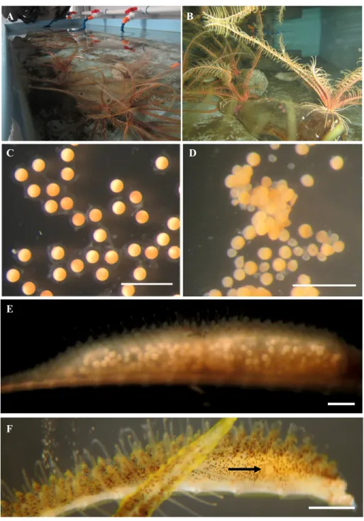
Longitude: -125.13720095) by SCUBA and transferred in plastic bags to BMSC where they were kept in the dark in flow through seawater tables (Figure 2.2A-B). Crinoids are diecious. To determined individual’s sex, a pinnule, which houses a tubular gonad, was removed from an arm and dissected out to find oocytes or sperm. To reduce the risk of natural spawning, males and females were kept in separate tables. Within an individual gonad gametogenesis is non-synchronous. Mature oocytes, which are round and non-sticky were separated from the
18
immature oocytes which adhere into clusters (Figure 2.2C-D) (Mladenov and Chia, 1983). Pinnules from each of the females were removed daily and checked for mature oocytes (Figure 2.2E-F). Sperm, also taken from pinnules by dissection, was in most cases mature and ready for fertilization. When mature oocytes were obtained, fertilization was conducted in small glass bowls with a minimal quantity of sperm, followed by several rinses with 50 µm filtered seawater. Individual cultures were kept at approximately 8 °C and placed on a shallow sea table, and washed every other day. A developmental timetable of the species is provided in Table 2.1. Doliolaria larvae were reared in clean glass bowls and bowls containing glass covered with a biofilm, coralline algae, rust, mussel shells and serpulid worm tubes. After several days of growth, feeding pentacrinoids were fed a mixture of Rhodomonas, Isocrysis and Chlorella every other day. The oldest larvae cultured were pentacrinoids of 56 day-old after fertilization (or 30 days old as pentacrinoids, for the quickest of the culture to metamorphose). This culture contained about 200 fertilized eggs. The majority survived and reached the pentacrinoid stage.
2.3.1 Ossicle isolation and observation
Larvae were collected and fixed in cold 2 % paraformaldehyde for one hour and then washed with Millipore filtered sea water. For ossicle observation, larvae were fixed in sodium borate solution between pH 8 and pH 10 and kept in a storage buffer (5.0 g/L sodium glycerophosphate powder in 70 % ethanol). Ossicles were isolated from living or fixed doliolaria, cystidean and pentacrinoid larvae by dissolving the tissues with one drop of 6 % bleach added to the drop of water pipetted with the larva directly on slides for microscopic observation, then rinsed with distilled water. Observations were made with an Olympus FluoViewTM 300 compound microscope equipped with polarizing filters and photographs were taken with a Coolsnap ProCF camera made by Roper Scientific Photometrics.
19
Table 2.1. Timetable of developmental events of F. serratissima, from fertilization to pentacrinoid, between 9.5 and 11.5 oC. All descriptions are taken from Mladenov and Chia (1983). F. serratissima development begins by free spawning of gametes into the water where fertilization takes place.
Age since fertilization
Stage Characteristics
before fertilization Ovulated egg Pale pink, perfectly spherical, diameter of 207 ± 6 µm
(Unfertilized)
0 - 3 min Fertilization Ridged fertilization membrane, perivitelline space Embryonic development
2 h - 2.25 h First cleavage Meridional, unilateral
2.25 h - 3.5 h Second cleavage Meridional, unilateral, right angle with first 3.5 h - 4.75 h Third cleavage Equatorial, radial, slightly unequal, 8-cell stage 4.75 h - 6 h Fourth cleavage Meridional, 16-cell stage, three pores
9 h Spherical embryo Only vegetal pore remains
12.5 h Coeloblastula About 200 cells, disappearance of vegetal pore 19 h Vegetal pole of blastula flattens into vegetal plate,
1000 cells
21 h Gastrula Gastrulation
27 h Cilia
Larval development
35 h to 47 h Pre-doliolaria Hatch from fertilization membrane
4 d Doliolaria Ciliated bands, vestibular invagination, antero-ventral adhesive pit, delicate glycocalyx. Sinusoidal swim path near the water surface 4.5 d Developed skeleton: 10 skeletal plates, primordia
of the oral and basal plates of the future cystidean. Exploration of substratum
Metamorphosis 4.6 d (possible delay
of 9 d)
Cystidean Attachment disk, loss of cilia, cuticle replaces glycocalyx, covering over the vestibular
invagination, 90 degree rotation of the vestibule, stalk. Gregarious settlement, non-feeding
16 d (possible delay
of 14 d) Pentacrinoid elongation. Feed on small food particles Extension of the 15 papillate tube feet, further stalk 4 months 10 adult arms. New ossicles: axillary, brachial,
costal, radial
6 months Arm span of 6.5 mm, cirri and pinnules not yet present
20
2.3.2 Settlement observations
In an effort to increase the settlement success of our larvae, so that a sufficient number of later developmental stages could be studied, five substrates were collected from the site of the adult population for settlement preference trials: i) shards of glass slides that had rested in sea water for four weeks (glass biofilm), ii) crustose coralline algae, iii) rust, iv) fragments of mussel shells and v) pieces of serpulid worm casings. Rust was taken from the wood pinches used to identify the bowls of culture. It was included in the analysis to see if fallen rust from pinches would affect larvae survival and settlement. For the rest of the study, pinches to identify cultures were new and lacked rust. One of each substrate was added to three replicate bowls, and an additional three control bowls, without substrate were prepared. At 5 days post-fertilization, 10 to 13 doliolaria larvae were transferred in each of the eighteen culture bowls. The number of settled larvae in each culture was counted at both 7 and 9 days post-fertilization. They were considered settled when they remained attached to the substrate following a light blast of water from a pipette. Settling proportions were compared using two one-way analysis of variance (ANOVA) tests, one test for day 7 and one test for day 9 after arcsine transformations of the data. The significance of the differences between treatments was determined using a Tukey post-hoc test.
After finding the greatest settlement success in the presence of coralline algae, we then compared the timing of settlement in a culture with branched coralline algae versus a culture without any substrate aside from a glass bowl. We divided a culture of embryos in two glass bowls just after fertilization. Branched coralline algae were added three days after fertilization, when the doliolariae were ciliated and swimming. Twenty-two larvae were present in the culture with algae and twenty-five larvae were in the culture without algae. Eleven days after fertilization, some larvae of the culture without algae were transferred to a bowl with algae to determine if they would settle. In other cultures, broken sea urchin tests and crustose coralline algae were added.
The diameters of doliolaria and cystidean ossicles at different ages were measured with a ± 5 µm precision. An ANCOVA test was conducted to see if the presence of branched coralline algae significantly affected the average ossicle diameter per larva depending on their age.
21
Also, five newly settled larvae were purposefully dislodged from the substrate with a sharp glass tool to see if they would grow normally without being attached.
2.3.3 Scanning electron microscopy (SEM) and electron-dispersive spectroscopy (EDS)
SEM and EDS analyses were performed on pentacrinoid ossicles at St. Francis-Xavier University. Ossicles were cleaned in 6 % bleach for two minutes on a slide until the soft tissues were dissolved. Ossicles were washed several times with distilled water, dried in 95 % ethanol, and transferred to an aluminium SEM stub. Ossicles were carbon coated using a Devon D205A sputter coater, analyzed and imaged using a JEOL JSM-6010LA SEM. EDS allowed the identification of elements present in the pentacrinoid ossicles and provided an estimate of their abundance. Twenty-six EDS analyses were performed on ten different plates, columnar ossicles and attachment disk of five pentacrinoids of 22 d old with an accelerate voltage of 10 to 20 kV, spot size of 50 to 60 and working distance of 8 to 11 mm.
2.3.4 Confocal Raman spectroscopy
Confocal Raman spectroscopy was used at St. Francis-Xavier University to identify the crystallization of CaCO3 on different ossicles of the pentacrinoids (see Methods in Cameron
and Bishop, 2012). A Renishaw InVia Raman miscrospectrometer attached to a Leica microscope with a 50X objective equipped with a deep depletion CCD detector, 1800 mm-1, holographic notch filter, argon ion 514.5 nm laser, 28 mW output and 5 mW on sample allowed us to acquire spectra of the samples.
2.4. Results
2.4.1 Ossicles in larvae of F. serratissima
The ossicles of approximately twenty larvae for each three larval stages were isolated for microscopy observations. The first spicules appeared in doliolariae at an age of four days after fertilization. There were normally ten columnar ossicles in doliolariae and the number increased during the later stages. Rare cases of 9 or 11 columnar ossicles in doliolaria were