Université du Québec INRS- Institut Armand-Frappier
Pseudoplusia includens densovirus (PiDNV)
genome organization and expression strategy
Par
Oanh Thi Hoang Huynh
Mémoire présenté pour l'obtention du grade de Maître ès sciences (M.Sc.) en Virologie et Immunologie Examinateur interne Examinateur externe Directeur de recherche Co-directeur de recherche Jury d'évaluation Angela Pearson, Ph.D. INRS-Institut Armand-Frappier Guy Lemay, Ph.D.
Département de microbiologie et immunologie Université de Montréal
Peter Tijssen, Ph.D.
INRS-Institut Armand-Frappier Tsuneyuki Ozaki, Ph.D.
INRS-Énergie, Matériaux et Télécommunication
_ _ A_c_know!_edgement<; - - - _ _ _ _ _ _ _ _
ACKNOWLEDGEMENTS
Last two years and a half is one of the most special joumeys that I have ever experienced. I would not have been possible to go to the end without the support of many kind people to whom I would like to show my deepest gratitude.
1 am truly thankful to the financial support of the Biotechnology center of Ho Chi Minh city and its scholarship program, which gave me the opportunity to do my Master in Canada. I would also like to thank the financial and academie support of the Armand-Frappier Institute, where I spent most of my studying time, and Dr. Ozaki (INRS-EMT) for financially supporting my virus inactivation work.
1 owe sincere and eamest thankfulness to my supervisor, Peter Tijssen and my co-supervisor, Tsuneyuki Ozaki who kindly gave me the chance to work in their laboratories and guided me throughout my study. 1 am sure that 1 would not have been able to finish without their support, inspiration, understanding and encouragement.
I am also sincerely grateful to the scientific support of my colleagues who I feel very lucky to work with. Your support really helped me develop my techniques as well as acquire knowledge in the laboratory.
1 am obliged to my family and my friends whose constantly moral support has been making my life more enjoyable.
It is again a great pleasure to acknowledge everyone who has ever helped me throughout my study.
Table of contents
---·---TABLE OF CONTENTS
ACKNOWLEDGEMENTS ...•.•...••.•...•...•.•...•...•...•.•...•• i TABLE OF CONTENTS ...••.•...•...•...••.•...•...•. ii LIST 0 F FI GU .RES ....•.•.•...•...•••••....•...•...•••..•...•..•.•.•••...•...•...•.•...•...••.••...•... vLIST OF TABLES ...•..•...•...•...
vii
LIST OF ABB.REVIATIONS .•.••.•••••••.•.•..•.••••••.••••.•...••..••••••••••...•...••••.•.••••....••.•••••••.••...••.••••.••..•..
viii
, SO MMAI.RE .RECAPITULA TIF ••••••....•...•.•.•••••••...•...•...•.•••••....•...•••...•.•..•••••.•..•.•...•..••. 1
INTRODUCTION ...•...•...••...•...•..••...••...•...•.•...•. 6
REVIEW OF LITERA TURE ..•.•...•••..•...•.•...•...•...•... 8
1. The family
Parvoviridae ..•..•.••.•...••.•...•...••••••...•.•••.•...•.•...
91.1. Classification ... 9
1.2. Virus particle ... 11
1.2.1. Viral capsid ... 11
1.2.2. Viral genome ... 12
1.2.2.1. Telomeres ... 13
1.2.2.2. Genome organization of subgroup A ambisense densoviruses ... 14
1.3. Virallife cycle ... 16
2.
Pseudoplusia includens
densovirus (PiDN'V) ... 18METHODOLOGY ....•.••...•...•.••.•••...•..•.•....•.•...•...•...•••.•...•. 20
1. Cloning and sequencing the genome of PiDNV .•..•...•.•..•.••••••...•.••••.•..•.•...•..•...•.•...• 21
1.1. Isolation and purification of PiDNV DNA from infected larvae ... 21
Table of contents
1.3. Agarose gel electrophoresis ... 21
1.4. Preparation of insert ... 22
1.5. Preparation ofvector ... 22
1.6. Ligation ... 23
1. 7. Transformation ... 24
1. 7 .1. Preparation of electrocompetent cells ... 24
1. 7 .2. Electroporation ... 24
1.8. Plasmid DNA extraction ... 25
1.9. Analysis of clones by restriction digestion ... 25
1.1 O. Sequencing ... 26
1.11. Cloning of the who le genome of PiDNV ... 27
1.12. Sequencing of hairpins in linear pJ AZZ clones ... 28
2. Bioinformatics ... 29
2.1. Primer design ... 29
2.2. Assembly of sequence ofPiDNV genome ... 29
2.3. Structure analysis of viral hairpins ... 29
2.4. Genome sequence analysis ... 29
2.5. Prediction of 3D structure of PiDNV capsid subunit and capsid ... 30
RESULTS ... 31
1. Cloning of PiDNV genome ... 32
1.1. Isolation and purification of PiDNV DNA ... 32
1.2. Cloning oftwo halves ofPiDNV genome ... 32
1.3. Cloning of viral hairpins ... 33
Table of contents
1.3.2. Cloning oftwo parts ofhairpins ... 35
1.4. Cloning the who1e genome of PiDNV ... 36
1.5. PCR to amplify the 5'-hairpin ... 38
2. Sequence and organization of PiDNV genome ... 39
2.1. Telomeres ofPiDNV genome ... 39
2.2. Coding sequences ofPiDNV genome ... 42
2.2.1. Non-structural proteins ... 43
2.2.2. Structural pro teins ... 44
3. Predicted structure of capsid subunit and capsid of PiDNV ... 46
D ISCUSSI 0 N ... 51
1. Cloning and sequencing of PiDNV genome ... 52
2. Nucleotide sequence of PiON V genome and comparison with other viruses in the family ... 53
3. Genome organization ofPiDNV and comparison with other densoviruses ... 53
4. Prediction of the 3D structure of the capsid subunit and capsid ofPiDNV ... 55
CONCLUSION ... 56
APPENDIX 1. PUBLICATION ... 58
APPENDIX 2. VIRUS INACTIVATION BY IMPULSIVE STIMULATED RAMAN SCATTERING (ISRS) ... 62
List of Figures
LIST OF FIGURES
Figure 1. Phylogenetic relationship among the pleiotropic NS 1 pro teins of members of the
family Parvoviridae ... 10
Figure 2. Morphology of parvoviruses ... 11
Figure 3. Genome organization of parvoviruses ... 12
Figure 4. Genome organization of ambisense densoviruses ... 15
Figure 5. Terminal hairpin of subgroup A densoviruses ... 15
Figure 6. Parvoviral "rolling-hairpin" DNA replication ... 18
Figure 7. DNA ofPiDNV ... 19
Figure 8. Vector pBluescript KS (+) ... 23
Figure 9. The pJAZ.Z-OC vector ... 28
Figure 10. Purified PiDNV DNA ... 32
Figure Il. Clones oftwo halves of PiDNV genome (digested with Xbal) ... 33
Figure 12. Cloning for sequencing ofhairpins ... 34
Figure 13. BamHl-BamHI subclone ... 34
Figure 14. Digestion ofBamHI-BamHI clones ... 35
Figure 15. Hairpin clones ( digested with Pvull) ... 36
Figure 16. Example of colon y PCR of the cloning of the who le genome of PiDNV ... 3 7 Figure 17. Positive linear clone of complete PiDNV genome (Notl) ... 37
Figure 18. First PCR of the 5'-hairpin from linear clones ... 38
Figure 19. Second (nested) PCR of the 5'-hairpin from linear clones ... 38
Figure 20. Alignment of two versions ( flip and flop) of 3 '-terminal hairpin ... 40
Figure 21. Hairpin ofPiDNV ... 41
List of Figu.'!!!_
Figure 23. Conserved domain within the ORF2 ... 45
Figure 24. Alignment of the conserved domain within ORF4 (30483) with the conserved domain of capsid protein VP4 of densoviruses ... 46
Figure 25. Alignment of phospholipase A2 domain in VPI of PiDNV and representative parvoviruses ... 46
Figure 26. Alignment of amino ac id sequence of VP of MVM and PPV ... 4 7 Figure 27. Alignment ofamino acid sequence ofVP4 ofPiDNV and GmDNV (COBALT) .... 48
Figure 28. Example of co-ordinates of last atoms in the protein string of capsid subunit of PiDNV ... 48
Figure 29. Reliability ofPiDNV capsid subunit mode!: QMEAN4 global scores ... 48
Figure 30. Reliability of PiDNV capsid subunit mode! according to Swiss-Model (online): Colouring by residue error ... 49
Figure 31. PiDNV capsid subunit.. ... 49
Figure 32. Mode! of capsid structure ofPiDNV ... 50
Figure 33. Viroses in inactivation experiments ... 70
Figure 34. Experimental setup ... 71
Figure 35. Example of a titration plate ... 75
Figure 36. Impact of ti me on virus inactivation ... 77
Figure 37. Impact of energy on virus inactivation ... 78
Figure 38. Impact oflaser pulse width on virus inactivation ... 79
Figure 39. Comparison of remaining virus infectivity between one- and two-colour irradiation.80 Figure 40. Virus inactivation with both 800 nm and 400 nm laser ... 81
Table 1. Table 2. Table 3. Table 4. Table 5. Table 6. List of Tables
LIST OF TABLES
Sequence of primers for sequencing PiDNV genome ... 26 BLAST Alignment ofPiDNV sequence and representative parvovirus sequences .. 39 Predicted promoter sequence and transcription factors for these promoters ... 42 Selected protein motifs and profiles present in ORF 1, ORF2, ORF3 and ORF4 ... 45 Representative inactivation technologies ... 69 Experiments with the 1 0 Hz laser ... 7 4
a a
AAV
ATP
CDC
CDD
DMSO
DNA
dNTP
EDTA
fsPiDNV genome organization and expression strategy: List of abbreviations
LIST OF ABBREVIATIONS
Amino acidAdeno-associated virus Adenosine triphosphate
Centers for Disease control and Prevention Conserved domains database
Dimethyl sulfoxide Deoxyribonucleic acid
Deoxyribonucleotide triphosphate Ethylenediaminetetraacetic acid Femtosecond
GmDNV :
Galleria mellonella densovirusHBV
Hepatitis 8 virusHCV
Hepatitis C virusHIV
Human immunodeficiency virusHTLV
Human T-lymphotropic virusITR
Inverted terminal repeatISRS
Impulsive Stimulated Raman ScatteringJcDNV
Junonia coenia densovirusL
B
Luria brothMVM
Minute virus of mi ceNS
Non-structural (protein)NID
Non-ionie detergentPiDNV genome organization and expression strategy: List of abbreviations
PCR Polymerase chain reaction Pfu Plaque fonning units
PIDNV Pefudensovirus
PiDNV Pseudoplusia includens densovirus
PLA2 Phospholipase A2
PPV Porcine parvovirus u Unit (of enzyme activity) VP Structural protein
WNV West Nile virus
PiDNV genome organization and expression strategy: Sommair_e récapitulatif
,
PiDNV geno'!_le organization and expression strategl: Sommaire récapitulatif
La famille de Parvoviridae regroupe des virus de petite taille (215-255 Â) sans enveloppe dont la capside est de symétrie icosaédrique et dont le génome est une molécule d'ADN simple brin d'une taille comprise entre 4 et 6 kb. Cette famille se subdivise en deux sous-familles, celles des Parvovirinae regroupant les virus qui infectent les vertébrés et celle des Densovirinae regroupant les virus pathogènes d'invertébrés (68). Environ la moitié seulement des densovirus isolés à ce jour sont classés, faute de caractérisation suffisante de leur génome. Le densovirus de Pseudoplusia includens (PiDNV), objet de notre étude, a été isolé par Chao et al. (13) à partir des larves de ce lépidoptère. Les particules de PiDNV sont icosaédriques et contiennent une molécule d'ADN linéaire simple brin de 6 kb. À chaque extrémité de la molécule une répétition terminale inversée (ITR) correspondant à 6-7% de la taille du génome permet, par appariement des séquences complémentaires, la formation d'une structure en forme de «poêle à frire» (panhandle-like structure) observable au microscope électronique (13). L'électrophorèse en gel dénaturant SDS-polyacrylamide a révélé 4 protéines virales VPI-4 de 87 à 46 kDa. Par immunodiffusion, une réaction croisée partielle a été observée entre le virus GmDNV et le PiDNV (13). Pseudoplusia includens, l'hôte du PiDNV, a une aire de répartition s'étendant sur tout le continent américain (Nord et Sud). Il s'agit d'une espèce polyphage pouvant se nourrir non seulement sur le soja mais aussi d'autres plantes économiquement importantes comme la patate douce, l'arachide, le coton, la tomate, les crucifères, etc. (12). En conséquence, le virus PiDNV peut être utilisé comme un agent de lutte biologique contre ce ravageur. Cependant, depuis la première caractérisation de Chao et al. (13), aucune caractérisation moléculaire de son génome n'a été pas faite. Nous reportons dans ce mémoire les résultats du clonage et du séquençage du génome du PiDNV ainsi que l'analyse de son organisation et de sa stratégie d'expression.
Nous avons purifié le PiDNV à partir des larves de Pseudoplusia includens, qui ont été fournies par le Dr. Yu-Chan Chao en 2000. Le génome du virus a été extrait et son analyse en gel d'agarose a permis de visualiser d'une bande de 6 kb. Ce génome a été coupé en deux moitiés par 1 'enzyme de restriction Clal et les deux moitiés ont été clonées dans le vecteur pBluescript KS ( ) et séquencées. Le séquençage de la région interne du génome, entre les répétitions terminales inversées (ITRs), n'a pas posé de problème. Par contre, le séquençage des ITRs dont l'appariement de séquences complémentaires génère des structures en « épingles à cheveux» (hairpins) s'est avéré plus difficile et on a dû faire appel à différentes stratégies pour obtenir ces séquences. Ainsi, l'épingle à cheveux à chaque extrémité du génome viral dans un clone a été coupée au site de restriction BstUI en deux moitiés et chaque moitié a été clonée puis séquencée séparément. Cette approche a permis d'obtenir la séquence de l'extrémité 3' mais pas celle de l'extrémité 5'. Le génome complet de PiDNV a été également cloné dans le vecteur linéaire pJAZZ-OC spécialement adapté au clonage d'ADN difficile à cloner (31). Seulement 6 clones positifs ont été obtenus à partir de 1848 colonies (0.3%). Ces clones ont été séquencés et leur séquençage a confirmé la séquence obtenue avec les séquences clonées dans pBluescript sans toutefois fournir la séquence de 1 'épingle à cheveux à 1 'extrémité 5'. Nous avons alors amplifié par PCR les ITRs en présence de trois additifs: la bétaine, le DMSO et le 7-deaza dGTP
PiDNV genome organi~ation and expression strategy: Sommaire récapitulatif
(47). Cette méthode a permis d'obtenir la séquence de l'ITR 5'. L'assemblage à l'aide du programme CAP3 de tous les contigs a généré une séquence de 5990 nucléotides correspondant au génome complet du PiDNV. BLAST a démontré une identité élevée entre le génome de PiDNV et les génomes des virus du sous-groupe A du genre Densovirus: JcDNV (87%), MlDNV (86%) and GmDNV (84%) et une identité beaucoup plus faible (<25%) avec les séquences des autres densovirus, y compris ceux appartenant aux autres sous-groupes du même genre. L'ITR 5' terminal et l'ITR 3' tenninal du PiDNV ont chacun une taille de 540 nucléotides et des séquences parfaitement complémentaires l'une de l'autre. Les 120 nucléotides aux extrémités de ces ITRs peuvent se replier pour créer les épingles à cheveux ayant une configuration «Y», semblable à celles des virus MlDNV, GmDNV et JcDNV (66). Comme chez ces derniers virus, les configurations «flip» et «flop» sont aussi observées dans les épingles à
cheveux de PiDNV. Dans ces deux configurations, les séquences formant la «tige» (stem) de 1' «Y» sont identique tandis que les deux petites palindromiques formant les deux «bras» de l'«Y» sont inversées dans la configuration «flip» par rapport à la configuration «flop». On a retrouvé également près de 1' extrémité de 3' des épingles à cheveux le motif hautement conservé (GAC)4 (24) chez tous les membres du sous-groupe A du genre Densovirus qui est considéré comme le site de fixation de la protéine NS 1 pour couper le génome et produire 1' origine de réplication. On a trouvé également dans la séquence des ITRs, les séquences spécifiques des deux promoteurs régulant l'expression des gènes non-structuraux (NS) sur un brin et structuraux (VP) sur le brin complémentaire avec leur «TATA box» localisées juste à la fin des ITRs.
Le programme ORF Finder (NCBI) a identifié quatre cadres ouverts de lecture (ORFs) dans le génome de PiDNV (ORF1-4). ORFl, ORF2 et ORF3 sont localisés sur la moitié 5' d'un brin tandis que l'ORF4 occupe la moitié 5' du brin complémentaire. Cette organisation est très semblable à celle du virus GmDNV, un densovirus ambisense dans le sous-groupe A du genre Densovirus. La distribution des ORF2, ORF3 et ORFl de PiDNV correspond respectivement à la distribution des protéines non-structurales NS 1, NS2 et NS3 du virus GmDNV. Il en est de même, pour l'ORF4 du PiDNV dont l'organisation correspond à l'ORF des protéines VP du virus GmDNV.
Les séquences codantes des protéines NS de PiDNV sont portées par les ORFl, ORF2 et ORF3. Le premier ORF (ORFl, nt 647 à 1348) code pour une protéine de 233 aa dont la séquence montre une forte homologie avec la séquence de la protéine NS3 des virus MlDNV (75%), GmDNV (73%) et JcDNV (72%). Le second ORF (ORF2, nt 1355 à 3019) code pour une protéine de 554 aa. Cette protéine possède un domaine hautement conservé de la protéine non-structurale NS 1 des parvovirus. Le troisième ORF (ORF3, nt 1362 à 2189) est inclus dans la séquence de l'ORF2 et code pour une protéine de 275 aa présentant une très forte homologie avec la protéine non-structurale NS3 des virus MlDNV (96%), JcDNV (93%) et GmDNV (94%). La forte homologie de séquence des génomes du PiDNV et du GmDNV et 1 'organisation similaire de leurs séquences codantes suggèrent que PiDNV utilise la même stratégie
d'expression que GmDNV. Ainsi ORFl du PiDNV qui porte la séquence NS3 est selon toute vraisemblance traduit à partir d'un transcrit NS épissé de la séquence NS3. L'ORF de NS2 est localisé à l'intérieur de l'ORF de NSl mais leurs codons d'initiation sont chacun dans un cadre de lecture différent et à peu de distance l'un de l'autre (ATG de NSl à la position 1355, ATG de NS2 à la position 1362). La traduction de NS2 résulte donc vraisemblablement d'un mécanisme «leaky scanning». Le signal de polyadénylation (AAT AAAA) localisé en position 3002, soit 17 nucléotides en amont du codon stop de la NS 1 est vraisemblablement fonctionnel.
L'ORF4 du PiDNV est le plus grand (2418 nucléotides) et a une capacité de codage de 805 aa. Comme pour les membres du sous-groupe A du genre Densovirus, la transcription de cet ORF génère vraisemblablement un seul messager à partir duquel les quatre protéines de capside (VP1-4) sont synthétisées par le mécanisme de «leaky scanning». Le fait que l'ORF4 commence à seulement 27 nucléotides en aval de la «TATA box» indique que les éléments essentiels du promoteur se trouvent dans les ITRs. Deux domaines conservés sont présents dans cet ORF. Le premier est celui de la protéine VP4 qui joue un rôle essentiel dans l'architecture de la capside des densovirus et le deuxième est la région N-terminale de la protéine VP 1 dans laquelle on retrouve chez quasiment tous les parvovirus deux motifs importants qui sont le motifPLA2 (phospholipase de type A2) (DxxAxxHDxxY) et Ca2+-binding loop (GPGN). Le motif de PLA2 est très conservé et joue un rôle essentiel dans la libération du virus de 1 'endosome tardif. Le Ca2+ -binding loop est en position 186 de la protéine VP 1 et 17 nucléotides en amont du site catalytique HD (nucléotide 207). En outre, la séquence PGYKYL en amont du motif GPGN est conservée dans PiDNV, GmDNV, MlDNV, MVM, PPV tandis que dans Bl9, seuls P, G, et Y sont conservés (PGxxYx).
La structure 3D de la sous-unité de la capside du PiDNV (protéine VP4) a été prédite à l'aide du modelage d'homologie Swiss-Model sur la base des fortes similarités (81%) existant entre les séquences des protéines VP4 du GmDNV et du PiDNV. La fiabilité de la structure a été estimée par «QMEAN4 global scores». Le modèle a un score brut de QMEAN4 très bas (0.308) et un score de QMEAN Z très négative (-7.76), c'est-à-dire une fiabilité très basse de la structure prédite. À l'aide du programme UCSF Chimera (50), un modèle identique à celui produit par Swiss-Model a été obtenu. Quand ce modèle a été utilisé pour l'assemblage d'une capside complète (soixante sous-unités de la symétrie T=l) du virus, il est apparu que les boucles extérieures peuvent se relier et s'entrelacer dans les canyons de la capside pour former une structure cohérente.
En résumé, le génome du virus PiDNV a été entièrement séquencé et a révélé une séquence de 5990 nucléotides. Le génome possède à chaque extrémité une longue répétition inversée (ITR) de 540 nts. Les 120 premiers nucléotides de chaque ITR peuvent se replier pour former une structure en épingle à cheveux suggérant un mécanisme de réplication de type de «rolling circle». Ce mécanisme, commun aux différents parvovirus, est à l'origine des deux orientations alternatives «flip» ou «flop» dans l'épingle. Le génome du PiDNV est ambisense c'est-à-dire, que les ORFs des protéines structurales (VP) et non-structurales (NS) sont chacun
PiDNV genome organization and expression strategy: Sommaire récapitulatif
localisé dans la moitié 5' des brins opposés. Sur un brin, 3 ORFs codent potentiellement les protéines NSl, NS2 et NS3. Par convention, les gènes NS sont représentés à gauche du génome viral. Sur le brin complémentaire, un ORF de grande taille occupe toute la moitié 5' et code les protéines structurales VPl, VP2, VP3 et VP4. La séquence nucléotide et l'organisation du génome de PiDNV justifient la classification de ce virus au sein du sous-groupe A du genre Densovirus de la sous-famille Densovirinae de la famille Parvoviridae.
~----
___
q_l_~_j
__
~:f_3.?
_____ _
Étudiante Date
·t
'.
a.· ··-'i
PiDNV genome organizatwn and expression strategy: Introduction
PiDNV genome organization and expression strategy: Introduction
The family Parvoviridae includes small (around 25 nm in diameter), non-enveloped icosahedral viroses whose genome is a single-stranded, linear DNA and about 4 to 6 kb in length. These viroses are classified into two subfamilies: Parvovirinae infecting vertebrates and Densovirinae infecting invertebrates. Densoviruses (DNVs) comprise viruses isolated from arthropods, mainly insects and cause high mortality in infected hosts. In addition to DNVs and all vertebrate parvoviruses with monosense genomic organization (structural and non-structural proteins encoding sequences present on the same strand), there are ambisense DNVs of which two representatives are GmDNV and JcDNV. All parvoviruses possess palindromic sequences at the extremities of the viral genome. Although these terminal sequences are dissimilar in length and in structure between different parvoviruses, they proved to play an important role in viral DNA replication. To date, the near-atomic structures of the three densoviruses (BmDNV, PstDNV and GmDNV) have been solved displaying a capsid composed of 60 subunits of identical proteins sorne of which include a domain with phospholipase Az (PLAz) activity.
Chao et al. (1985) (13) isolated a densovirus, PiDNV, from larvae of soybean loopers (Pseudoplusia includens). Up until now, only the physicochemical and serological characteristics of this virus were resolved (13). Because PiDNV can be used as a biological control against soybean loopers, an important agriculture pest, we would like to determine further molecular characteristics of this virus. The four objectives of the work were the following:
l. Cloning and sequencing the genome of PiDNV. In this objective, we would like to have a clone with the whole viral genome, including the complete inverted terminal repeats (ITRs) at two extremities of the genome in a plasmid vector. Then the complete nucleotide sequence of PiDNV genome will be determined by sequencing.
2. Determining the organization of the genome of PiDNV from which expression strategy of the virus can be predicted.
3. Comparing the genomic organization of PiDNV with other densoviruses.
PiDNV genome organization and expression strategy: Review ofliterature
PiDNV genome organization and expression stratf!_gy: Review ofliterature
1. The family
Parvoviridae
1.1. Classification
From the first parvovirus isolation (Kilham rat virus) (39) in 1959, the family
Parvoviridae has been rapidly expanding to include all viroses that are small, isometric, non-enveloped and have linear single-stranded DNA. This family is divided into two subfamilies based on host range: Parvovirinae which infect vertebrates and Densovirinae which infect invertebrates. Phylogenetic analysis and molecular characterization allows further classification of members within a subfamily into specifie genera and species. Up until now, there are five genera in the subfamily Parvovirinae and four genera within the subfamily Densovirinae. These genera involve 37 accepted species and many unassigned or tentatively assigned to a genus or species (65).
Although also isolated from Crustacea, most of densoviruses (DNVs) are from insects including Lepidoptera, Diptera, Orthoptera, Dictyoptera and Odonata (66). According to the phylogeny of the most conserved non-structural protein sequences (Figure 1 ), the individual species within the subfamily differ by at least 5 percent. In addition, natural infection of each species seems usually to be confined to single host species. Only half of around 30 reported DNV isolates are presently classified due to the lack of sufficient genomic characterization. In general, DNVs were classified to four genera: Densovirus, lteravirus, Brevidensovirus and
Pefudensovirus. Unlike iteraviruses and brevidensoviruses with monosense genome organization in the Densovirinae subfamily, genomes of members in the Densovirus and Pefudensovirus genera are ambisense, meaning that their non-structural (NS) proteins are encoded on the left half of the genomic DNA strand and structural (VP) proteins on the right half of the complementary strand of the single-stranded DNA genome. Upon infection a double-stranded genome is created by self-priming from its terminal hairpin so that both gene cassettes are present on the same molecule. The ambisense densoviruses are further subdivided into 3 subgroups: subgroup A, B and C. Subgroup A of the genus Densovirus is composed of viruses infecting Lepidoptera (butterflies and moths), represented by Junonia coenia DNV (JcDNV) (24) and Galleria mellonella DNV (GmDNV) while two other groups encompass viroses isolated from different orders of insects (68). In spite the fact that ali viruses in the genus have ambisense organization, viroses in subgroup A display high sequence identity (80-90 percent)
PiDNV genome organization and expression strategy: Review of litera ture
while those in subgroup B show very low sequence identities (10-30 percent) maybe because they adapted to hosts of different insect orders (66).
4S
PslDNV
7
tot
Dep ndovirus
Possible new genus
Erythrovirus Amdovirus• Bocav1rus Possible new genus• P~NO\il/l/S Brevfdensovfrus• llemvfrus
Possible new species Possible new genus
Den
Poss1ble new genus
~
~ <...
.
3C :t ::s 0 Q) ::a Ill 0x:
::ax:
Figure 1. Phylogenetic relationship among the pleiotropic NSl proteins of members of the family Parvoviridae. The tree was constructed with the programs included in the Phylip package at the http://mobyle.pasteur.fr/cgi-bin/portal.py website (ClustalW-multialign, Phylip distance matrix, PROTDIST, Neighbor Joining method, and phylogenetic tree drawing). The percentage of replicate trees in which the associated taxa clustered together in the bootstrap test (1000 replicates) is shown next to the branches. The scale bar represents the rate of amino acid substitutions. This phylogenetic analysis distinguishes the two subfamilies, as weil as the monosense and ambisense densoviruses, and recognizes the different genera as separate clades. Severa! other clades that are possible new genera are also recognized. The asterisk indicates clades of viruses that do not contain the phospholipase A2 motif in their capsid proteins. Reprinted from reference (68).
PiDNV genome organization and expression strategy: Review ofliterature
1.2. Virus particle
1.2.1. Viral capsid
Figure 2. Morphology of parvoviruses. (A) Side-view, at a resolution of 3.5 A, of a tilted model of a porcine parvovirus (PPV) capsid with the top five trimers translated 120 A along its 5-fold axis from the middle body of 10 trimers (five shown in dark blue and five in light blue); the bottom five trimers are not shown. The 5-fold axes are located at the intersection of five trimers (e.g. at arrow: green, magenta, two dark blue and one light blue colored trimer). (B) Top view of model shown in (A), without light-blue trimers, along the 5-fold axis. The channels at the 5-fold axes are clearly visible (arrow indicates same 5-fold axis as shown by arrow in (A)). (C) Structure of a PPV trimer. The arrows indicate the intertwining of the GH-loop (between B strands G and H) in the counter-clockwise located proteins from near the 3-fold axis towards the 2-fold axis (arrows). The GH-loop actually consists of two loops of which one (loop 3, yellow), running to the 2-fold axis, is partly covered by loop 4 (cyan) near the 3-fold axis (oval arrow). (D) Parvoviruses may have remarkably similar structures despite low sequence identities. This figure shows the alignment of minute virus ofmice (MVM, in red; 549 amino acid residues, PDB lmvm) and PPV
(in blue; 542 amino acid residues, PDB Ik3v) structural proteins that have otùy 52% sequence identity. Nevertheless, 528 CV's (97%) of the residue pairs occupy the same position in the caps id (root-mean-square error of 1.0 A). (E) Negative contrast electron micrograph of empty and full PPV particles (bar = 50 mn) and space-filling models of the capsid structures ofPPV, Galleria mellonella densovirus (GmDNV; PDB: lDNV), human B19 virus (B 19V; PDB: ls58), and adenoassociated virus-2 (AA V -2; PDB: llp3) shown at a resolution of 4
A.
In each case, the view is down a 2-fold axis at the centre of the virus, with 3-fold axes left and right of centre, and 5-fold axes above and below. Models (A-D) have been rendered by PyMOL and the space-filling models by CHIMERA (Multiscale Models). Reprinted from (68).PiDNV genome organization and expression strategy: Review of litera ture
Parvoviral virions are very compact ("parvo" meaning "small"). They are 215-255
A
in diameter, non-enveloped and icosahedral (six 5-folds, ten 3-folds and fifteen 2-folds) with T=1 symmetry (Figure 2). Sorne parvoviruses (GmDNV) have smooth structures, sorne (AAV-2, PstDNV) have spiky ones (3- or 5-fold symmetry axes). In general, parvoviral particles are stable and can survive at room temperature for months or years with little loss of infectivity. The rugged virion is made up of structural proteins VP1-4 to protect the vulnerable DNA genome inside. Its molecular mass is about 5.5-6.2 x 10⁶, buoyant density in aqueous isopycnic cesium chloride gradient is 1.39 -1.43 glcm3, and sedimentation coefficients (S2ow) is approximately 110-120 S. While spermidine, spermine and putrescine were found in sorne insect viroses (GmDNV, JcDNV and BmDNV-1), cellular enzymes or chromatin components were not found in parvoviral particles. Ali parvoviruses, except brevidensoviruses, amdovirus, chicken and turkey parvoviruses and PmDNV, have a PLA2 domain in the minor structural protein which can play important role in virus entry and infection (68).1.2.2. Viral genome
Parvowus ~ ~___Il
(MVM)~~---
,
-
~~
---,r
LNS-.
l_V~
OensoVJrus (GnONV)~~---~
v-,
NS·<
VP 1~
Figure 3. Genome organization of parvoviruses. Genomes of representatives of four out of the nine genera of Parvoviridae. Their terminal hairpin structures are magnified around 10-fold of the single-stranded region. The promoters are indicated by arrows, major gene blocks by open arrows with N- to C- temtinal direction. Reprinted from (18).
PiDNV genome organization and expressio_n strategy: Review of/iterature
As a result of the tiny size of the viral particle which may confer the virus the capacity to enter the nucleus of host cell, only a limited size of the parvoviral genomes can be tolerated. In fact, they are exclusively 4-6 kb in length and comprise roughly 25 percent (1.5-2 x 10⁶) of the particle mass. These genomes are single-stranded linear DNA which terminate in
short palindromic sequences at two ends (Figure 3) (18). Within a viral particle, either positive-or negative-sense DNA can be packaged. The ratio of these two kinds of particles depends on the specifie virus, either equal polarity or predominantly minus. All parvoviruses possess two gene cassettes which are, by convention, non-structural proteins on the left hand and structural proteins on the right hand. In addition to overlapping reading frames, parvoviruses also have transcriptional promoters and splicing signais integrated in the same primary sequence. Furthermore, many parvoviral genomes are highly repetitive maybe due to the unidirectional, strand-displacement process of their genome replication. These repeats have their own benefits, for example, they can be binding sites for NS 1 proteins (5' -ACCA-3' motif) (18).
1.2.2.1. Telomeres
The telomeres are the hallmark of the Parvoviridae family. These imperfect palindromes of about 120-550 nucleotides are short imperfect palindromes. These sequences are able to fold back on themselves to create duplex secondary structures that vary in size, sequence and shape between genera or even between species in the genus Dependovirus. The terminal hairpins can be different in sorne viroses, such as left and right hairpins of Minute Virus of Mi ce (MVM), while in others they are part of inverted repeats at two extremities so that their sequences are closely related or identical like the hairpins of viroses in the genus Densovirus. These terminal palindromes can be small internai palindromes forming Y -shape, for example the hairpins of GmDNV, or T-shape like the left-hand end of AAV. On the other hand, these sequences maybe severely asymmetric or contain one or more clusters of mismatched nucleotides near the axis of the palindrome ( 18).
Despite the variety in size, sequence and shape, all parvoviral telomeres have certain functions in viral life cycle. First of all, these hairpins contain cis-acting information for the viral genome to be replicated and packaged. Parvoviruses have a special mechanism of DNA genomic replication which is called "rolling-hairpin replication" (RHR). In this process, parvoviruses use the terminal hairpins as primers for synthesizing the complementary strand.
PiDNV genome organizatJOn and expresswn strategy: Review of/iterature
After monomer or replicative intermediates were formed, viral replication origin within the hairpin is exposed to allow a protein (NS 1 or Rep68/78) to nick the DNA to separate unit-length genomes and also allow the termini to be co pied. Resulting from this process, an inversion of the terminus occurred when each round of replication ends. As all parvoviral hairpins are imperfect, the synthesized hairpins exhibit two configurations, which are named "tlip" and its reverse-complementary version "flop". In the genome replication, these palindromes can also act as hinges where the direction of replication is reversed at the end of each unit-length genome. This ability seems to depend on the imperfect or base-paired regions around the axis of each terminus which can provide favourable environment in energy for the hairpins to unfold and refold. Moreover, transcriptional control elements for nearby viral promoters were found in telomere sequences. Besides, viral telomeres contain sequences or structures that promote interactions between viral genome and capsid in viral packaging. Viruses with identical telomeres package their positive- or negative-sense DNA genome with equal ratio whereas those with different telomeres usually package their genomes disproportionately (except Lulli virus) (18).
1.2.2.2. Genome organization of subgroup A ambisense densoviruses
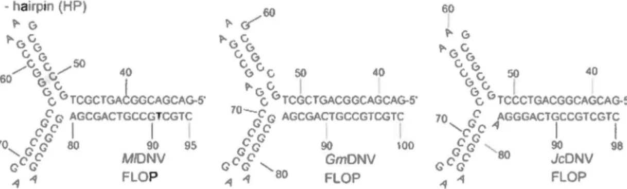
Genomes of viroses in this group are about 6 kb in length including two long inverted terminal repeats (ITRs) which are approximately 550 nucleotides, making up around 18 percent of the genome (Figure 4). About 130 nucleotides at the extremities can fold back into themselves to form hairpins with typical Y -shapes (Figure 5). Additionally, flip/tlop configurations are also found at both ends of the genomes (66). The remaining sequence ofiTRs contains TATA boxes and upstream promo ter elements for NS and VP genes ( 67).
Unlike vertebrate parvoviruses, ambisense densoviruses have three different NS proteins (NSl-3, with molecular mass 20, 30 and 60 kDa) which are encoded in similar ways among viruses within the group. In fact, the 5'-half of open reading frame (ORF) of NSl overlaps the ORF of NS2 although the proteins are encoded in different reading frames. Just upstream of the NSl/2 cassette is NS3 ORF (66). Transcript mapping of GmDNV (67) and MlDNV (25) revealed the expression strategy employed by these ambisense densoviruses. The 20-kDa NS3 protein is translated from a 2.5 kb transcript initiated at the upstream promoter within the 5' ITR while NS 1 and NS2 proteins are translated from a 1.8 kb transcript after splicing out the NS3 coding sequence from the initial 2.5 kb transcript. The translation of NS2
..
.
·....
..
..
,
..
'.
. :.PiDNV genome organization and expression st~ategy: Review of/iterature
proteins appears to be done via leaky scanning mechanism because the upstream initiation codon ofNS1 is in a poor context (YnnAUG). In addition, the proximity of the initiation codon ofNS1 and that of NS2 possibly promotes alternative initiation. Ding et al. (22) showed that NS 1 of
JcDNV recognizes and binds the (GAC)4 sequence in the stem of the hairpin. This enzyme
subsequently nicks the single-stranded forms of the hairpin preferentially at two sites (G*TAT*TG). It was demonstrated that this protein possesses helicase activity with less conserved Walker A-motif (GxxxxGK [T/S]). Another non-structural protein, the NS3 of JcDNV contains two zinger-finger motifs, and was shown to be essential for viral replication (1). NS 1 is the most conserved protein among ambisense densoviruses whereas no highly conserved domains were found in NS2 or NS3.
Ambisense densoviruses SubgroupA HP rm VPl
!'??
.
HPFigure 4. Genome organization of ambisense densoviruses. AJl viruses have ITRs and their structural proteins have a PLA2 motif. The large NS proteins contain NTPase (ATPase) and rolling circle replication (RCR) motifs (66). 60 ~ G ~6" vG "6" 50 40 6 6v 1 c. 6 TCCCTGACGGCAGCAG-5' 70 cF AGGGACTGCCGTCGTC 'cc"'~ 1 1 cFcf 90 98 G c Ge 80 JcONV .q -1 FLOP
Figure 5. Terminal hairpin of subgroup A densoviruses. Ali of them have Y-shape and each have two configurations: flip and flop (66), e.g. nucleotides 48-79 in MlDNV have the reverse-complement sequence in the flip configuration.
In contrast to non-structural proteins, structural proteins of subgroup A densovirus are encoded by only one ORF. After the VP mRNA is transcribed, the four VP proteins (45, 53, 58 and 89 kDa) are generated by a leaky scanning mechanism (25, 67). The two most important
PiDNV genome organization and expression strategy: Review of/iterature
conserved motifs were detected in ail ambisense densoviruses as weil as in iteraviruses and vertebrate parvoviruses (81). These are the Ca2+-binding loop (GPGN) and PLA2 (DxxAxxHDxxY) motifs, even though the specifie activity of densovirus PLA2 is significantly lower than that of iteravirus or vertebrate parvoviruses (25, 26, 42, 67, 81 ). Similar to other PLA2s, the distance between the Ca2+ -binding loop and the active site (HD) varies slightly in the densoviruses. Additionally, all the PLA2 identified up until now have been on the least abundant capsid protein VP 1 ( 66).
1.3. Virallife cycle
As tiny particles and thus with limited genetic capacity, parvoviruses must equip themselves with numero us mechanisms to take advantage of the host ceil throughout the who le virus life cycle from entry to egress (77). The earliest step in virus infection is attachment to the host cell, which is mediated by interactions between viral capsid molecules and receptors and/or coreceptors on host cell surface. The parvovirus capsid proteins involve a highly conserved ~
barrel motif and hypervariable regions connecting the eight strands of the ~-barrel. These variable regions may be responsible for usage of cellular receptors which may be heparan sulfate proteoglycan (AAV2) (63), sialic acid (AA V4, AAV5, AMDV, BPV, MVM) (17, 75), erythrocyte P antigen globoside (819) (8) or transferrin (CPV, FPV) (49). Following attachment, entry of parvoviruses to host cell is thought to be accomplished via receptor-mediated endocytosis. However, how these viruses escape endosomes remain unclear (77). Moreover, there are multiple pathways of intercellular trafficking in different cell types and hence the specifie way employed depends on the distinct virus and its host cell. Even though parvoviruses have to bring their genomes to the nucleus for replication, the nuclear transport mechanism is still not known. Nevertheless, viral capsid appears to play important role in translocation of viral particle to the nucleus as weil as in uncoating of its genome. Specifically, the VP1 protein with PLA2 activity on its unique N-terminus is required for these events (30, 81).
Once released into the nucleus, DNA of autonomous parvoviruses wait for the cell to enter S-phase to promote the conversion of single-stranded DNA molecule into double-stranded DNA. This DNA can then be a template for transcription and subsequent replication.
The first genes to be transcribed are non-structural genes (NS 1, NS2 and NS3 for densoviruses, NS 1 and NS2 for autonomous parvoviruses, and Rep52, Rep68 and Rep78 for
PiDNV genome organization and expression strategy: Review of literature
dependoparvoviruses (AA V)) which are required for viral DNA replication. After these gene products accumulate, viral DNA amplification begins (77). Parvoviruses exert a novel mechanism of replication of their genomes, which is called "Rolling-hairpin replication" (RHR) as depicted in Figure 6 (19). This unidirectional, strand-displacement process is unique in using the short imperfect palindromic sequences at the ends of the linear single-stranded DNA as replication origin, as weil as self-primers, and also in the creation of series of duplex intermediate replicative forms. As presented in step (i) of Figure 6, the left hairpin of the negative-strand DNA primes the synthesis of the complementary strand to yield the first duplex intermediate (ii). NS 1 protein then cornes to nick the covalently-closed right hairpin (iii) and attaches covalently to the S'end at the nick. Next, the replication fork unfolds and copies the right hairpin so that the original sequence (R) is replaced by its inverted complement (r), as shown in step (iv) of Figure 6. Because terminal hairpins are imperfect and this inversion occurs at every replication round, progeny genomes have two configurations, designated "flip" and its reverse-complement "flop". This process is called "terminal resolution" which is used generally by parvoviruses with identical telomeres including group A ambisense densoviruses, AAVs and 819. In contrast, viroses with unlike termini, e.g. MVM, replicate their left telomere by terminal resolution and their right telomere by using "junction resolution" which conserves a single sequence orientation and activates NS 1 in different manner. At the end of step (iv), extended form duplex termini are created which are then melted out causing single strand to fold back on themselves to create "rabbit ear" hairpins (v). This formation allows strand-switching and self-priming to synthesize additional sequences (vi), giving rise to duplex dîmer (vii). Step (viii) is repeat of step (v) and after another extension, concatemers of viral genome are obtained. NSI can finally excise the multimer to produce monomers of viral genome, sorne of which will have flip, sorne will have flop orientation. These genomes are ready to be packaged to generate new progeny virions. This step in viral life cycle happens in the nucleus and viral ITRs appears to play an important role in encapsidation (76, 79).
PiDNV genome organization and expression strategy: Review of/iterature --=) A L
c=====-
R L A LC!
+A (i) (ii) (iîi) (iv) rL
~
r L R (vi) A (v) Aofl
l !) R l (viii) (ix) l R L RFigure 6. Parvoviral "rolling-hairpin" DNA replication. The parvoviral genome is represented by continuous tine, blue for original genome, yellow for progeny genome. Black bar with an arrow at its 3'end is newly sythesized DNA. NS 1 is represented by green circle. L and R are palindromic sequences at each terminus and its reverse complement are depicted by 1 and r, respectively (19).
2.
Pseudoplusia includens
densovirus (PiDNV)
Chao et al. (13) isolated two full virus-like particles from field-collected
Pseudoplusia includens larvae (also called: soybean looper or Chrysodeixis includens (Hübner) (Noctuidae, Plusiinae, Lepidoptera)). Under the electron microscope, their sizes were determined, one is 20 nm and the other is 25 nm. When injected separately to Pseudoplusia includens larvae, each caused high mortality for its hosts. The 25 nm particles were later identified as an insect picornavirus while the 20 nm particles were believed to be densovirus. The latter particles were thus named Pseudoplusia includens densovirus (PiDNV) and their physicochemical and serological characteristics were determined.
PiDNV particles, as shown by electron microscopy, are icosahedral (13). They have a sedimentation coefficient of about 120 Sand a buoyant density of 1.40 g/cm3. Absorption spectrum indicates that the viral particle is composed of nucleoprotein. Furthermore, positive diphenylamine reaction and negative orcinol reaction demonstrate that PiDNV includes only D A, making up 37.8% of the total particle. Additionally, acridine orange staining as well as reaction to formaldehyde indicate that viral DNA is single-stranded. Agarose gel electrophoresis gave one sharp band on the gel which is about 6 kb in length (Figure 7C). Interestingly, inverted
PiDNV genome organiz~ion and expression strategy: Review of literature
tenninal repeats (ITRs) were observed as panhandle-like structure (Figure 7A and 78) by electron microscope and consisted of 6-7 % of the genome. SDS-polyacrylamide gel electrophoresis showed four viral proteins (VP1-4) with molecular weights of 46.5, 54, 64 and 87 x 103• Finally, immunodiffusion indicated that antiserum of GmDNV reacted with both
GmDNV antigen and that of PiDNV but they are not identical while antiserum of PiDNV reacted only with PiDNV antigen (13).
Soybean looper, the host of PiDNV, can be found in Canada, Mexico and the United States and can feed not only on soybean but also on a wide range of important cultivated plants such as sweet potato, peanut, cotton, tomato, crucifers, etc. (12). PiDNV can thus be used as a biological control against these harmful pests. However, since the first characterization of this virus by Chao et al. (13), molecular characteristics ofPiDNV remained to be unsolved. Here we reported the characterization of PiDNV genome with an analysis on its organization and expression strategy.
B
Figure 7. DNA of PiDNV. A. Single-stranded DNA. B. Double-stranded DNA. Both DNA in A and B have panhandle-like structure at two extremities, an evidence of ITRs. C. Agarose gel electrophoresis of PiDNV DNA with a band of around 6kb on lane l and lane 2 is a DNA marker (13).
PiDNV genome o!ganization and expression strategy: Methodo/ogy
PiDNV genome organization and expression strategy_~ Methodology
1. Cloning and sequencing the genome of PiDNV
1.1. Isolation and purification of PiDNV DNA from infected larvae
The infected soybean loopers larvae were provided by Dr. Yu-Chan Chao, University of Arkansas, in 2000. They have been stored at -20°C since then. To isolate viral DNA, one infected larva was ground with a pestle in a mortar. 500 J.ll of sterile H20 was then
added and the grinding was continued until a homogeneous suspension was achieved. Next, the suspension was transferred to an Eppendorf tube, 500 J.ll of sterile H20 was used to wash the
mortar and pestle and all the mixture was transferred to the same tube prior to be centrifuged at 3000 rpm for 10 minutes. The supernatant, which contains the virus, was transferred to a new tube. 200 J.ll of this suspension was used to extract viral DNA by mixing with 300 J.ll of lysis buffer (6 M guanidine-HCI, 10 mM Tris-HCI, 20% Triton X-100, pH 4.4, 80 J.lg/mL poly(A) carrier RNA and 0.2 mg/mL proteinase K). After 10 minutes of incubation at 70°C, 125 J.ll of isopropanol was added and vortexed. The solution was then transferred to a spin column (Qiagen) and centrifuged at 6000 rpm for 2 minutes. The flow-through was discarded and the column was washed twice with 70% ethanol. The column was then centrifuged at 9000 rpm for 1 minute to remove ali traces of ethanol before 50 J.ll of H20 was added to the center of the
membrane in the column. 40 J.ll of viral DNA-containing solution was collected after centrifugation at 9000 rpm for 2 minutes. This was finally stored at -20°C for downstream applications.
1.2. Digestion of
PiDNV DNA
with the Cial enzyme
500 ng ofthe genomic DNA ofPiDNV was digested with 1 u ofClai (lnvitrogen) in the presence of lX buffer M (10 mM Tris-HCl, pH 7.5; 10 mM MgCb; 1 mM dithiothreitol; 50 mM NaCl) at 37°C for 2h. The digest was then run on agarose gel electrophoresis to see if the enzyme can eut the DNA.
1.3. Agarose gel electrophoresis
In general, 0.8% agarose gel was prepared in buffer T AE ( 40 mM Tris base; 1 mM ethylenediaminetetraacetic acid (EDT A), di sodium salt, pH 8.0 and 0.114 % glacial acetic
PiDNV genome organization C!nd expression strategy: Methodology
acid). The DNA was loaded into a well on the gel by using a loading buffer (0.25% of bromophenol blue, 0.25% of xylene cyanol FF, 30% of glycerol). To estimate the size of the DNA of interest, a DNA ladder (lkb or lOObp, Gibco®BRL) was also used. 100 V was then applied for 45 minutes using the Biorad agarose electrophoresis apparatus. After electrophoresis, the gel was stained with ethidium bromide and the DNA bands were visualized under an UV transilluminator.
1.4. Preparation of insert
The viral DNA (2 /-lg) was first blunt-ended with 2 u of Klenow (the large fragment of DNA polymerase I) (New England Biolabs, NEB) in the presence of 100 !-lM dNTPs (Invitrogen) and lX buffer 2 (NEB; 50 mM NaCl, 10 mM Tris-HCl, 10 mM MgCh, 1 mM dithiothreitol, pH 7.9 at 25°C) at 25°C for 15 minutes.
After that, ethanol precipitation was carried out. 2-2.5 volume of ice-cold ethanol 100% and 1/10 volume of3M sodium acetate was added to the Klenow reaction and the mixture was let stand for 20 minutes at -20°C before being centrifuged at 15 000 rpm for 10 minutes. The supematant was then decanted. After being washed with ethanol 70%, the pellet was air-dried for around 1 hour and then DNA was suspended in 20 !-1.1 of sterile H20.
For cloning of two halves of PiDNV genome, the blunt-ended genome was digested with 5 units of Clal (lnvitrogen) at 37°C for 5 hours. Next, the digest was run on agarose gel electrophoresis to separate digested fragments following the same protocol as described above except that only 75 V and 90 minutes were used. The distinct bands, around 3.2 kb and 2.8 kb in case of Clal-digested PiDNV DNA, were excised from the gel and placed in separate tubes.
To retrieve the DNA from gel, 300 !-1.1 of the solution of Minipreps Express Matrix TM {Qbiogene) was added to one volume of the gel (100 mg) and incubated at 65°C for 5 minutes. The DNA was then precipitated using ethanol as described above.
1.
5. Preparation of vector
The circular vector pBluescript KS (+) (Figure 8) was used to clone separately two halves of PiDNV genome. This 2.9 kb vectorisa high-copy plasmid and has the ampicillin
PiDNV genome organization and expression strategy: Methodology
resistance gene, lacZ (blue/white screening) and a multiple cloning site including Clal (5
'-overhang) and Smal (blunt enzyme, 5' -CCCAGGG-3 '). To prepare for cloning, 2 j..tg of the
plasmid was digested simultaneously with the two enzymes (Invitrogen), Clal (5 units) and Smal (5 units) in 1 X buffer React 4 (20 mM Tris-HCl, pH 7.4; 5 mM MgCh; 50 mM KCl) at 37°C for 8 hours so that the vector had a Smal-blunt end and a Clal-sticky end. The digest was then
incubated at 65°C for 20 minutes to inactivate the enzymes.
.
-
-
..;lSiue-Scnpt 1 KS1
-2S<:1t:;:
Figure 8. Vector pBiuescript KS (+) (Stratagene).
1
.6.
L
igation
1•_
.. .. ,.'"~ :. .. ·; : .fll"" l-3:: 3 • ·::c..,:_: :_t ...... :. ' · !_":r ?·:::_c·-e-• - cr:ll':·:• si:1 !:-s~: ::4 \·• ·--El'; ~-: ·t:l !--Ste !~:: tE .ti:• c•S,. c· 'l" V E3rr!- ~~? -'(f!"3 :?~ Srr1 ~9-=:t -:~ ===~·
:
-Ex:;. .. -·~ r-Lr: '"'•? Ac:l -~3 '\tr -:;; -;_,,:f"" =·~· • : -• e-•.ae t - ~· • ::t-:_.-.-_c"-&"' i ~_,,:-:·:·To ligate the Clai-Smai digested vector with each half of PiDNV DNA (Klenow and Cial), a 1 0 j..tl reaction was prepared which con tains 50 ng of vector, 250 ng of insert, 1 j..tl of T4 DNA ligase (NEB), 1 X buffer for T4 DNA ligase and sterile H20. lt is then incubated at 4°C overnight.
PiDNV genome organizatton and expression strategy: Methodology
1.7. Transformation
1.7.1. Prepare electrocompetent cells
The bacterial cells used are SURE 2 strain (Stratagene). As indicated by its name, SURE (Stop Unwanted Rearrangement Events), the strain lacks the components of pathways that catalyse the rearrangement and deletion of nonstandard secondary and tertiary structures in conventional E.coli strains. In fact, they are restriction minus (McrA-, McrCB-, McrF-, Mrr-, HsdR-), endonuclease (endA) deficient, and recombination (recB recJ) deficient. Because PiDNV genome is expected to contain many secondary structures, especially in the ITRs at the two extremities, we used this strain to ensure that the whole sequence of PiDNV is amplified completely within the bacterial cells. For antibiotic resistance, SURE 2 cells are resistant to kanamycin and tetracycline. Furthermore, they have the laclqZLJMJ 5 gene, on the F' episome, which allows blue-white screening and thus facilitates the selection of positive clones.
To prepare electrocompetent SURE 2 cells, 2 ml of LB was inoculated with a single colon y of SURE 2 in the presence of 10 J.lg/ml tetracycline and 50 J.Lllml kanamycin and cultured ovemight at 30°C, with shaking at 200 rpm. After that, the 2 ml ovemight culture was used to inoculate 250 ml LB with 10 J.lg/ml tetracycline and 50 J.1l/ml kanamycin in an Erlenmeyer flask. This culture was incubated at 30°C with shaking at 300 rpm up until its OD-600 was around 0.6. 1t was then incubated on ice for 30 minutes before being centrifuged at 2500 rpm for 20 minutes at 4°C. The pellets were subsequently washed twice with 250 ml of ice-cold sterile H20. Next, 10 ml of ice-ice-cold 10% sterile glycerol was added, the mixture was mixed gently and centrifuged at 2500 rpm at 4°C for 20 minutes. 1 ml of sterile GYT (1 0% g1ycerol; 0.125% yeast extract; 0.25% tryptone) was added to resuspend gently the pellet, and the competent bacteria were aliquoted to Eppendorf tubes and stored at -80°C. The efficiency as tested with pBluescript KS (+) is 7 x 10⁷ cfu!J.lg DNA.
1.7.2. Electroporation
The recombinant plasmid was introduced into SURE 2 cells by electroporation. In addition, controls, which comprise (i) only bacteria and (ii) only vector (0.1 ng), were also included in the ex periment. Firstly 50 J.1l of electrocompetent cells were thawed on ice and 1 J.1l of ligation mixture was added. This mix was transferred to a cold cuvette (BTX disposable
..
PiDNV genome organization and expression s_trategy: Methodology
cuvette, 1 mm) and the cuvette was then placed in an electroporator (BXT ECM 395 Electroporation System). 1.8 kVolts was applied for 5 milliseconds. The mixture was then transferred to a tube containing 950 Ill of SOC (20 mM glucose; 20% tryptone; 2% tryptone; 0.5% yeast extract; 0.05% NaCl; 10 mM MgCh and 10 mM MgS04), and was cultured at 30°C
(200 rpm) for an hour to recover the ability of the recombinant plasmid to resist antibiotics. Subsequently, 100 J . .tl or 200 j..ll of the SOC culture was spread on a LB agar plate containing 50 j..lg/ml ampicillin (recombinant pBluescript plasmids are resistant to ampicillin), 80 j..lg/ml of X-gal (5-bromo-4-chloro-3-indolyl-B-D-X-galactopyranoside) and 20 mM ofiPTG (isopropyl-1-thio-B-D-galactopyranoside) (for blue/white screening). The plate was incubated overnight at 30°C.
1.8. Plasmid DNA extraction
Single white colonies were cultured overnight (30°C, 200 rpm) in LB-Ampicillin (50 j..lg/ml). Plasmid DNA was then extracted from the culture by minipreps. In order to do so, 1 ml of overnight culture was transferred to an Eppendorf tube which was centrifuged at 15 000 rpm for 1 minute. The pellet was mixed vigorously with 100 Ill of ice-cold solution I (50 mM glucose; 25 mM Tris-HCI; 10 mM EDTA; 10 j..lg/ml RNAse A) before 200 j..ll of solution II (0.2 N NaOH; 1% SDS) was added and the suspension was mixed thoroughly. Next, 150 Ill of ice-co1d solution III (5 M potassium acetate; 11.5% glacial acetic acid) was added, the suspension was mixed and incubated on ice for 10 minutes before being centrifuged at 15 000 x g for 20 minutes. The supernatant containing the DNA was transferred to another tube to which 0.8 volume of isopropanol was added. After mixing, the tube was centrifuged at 13 000 rpm for 15 minutes, the supernatant was decanted and the pellet was washed with 70% ethanol. Then the pellet was air-dried for about an hour before the DNA was suspended in 30 j..ll of sterile H20. Five j..ll of the extracted DNA were run on agarose gel electrophoresis, as mentioned above, so that its size can be compared with that of a vector and bence the plasmid with an appropriate insert and can be differentiated from an empty plasmid.
1.9. Analysis of clones by restriction digestion
To confirm that the recombinant plasmid contains the proper insert, a restriction digestion was performed. In the case of the clones of two hal v es of PiDNV genome, Xba 1 ( 5 '-T"CTAGA-3') was used. The restriction site of this enzyme is present on both the vector and the
,
'
.
PiDNV genome organization and expression strategy: Methodology
insert. After cutting with this enzyme, the clone with one half of the PiDNV genome would produce 2 bands of 1.5 kb and 4.6 kb, and the clone with the other half would generate 1.1 kb and 4.6 kb bands on an agarose gel. The restriction digestion was composed of 7 ~1 of minipreps DNA; 5 units of Xbal (Invitrogen); 1 X buffer React 2 (50 mM Tris-HCl, pH 8.0; 10 mM MgCh; 50 mM NaCl). The reactions were incubated at 37°C for 3h, and the entire digest was loaded on a 0.8% agarose gel and electrophoresis was run as described above.
1.10. Sequencing
-:ro- ... ,-.-- - - ::' -.-- •• ----~. - - - - ... - ~---;-: ... -.. ,. .----~ - •• -,.. · - - - -~---- -. - · - --...,... '"
[, · _ Table l.Seque~~e,~f primers for seque~c~~~~Pi~~V genom~. ·
5'-3' 3'-5' GTAAAACGACGGCCAG CATTGGGTATATAAACAACCACGT CGGTATGTCCAACTCAAGAATTGC ACCACAATAGAAGACTAGCTGG AACCGCACTGTATTTGCTGAAG TGCTATACCTCGACCTTTCTC GCGCATATATCAGCGATGTC TTGGCCTTGTTTAGCTGAAC AAGAAAGCCTATTCGGCAGG AGAAGCTACCACTAATCCTGG TGTCTCCTCCAAGTGCTGG CAATTTTACTGATGAAAAGGAAC TACCAGGATTAGTGGTAGCTTCTG TGCCCAGTAACGTATCCAAAG TGTTCAGCTAAACAAGGCCAAC TAGATGTTTACTCGACGACTGC GCCATGGTACTTCAGCTATATCAG AGCGGATAACAATTTCACACAGGAA TCAGACCAGCTCTGAAAGAG TCCTCAGGAAATGCCCTATG CGCGCGATGTATTAACCCTC
Recombinant plasmids containing two halves of PiDNV genome were extracted following the protocol mentioned above except that Minipreps Express™ Matrix (Qbiogene) was used to purify DN A instead of isopropanol precipitation. Here 400 ~1 of Minipreps Express Matrix ™ was added to the supernatant containing DNA (after adding 3 solutions) and the tube was spun for 1 minute (DNA binds instantaneously to the silica in the solution). After the supematant was decanted, 500 ~1 of 80% ethanol was added and mixed. The tube was then spun for 10 minutes, the supematant was removed and the pellet with DNA was air-dried ovemight. 60 ~1 of sterile H20 was used to resuspend the DNA. The tube was spun for 1 minute and the supematant containing the D A was transferred to another tube.
In general, 10 ~1 of purified DNA (50-100 ng/~1) and 10 ~1 of primer (5 ~M) (IDT) were sent to MCLAB (USA) for sequencing by automated DNA sequencing (the Sanger's method and primer-walking method (57)). Two clones of two halves of PiDNV genome, which