Université de Sherbrooke
Régulation de l’immunogénicité tumorale et activation des lymphocytes cytotoxiques anti-tumoraux pour l’immunothérapie du cancer
Par
Galaxia Maria Rodriguez Programme d’Immunologie
Thèse présentée à la Faculté de Médecine et des Sciences de la Santé en vue de l’obtention du grade de philosophiae doctor (Ph. D.)
en Immunologie
Sherbrooke, Québec, Canada Juin, 2017
Membres du jury d’évaluation :
Dr Subburaj Ilangumaran: Directeur de recherche, Programme d’immunologie Dr Denis Gris: Examinateur interne au programme, Programme d’immunologie Dr Viktor Steimle: Examinateur externe au programme, Département de biologie Dr John Stagg : Examinateur externe, Département de microbiologie et immunologie,
Université de Montréal
[Epigraph]
‘‘With ordinary talent and extraordinary perseverance, all things are attainable’’ Thomas Fowell Buxton
Résumé
Régulation de l’immunogénicité tumorale et activation des lymphocytes cytotoxiques anti-tumoraux pour l’immunothérapie du cancer
Par
Galaxia Maria Rodriguez Programme d ’Immunologie
Thèse présentée à la Faculté de médecine et des sciences de la santé en vue de l’obtention du diplôme de philosophiae doctor (Ph. D.) en Immunologie,
Faculté de médecine et des sciences de la santé, Université de Sherbrooke, Sherbrooke, Québec, Canada, J 1H 5N4
Les cellules T CD8+ peuvent être programmées à leur état naïf avec des cytokines pro-inflammatoires telles que l’IL-15 et l’IL-21. IL-15 induit la prolifération de cellules T CD8+ et permet la génération de cellules T CD8+ mémoire. IL-21 programme les cellules T CD8+ à devenir plus cytotoxiques tout en conservant un phénotype de type mémoire. Dans le laboratoire, nous avons étudié l’effet de ces deux cytokines dans différent contextes en utilisant le modèle de souris transgénique Pmel-1 qui possède des récepteurs de cellules T (TCR) spécifiques envers le peptide gp10025-33, exprimé par des cellules de mélanome et aussi par des mélanocytes.
Premièrement, nous avons élucidé l’effet de l’IL-15 sur les cellules T CD8+ dans le modèle transgénique Pmel-1 déficient dans la protéine Suppressor of cytokine signaling 1 (SOCS1). SOCS1 est un régulateur critique de l’homéostasie de lymphocytes T. Nous avons trouvé que ces souris ont de cellules T CD8+ exprimant des protéines de surface caractéristique de cellules T mémoire (CD44, Ly6C, CD122 et CD62L). Cependant, ces cellules diminuent l’expression du TCR et augmentent celle de CD5, ce qui témoigne d’une activation du TCR in vivo. Lorsque stimulées in
vitro, ces cellules montrent un phénotype hautement cytotoxique mais une prolifération très
faible. Lorsque nous avons fait des études de transfert cellulaire adoptive dans de souris Rag1-/-, ces cellules ont proliféré de façon importante causant des lésions inflammatoires sévères dans la peau, les extrémités et les yeux. Cette étude démontre l’importance de l’IL-15 et de SOCS1 dans la régulation de cellules auto-réactives qui peuvent être activées sous des conditions lymphopéniques et qui peuvent causer de maladies auto-immunitaires.
Deuxièmement, nous avons étudié l’effet synergique d’IL-15 et d’IL-21 dans les cellules T CD8+ naïves pour l’immunothérapie du cancer. Nous avons utilisé le modèle B16-F10 (B16) de mélanome de souris qui exprime très faiblement des molécules de CMH-I. En parallèle, nous avons étudié l’effet de la surexpression de NOD-like receptor CARD domain containing 5 (NLRC5), le trans-activateur de gènes de CMH-I dans les cellules B16, dans le but d’augmenter leur immunogénicité et de restaurer l’immunité anti-tumorale. Nous avons généré des lignées stables de cellules B16 exprimant NLRC5 5); la molécule co-stimulatrice de cellules T, CD80 (B16-CD80), ou les deux (B16-5 / 80). Les cellules sur-exprimant NLRC5 ont régulé positivement de manière constitutive les gènes MHC-I et LMP2, LMP7 et TAP1. Les cellules B16-5 ont efficacement présenté gp10025-33 aux cellules T CD8+ Pmel-1 et ont induit leur prolifération. Cette prolifération a été très robuste lorsque les cellules naïves Pmel-1 étaient pré-stimulées avec IL-15 et IL-21 avant leur activation. En présence de CD80, les cellules B16-5 stimulent les cellules Pmel-1 même sans l'addition de gp100, ce qui indique que NLRC5 facilite l’apprêtement et la présentation des antigènes tumoraux endogènes. Lors de l'implantation sous-cutanée, les cellules B16-5 ont montré une réduction significative de la croissance tumorale chez des hôtes C57BL/6, mais pas chez des hôtes immuno-déficients, ce qui indique que les cellules tumorales exprimant NLRC5 ont provoqué une immunité anti-tumorale. Cette réponse est dépendante de cellules T CD8+ puisque chez les souris déplétées de ces dernières, les cellules B16-5 ont formé de grandes tumeurs sous-cutanées et pulmonaires. Enfin, l'immunisation avec des cellules B16-5 irradiées a permis une protection anti-tumorale lors de la réimplantation des cellules B16 parentales. Collectivement, nos résultats indiquent que NLRC5 pourrait être exploité pour restaurer l'immunogénicité tumorale et pour stimuler l’immunité anti-tumorale protectrice.
Mots clés: cellules T CD8+, Pmel-1, SOCS1, IL-15, IL-21, B16-F10, CMH-I, NLRC5, immunothérapie du cancer.
Summary
Regulation of tumor immunogenicity and activation of antitumor cytotoxic lymphocytes for immunotherapy of cancer
By
Galaxia Maria Rodriguez Program of Immunology
Thesis presented at the Faculty of medicine and health sciences for the obtention of Doctor degree diploma philosophiae doctor (Ph.D.) in Immunology,
Faculty of medicine and health sciences, Université de Sherbrooke, Sherbrooke, Québec, Canada, J1H 5N4
CD8 + T cells can be programmed in their naïve state with pro-inflammatory cytokines such as IL-15 and IL-21. IL-15 induces the proliferation of CD8 + T cells and allows the generation of memory cells. IL-21 programs CD8 + T cells to become more cytotoxic while retaining a memory type phenotype. In the laboratory, we studied the effect of these two cytokines in different contexts by using the mouse model MHC-I-restricted Pmel-1 transgenic TCR specific to the melanoma-derived gp10025-33 antigen, which is also
expressed by normal melanocytes.
First, we elucidated the effect of IL-15 on CD8 + T cells in the Pmel-1 transgenic model lacking the protein Suppressor of cytokine signaling 1 (SOCS1). SOCS1 is a critical regulator of T cell homeostasis. We have found that these mice have CD8 + T-cells expressing surface proteins characteristic of memory T cells (CD44, Ly6C, CD122 and CD62L). However, these cells decrease the expression of the TCR and increase that of CD5, indicative of TCR activation in vivo. When stimulated in vitro, these cells displayed a highly cytotoxic phenotype but very low proliferation. Adoptive cell transfer studies in Rag1 - / - mice showed that these cells can undergo homeostatic proliferation under lymphopenic conditions. This proliferation was characterized by severe inflammatory lesions in the skin, extremities and eyes. This study demonstrates the importance of IL-15 and SOCS1 in the regulation of self-reactive cells that can be activated under lymphopenic conditions and can cause autoimmune diseases.
Second, we studied the synergistic effect of IL-15 and IL-21 in native CD8+ T cells for cancer immunotherapy. We used the mouse melanoma model B16-F10 (B16) which expresses very weakly MHC-I molecules. In parallel, we studied the effect of NOD-like receptor CARD domain containing 5 (NLRC5) overexpression, the trans-activator of MHC-I genes, in B16 cells in order to increase their immunogenicity and restore anti-tumor immunity. We generated stable lines of B16 cells expressing NLRC5 (B16-5); the co-stimulatory molecule of T cells, CD80 (B16-CD80), or both (B16-5 / 80). The over-expressing NLRC5 cells positively regulated the MHC-I and LMP2, LMP7 and TAP1 genes. B16-5 cells efficiently presented gp10025-33 to CD8+ Pmel-1 T cells and induced their
proliferation. This proliferation was very robust when Pmel-1 naive cells were pre-stimulated with IL-15 and IL-21 prior to activation. In the presence of CD80, B16-5 cells stimulate Pmel-1 cells even without the addition of gp100, indicating that NLRC5 facilitates the treatment and presentation of endogenous tumor antigens. During subcutaneous implantation, B16-5 cells showed a significant reduction in tumor growth in C57BL/6 hosts but not in immunodeficient hosts, indicating that tumor cells expressing NLRC5 generated an anti-tumor immunity. This response is dependent on CD8 + T cells since in mice depleted of these cells, B16-5 cells formed large subcutaneous and pulmonary tumors. Finally, immunization with irradiated B16-5 cells allowed anti-tumor protection during challenge of parental B16 cells. Collectively, our results indicate that NLRC5 could be exploited to restore tumor immunogenicity and to stimulate protective antitumor immunity.
Keywords: CD8 + T cells, PmeI-1, SOCS1, IL-15, IL-21, B16-F10, CMH-I, NLRC5, cancer immunotherapy.
TABLE OF CONTENTS
Résumé ... IV Summary... VI TABLE OF CONTENTS ... VIII List of Tables ... XII List of Figures ... XIII List of Abbreviations ... XIV
CHAPTER 1- INTRODUCTION ... 17
1. The immune system ... 17
1.1. The innate immune system ... 17
1.1.1. The pattern recognition receptors ... 18
1.1.1.1. NOD-like receptors ... 19
1.1.1.1.1. The master regulators of major histocompatibility complexes class I and II expression ... 20
1.1.1.1.1.1. CIITA ... 22
1.1.1.1.1.2. NLRC5 ... 23
1.1.1.1.1.2.1. MHC-I gene and NLRC5 structure ... 23
1.1.1.1.1.2.2. Expression and biological functions ... 26
1.1.1.1.1.3. Antigen cross-presentation by MHC-I ... 29
1.2. The adaptive immune system ... 30
1.2.1. CD8+ T lymphocytes ... 30
1.2.1.1. CD8+ T cells homing receptors ... 31
1.2.1.2.1. Interleukin 2 ... 35
1.2.1.2.2. Interleukin 7 ... 36
1.2.1.2.3. Interleukin 15 ... 37
1.2.1.2.3.1. Structure and Expression ... 37
1.2.1.2.3.2. Functions ... 40
1.2.1.2.4. Interleukin 21 ... 41
1.2.1.2.4.1. Structure and expression ... 41
1.2.1.2.4.2. Biological functions ... 43
1.2.1.3. Activation of CD8+ T cells ... 47
1.2.1.3.1. The three signals model... 48
1.2.1.4. Memory CD8+ T cells ... 50
1.2.1.5. Cytokine priming of naive CD8+ T cells ... 54
1.2.1.5.1. Synergistic effects of IL-15 and IL-21 on CD8+ T cells ... 54
1.2.1.6. Inhibition of cytokine signaling pathways by intracellular proteins on T lymphocytes ... 55
1.2.1.6.1 Suppressor of cytokine signaling 1 (SOCS1) and CD8+ T cells ... 56
2. Cancer ... 57
2.1. Cancer and inflammation ... 59
2.2. The tumor microenvironment ... 60
2.2.1. Infiltrating immune cells ... 60
2.2.1.1. The main players of immunosurveillance mechanisms ... 62
2.2.1.1.1. NK and NKT cells ... 62
2.2.1.1.2. T lymphocytes ... 63
2.3.1. Tumor immune evasion mechanisms ... 67
2.3.1.1. Failure to recruit immune effector cells ... 67
2.3.1.2. Evasion of immune recognition ... 68
2.3.1.2.1. Central and peripheral tolerance mechanisms limit antitumoral T cell responses ... 68
2.3.1.2.2. Suppression of the antitumoral immune responses ... 69
2.3.1.3. Loss of tumor immunogenicity ... 69
2.3.1.4. The tumor associated antigens ... 71
2.4. Cancer immunotherapies ... 72
2.4.1. Using pro-inflammatory cytokines to increase antitumoral CTL responses ... 74
2.4.1.1. The antitumoral role of IL-2 ... 76
2.4.1.2. The antitumoral role of IL-15 ... 77
2.4.1.3. The antitumoral role of IL-21 ... 79
2.4.1.4. The antitumoral synergistic effect of IL-15 and IL-21 ... 81
The thesis outline ... 83
CHAPTER 2- ARTICLE 1... 84 RÉSUMÉ ... 85 ABSTRACT... 87 Introduction ... 87 Results ... 89 Discussion ... 101
Materials and methods ... 105
Acknowledgments ... 107
CHAPTER 3 - ARTICLE 2 ... 115 RÉSUMÉ ... 116 ABSTRACT... 119 Introduction ... 119 Results ... 122 Discussion ... 139
Materials and methods ... 141
Acknowledgment ... 144 References ... 144 CHAPTER 4 - DISCUSSION ... 153 CHAPTER 5 - CONCLUSIONS ... 168 ACKNOWLEDGMENTS ... 169 REFERENCES ... 170
List of Tables
CHAPTER 1Table 1.1: MHC-I and class II antigen processing and presentation pathways………...20 -chain cytokines deficiencies in mouse models…...34 Table 1.3: Functional effects of IL-15 on immune cells (besides T cells)……….40 Table 1.4: IL-21 effects on immune cells (besides CD8+ T cells)……….43 Table 1.5: The main subsets of innate and adaptive immune cells in the TME………61 Table 1.6: Extrinsic mechanism in the TME shutting down antitumor T cell responses……66 Table 1.7: Antitumoral effects of IL-15 and IL-21 on CD8+ T cells………78 Table 1.8: Synergistic Antitumoral properties of IL-15 and IL-21………..81 CHAPTER 3
Table 1: The primer sequences used in gene expression analysis……….………148 Table 2: Antibodies used for flow cytometry………...149
List of Figures
CHAPTER 1Figure 1.1: IFNγ upregulates NLRC5 expression to activate MHC-I gene expression………..24
Figure 1.2: Comparison of MHC master regulators CIITA and NLRC5/CITA………26
Figure 1.3: Common γ-chain receptor family of cytokines……….……33
Figure 1.4: Modulation of CD8+ T cell responses by IL-21……….….44
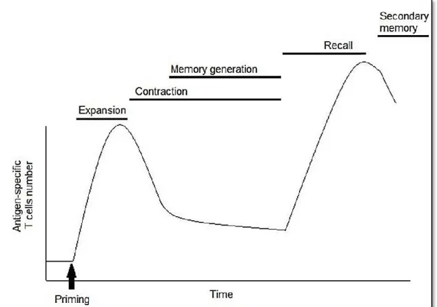
Figure 1.5: Antigen-specific expansion of CD8+ T cells during an acute immune response48 Figure 1.6: The hallmarks of cancer………58
Figure 1.7: Cancer immunoediting model……….65
Figure 1.8: The cancer-immunity cycle and possible therapy approaches……….73
Figure 1.9: Generation of anti-tumour T cells for adoptive cell therapy……….….75
CHAPTER 2 Figure.1:Socs1-/-Pmel-1 cells display signs of in vivo antigen stimulation………...90
Figure 2: Socs1-/- CD8+ T cells show impaired antigen (Ag)-induced proliferation………...92
Figure 3: Socs1-/-Pmel-1 cells display increased antigen (Ag)-specific cytolytic activity after Ag or cytokine stimulation………...95
Figure 4: Socs1-/-Pmel-1 cells undergo massive lymphopenia-induced proliferation……….97
Figure 5: Socs1-/-Pmel-1 cells cause severe skin lesions following lymphopenia-induced proliferation………99
Figure 6: Socs1-/-Pmel-1 cells show increased Blimp-1 expression after antigen stimulation………...101
Figure S1: Proximal TCR signaling and calcium response are not impaired in Socs1-/-Pmel-1 cells………112
Figure S2: Dermatologic and ocular lesions caused by lymphopenia-induced proliferation of SOCS1 deficient Pmel-1 cells………..113
CHAPTER 3 Figure 1: Stable expression of NLRC5 induces MHC-I and a subset of antigen processing pathway genes in B16-F10 melanoma cells………..123
Figure 2: NLRC5 increases the antigen-presenting capacity of B16 cells and enables presentation of endogenous tumor antigenic peptide……….127
Figure 3: B16 cells expressing both NLRC5 and CD80 efficiently activate Pmel-1 CD8+ T cells………...130
Figure 4: NLRC5 expression reduces the tumor forming ability of B16 cells………..133
Figure 5: Rapid growth of NLRC5-expressing B16 cells in mice depleted of CD8+ T cells………..…….135
Figure 6: NLRC5 expression in B16 cells enables cross-presentation of endogenous mgp100 and elicits protective antitumor immunity………...137
Figure S1: NLRC5 gene expression in mouse tumor cell lines………..151
List of Abbreviations
ACT Adoptive cell transferAg Antigen
AICD Activation induced cell death APC Antigen presenting cell
APM Antigen processing and presentation machinery Arg-1 Arginase 1
ATF1 Activation transcription factor 1 AVS Angiogenic vascular cells
B16 B16-F10 mouse melanoma cells
BATF Basic leucine transcription factor ATF-like Bcl-6 B-cell lymphoma 6
BL6 C57BL/6 mouse strain
Blimp1 B lymphocyte-induced maturation protein 1 CAF Cancer-associated fibroblastic cells
CAR Chimeric antigen receptor CARD Caspase recruitment domain CCR7 CC-chemokine receptor 7 CD Cluster of differentiation CIITA MHC-II trans-activator CITA MHC-I trans-activator
CPM Counts per minute, a unit of radioactivity CTL Cytotoxic T lymphocyte
CTLA-4 Cytotoxic T-lymphocyte-associated protein 4 (CD172) DAMPs Danger-associated molecular patterns
DC Dendritic cell
dLNs Draining lymph nodes EGF Epidermal growth factor
Eomes Eomesodermin
ER Endoplasmic reticulum
GAS gamma-interferon-activation sites
GM-CSF Granulocyte macrophage colony-stimulating factor
gp100 glycoprotein 100 melanoma antigen of amino acid residues 209-217 Gy Gray, a unit of radioactivity
HEV High endothelial venules
hgp100 human premelanosome protein gp100 HLA Human leukocyte antigen (human MHC) HMGB1 High mobility group box 1
IDO Indoleamine 2,3-dioxygenase IELs Intraepithetial lymphocytes
IFN Interferon
Ig Immunoglobulin
IL Interleukine
IL-15Rα Alpha-subunit of the IL-15 receptor (CD215) IL-2R Beta-subunit of IL-2 receptor (CD122) IL-2R Gamma-subunit of IL-2 receptor (CD132) IL-2Rα Alpha-subunit of the IL-2 receptor (CD25) IL-7Rα Alpha subunit of the IL-7 receptor (CD127) iMCs Immature myeloid cells
iNOS Inducible nitric oxide synthase IRF1 Interferon regulatory factor 1
ISRE Interferon-sensitive response element
JAK Janus kinase
LAG-3 Lymphocyte-activation gene 3 (CD223) LCMV Lymphocytic Choriomeningitis Virus LEF-1 Lymphoid enhancer-binding factor 1
LNs Lymph nodes
LRR Leucine-rich repeats LSP Long signal peptide
MAPK Mitogen-activated protein kinase MDSCs Myeloid derived suppressor cells MFI Mean fluorescence intensity
mgp100 Mouse pre-melanosome protein gp100 MHC-I Major histocompatibility complex type I MHC-II Major histocompatibility complex type II
MIIC MHC-II compartment
NBD Nucleotide binding domain NF-B Nuclear factor kappa B
NFY Nuclear factor Y
NK Natural killer cell
NKT Natural killer T lymphocyte NLR (Nod)-like receptor
NLRC5 NOD-like receptor family CARD domain containing protein 5 NOD Nucleotide-binding oligomerization domain
P14 cells CD8+ transgenic TCR specific for is specific for the peptide gp33-41 from the
lymphocytic choriomeningitis virus (LCMV) presented by the MHC-I molecule H-2Db
PAMPs Pathogen-associated molecular patterns PBS Phosphate Buffered Saline
PD-1 Programmed cell death 1 (CD279) PD-L1 Programmed death-ligand 1 (CD274) PI3K Phosphoinositide 3-kinase
PLC Peptide loading complex
Pmel-1 cells CD8+ Transgenic TCR cells specific for gp10025-33 peptide presented by the
MHC-I molecule H-2Db
RFX Regulatory factor X
RIG-I Retinoic acid-inducible gene I RLHs RIG-I like helicases
ROS Reactive oxygen species
SCID Severe combined immunodeficiency SOCS1 Suppressor of cytokine signaling protein 1 SSP Short signal peptide
STAT Signal transducers and activators of transcription TAA Tumor associated antigen
TAMs Tumor associated macrophages
TAP Transporter associated with antigen processing protein TCM Central memory CD8+ T lymphocyte
TCR T cell receptor
TEFF Effector CD8+ T lymphocyte
TEM Effector/memory CD8+ T lymphocyte
Tfh Follicular helper T cells Tfr Follicular regulatory T cells TGF Transforming growth factor beta
Th T helper cell
TILs Tumor infiltrating lymphocytes TLRs Toll-Like receptors
TME Tumor microenvironment
TMEM Memory CD8+ T lymphocyte
TRAIL TNF-related apoptosis-inducing ligand Treg Regulatory T lymphocyte
TSCM Memory stem CD8+ T lymphocyte
VEGF Vascular endothelial growth factor β2m beta2-microglobulin
CHAPTER 1- INTRODUCTION
1. The immune systemDuring the course of evolution, human beings have evolved in an environment governed by several types of microorganisms such as fungi, archaea, bacteria, protozoa and viruses. In order to coexist with all these living or, in the case of viruses, opportunistic organisms, our body has developed a complex system that allows it to cohabit in equilibrium, yet also eliminate potentially harmful pathogens with minimal damage to self (Hoffmann et al., 1999). This complex defense system is known as the immune system, which is composed of different types of specialized cells that are able to recognize and mediate efficient responses against infection and cancer.
Immune cells circulate through the blood as well as through the lymphatic system that infiltrates almost all of the tissues in the body. This network includes lymph nodes (LNs) and other organs, such as the spleen, thymus, tonsils and adenoids. The thymus and the bone marrow are the niche for the development and maturation of immune progenitor cells and they are, therefore, known as primary immune organs. The rest of the immune organs mentioned are known as secondary immune organs.
The immune system consists of two interrelated arms, known as the innate and adaptive immune systems, which are formed of different types of cells, depending on their capability to be recruited and respond to a specific stress.
1.1. The innate immune system
The innate immune system is the first line of defense characterized by rapid pleiotropic responses after exposure to microorganisms. All cells of a given type in our bodies express different types of receptors that are able to recognize self and non-self-patterns (Medzhitov and Janeway, 1997). However, innate immune responses are not specific to a particular pathogen. Upon infection, innate immune cells are recruited to the site of stress as infected cells are able to recognize unique molecular patterns exposed by
microorganisms. Cells are able to identify most conserved features of pathogens, known as the pathogen-associated molecular patterns (PAMPs) (Janeway, 1989), and cell components that are released during cell damage or death, danger-associated molecular patterns (DAMPs) (Matzinger, 1994), through the expression of germline-encoded receptors, the pattern recognition receptors (PRRs) (Medzhitov and Janeway, 1997). In order to detect the presence of any type of microbial infection or endogenous danger signals, different PRRs react with specific PAMPs and DAMPs activating specific signaling pathways to initiate an adequate innate immune response.
1.1.1. The pattern recognition receptors
There are several types of PRRs that are broadly classified based on structural homology and the requirement of different adaptor proteins that ensure their function and down-stream signal transduction (Hansen et al., 2011). Transmembrane PRRs are composed of twelve members of Toll like-receptors (TLRs) that recognize bacterial cell wall components, including viral nucleic acids in endosomes, etc., from Gram negative and Gram positive bacteria, DNA and RNA viruses, protozoa and fungi (Iwasaki and Medzhitov, 2004; Pasare and Medzhitov, 2005), as well as C-type lectins, which recognize fungal cell wall components, such as -glucan (Hardison and Brown, 2012; Taylor et al., 2006). While, some TLRs (TLRs 1, 2, 4, 5, and 6) are expressed on the cell surface, others (TLRs 3, 7, 8, and 9) are found almost exclusively in intracellular compartments such as endosomes, and their ligands, principally nucleic acids, need internalization by the endosome (Kawai and Akira, 2011). Antigen presenting cells (APCs), such as dendritic cells (DCs) and macrophages, that are able to engulf and degrade pathogens, prominently express TLRs. Activation of PRRs triggers downstream signaling cascades and production of pro-inflammatory cytokines and chemokines (Akira et al., 2006). After activation, APCs migrate from the site of infection to the tissue draining LNs (dLNs). On site, APCs present processed peptides, known as antigens (Ag), to T and B lymphocytes.
In contrast to transmembrane PRRs, cytosolic PRRs can differentiate between intracellular and extracellular infections (Palm and Medzhitov, 2009). Cytosolic PRRs include the nucleotide-binding domain, leucine-rich repeat containing proteins (NLRs) (Wen et al., 2013; Yeretssian, 2012), retinoic acid-inducible gene (RIG) I-like RNA helicases (Goubau et al., 2013), and AIM2-like receptors (ALRs) (Ratsimandresy et al., 2013). On one hand, members of this class of PRRs directly detect cytosolic PAMPs and activate several signaling cascades that induce anti-viral mediators like type I interferons (IFNs) and pro-inflammatory cytokines (Meylan et al., 2006; Takeuchi and Akira, 2007). On the other hand, other cytosolic PRRs consists of members of the NLR family involved in the formation and activation of inflammasomes, which are large multimeric protein complexes that control the activation of caspase-1, and the secretion of caspase-1-dependent pro-inflammatory cytokines IL1α, IL-1β and IL-18 (Martinon and Tschopp, 2004). Some members of this class of cytosolic PRRs, such as Nacht domain-, leucine-rich repeats-, and pyrin domain-containing protein 3 (NALP3), can be activated by potassium efflux, which can result from microbial infection rather than cytosolic PAMPs (Palm and Medzhitov, 2009).
1.1.1.1. NOD-like receptors
NLRs can perceive microbes as well as homeostatic perturbations by recognizing PAMPs and DAMPs (Ting et al., 2008). To date, there are 22 known members of human NLRs that are grouped into four subfamilies depending on their N-terminal effector domains: NLRA has an acidic transactivation domain, NLRB a baculoviral inhibition of apoptosis protein repeat (BIR)-like domain, NLRC a caspase recruitment domain (CARD), and NLRP a pyrin domain (PYD); NLRX, does not present strong N-terminal homology to any other NLR member (Ting et al., 2008). All NLRs contain a central NACHT domain that facilitates oligomerization, and multiple leucine-rich repeats (LRRs) on their C-terminal end for ligand sensing (Harton et al., 2002).
1.1.1.1.1. The master regulators of major histocompatibility complexes class I and II expression
During infections, innate immune cells such as macrophages and DCs will phagocytise pathogens and infected cells in order to process their content to generate antigens that will be presented via major histocompatibility complexes (MHC) class I (MHC-I) and II (MHC-II) to lymphocytes (Germain, 1994). Antigen presentation by APCs will make the bridge with the adaptive immune system allowing the generation of a specific immune response. While, MHC-I will present antigens to the CD8+ T cells, MHC-II will allow antigen-presentation to CD4+ T cells (Germain, 1994). T lymphocytes specifically recognize antigens by their T cell receptor (TCR) (Krogsgaard and Davis, 2005), as will be discussed later.
NOD-like receptor CARD domain containing 5 (NLRC5) and NLRA/ Class II trans-activator (CIITA) are important in the biology of T lymphocytes considering they have key functions in their development, maturation, homeostasis and activation. NLRC5 and CIITA are the master regulators of MHC-I and MHC-II, respectively. MHC molecules are differently expressed on the surface of cells in the human body, and can be upregulated under inflammatory conditions in order to present extrinsic or intrinsic antigens to APCs and T lymphocytes (Table 1.1.1).
Characteristic MHC-I pathway MHC-II pathway Reference
Master regulator of gene expression NLRC5 (CITA) CIITA (Meissner et al., 2010; Steimle et al., 1993) MHC complex
Polymorphic α chain non-covalently linked to a non-polymorphic invariant chain β2m, accommodates peptides of 8–11 amino acids
Polymorphic chains α and β, peptide binds to both,
accommodates peptides of 12 or more amino acids
(Neefjes et al., 2011) Masternak et al., 2000)
MHC types
Classical MHC-Ia genes are highly
Classical MHC-II genes HLA-DP, HLA-DQ, HLA-DR. Non-classical
(Gao et al., 2000; Parham
polymorphic: HLA-A, -B, -C. Non-classical MHC-Ib genes are homologous to classical Ia but less polymorphic: HLA-E, -G, -F.
MHC-II genes DM and HLA-DO
et al., 1988; Steimle et al., 1993)
MHC
expression Constitutively expressed on all nucleated cells
Constitutively: DCs,
macrophages, B lymphocytes, Inducible: other innate immune cells (basophiles, eosinophils, mast cells, ILC3), some endothelial cells, LN stromal cells, epithelium of thymus
(van den Elsen et al., 2004; Kambayashi and Laufer, 2014); Inducible MHC expression
Type I and II interferons (IFNα/ and
, respectively) Type II interferon (IFN)
(Steimle et al., 1994; (Benko et al., 2010; Kuenzel et al., 2010; Meissner et al., 2010; Staehli et al., 2012) Responding T
lymphocytes CD8+ T lymphocytes CD4+ T lymphocytes
(Germain, 1994; Wang and Reinherz, 2002) Origin of antigenic proteins
cytosolic proteins (mostly synthetized by the cell; may also enter from the extracellular medium via phagosomes)
Proteins present in endosomes or lysosomes (mostly internalized from extracellular medium)
(Krogsgaard and Davis, 2005) Location of loading the peptide on the MHC molecule ER Specialized vesicular compartment (Downs et al., 2016) Molecules implicated in transporting the peptides and loading them on the MHC molecules TAP1/TAP2 loaded on to MHC-I by PLC
(consisting of the MHC-I heavy chain and β2m, TAP1, TAP2, tapasin, calreticulin, and ERp57)
DM, invariant chain (Harding and Geuze, 1993; Blum et al., 2013; Downs et al., 2016)
MHC-dependent immune responses
Inflammatory processes during viral infection, intracellular bacterial infection, organ transplantation and cancers
Induced under inflammation by extracellular pathogens
(Harding and Geuze, 1993; Yoshihama et al., 2016)
Table 1.1: MHC-I and class II antigen processing and presentation pathways. ILC3 (Innate lymphocyte cells), ER (endoplasmic reticulum), TAP1/TAP2 (Transporter associated with Antigen Processing 1/2), PLC (peptide loading complex), β2m (β2-microglobulin).
1.1.1.1.1.1. CIITA
NLRA, better known as the MHC-II trans-activator (CIITA), is a key molecule in the regulation of adaptive immune responses. It is essential for MHC-II gene expression and functions as a trans-activator of MHC-II antigen presentation genes (Meissner et al., 2010; Steimle et al., 1993). MHC-II molecules are cell-surface glycoproteins displayed as heterodimeric chains (α chain/β chain), whose function is to present antigenic derived peptides from exogenous origin to CD4+ T helper cells. As mentioned before, these molecules are essential for the initiation of CD4+ T-cell-mediated immune responses, and also in the development and homeostasis of the CD4+T-cell population (Masternak et al., 2000). MHC-II molecules are expressed constitutively in only a few cell types of the immune system, such as DCs, macrophages, and B lymphocytes. However, they are induced in the majority of cells after interferon-gamma (IFN-) stimulation (Steimle et al., 1994). In the same way, CIITA is expressed in a cell type-specific manner, predominantly in hematopoietic cells, and the pattern of its expression correlates with that of MHC-II molecules in several cell lines and tissues (Steimle, 1993). CIITA regulates the transcription of MHC-II molecules HLA-DM, HLA-DQ, HLA-DR as well as other related proteins in the MHC-II presentation pathway, such as the invariant chain (li) (Steimle et al., 1993).
In order to activate the transcription of MHC-II gene loci, CIITA does not possess a DNA binding domain nor directly binds DNA, but instead forms an heteromultimeric protein complex called MHC II enhanceosome with transcription factors such as regulatory factor X (RFX) complex, CREB/ATF1 and nuclear factor Y (NFY) that associate with the X1, X2, and Y cis-elements, respectively, to transactivate promoter activity (Hake et al., 2000;
Masternak et al., 2000; Raval et al., 2001; Reith and Mach, 2001; Reith et al., 1988; Scholl et al., 1997; V Steimle et al., 1993). Moreover, CIITA also binds to the SXY module of MHC-I and 2M to induce MHC-MHC-I transcription activation (Gobin et al., 1997; Martin et al., 1997). However, in mice lacking CIITA, MHC-I expression was not impaired indicating the presence of other regulators that control MHC-I expression (Chang et al., 1996; Itoh-Lindstrom et al., 1999; Williams et al., 1998).
1.1.1.1.1.2. NLRC5
1.1.1.1.1.2.1. MHC-I gene and NLRC5 structure
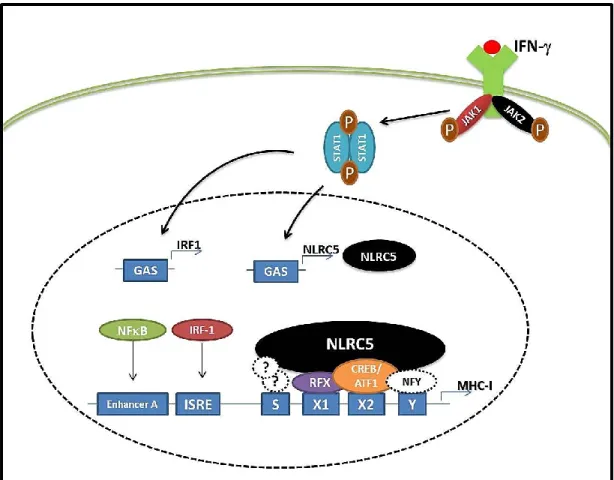
MHC-I expression is closely regulated at the transcriptional level by several conserved regulatory elements in the proximal promoter region, which contain cis-regulatory elements that play a crucial role in both constitutive and inducible expression (Figure 1). On one hand, enhancer A contains one or two nuclear factor-κB (B) binding sites. NF-B binding to Enhancer A in the promoter of the HLA-A gene induces stronger transcriptional activation (Gobin et al., 1998). On the other hand, interferon regulatory factor 1 (IRF1) can bind to the ISRE region to induce MHC-I expression (Hobart et al., 1997). Similarly to MHC-I, B2m and MHC-II promoters contain a SXY module, where regulatory factor X complex (on X1 box), CREB or activating transcription factor 1 (ATF1) (on X2 box), and NFY complex (on Y box) are able to bind and assemble to form a multi-protein transcriptional activation complex (Gobin et al., 2001; Moreno et al., 1999, 1995). MHC-I products are cell-surface glycoproteins composed of a polymorphic heavy chain (α1, α2, α3 domains) associated with the non-polymorphic β2-microglobulin (β2m). The third component crucial for the structural stability of the MHC-I complex is a peptide, composed of 8- 10 amino acids, which is loaded in the ER. At the cell surface, the MHC-I/peptide complex presents antigens to the TCR of CD8+ T lymphocytes (Neefjes et al., 2011).
Figure 1.1: IFN upregulates NLRC5 expression to activate MHC-I gene expression. Upon IFN stimulation, STAT1 becomes activated and translocates to the nucleus where it binds the GAS elements in the NLRC5 promoter to activate its expression. NLRC5 protein interacts with other transcription factors including RFX components, CREB, ATF1 and NFY factors, to form an enhanceosome to activate MHC-I gene transcription. IFN also activates IRF1 and NF-B, which modulate MHC-I transcription activity by interacting with ISRE and NF-B elements, respectively. STAT (signal transducer and activator of transcription), GAS (gamma-interferon-activation sites), CITA (class I transactivator), IRF1 (interferon regulatory factor 1), NF-B (nuclear factor-kappa B), ISRE (interferon-sensitive response element) (Adapted from Downs et al., 2016).
The largest member of the NLR protein family, NLR CARD domain containing 5 (NLRC5 or CITA, NOD4, NOD27), has recently been identified as a critical regulator of MHC-I genes as well as some related genes involved in MHC-I-dependent antigen processing and presentation pathway (Meissner et al., 2010). NLRC5 has been shown to induce the expression of classical MHC-I (i.e., HLA-A, HLA-B, HLA-C), non-classical class I (i.e., HLA-E, HLA-F, HLA-G), beta subunit (B2M), immunoproteasome component (PSMB9, i.e., LMP2) and peptide transporter (TAP1) (Biswas et al., 2012; Ludigs et al., 2015; Meissner et al.,
2012, 2010). Similar to CIITA, NLRC5 associates with the RFX complex and CREB/ATF1 in the nucleus to form a transcriptional enhanceosome onto SXY modules of MHC-I and B2M genes in order to induce their expression (Figure 1) (Meissner et al., 2012a, 2012b; Neerincx et al., 2012; Robbins et al., 2012).
NLRC5 possess three structural domains, including an effector domain in the atypical N-terminal caspase activation and recruitment domain (CARD), a central nucleotide binding domain (NBD) and a C-terminal domain containing 27 leucine-rich repeats (LRRs), which makes it the largest member of the NLR family with a predicted size of more than 200 kDa. NLRC5 also possesses a nuclear localization signal between the CARD and NBD domains (Meissner et al., 2012a) (Figure 1.2). Furthermore, at its N-terminal extremity, NLRC5 possesses an atypical CARD consisting of a death motif (DD) fold that shares little similarity with the CARD or PYD domains from other NLR family members. DD motifs in this extremity are commonly implicated in interactions with other protein family members in order to accomplish their effector functions by homotypic interaction. However, as the DD motif of NLRC5 does not display a defined homology of CARD, PYD and DD motifs of other known adaptor and effector proteins, it is difficult to predict NLRC5 binding partners and signaling pathways (Lamkanfi and Kanneganti, 2010). Interestingly, the N-terminal domain composed of the first 139 amino acids of human NLRC5 was shown to have intrinsic transcriptional activity toward MHC-I and MHC-II promoters. Importantly, the nuclear localization signals (NLS) located in this effector domain (121–122; 132–134 amino acids) are needed for its functionality (Neerincx et al., 2014). Moreover, the NACHT domains of NLRC5 act as a single functional unit with the LRR extremity to induce transcriptional activity of MHC promoters (Neerincx et al., 2014).
Figure 1.2: Comparison of MHC master regulators CIITA and NLRC5/CITA. CIITA exists in three different isoforms found in DCs, B cells, and IFN stimulated cells, respectively. Both, CIITA and NLRC5 possess nuclear localization signals (NLS) allowing them to translocate into the nucleus. Both NLRC5 (1866 a.a.) and CIITA (1130 a.a.) contain a central nucleotide-binding domain (NBD) critical for their function. Their C-terminus extremity contains leucine-rich repeats (LRRs) (Adapted from Downs et al., 2016).
In humans, NLRC5 is located on chromosome 16q13, covering a region of 96 Kbp and consisting of 1866 amino acids. In mice, it is located on chromosome 8 and consists of 1915 amino acids (Kobayashi and van den Elsen, 2012; Meissner et al., 2010). Humans and mice share a 64% identity (Yao and Qian, 2013). Based on structural analysis from the Ensembl database, there are at least 10 protein coding isoforms of NLRC5 that display shorter LRRs. The mRNA levels of some NLRC5 isoforms have been detected in different tissues and cell lines, but their function is currently unknown (Neerincx et al., 2013, 2010; Staehli et al., 2012).
1.1.1.1.1.2.2. Expression and biological functions
NLCR5 expression has been observed in several human and mouse tissues. This protein is mainly expressed constitutively in immune tissues such as the thymus, the bone marrow, the lymph nodes and the spleen (Benko et al., 2010; Neerincx et al., 2010). Among the lymphoid tissues, high concentrations of NLRC5 mRNA have been mainly reported in B and T lymphocytes. NLRC5 transcripts are also detected in some mucosal-surface tissues such as the lung, small intestine, colon and uterus (Benko et al., 2010; Kuenzel et al.,
2010). This expression pattern suggests that NLRC5 could be involved in the immune regulation of the host defence at these interfaces. Accordingly, NLRC5 expression has been reported in many cell lines including human THP-1 and murine RAW264.7 monocytic cell lines, the Jurkat T and Raji B cell lines, as well as the human cervical carcinoma cell line HeLa (Benko et al., 2010; Cui et al., 2010; Davis et al., 2011; Neerincx et al., 2010; Staehli et al., 2012). Interestingly, NLRC5 mRNA expression levels positively correlates with constitutive MHC-I expression level in several cell lines and tissues (Neerincx et al., 2012). Moreover, ectopic NLRC5 expression is able to activate MHC-I promoter and knockdown of NLRC5 reduces MHC-I transcription both in immortalized cell lines and primary cells (Neerincx et al., 2012).
NLRC5 is strongly induced by type I and type II interferons (Benko et al., 2010; Kuenzel et al., 2010; Meissner et al., 2010; Staehli et al., 2012). Indeed, two signal transducer and activator of transcription (STAT) binding sites are located within NLRC5’s promoter. Also, NLRC5 can be upregulated upon exposure to PAMP stimuli such as poly (I:C), LPS, CpG as well as by viral pathogens such as influenza, respiratory syncytial virus and Sendai virus (Benko et al., 2010; Guo et al., 2015; Neerincx et al., 2010; Ranjan et al., 2015). Recently, an interesting observation was made in an epigenome-wide differential DNA methylation study between HIV-infected and uninfected individuals (Zhang et al., 2016). This study found that two CpGs in the promoter of NLRC5 were significantly hypo-methylated in HIV-infected subjects in comparison with the unHIV-infected subjects, thus implying an important role for NLRC5 in HIV pathogenesis (Zhang et al., 2016).
NLRC5 also associates with chromatin remodeling enzymes, such as histone acetyltransferases, to epigenetically control MHC-I promoter activity (Meissner et al., 2012b; Robbins et al., 2012). Recently, a study showed that NLRC5 interacts with miR-34a, a potent tumor suppressor, to inhibit NF-B signaling in cells infected with HPV16 in order to promote virus persistence (Li et al., 2016). These studies suggest NLRC5 as a target of immune evasion in viral infections (Benko et al., 2010; Guo et al., 2015; Li et al., 2016; Neerincx et al., 2010; Ranjan et al., 2015; Zhang et al., 2016).
While the functions of NLRC5 in the adaptive immune response are well established, its role in innate immune responses is quite controversial. Some studies have reported NLRC5 as a negative modulator of inflammatory pathways. In vitro studies have shown that NLRC5 downregulates NF-B activation and thus interferes downstream of the TLR4 signaling (Benko et al., 2010). NLRC5 inhibited NF-B-dependent responses by interacting with IKKα and IKK and blocking their phosphorylation as well as inhibiting type I interferon responses by interacting with RIG-I (Cui et al., 2010). In contrast, other studies showed that type I interferon production is increased by NLRC5 (Kuenzel et al., 2010; Neerincx et al., 2010), and that Nlrc5-defiency does not impair TLR2, TLR4 and TLR9 signaling pathways (Kumar et al., 2011; Staehli et al., 2012; Yao et al., 2012). Interestingly, NLRC5 is crucial for the elimination of intracellular bacterial infection such as Listeria monocytogenes in mice. Also in chickens, NLRC5 is needed for Salmonella pullorum elimination via STAT1 activation (Biswas et al., 2012; Qiu et al., 2017). In addition, some studies have reported that NLRC5 can interact with the NLRP3 inflammasome to activate IL-1 and IL-18 production (Davis et al., 2011), although another study showed that Nlrc5 deficiency does not impair inflammasome activation nor IL-1 and IL-18 production (Kumar et al., 2011).
In order to resolve discordances in NLRC5 studies, Dr. Guarda and Dr. Tschopp’s groups generated Nlrc5-deficient mice (Staehli et al., 2012). General observations from this mouse model showed that NLRC5 deficiency does not affect MHC-I expression completely, but reduces it in APCs. Moreover, T cell development and activation were not impaired in these mice, other than a small decrease in the number of CD8+ T cells in the spleen, blood and LNs. However, NLRC5 deficiency dramatically impaired the basal expression of MHC-I in T, natural killer (NK), and NKT lymphocytes. In contrast, B cells and conventional DCs displayed an intermediate and a slight reduction of MHC-I expression, respectively. NLRC5 expression is important for the cognate recognition of target cells by cytotoxic T lymphocytes (CTLs). Interestingly, this study also described low NLRC5 expression in several murine and human lymphoid-derived tumor cell lines. The authors found a correlation between the presence of NLRC5 and the abundance of the MHC I
α-chain mRNA for some tested cell lines (Staehli et al., 2012). Additionally, recent publications have highlighted the role of NLRC5 in activated DCs upon stimulation with different inflammatory stimuli. Indeed, DCs deficient in NLRC5 have decreased neo-synthesized MHC-I and are defective at displaying endogenous antigens (Rota et al., 2016). Surprisingly, this impairment does not affect T cell priming by endogenous antigens or cross-priming capability of DCs (Rota et al., 2016). In contrast, NLRC5 deficiency in NK cells reveals moderate alterations in inhibitory receptor expression and ability to eliminate target cells (Ludigs et al., 2016). Under inflammatory conditions, such as LCMV infection, T lymphocytes deficient in NLRC5 are eliminated by NK cells. Thus, NLRC5 expression in T lymphocytes seems to be crucial to prevent them from NK-cell-mediated elimination (Ludigs et al., 2016).
Hence, despite its crucial role as the master regulator of MHC-I and related genes, the implications of NLRC5 in NF-B activation, type I IFN signaling and inflammasome activation remain to be elucidated.
1.1.1.1.1.3. Antigen cross-presentation by MHC-I
Through the activation of CD8+ T cells, MHC-I-dependent responses elicit and induce significant adaptive immunity against viral infections, intracellular-bacterial infections, transplant rejection and cancers. Indeed, some professional APCs, such as DCs, possess the capability to present exogenous antigens on MHC-I molecules by a process termed antigen cross-presentation (Bevan, 1976; Nair-Gupta et al., 2014). During this process, exogenous antigenic peptides derived from phagocytosed microbes, infected cells, or tumor cells are loaded onto MHC-I and presented to CD8+ T cells (Bevan, 1976; Nair-Gupta et al., 2014). Cross-presentation seems to be possible because MHC-I are recruited from an endosomal recycling compartment (Nair-Gupta et al., 2014). Furthermore, this process has a key role in cancer immunosurveillance through processing and presentation of tumor associated antigens (TAAs) (Mellman et al., 2011).
Cross-presentation process can be influenced by several factors such as the type of antigen, DC location and activation status and the presence and timing of inflammatory signals (Fehres et al., 2014; Nierkens et al., 2013). In most cases, cross-presentation is TAP- and proteasome-dependent (Kovacsovics-Bankowski and Rock, 1995) but other study also showed that this process can be TAP-independent as well (Merzougui et al., 2011).
1.2. The adaptive immune system
When professional APCs such as DCs are activated by infection they present foreign antigens loaded on MHC molecules to a TCR on T cells in the dLNs of the affected tissue (Steinman, 2012). This tri-molecular interaction of MHC–peptide–TCR is central to the generation of antigen-specific immune responses. In contrast to the innate immune responses, the adaptive immune responses are generated against a specific pathogen to create memory and long-lasting immune protection (Dempsey et al., 2003). T and B lymphocytes are the main players of the adaptive immune responses displaying unique antigen-specific receptors that have been created through somatic DNA recombination (>1015 distinct TCRs are possible) (Davis and Bjorkman, 1988; Litman et al., 1999). The
large number of receptors on lymphocytes increases the probability of recognizing and proliferating in response to a specific antigen. B lymphocytes originate from bone marrow precursors and generate antibody responses, whereas T lymphocytes development occurs in the thymus and generates cell-mediated immune responses (Rajewsky and von Boehmer, 2008) .
1.2.1. CD8+ T lymphocytes
CD8+ T cells, also known as CTLs after their TCR activation, are key players of the adaptive immune system that fight against intracellular pathogens as well as cancer cells (Zhang and Bevan, 2011).
The term ‘‘naive’’ represents a state of quiescence for mature T lymphocytes that have not been activated via their TCR in the periphery (Sprent and Tough, 1994). In fact, naïve T
cells are long-lived cells that remain in the interphase, recirculating through the secondary lymphoid tissues waiting to be activated by their cognate peptide-MHC complex (Sprent and Tough, 1994). However, naive T lymphocytes are maintained by signals from the TCR engaging the self-peptide-MHC complexes. Importantly, cytokines such as interleukin-7 (IL-7) and IL-15 are also essential for their homeostasis and survival (Berard et al., 2003; Takada and Jameson, 2009).
Naive T lymphocytes have the ability to circulate through the LNs to look for their cognate antigen, maximizing their chances of being activated and returning to the blood stream (Masopust and Schenkel, 2013). CD8+ T cell subsets are characterized by the expression of surface proteins allowing them to perform diverse functions and to home to different tissues (Masopust and Schenkel, 2013). Among those proteins, some are used as markers to distinguish phenotypic and functional profiles relying on the stage of T cell differentiation. Resting CD8+ T cells displaying a naive phenotype are characterized by the expression of high (hi) levels of CD62L (L-selectin), CC-chemokine receptor 7 (CCR7), CD127 (IL-7 receptor alpha), and low (lo) levels of CD44 (an activation marker).
1.2.1.1. CD8+ T cells homing receptors
CD62L and CCR7 are LN homing receptors allowing naïve CD8+ T cells to accomplish their immunosurveillance role (Gallatin et al., 1983; Rosen, 2004). CD62L is a protein that enables naive CD8+ T cells to tether to the high endothelial venules (HEVs), a specialized type of post-capillary vascular endothelium existing within LN paracortical areas (T cell zone), to enter to the LNs (Gallatin et al., 1983; Rosen, 2004). This is accomplished via short-term interactions between CD62L on CD8+ T cells and 6-sulpho sialyl Lewis X oligosaccharides covering several proteins expressed on HEVs (Gallatin et al., 1983; Rosen, 2004). Then, a G-protein coupled chemokine receptor named CCR7 recognizes its ligands CCL21 and CCL19 immobilized on the HEVs via binding to heparin glycosaminoglycans. Finally, CCR7-signaling activates LFA1 (lymphocyte function-associated antigen 1), which interacts with ICAM1 (intercellular adhesion molecule 1) to arrest the naive T cell, allowing its passage into the LN (Moschovakis and Förster, 2012). Therefore, CD62L, CCR7
and LFA1 together are essential for the rolling, activating and arresting processes enabling naive T cells to selectively go into lymph nodes (Bao et al., 2010; Masopust and Schenkel, 2013; Springer, 1994).
1.2.1.2. Effect of some cytokines of the -chain family on CD8+ T cells
Cytokines are small proteins (~5–20 kDa) that serve as intracellular messengers between cells within and outside the immune system. They are involved in autocrine, paracrine and endocrine signalling, and act as immunomodulating agents (Leonard et al., 1999). Cytokines have essential functions in modulating cellular proliferation, differentiation and survival as well as in homeostasis (Overwijk and Schluns, 2009). Cytokines are divided depending on their effector function, cellular origin, or target of action, and include chemokines, interferons, interleukins, and tumour necrosis factors (Cameron and Kelvin, 2000). Cytokines are classified depending on their tridimensional structures (Leonard, 2001). Type I cytokines have a four α‑helical bundles structure that includes several interleukins and some growth and hematopoietic factors (Leonard, 2001). One family of cytokines belonging to the type I has the particularity to share a common receptor subunit. Among them, a crucial family that plays a key role in T lymphocyte homeostasis is the common cytokine receptor chain (c), which is denoted in this manner because the receptor chain (also known as IL-2R or CD132) is shared by several cytokines. The c family includes IL‑2, IL‑4, IL‑7, IL‑9, IL‑15 and IL‑21 (Leonard, 2001; Spolski and Leonard, 2008) (Figure 1.3).
Figure 1.3: Common -chain receptor family of cytokines. When a c family interleukin binds its receptor, c receptor (CD132 hereafter) cytoplasmic tail initiates signaling via to the Janus kinase- signal transducer and activator of transcription (JAK-STAT) pathway. These pathways culminate in the transcription of several genes involved in immunity, proliferation, differentiation, apoptosis and oncogenesis (Aaronson and Horvath, 2002). Inspired from Liao et al., 2013.
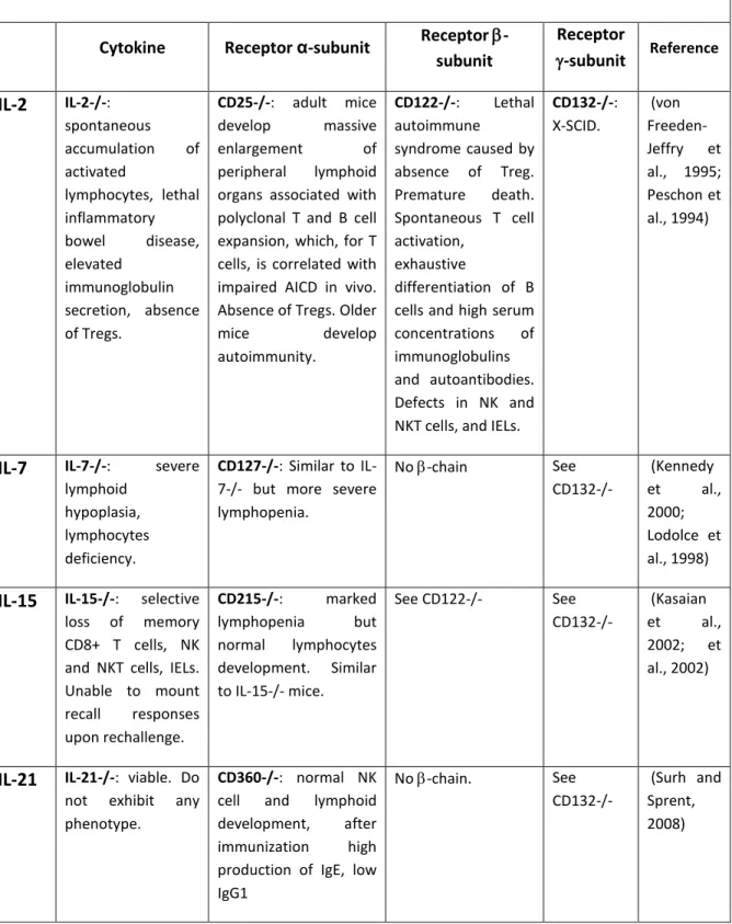
The importance of the c cytokine family was revealed by a disease that accounts for about half of the cases of severe combined immunodeficiency (SCID), the X-linked (X-SCID), in which both cellular and humoral immunity are profoundly compromised (Table 1.2). This syndrome is a consequence of mutations in the c chain. The disease is characterized by an absence of T cells and NK cells, highlighting the crucial role of these cytokines in the development of these cells. B cells are normal in number but are non-functional (Conley, 1992; Leonard, 1996; Noguchi et al., 1993).
Knockout phenotypes of some gamma chain family cytokines/receptors
Cytokine Receptor α-subunit Receptor -subunit Receptor -subunit Reference IL-2 IL-2-/-: spontaneous accumulation of activated lymphocytes, lethal inflammatory bowel disease, elevated immunoglobulin secretion, absence of Tregs. CD25-/-: adult mice develop massive enlargement of peripheral lymphoid organs associated with polyclonal T and B cell expansion, which, for T cells, is correlated with impaired AICD in vivo. Absence of Tregs. Older
mice develop autoimmunity. CD122-/-: Lethal autoimmune syndrome caused by absence of Treg. Premature death. Spontaneous T cell activation, exhaustive differentiation of B cells and high serum concentrations of immunoglobulins and autoantibodies. Defects in NK and NKT cells, and IELs.
CD132-/-: X-SCID. (von Freeden-Jeffry et al., 1995; Peschon et al., 1994)
IL-7 IL-7-/-: severe lymphoid
hypoplasia, lymphocytes deficiency.
CD127-/-: Similar to IL-7-/- but more severe lymphopenia. No -chain See CD132-/- (Kennedy et al., 2000; Lodolce et al., 1998)
IL-15 IL-15-/-: selective loss of memory CD8+ T cells, NK and NKT cells, IELs. Unable to mount recall responses upon rechallenge. CD215-/-: marked lymphopenia but normal lymphocytes development. Similar to IL-15-/- mice. See CD122-/- See CD132-/- (Kasaian et al., 2002; et al., 2002)
IL-21 IL-21-/-: viable. Do not exhibit any phenotype.
CD360-/-: normal NK cell and lymphoid development, after immunization high production of IgE, low IgG1 No -chain. See CD132-/- (Surh and Sprent, 2008)
1.2.1.2.1. Interleukin 2
The cytokine IL-2 is a powerful T cell mitogen of activated T cells that influences several lymphocyte subsets during differentiation, immune responses, and homeostasis. IL-2 has essential roles in protective immunity but especially in peripheral immune tolerance. Indeed, the functions of IL-2 are almost exclusively restricted to T cells and plays crucial roles in the differentiation of regulatory CD4+ T cells (Treg) (Malek, 2008), as highlighted by IL-2/IL-2R deficiencies in mouse models (Table 1.2).
Under physiological conditions, IL-2 is produced mainly by CD4+ T helper cells that are activated by pathogens or self-peptide-MHC-II complexes on DCs in secondary lymphoid organs (Setoguchi et al., 2005). IL-2 is strongly induced after TCR stimulation and is also, to a lesser extent, produced by CTLs, NK and NKT cells (Malek, 2008), activated DCs (Granucci et al., 2001) and mast cells (Hershko et al., 2011). Its production is tightly regulated by the transcription factor B lymphocyte-induced maturation protein 1 (BLIMP1 or PRDM1). Blimp1 is upregulated following IL-2 signaling, which in turn negatively regulates IL-2 transcription, thus weakening T cell proliferation and survival (Martins et al., 2008).
The main consumers of IL-2 are Tregs CD25+ and to a lesser extent CD4+ and CD8+ T cells (Malek, 2008). Activated DCs also express CD25 and can bind IL-2 to trans-present this complex to T lymphocytes. Similar to IL-15, IL-2 possesses a high binding affinity with the trimeric IL-2R (CD25/CD122/CD132) and a low affinity to the dimeric form (CD122/CD132) (Taniguchi and Minami, 1993). Interestingly, the low affinity IL-2R is weakly expressed on naive CD4+ T cells, moderately on naive CD8+ T cells and memory CD4+ T cells, and strongly on memory CD8+ T cells and NK cells. The robust expression of the dimeric form of IL-2R renders the latter cells more sensitive to low concentrations of IL-2 (Boyman et al., 2006a). CD25 is constitutively expressed on Tregs, but is upregulated on CD4+ and CD8+ T cells upon TCR activation and IL-2 signaling (Malek, 2008).
IL-2 binds CD25 to eventually recruit CD122/CD132. This quaternary complex activates the transcription of CD25 through several signaling pathways, such as the JAK1-3/STAT5, phosphoinositide 3-kinase (PI3K)-AKT and mitogen-activated protein kinase (MAPK) pathways. After IL-2 binding, the quaternary complex is internalized and degraded but CD25 can be recycled to the cell surface (Malek, 2008).
IL-2 mainly induces robust proliferation of CTLs by autocrine production but can also induce proliferation by paracrine production from CD4+ T cells or DCs (Feau et al., 2011). However, this strong stimulation leads CTLs to differentiate into short-lived effector cells that rapidly die by apoptosis, or into long-lived memory T cells that decrease CD25 expression early during the expansion stage (Kalia et al., 2010; Obar et al., 2010; Pipkin et al., 2010). Indeed, prolonged exposure to IL-2 after TCR activation promotes terminal effector differentiation of CD8+ T cells (Kalia et al., 2010; Lenardo, 1991). Moreover, persistent T cell stimulation of TCR by foreign or self-peptides along with IL-2 induces activation-induced cell death (AICD), which is mediated by Fas (CD95) and FasL (CD95L) on T cells (Lenardo, 1991; Van Parijs and Abbas, 1998).
IL-2 signaling also induces Eomesodermin (Eomes) transcription factor and upregulates perforin (Prf1) transcription (Pipkin et al., 2010). Conversely, IL-2 repressed the expression of B-cell lymphoma 6 (Bcl-6) and CD127, which are known as memory markers and will be discussed in further detail in subsequent sections (Pipkin et al., 2010).
1.2.1.2.2. Interleukin 7
In order for naive T lymphocytes to remain viable, they require pro-survival signals from their environment through TCR contact with self-peptide/MHC complexes provided by DCs as well as by IL-7, secreted by fibroblastic reticular cells in the secondary lymphoid organs (Surh and Sprent, 2008). Upon IL-7 binding, JAK-3, which is associated with c, and JAK-1, associated with IL-7R α chain, CD127, become phosphorylated which in turn activate STAT5 protein (Figure 1.3). Once activated, STAT dimers recognize a target promoter and increase its transcription.
IL-7 is essential for T cell development in humans and mice and is a potent survival factor for T cells (Table 1.2) (Mazzucchelli and Durum, 2007). A unique feature of IL-7, in comparison with other c cytokines, is that it is constitutively produced by stromal and epithelial cells in the bone marrow and thymus and by fibroblastic reticular cells in the T-cell zones of secondary lymphoid organs (Link et al., 2007; Surh and Sprent, 2008). Moreover, its receptor CD127 is downregulated after TCR activation. Due to the fact that IL-7 is essential for T cell survival, CD127 is highly expressed on naive and memory T lymphocytes but downregulated in activated effector T lymphocytes (Alves et al., 2008; Schluns et al., 2000). This phenomenon seems to support the idea that reducing CD127 expression on T cells that have already received cytokine-mediated survival signals, as with TCR/IL-2 or IL-15, will not compete for the remaining IL-7, making it available to unsignaled naive T cells (Park et al., 2004).
In addition, naive CD8 +T cells are also partly dependent on signals from IL-15 expressed by different types of stromal cells for their survival and homeostasis (Surh and Sprent, 2008).
1.2.1.2.3. Interleukin 15
1.2.1.2.3.1. Structure and Expression
IL-15 is a pleiotropic cytokine with unique characteristics in its synthesis, presentation, secretion and cytokine-induced signaling events. In 1994, IL-15 was discovered simultaneously by two teams as a T cell growth factor sharing many biological properties with IL-2 as well as two of its sub-units receptors IL-2R/IL-15R (CD122) and the c CD132 (Burton et al., 1994; Grabstein et al., 1994). IL-15 and IL-2, share the particularity of binding to three receptor subunits, with a unique ligand-binding subunit IL-15Rα (CD215) and IL-2Rα (CD25), respectively (Giri et al., 1995) (Figure 1.3). This feature allows them to share the same signaling pathway mediated by JAK1-JAK3/STAT5 and also several functions, including T cell proliferation, generation of CTLs, stimulation of immunoglobulin
synthesis by B cells and the generation and homeostasis of NK cells (Steel et al., 2012; Waldmann and Tagaya, 1999).
IL-15 is a small glycoprotein of 14-15 KDa with two known mRNA isoforms produced by alternative splicing, containing either a long signal peptide (LSP) (48 amino acids) or a short signal peptide (SSP) (21 amino acids) (Meazza et al., 1997, 1996; Nishimura et al., 1998; Onu et al., 1997; Tagaya et al., 1996).The two isoforms show a distinct intracellular trafficking but produce the same mature protein in both human and mouse. While one isoform is secreted through the secretory pathway, the other remains located near the nucleus, potentially due to its regulatory roles (Tagaya et al., 1997). In fact, one study reported that the non-secreted SSP IL-15 is found in the nucleus associated with CD215, where it decreases IL-15 gene expression by an autocrine regulatory mechanism (Nishimura et al., 2005). As a result of different intracellular processing, the SSP IL-15 can also be secreted in complex with CD215 but display a shorter half-life in comparison to the LSP IL-15 (Bergamaschi et al., 2009). Furthermore, IL-15 promoter possesses several different motifs regulated by transcription factors, including NF-B, IRF-E, γIRE, NF-IL-6, myb, αIFN-2 and GCF (Azimi et al., 1998; Budagian et al., 2006).
IL-15 is produced by many cell types. Indeed, its mRNA is constitutively expressed by several organs, including the placenta, skeletal muscle, and kidney (Grabstein et al., 1994). However, IL-15 mRNA can also be found in fibroblasts, keratinocytes and epidermal skin cells, nerve cells, lung, heart as well as epithelial cells from different tissues (Budagian et al., 2006). Conversely, the IL-15 protein is only found in a small subset of cells including thymic epithelial cells, bone marrow stromal cells, lymph node stromal cells, blood endothelial cells, monocytes/macrophages and CD8+ DCs, making it difficult to detect in in vitro cultures and indicating its regulation at a post-transcriptional level (Cui et al., 2014). IL-15 is a potent pro-inflammatory cytokine enabling the induction of other strong inflammatory cytokines, such as tumor necrosis factor alpha (TNF-α), IL-1β, and IFN-, among others. Consistent with this, autoimmune disorders, such rheumatoid arthritis and
celiac disease, are associated with IL-15 dysregulation (Waldmann, 2004; McInnes et al., 1997; Mention et al., 2003).
Another particularity of IL-15 is its trans-presentation to activate signaling on target cells. IL-15 cytokine binds CD215 on an APC, such as DC, and this complex is trans-presented to nearby T lymphocytes or NK cells bearing CD122/CD132 subunit receptors. This trans-presentation is believed to avoid aberrant immune stimulation of uncontrolled IL-15 exposure (Dubois et al., 2002; Kobayashi et al., 2005). The primary role of the alpha subunits CD215 and CD25 (for IL-2) is to confer high affinity binding to their cognate cytokines. Importantly, despite the fact that CD215 has a 1000-fold higher affinity for IL-15 (Ka greater or equal to 1011 M−1) than CD25 for IL-2 (Ka ∼108 M−1) in the absence of the
other subunits (Wang et al., 2005), both cytokines are able to signal through complex CD122/CD132 in the absence of their alpha sub-units (Anderson et al., 1995). In fact, IL-15 displays an intermediate affinity (Ka ∼109 M−1) towards the CD122/CD132 complex (Giri et
al., 1994). Interestingly, CD215 has an expression pattern very similar to that of IL-15, suggesting that both of them may bind internally before being sent to the cell surface (Giri et al., 1995; Rückert et al., 2003).
IL-15 trans-presentation/binding into CD122/CD132 complex on T lymphocytes leads to their oligomerization in order to cross-phosphorylate and activate JAK1/JAK3, which in turn activates STAT3/STAT5 (Johnston et al., 1995) transcription factors allowing them to form homo- and/or heterodimers that will translocate to the nucleus to activate gene expression (Leonard, 2001a). Additionally, this signaling pathway allows the activation of Lck kinase and Shc, which activates the PI3K/Akt and Ras/Raf/MAPK signaling cascades. These pathways enable the subsequent expression of bcl-2, c-myc and c-fos/jun genes and NF-B activation (Bianchi et al., 2006; Gu et al., 2000; Miyazaki et al., 1995; Zhu et al., 1994).
Overall, in vivo studies have reported evidence demonstrating that trans-presentation is the main mechanism that mediates IL-15 functions being used potentially by several cell types. However, some target cells for IL-15 can express all three receptor subunits, such