UNCORRECTED
PROOF
General and Comparative Endocrinology
journal homepage: http://ees.elsevier.com
Consequences of steroid-5α-reductase deficiency and inhibition in vertebrates
JulieRobitaille, Valerie S.Langlois
⁎
Centre Eau Terre Environnement, Institut national de la recherche scientifique (INRS), Quebec City, QC, Canada
A R T I C L E I N F O
Keywords
Steroid-5α-reductase Dihydrotestosterone Allopregnanolone N-glycosylation 5α-reductase inhibitorsA B S T R A C T
In 1974, a lack of 5α-dihydrotestosterone (5α-DHT), the most potent androgen across species except for fish, was shown to be the origin of a type of pseudohermaphrodism in which boys have female-like external genitalia. This human intersex condition is linked to a mutation in the steroid-5α-reductase type 2 (SRD5α2) gene, which usually produces an important enzyme capable of reducing the Δ4-ene of steroid C-19 and C-21 into a 5α-stereoisomer.
Seeing the potential of SRD5α2 as a target for androgen synthesis, pharmaceutical companies developed 5α-re-ductase inhibitors (5ARIs), such as finasteride (FIN) and dutasteride (DUT) to target SRD5α2 in benign prostatic hyperplasia and androgenic alopecia. In addition to human treatment, the development of 5ARIs also enabled further research of SRD5α functions. Therefore, this review details the morphological, physiological, and molecu-lar effects of the lack of SRD5α activity induced by both SRD5α mutations and inhibitor exposures across species. More specifically, data highlights 1) the role of 5α-DHT in the development of male secondary sexual organs in vertebrates and sex determination in non-mammalian vertebrates, 2) the role of SRD5α1 in the synthesis of the neurosteroid allopregnanolone (ALLO) and 5α-androstane-3α,17β-diol (3α-diol), which are involved in anx-iety and sexual behavior, respectively, and 3) the role of SRD5α3 in N-glycosylation. This review also features the lesser known functions of SRD5αs in steroid degradation in the uterus during pregnancy and glucocorticoid clearance in the liver. Additionally, the review describes the regulation of SRD5αs by the receptors of androgens, progesterone, estrogen, and thyroid hormones, as well as their differential DNA methylation. Factors known to be involved in their differential methylation are age, inflammation, and mental stimulation. Overall, this review helps shed light on the various essential functions of SRD5αs across species.
1. Introduction
In the 1950s, various case studies of men with pseudo-female
exter-nal genitalia and enlarged clitorises were observed (Nowakowski and
Lenz, 1961). These men exhibited a masculine body type, no breast,
normal epididymis, deferent duct, and seminal vesicles. This condition
was thought to originate from a single gene mutation (Nowakowski
and Lenz, 1961). The condition was classified as pseudovaginal
penis-crotal hypospadias. A few years later, a small population of men
ex-hibiting external genitalia ambiguity at young age was also described.
The men were raised as girls and when they reached puberty, the men
showed signs of virilisation, such as deepening of the voice and an
in-crease in muscle mass (Imperato-McGinley et al., 1974). Like the
cases from the 1950s, they also had normal epididymis and vas
defer-ens, as well as testes showing signs of functional spermatogenesis.
Im-perato-McGinley et al. (1974) showed the condition was linked to
a decrease in production of 5α-dihydrotestosterone (5α-DHT). 5α-DHT
has a higher affinity for the androgen receptor (AR) than testosterone
(T) (Deslypere et al., 1992), except in fish in which 11-ketotestos
terone (11KT) is the more potent androgen (Martyniuk et al., 2013).
This decline in 5α-DHT’s production in those men is now known to be
linked to a deficiency in steroid-5α-reductase type 2 (SRD5α2), which
reduces T into 5α-DHT.
SRD5α2 is part of the SRD5α enzyme family, which has the
abil-ity to reduce the Δ
4-ene of steroid C-19 and C-21 into a
5α-stereoiso-mer (Fig. 1A) and are NADPH-dependent oxidoreductase (Russell and
Wilson, 1994). There are five members of the SRD5α family: SRD5α1,
SRD5α2, SRD5α3, glycoprotein synaptic 2 (GSPN2), and GSPN2-like, as
shown by phylogenetic analysis (Cantagrel et al., 2010; Langlois et
al., 2010a). SRD5αs are bound to endoplasmic reticulum membrane
(Liang et al., 1985; Yokoi et al., 1996; Cantagrel et al., 2010;
Scaglione et al., 2017), which can be explained by the predicted
α-helices in their structure (Bhattacharjee et al., 2011; Jayadeepa
and Sharma, 2011; Shamsara, 2018). Even though SRD5αs can
per-form the same reaction, they are expressed differently in the body
(Table 1) and exert different functions: SRD5α1 is mainly linked to the
production of neurosteroids, such as allopregnanolone (ALLO), which
can decrease anxiety (Darbra and Pallarès, 2010; Darbra et
⁎Corresponding author at: Canada Research Chair in Ecotoxicogenomics and Endocrine Disruption, Institut national de la recherche scientifique (INRS), Centre Eau Terre Environnement,490 de la Couronne, Quebec City, QC, Canada. E-mail address: valerie.langlois@inrs.ca (V.S. Langlois) https://doi.org/10.1016/j.ygcen.2020.113400
UNCORRECTED
PROOF
Fig. 1. Scheme of reactions performed by steroid-5α-reductase. (A) Scheme of the general reaction performed by SRD5α and SRD5β enzymes. The structure of the Δ4-3-ketosteroid is annotated with the nomenclature of steroids with the A, B, C and D ring and the numbering of each carbon atom. 5α-isomers are planar, while 5β-isomers have a 90° angle at the junction of the A and B ring. (B) Synthesis of neurosteroid allopregnanolone (ALLO) by reduction of progesterone into 5α-DHP by a SRD5α and into ALLO by 3α-HSD; (C) synthesis of neurosteroid 5α-androstane-3α,17β-diol (3α-diol) in the same way, testosterone is transformed into 5α-DHT, and finally in 3α-diol; (D) reduction of polyprenol into dolichol by SRD5α3.
al., 2013; SRD5α1/KO: Frye et al., 2004; Koonce and Frye, 2013),
SRD5α2 is implicated mostly in the synthesis of the androgen 5α-DHT,
and SRD5α3 is involved in N-glycosylation (Cantagrel et al., 2010).
In order to differentiate between isoforms, studies rely on the difference
in the optimum pH of their activity. SRD5α1 is known to have a broad
spectrum pH optimum from pH 6–8.5, whereas SRD5α2′s optimum
ac-tivity is more acidic around pH 5–5.5 (dog: Span et al., 1998; monkey:
Levy et al., 1995; Ellsworth et al., 1998; hamster: Ramos et al.,
2010; rat: Normington and Russell, 1992), and SRD5α3′s optimum
pH is 6.5 (human: Titus et al., 2014).
2. History of 5α-reductase inhibitors (5ARIs)
The discovery of 5α-DHT’s function in the development of secondary
sexual male organs led to the development of finasteride (FIN), an
ir-reversible inhibitor of SRD5α2. FIN was approved by the United States
Food and Drug Administration in 1992 for the treatment of benign
prostatic hyperplasia (BPH), and in 1997 for the treatment of
andro-genic alopecia, a loss of hair related to androgens. Since FIN’s
dis-covery, other 5α-reductase inhibitors (5ARIs) have been developed
(re-viewed by Aggarwal et al., 2010). Among all 5ARIs, FIN and dutas
teride (DUT) are the most studied. FIN and DUT possess their own
speci-ficity for SRD5α isoforms. FIN is more specific to srd5α2 in primates
(Levy et al., 1995; Yamana et al., 2010), since it induces the
for-mation of an NADP-dihydroFIN adduct in the active site of SRD5α2 and
inactivates the enzyme (Bull et al., 1996). However, FIN can also
in-hibit SRD5α1 in rats (Normington and Russell, 1992; Azzolina et
al., 1997), and could also inhibit SRD5α3, as shown in human cells
(Yamana et al., 2010). In contrast, DUT can inhibit both SRD5α1 and
SRD5α2 in all mammals (Frye, 2006; Cabeza et al., 2016), and
po-tentially SRD5α3 as well (Yamana et al., 2010; Titus et al., 2014).
Altogether, 5ARIs can inhibit more than one isoform of SRD5αs,
which increases the possibility of developing unwanted side-effects
when such compounds are administered. Men taking 5ARIs are known
to develop erectile dysfunction and/or lose their libido (Gacci et al.,
2014; Liu et al., 2016; Corona et al., 2017). Based on mouse
studies, 5ARIs could increase anxiety and risk of seizure, which are
linked to the loss of ALLO in the brain (Kokate et al., 1999; Frye
et al., 2013). Moreover, the inhibition of SRD5α3 could lead to
un-foreseen side-effects, since humans lacking this isoform suffer from
ma-jor morphological and physiological defects, such as heart malformation
UNCORRECTED
PROOF
Species Age andsex Type ofdetection Localization Reference
Primate Homo sapiens Fetus,
newborn, adolescent, adult, ♂
WB, NB SRD5α1: liver (except fetus), skin, balding scalp; adult: brain, chest skin; ND: adrenal gland, epididymis, kidney, prostate, testes, seminal vesicle, skeletal muscle
Thigpen et al., 1993
SRD5α2: liver (except fetus), prostate (fetus not tested); adult: epididymis, prostate, seminal vesicle; ND: balding scalp, adrenal gland, kidney, skeletal muscle, chest skin, testes
Fetus and
adult, ♂ IH SRD5α2: fetus: stroma of prostate,seminal vesicle, ejaculatory ducts, prostatic urethra, corpus cavernosum, spongiosum, pendulous urethra, scrotal skin, dorsal vein complex, external sphincter, Cowper’s gland; ND: testes, epididymis, nerves of corpora cavernosum; adult prostate: stromal cells, basal/ luminal epithelial cells of prostatic acini and prostatic fibroblast
Levine et al., 1996
Adult, ♀♂ IH, NB,
EA SRD5α1and SRD5α2: scalp of AGApatients: ↑frontal than occipital follicles, SRD5α1 ↑ than SRD5α2 in ♀frontal follicles, both in outer root sheath and ↓dermal papilla
Sawaya and Price, 1997
Adult, ♂ NB, RT-PCR with a gel
SRD5α1: testes, constant and ↓
throughout epididymis Mahony etal., 1998 SRD5α2: ↑ in caput and distal
caput of epididymis than other region, follow a gradient, ND in testes Prostate of patients; preputial skin from 3 to 9 y.o, ♂
ISH SRD5α1 and SRD5α2: prostate (adult): epithelial cells more than stroma;
preputial skin (3–9 y.o): ↑ epidermis layers except stratum corneum, ↓ fibroblasts, secretory cells of sebaceous glands and excretory duct cells of sweat glands
Pelletier et al., 1998
Adult, ♂ IH No difference between normal
scalp and AGA scalp Bayne etal., 1999 SRD5α1: sebaceous gland, hair
follicles, epidermis
SRD5α2: hair follicles within the innermost layer of the outer root sheath, proximal region of hair follicles of ten extending into the inner root sheath, hair infundibular region, granular layer of epidermis, nerve sheath. Not dependent of follicle’s phase Fetus,
neonate, 6 y.o, ♂
IH,
RT-qPCR SRD5α1: prostate epithelium morethan stroma SRD5α2: prostate stroma more than epithelium
Both ↑ at week 21 of gestation, ↓ until birth, ↑ at day 14 and ↓ until 4 months to 6 y.o
Lunacek et al., 2007
SRD5α2: prostate stroma more than epithelium Both ↑ at week 21 of gestation, ↓ until birth, ↑ at day 14 and ↓ until 4 months to 6 y.o
UNCORRECTED
PROOF
Table 1 (Continued)Species Age andsex Type ofdetection Localization Reference
Homo sapiens Adult
tissue, ♀♂ RT-qPCR SRD5α3: brain (↑, also in fetus),liver, kidney, lung, heart, duodenum, retina, spleen, testes, placenta; brain: frontal, parietal and occipital cortex, cerebellum (↑), striatum, hippocampus (↑), thalamus, brain stem, spinal cord
Morava et al., 2010
Adult
tissue, ♀♂ RT-qPCR SRD5α1: liver, kidney; ↑: brain(frontal cortex), skin (dermis, epidermis); ↓: lung, heart, muscle, pancreas, stomach, colon, small intestine, spleen, testes, prostate, ovaries, cervix, mammary gland
Yamana et al., 2010
SRD5α2: liver, kidney, muscle (↑), prostate (↑ in BPH patient); ↓: lung, heart, pancreas, stomach, colon, small intestine, testes, ovaries, cervix, spleen; ↓ to ND: brain, skin, mammary gland SRD5α3: liver, lung, muscle, colon; ↑: brain (frontal cortex), kidney, heart, stomach, small intestine, pancreas, spleen, skin (dermis, epidermis), testes, prostate, ovaries, cervix, mammary gland Benign and
malignant tissues, ♀♂
IH SRD5α3: brain, thyroid, testes, breast, stomach, colon, esophagus, adrenal gland; ↑: liver, kidney, uterus, pancreas, skeletal muscle, skin; detection dependent on cancer type: bladder, lung, and ovaries; ND: spleen, endometrium, and tonsil
Godoy et al., 2011
Macaca fascicularis Adult, ♂ RT-PCR
with a.gel SRD5α1: decreasing gradient inthe epididymis, testes Mahony etal., 1997 SRD5α2: constant throughout
epididymis and ↑ than SRD5α1, ND in testes
Macaca mulatta Fetus and
2 y.o, ♂ EA Fetus: SRD5α1: ↓: brain, liver,scalp and back skin, testes; SRD5α2: ↑: prostate, external genitalia, seminal vesicles; ↓: kidney, scrotal skin
Prahalada et al., 1997
2 y.o: SRD5α1: scalp skin; SRD5α2: prostate
Rats Sprague-Dawley 7w.o, ♀♂ NB SRD5α1: brain, liver (↑), kidney,
lung, muscle, stomach (↓), intestine (↑), colon, spleen (↓), adrenal gland, epididymis (↓ gradient); ↓: testes, vas deferens, seminal vesicle ovaries; ND:heart, prostate
Normington and Russell, 1992
SRD5α2: adrenal gland (↓), colon (↓), intestine (↓), testes, epididymis (↑, small ↓ gradient throughout tissue), vas deferens, prostate, seminal vesicle; ND: brain, liver, kidney, lung, heart, muscle, stomach, spleen, ovaries
UNCORRECTED
PROOF
Sprague-Dawley GD17 and 21, ♀♂ ISH,IH (only SRD5α1) SRD5α1: ♂: epithelial cells of urogenital sinus, bladder, ureter (GD21), ventral and dorsal prostates (GD21), mesenchyme surrounding seminal vesicle anlage, not in epithelial cells of Wolffian and Mullerian ducts (GD17), ejaculatory ducts and vas deferens (GD21); ♀: epithelia of the urogenital sinus, bladder, rectum, Mullerian duct-derived portion (GD21) and vaginal plate (GD21)Berman et al., 1995
SRD5α2: GD17: ♂: mesenchyme of the urogenital sinus, urorectal septum (GD17), seminal vesicle; ND: mesenchyme surrounding bladder epithelium; ♀: mesenchyme urogenital sinus, rectum and symphis pubis (GD17) and mesenchyme of urethra and vagina (GD21)
7–91 d.o, ♀♂
NB, IH SRD5α1: liver (↑), kidney, testes (Leydig cells), ND in epididymis. In testes, ↓ from day 7–14, ↑ at day 21–28 and ↓ progressively with age Viger and Robaire, 1995 SRD5α2: epididymis; ND: liver, testes GD 14–18, PN 2–28, adulthood, ♀♂ RT-PCR
with SB SRD5α1: prostate, constant inbrain for all age tested Poletti etal., 1998 SRD5α2: prostate, not expressed
in the brain at GD14 and 16, ↑ in between GD18-PN2 and ND in adulthood
10–160
d.o, ♂ RT-qPCR,EA SRD5α1 and SRD5α2: in testes, ↑from day 20 to 40 (puberty) and ↓ with age. SRD5α1 always ↑ than SRD5α2
Killian et al., 2003 Adult, ♂
RT-PCR + SB SRD5α1: constant in brain, spinalcord and prostate Pozzi et al.,2003 SRD5α2: ↑: spinal cord,
prostate; ↓: brain (motor neurones of the anterior horn and perinuclear region) Adult, ♂ IH SRD5α2: brain: olfactory bulb,
cortical area, basal ganglia, septum, amygdala, hippocampus, habenula, thalamus,
hypothalamus, midbrain (↓), pons (↓), rhombencephalon, cerebellum (high only in Purkinje cells). Type of neurons: Purkinje cells, pyramidal and GABAergic neurons
Castelli et al., 2013
Wistar Adult, ♀♂ WB, EA,
IH SRD5α1: olfactory bulb(astrocytes, oligodendrocytes and olfactory ensheathing cells), liver
Kiyokage et al., 2005 P0- 6 w.o,
♀♂ WB, EA,IH SRD5α1: cerebellum (Bergmannglia, astrocytes, oligodendrocytes) Kiyokage etal., 2014 SRD5α1: liver (↑), skeletal muscle,
adipose tissue
SRD5α2: liver, adipose tissue
Mice C57BL/6J /129Sv Adult, ♀♂ NB SRD5α1: liver (↑), kidney, skin,
epididymis, testes, vas deferens, ovaries, uterus; ↓: brain, adrenal gland, testes, vas deferens
Mahendroo et al., 1996
UNCORRECTED
PROOF
Table 1 (Continued)Species Age andsex Type ofdetection Localization Reference
SRD5α2: adrenal gland (↓ in ♀), kidney, prostate, epididymis, vas deferens, ND: brain, liver, skin, testes
Heterozygous SRD5α3 mutant with LacZ gene GD
10.5–15.5 and PN 5 Whole-mount β-gal coloration SRD5α3: GD10.5–12.5: eye, branchial archs, limbs, umbilical cord, yolk sac, heart and neural tube; GD15.5: same but lower in gut and choroid plexus; PN5: ↓ in hippocampus
Cantagrel et al., 2010
Heterozygous SRD5α3 mutant with LacZ gene Adult, ♀♂ RT-qPCR SRD5α1: bone, heart; ↑: liver,
epididymis, prostate Windahl etal., 2011 SRD5α2: liver; ↑: epididymis,
prostate; ↓: bones, heart SRD5α3: ↑: liver, bones, heart, epididymis, prostate
Swiss-Webster Adult, ♂ IH SRD5α1: brain: cortex,
hippocampus, olfactory bulb, striatum, thalamus, amygdala and cerebellum
Agis-Balboa et al., 2006
Hamster (Mesocricetus auratus) 12 w.o, ♀♂ RT-qPCR SRD5α3: adrenal gland,
epididymis, liver, pancreas, seminal vesicles, ↑: brain (cerebellum), Harderian gland, testes; ↓: lung, ovaries, uterus, spleen
Chávez et al., 2015
Muskrat (Ondatra zibethicus) Adult, ♂ WB,
RT-qPCR, IH
SRD5α2: testes, scented gland Han et al., 2017
Beagle dog Adult, ♂ RT-PCR
with SB, EA
SRD5α1: mRNA: cardiac muscle, adrenal gland, prostate, brain (cerebrum and cerebellum), liver, lung, testes (↑), pectoral muscle, ND: bladder; enzyme: ↑: kidney, epididymis, prostate; ↓: brain (cerebrum and cerebellum), adrenal gland, liver, testes; ND: cardiac muscle, lung, spleen, bladder wall, pectoral muscle
Span et al., 2000
SRD5α2: mRNA: liver (↑); ↓: brain (cerebrum and cerebellum), bladder, testes, pectoral muscle; ND: cardiac muscle, adrenal gland, prostate; enzyme: ↑: kidney, epididymis, prostate; ↓: brain (cerebrum and cerebellum), adrenal gland, liver; ND: cardiac muscle, lung, spleen, bladder wall, pectoral muscle, testes
Adult, ♀♂ RT-qPCR SRD5α1: prostate (↓), skin (↓, except in ♀ thorax skin in which ↑) Bernardi de Souza et al., 2015 SRD5α2: prostate (↓), ND in skin SRD5α3: prostate (↓), skin (↓, except in ♀ thigh and thorax skin in which ↑)
Bird Coturnix cortunix japonica Adult, ♀♂ EA SRD5α: brain: nucleus taenia,
hyperstriatum (↓), septum, anterior hypothalamusipreoptic area, posterior hypothalamus, midbrain containing nucleus intercollicularis (↓), cerebellum (↓) and pituitary Schlinger and Callard, 1987
UNCORRECTED
PROOF
Adult, ♂ EA SRD5α: nucleus preopticusdorsolateralis, nucleus preopticus medialis, nucleus anterior hypothalamic, area lateralis hypothalamic, nucleus paraventricularis, nucleus ventromedialis hypothalamic, bed nucleus pallial commissure, nucleus septalis medialis, nucleus septalis lateralis, archistriatum pars ventralis, nucleus rotundus
Schumacher and Balthazart, 1987
Phalaropus tricolor Adult, ♀♂ EA SRD5α: brain (anterior
hypothalamus/preoptic area, posterior hypothalamus, septum, archistriatum, hyperstriatum, pituitary), scapular skin (↑in ♀)
Schlinger et al., 1989
Parus major Juvenile
and adult, ♂
EA SRD5α: brain: anterior and posterior hypothalamus, cerebellum Silverin and Deviche, 1991
Melospiza melodia morphna Adult, ♂ EA SRD5α: brain: hippocampus (↑),
ventromedial telencephalon, caudomedial neostriatum and diencephalon (DIEN)
Soma et al., 2003
Manacus vitellinus Adult, ♀♂ RT-qPCR SRD5α1: spinal cord (cervical,
thoracic and lumbar; always ↑ in ♂), muscle (supracoracoideus (↓ in ♀), scapulohumeralis caudalis, pectoralis)
Fuxjager et al., 2016
SRD5α2: spinal cord (cervical, thoracic and lumbar), muscle (supracoracoideus, scapulohumeralis caudalis (↑ in ♀), pectoralis)
Taenoygia guttata Adult, ♀♂ RT-qPCR SRD5α1: spinal cord (cervical,
thoracic and lumbar), muscle (supracoracoideus, scapulohumeralis caudalis (↓ in ♀), pectoralis)
Fuxjager et al., 2016 SRD5α2: spinal cord (cervical,
thoracic and lumbar), muscle (supracoracoideus, scapulohumeralis caudalis (↑), pectoralis)
Lizard Anolis carolinensis PN0-PN50 ISH SRD5α1 and SRD5α2: brain:
anterior dorsal ventricule ridge (↑), dorsal cortex (↑), nucleus accumbens, bed nucleus of the stria terminalis (SRD5α1: ND; SRD5α2: ↓), preoptic area, ventromedial amygdala, spetum (↓), ventromedial hypothalamus, torus semicircularis, oculomotor nucleus, trigeminal motor regions (↑), trochlear nucleus, nucleus ambiguus, spinal accessory and hypoglossal nuclei
Cohen and Wade, 2012
Frog Xenopus laevis NF12-66 RT-PCR
with a.gel NF59: SRD5α1: brain, liver,kidney, heart, gonads, spleen; SRD5α2: gonads (↑ in ♂), brain (↓) and kidney, ND in heart, liver, spleen Urbatzka et al., 2007 NF12-48: SRD5α1 ↑ at NF12, ↓ with development; SRD5α2 little ↑ at NF39-44 and ↓ NF48-66: SRD5α1 ↓ in kidney from NF 48–54 and stay stable; ↑ a little in brain during development; SRD5α2 ↓ from NF 48–54 and ↑ in ♂ kidney after differentiation and in the brain, ↓ from NF48-54
UNCORRECTED
PROOF
Table 1 (Continued)Species Age andsex Type ofdetection Localization Reference
Rana esculenta Brain,
stage IV-XXV
IH SRD5α1: Stage IV: anterior olfactory nucleus, ↓: glomerular layer of olfactory bulb, nucleus of Broca’s diagonal band, anterior preoptic area.
Stages V–IX: similar as before and dorsal pineal gland, medial longitudinal fascicle
Stages X–XII: similar as before and telencephalon (dorsal pallium), hypothalamus (preoptic nucleus), mesencephalon (posterocentral nucleus), cerebellum (Purkinje cells) and pituitary (distal lobe) Stages XIII–XVIII: similar as before and telencephalon (septum, ventral striatum medial and lateral amygdala), diencephalon (anterior preoptic area, ventrolateral area of the thalamus, ventral and dorsal infundibular nuclei), mesencephalon (anteroventral nucleus, anterodorsal nucleus, semicircular torus, posterolateral nucleus, optic tectum, interpeduncular nucleus) Stages XIX–XXV: similar as previous stage
Bruzzone et al., 2010
Stages V–IX: similar as before and dorsal pineal gland, medial longitudinal fascicle
Stages X–XII: similar as before and telencephalon (dorsal pallium), hypothalamus (preoptic nucleus), mesencephalon (posterocentral nucleus), cerebellum (Purkinje cells) and pituitary (distal lobe) Stages XIII–XVIII: similar as before and telencephalon (septum, ventral striatum medial and lateral amygdala), diencephalon (anterior preoptic area, ventrolateral area of the thalamus, ventral and dorsal infundibular nuclei), mesencephalon (anteroventral nucleus, anterodorsal nucleus, semicircular torus, posterolateral nucleus, optic tectum, interpeduncular nucleus) Stages XIX–XXV: similar as previous stage
Rana rugosa Stage 25, I,
III and V, ♀♂
RT-qPCR SRD5α1: present in both sex at all
stages in similar small quantities Maruo etal., 2008
Silurana tropicalis Adult, ♀♂ RT-qPCR SRD5α1: ↑: liver; ↓: brain and
gonads Bisseggerand
Langlois, 2016b SRD5α2: ↑: testes; ↓: ovaries; ND:
brain and liver
SRD5α3: ↑: liver and testes; ↓: brain and ovaries
NF2-46 Whole mount ISH
SRD5α1: brain (telencephalon, midbrain), spinal cord, neural plate, notochord, liver (↑), hepatic diverticulum, pronephric kidney, heart, stomach, hindgut, intestine, otic vesicle, pharyngeal pouch, yolky endoderm
Bissegger and Langlois, 2016a
UNCORRECTED
PROOF
SRD5α2: brain (↓), spinal cord, neural plate, liver, cloaca, heart, stomach, intestine, otic vesicle, pharyngeal pouch, yolky endodermSRD5α3: brain (midbrain, forebrain), spinal cord, neural tube, liver, heart, heart anlage, visceral pouch, stomach, intestine, otic vesicle, pharyngeal pouch, gills, yolky endoderm
Fish Pimephales promelas 1–14 dpf
and adult, ♀♂
RT-qPCR SRD5α1: 1dpf: high ; 3–14 dpf: low; adult: brain, liver, ovary and testis
Martyniuk et al., 2013 SRD5α2: relatively low from 1 to
14 dpf in comparison to other SRD5α, ↑ a little at 6 dpf; adult: brain (higher than other isoform), liver (higher in ♂ and higher than other isoform), ovary and testis (higher than in ovary) SRD5α3: 1dpf: high; 3–14 dpf: low; adult: brain, liver, ovary (higher than in testis) and testis
Protopterus annectens Adult, ♂ IH SRD5α1: Brain: telencephalon
(intercalate nucleus, subpallium, medial, dorsal, and lateral pallium), diencephalon (periventricular preoptic nucleus, ventral and dorsal hypothalamic nuclei, dorsal and ventral thalamus), mesencephalon (caudal part of the tectum, periaqueductal gray), rhombencephalon (visceral area), pituitary (pars distalis). Label ependymocytes and neurons; testes: Leydig cells
Mathieu et al., 2001
Carassius auratus Adult, ♀♂ EA SRD5α: ↓ gonads and muscle;
brain: pituitary, telencephalon, hypothalamus and preoptic area, midbrain, cerebellum, spinal cord
Pasmanik and Callard, 1985
Opsanus tau Adult, ♀♂ EA SRD5α: ↓ to undetectable in every
part of the brain, gonads and muscle
Pasmanik and Callard, 1985 Abbreviations: AGA: androgenic alopecia; a.gel: agarose gel; d.o: days old; dpf: day post fertilization; EA: Enzymatic assay; GD: gestational days; IH: Immuno-histochemistry; ISH: in situ hybridization; NB: Northern blot; ND: not detected; NF: Nieuwkoop-Faber stages; PN: postnatal days; SB: Southern blot; w.o: week old; WB: Western blot; y.o: year old.
and mental retardation (Cantagrel et al., 2010). These examples show
a glimpse of the importance of SRD5αs. In the last decades, significant
progress has been made in understanding SRD5αs’ roles, nevertheless
there are still numerous research questions related to their functions,
es-pecially in non-mammalian species. To identify the many functions of
SRD5αs across vertebrates, this review compiles literature on SRD5α
de-ficiency induced by both mutation and inhibition with 5ARIs. The
re-view highlights the molecular and physiological mechanisms by which
SRD5αs are involved in male development via 5α-DHT, the role of ALLO
in behaviour, as well as lesser known functions of SRD5αs such as their
role in pregnancy and childbirth, glucocorticoid clearance and
N-gly-cosylation. The functions of SRD5αs described in the review are
sum-marised in Figure 2. The review also explores the known mechanism of
regulation of SRD5αs by transcription factor and epigenetic regulation
and highlights which pathways should be explored further to improve
our knowledge of SRD5αs’ regulation across tissues and species.
3. Consequences of a lack of 5α-DHT
3.1. Impacts on male secondary sexual organs
T is converted into 5α-DHT directly in the targeted tissue, which is
the reason why SRD5α2 is expressed throughout the body, but mainly
in male secondary sexual organs such as the prostate, epididymis and
penis (Table 1). A lack of SRD5α2 activity decreases 5α-DHT levels in
those tissues, which decreases overall 5α-DHT levels in the serum of
vertebrates (human: Park and Choi, 2014; monkey: Rhodes et al.,
1994; rat: George et al., 1989; George, 1997; Prahalada et al.,
1998; Pinsky et al., 2011; Garcia et al., 2012; Zhang et al., 2012;
Enatsu et al., 2017; SRD5α2/KO mice: Mahendroo et al., 2001; fish:
García-García et al., 2017). As 5α-DHT promotes the development of
secondary sexual organs, a decrease in its synthesis can lead to adverse
effects in androgen-dependent organs as described in the following
sec-tions.
UNCORRECTED
PROOF
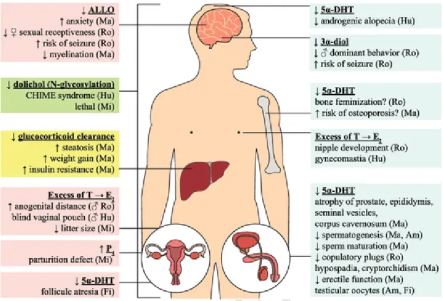
Fig. 2. Comparative physiological consequences of a lack of SRD5α in vertebrates (amphibians: Am; fish: Fi; humans: Hu; mammals: Ma; mice: Mi; rodents: Ro). Lack of SRD5α can de-crease 5α-DHT and adversely affect secondary sexual tissues (penis, scrotum, prostate, epididymis, and seminal vesicles), hair growth and bone structure, as well as cause female biased sex ratio in amphibians. Simultaneously, the excess of T can be converted to E2, which can increase breast development, anogenital distance, vaginal development and decrease litter size.
The absence of SRD5α activity can also decrease the degradation of P4involved in mice parturition, as well as slowing glucocorticoid clearance which increases steatosis, weight gain and
insulin resistance. SRD5αs are also implicated in ALLO synthesis in the brain. A decrease in ALLO increases anxiety, risk of seizure, and decreases myelination. Also, lower levels of 3α-diol in the brain can lead to a decrease in aggressiveness and dominance. Finally, SRD5α3 deficiency leads to a decrease in dolichol, which impacts N-glycosylation and leads to a CHIME syn-drome in human and to lethality in mice. Of note, the human schematic was only used as an example. Abbreviations: 3α-diol: 5α-androstane-3α,17β-diol; 5α-DHT: 5α-dihydrotestosterone; ALLO: allopregnanolone; CHIME syndrome: ocular colobomas, heart defects, ichthyosiform dermatosis, mental retardation and ear defects or epilepsy; E2: estrogen; P4: progesterone; T:
testosterone.
3.1.1. Atrophy of androgen-dependent organs
The most well-documented consequence of a decrease in 5α-DHT
in mammalian species is the reduction of the prostate weight (human:
Kang et al., 2014; Park and Choi, 2014; Mendonca et al., 2016;
dog: Juniewicz et al., 1993; Laroque et al., 1994; rats: George et
al., 1989; Imperato-McGinley et al., 1992; Clark et al., 1993;
Shao et al., 1993; Prahalada et al., 1998; Cayatte et al., 2006;
Zhang et al., 2012; Giatti et al., 2016; Enatsu et al., 2017; mice:
Mahendroo et al., 2001; wallaby: Ryhorchuk et al., 1997). The
ab-sence of 5α-DHT can also lead to weight loss of androgen-dependent
organs, such as the epididymis (rat: George et al., 1989; Cayatte
et al., 2006; Garcia et al., 2012), seminal vesicles (rat: George et
al., 1989; Imperato-McGinley et al., 1992; Cayatte et al., 2006;
Enatsu et al., 2017), corpus cavernosum (rat: Zhang et al., 2012;
Enatsu et al., 2017), and Cowper’s gland (rat: Cayatte et al., 2006).
However, in some cases this weight reduction is not seen (e.g.,
epi-didymis: men: Kang et al., 2014; Mendonca et al., 2016; dog:
Ju-niewicz et al., 1993; Laroque et al., 1994; rat:
Imperato-McGin-ley et al., 1992; seminal vesicle: men: Kang et al., 2014; Mendonca
et al., 2016; monkey: Prahalada et al., 1997). Also, the vas
defer-ens weight does not seem to be affected by a lack of 5α-DHT (men:
Kang et al., 2014; Mendonca et al., 2016; rat:
Imperato-McGin-ley et al., 1992). Some of the prostate studies have suggested that
this decrease in androgen-dependent organ weight is the result of the
tissue atrophy (dog: Juniewicz et al., 1993; Laroque et al., 1994;
rat: Cayatte et al., 2006; Enatsu et al., 2017), which is showed
by a significant decrease of cell proliferation (rat: Cayatte et al.,
2006), the presence of fibrosis (rat: Enatsu et al., 2017), the increase
in apoptotic prostatic cells in ejaculate (dog: Sirinarumitr et al.,
2002), and the alteration of tissue morphology (rat: Enatsu et al.,
2017; gerbil: Corradi et al., 2004). Similar signs of fibrosis and
at-rophy can be observed in the corpus cavernosum (rat: Pinsky et al.,
2011; Enatsu et al., 2017). On the molecular level, the absence of cell
proliferation is related to a lesser activation of AR receptors. In the
epi-didymis of FIN-treated rats, ARs are localized in the cytoplasm, in
con-trast to controls in which ARs are mainly located in the nucleus (Trybek
et al., 2005). Hence, ARs are not able to translocate to the nucleus due
to a lack of 5α-DHT, which prevents activation of transcription needed
for cell proliferation in androgen-dependent organs and explains the
ob-served weight loss.
3.1.2. Developmental defects
A lack of 5α-DHT can also lead to a defect in the male genital
tu-bercle development. The genital tutu-bercle has the potential to develop
either into a penis and scrotum in males, or a clitoris and vagina in
fe-males (Yamada et al., 2003; Blaschko et al., 2012). In fe-males, the
differentiation of the structure is regulated by androgens. The loss of
5α-DHT production consequently leads to hypospadia, a congenital
dis-order causing the urethra to be mislocated on the underside of the penis
(human: Kang et al., 2014; Mendonca et al., 2016; monkey:
Praha-lada et al., 1997; rats: Clark et al., 1990; Imperato-McGinley et
al., 1992; Clark et al., 1993; rabbit: Kurzrock et al., 2000). The
hy-pospadia condition is the result of a failure during the fusion process
be-tween the urethral groove and the genital tubercle, which, normally in
the presence of 5α-DHT, would form the penile urethra (Yamada et al.,
2003; Blaschko et al., 2012). In addition to hypospadia,
SRD5α2-de-ficient men can also exhibit a bifid scrotum (
UNCORRECTED
PROOF
in rats (Clark et al., 1990; Imperato-McGinley et al., 1992; Clark
et al., 1993). The most sensitive period for FIN to induce such effects in
rats is from the gestational days 16–17 (Clark et al., 1993), which
cor-relates with the development of the genital tubercle, occurring during
weeks 8 to 12 of pregnancy in humans, and gestational days 11–16 in
mice (Blaschko et al., 2012). During these stages of development, the
fate of the male genital tubercle appears to depend on the expression of
SRD5α2 in mesenchymal cells of the tubercle. Those cells seem to play
an instructive role for the epithelial cells as suggested by Berman et
al. (1995). This correlates with the expression pattern of SRD5α1 and
SRD5α2 in the urogenital tract where SRD5α1 is confined in epithelial
cells and SRD5α2 to the mesenchymal cells (Table 1).
Additionally, the lack of SRD5α2 activity increases T levels (human:
Park and Choi, 2014; monkey: Rhodes et al., 1994; rat: George
et al., 1989; George, 1997; Prahalada et al., 1998; Ribeiro and
Pereira, 2005; fish: García-García et al., 2017). During fetal
devel-opment T can be converted in E2 by aromatase and promote the
differ-entiation of the urogenital sinus in females (Blaschko et al., 2012).
Indeed, SRD5a2 deficiency leads to the development of a blind shallow
vaginal pouch in men (Kang et al., 2014) and to a decrease of the
anogenital distance (rat: Clark et al., 1990; Clark et al., 1993; rabbit:
Kurzrock et al., 2000); however, this condition is not always observed
(monkey: Prahalada et al., 1997; rats: Ribeiro and Pereira, 2005).
Moreover, the produced E
2induces nipple development in rats (Clark
et al., 1990; Imperato-McGinley et al., 1992; Clark et al., 1993)
and leads, on rare occasion, to gynecomastia at puberty in
SRD5α2-defi-cient men (Mendonca et al., 2016).
Another developmental defect due to a lack of 5α-DHT is
cryp-torchidism, in which testes are undescended (SRD5α2-deficient men:
Kang et al., 2014; Mendonca et al., 2016; monkey: Prahalada et
al., 1997). However, cryptorchidism is rarely observed in rats (Clark
et al., 1990; Imperato-McGinley et al., 1992). The difference
be-tween primates (including humans) and rodents can be explained by a
difference of periods when the testicular descent takes place. The
testic-ular descent is split into two phases: the transabdominal phase and the
inguinoscrotal phase. The transabdominal phase consists of testes
migra-tion through the iguinal region, which is an insulin-like hormone 3
(IN-SL3)-dependent process. This phase usually takes place during week 10
to 15 of gestation in humans, while in rodents it happens around
gesta-tional days 13–17. In the inguinoscrotal phase, testes descend through
the inguinal canal to the scrotum using an androgen-dependent
mecha-nism, which occurs from gestational week 25 to 35 in humans and
dur-ing postnatal week 3–4 in rodents (Hutson et al., 2013). Since the
androgen-dependent phase occurs after birth in rodents, inhibition of
5α-DHT production during gestation does not affect as strongly the
tes-ticular descent in rats in comparison to primates.
3.1.3. Disruption of erectile function
Men treated for BPH have up to 156% increased risk of developing
erectile dysfunction (Gacci et al., 2014; Liu et al., 2016; Corona
et al., 2017), while no significant side effects on erectile dysfunction
were found in cohorts of patients treated for androgenic alopecia (Liu et
al., 2016). However, the assessment of 5ARIs’ impact on erectile
dys-function in men is currently limited by a lack of method standardisation
as highlighted by three recent meta-analyses on the subject (Gacci et
al., 2014; Liu et al., 2016; Corona et al., 2017). In 5ARI-treated
rats, erectile function is also decreased (Pinsky et al., 2011). This
loss of function can be in part due to tissue atrophy as discussed
ear-lier (rat: Pinsky et al., 2011; Enatsu et al., 2017), as well as to
a decrease in neuronal nitric oxide synthase (NOS), essential for
pe-nile erection (Pinsky et al., 2011). Indeed, lower neuronal NOS ac
ducible NOS, to try to counter fibrosis and oxidative stress in the penile
tissue. However, the production of inducible NOS does not seem to be
enough in rats to prevent a loss of function (Pinsky et al., 2011). It is
possible the same phenomenon happens in other mammals, suggesting a
constant exposure to 5ARIs could lead to permanent damage to the
pe-nile tissue.
3.1.4. Effects on spermatogenesis and the structure of seminiferous tubules
A decrease in 5α-DHT has no effects on testis weight (dog:
Ju-niewicz et al., 1993; Laroque et al., 1994; rat: Rhoden et al.,
2002; SRD5α2/KO mice: Mahendroo et al., 2001; wallaby:
Ry-horchuk et al., 1997), nor on the presence of spermatogonia
(SRD5α2-deficient men: Kang et al., 2014; Mendonca et al., 2016;
wallaby: Ryhorchuk et al., 1997; amphibian: Urbatzka et al., 2009;
fish: Margiotta-Casaluci et al., 2013). However, spermatogenesis
progression was impaired as confirmed by a decrease in the production
of spermatocytes and spermatids (SRD5α2-deficient men: Kang et al.,
2014, amphibian: Urbatzka et al., 2009). Additionally, hypotrophy
of seminiferous tubules can occur (hamster: Vidigal et al., 2008;
wal-laby: Ryhorchuk et al., 1997). In FIN-treated amphibians (Xenopus
lae-vis), testes are immature and exhibit empty spermatocyst cavities
(Ur-batzka et al., 2009). This suggests that a decrease in 5α-DHT can affect
spermatogenesis, as well as the structure of seminiferous
tubules/sper-matocysts. These effects indicate that Sertoli cells are mainly affected
by the lack of 5α-DHT, since they are involved in spermatogenesis by
supporting germ cells’ development, as well as playing an important
role in the structure of the seminiferous tubules and the maintenance
of the blood-testis barrier (Wang et al., 2009; Huhtaniemi, 2018).
The impact on Sertoli cells could be linked to the high intratesticular T
concentration found in the testes, which is secreted by Leydig cells and
known to promote spermatogenesis (Wang et al., 2009; Huhtaniemi,
2018). This high intratesticular T concentration is probably converted
locally into 5α-DHT in the Sertoli cells which express SRD5α2 (Table 1).
Hence, 5α-DHT is implicated in spermatogenesis and the maintenance of
the seminiferous tubules in Sertoli cells. The only exception to this rule
being 5ARIs-treated fish in which spermatogenesis is unaltered or even
increased because levels of T and 11KT, the most potent androgen in
fish, are increased (Margiotta-Casaluci et al., 2013; García-García
et al., 2017).
3.1.5. Effects on sperm maturation and fertilization
After spermiation in the testes, the spermatozoids are matured in
the epididymis to acquire motility and increase their fertilization
poten-tial. This process is known to be androgen-dependent (Cornwall, 2008;
Sullivan and Mieusset, 2016). Indeed, SRD5α2 (Table 1), AR and
the androgen binding protein, which transports T to the epididymis, are
expressed throughout the epididymis (Robaire and Hamzeh, 2011).
Hence, a lack of 5α-DHT leads to the atrophy of the epididymis, as
discussed previously. In FIN-treated rats, daily sperm production was
not altered (George et al., 1989; Rhoden et al., 2002; Ribeiro
and Pereira, 2005; Garcia et al., 2012), but their sperm
motil-ity was impaired by the atrophy (Ribeiro and Pereira, 2005;
Gar-cia et al., 2012). Additionally, the motility was not recovered after
FIN-withdrawal (Garcia et al., 2012), which suggests a long term
im-pact on fertility. Similarly, SRD5α2-deficient men exhibited decreased
sperm motility (Kang et al., 2014; Mendonca et al., 2016), which
is rarely observed in men treated with 5ARIs (Amory et al., 2007).
Overall, 5α-DHT plays a crucial role in the epididymal structure by
maintaining cell proliferation, epithelial cell height and lumen
diame-ter (Robaire and Hamzeh, 2011). To preserve the structure of the
epididymis, 5α-DHT regulates gene expression through ge
UNCORRECTED
PROOF
nomic response via AR and rapid non-genomic response via the
extracel-lular receptor kinase pathway (Hamzeh and Robaire, 2011). Among
the genes regulated by 5α-DHT, the insulin-like growth factor-I and the
epidermal growth factor appear to play a main part in the promotion of
cell proliferation (Hamzeh and Robaire, 2010).
To ensure fertilization success, the ejaculate of some species
(no-tably rodents) coagulates into copulatory plugs in the vagina of females.
As well as indicating successful coitus, copulatory plugs gradually
re-lease spermatozoids, promote sperm transport, and can prevent other
males from mating the same female (Schneider et al., 2016). In rats,
FIN treatment decreased the capacity to form copulatory plugs by 50%,
which impacted fertilisation rates (Cukierski et al., 1991). The
ob-served decrease originates from the atrophy of the prostate and the
sem-inal vesicle, which produce the proteins responsible for the formation of
copulatory plugs (Schneider et al., 2016).
3.1.6. Effects on sex determination in non-mammalian vertebrates
A lack of 5α-DHT can affect testes differentiation in genetic males
of non-mammalian species, while mammalian gonads still differentiate
into testes. In male common toad (Bufo bufo), treatment with
β4-an-drostene-3-one-17β-carboxylic acid (17βC) (another 5ARI) led to larger
oocytes in Bidder’s organs (a rudimentary ovary found in the gonad of
both sexes), and to sex reversal of the actual testes (Petrini and
Zac-canti, 1998). In the western clawed frog (Silurana tropicalis), chronic
exposure to FIN induced a female biased sex ratio; with 27% males,
53% females, and 20% exhibiting testicular oocytes, an intersex
con-dition (Duarte-Guterman et al., 2009; Langlois et al., 2011). In
male medaka (Oryzias latipes), testicular degeneration and presence of
testicular oocytes were observed when exposed to FIN from the
fertil-ized egg stage to sexual maturity (Lee et al., 2015). These results
sug-gest 5α-DHT can affect the sex determination of gonads. This contrasts
with mammals in which sex determination is independent of hormones,
except for marsupials (Hayes, 1998; Nakamura, 2013; Trukhina et
al., 2013; Capel, 2017).
3.1.7. Impaired bone development and retention
In mice, SRD5α1/KO males have showed reduced bone mass and
forelimb muscle grip strength. The observed partial feminization of the
skeleton suggests 5α-DHT is needed for normal development of bone and
muscle in male mice (Windahl et al., 2011). In contrast,
SRD5α2-de-ficient men (Mendonca et al., 2016) and FIN-treated men (Amory et
al., 2008; Macukat et al., 2014) do not exhibit differences in bone
density. DUT could even have osteoprotective effects based on increased
bone density observed in a patient cohort (Macukat et al., 2014). The
osteoprotective effect is probably time dependent, since no effects on
bone density were observed after 52 weeks of treatment (Amory et al.,
2008), but an increase in density was observed after 24 to 48 months
(Macukat et al., 2014). In contrast, FIN-treated patients have an
in-creased risk to develop osteoporosis (Lin et al., 2015). It has also been
suggested that the lack of SRD5α activity would impact the levels of
glucocorticoids, which are also involved in bone maturation (Warriner
and Saag, 2013). However, 5α-DHT has more potential to be linked to
the role of SRD5αs in bone development since in vitro, 5α-DHT increases
the proliferation of epiphyseal chondrocytes and osteoblasts, both
impli-cated in longitudinal bone growth (Gori et al., 1999; Krohn et al.,
2003; Raz et al., 2005). The observed increase in cell proliferation
is mediated by the 5α-DHT-induced synthesis of insulin-like growth
fac-tor-I (Gori et al., 1999; Krohn et al., 2003).
3.1.8. Effects on the abundance and distribution of body and scalp hair
SRD5α2-deficient men exhibit less body hair growth than other men
and their hair distribution follows a female pattern (Randall, 2008).
In contrast, women overproducing 5α-DHT in their skin suffer from hir
sutism, a condition in which body hair overgrows in regions
associ-ated with a male pattern. Hirsutism can be treassoci-ated with FIN (van
Zu-uren and Fedorowicz, 2016; Barrionuevo et al., 2018), indicating
5α-DHT has a role in body hair growth. At puberty, 5α-DHT stimulates
hair growth of small and colorless hair, called vellus follicles, which
will grow into thick, long, and pigmented hair known as terminal
fol-licles as can be found on the chin, chest, pubis, arms, legs, and back
of post-pubescent men (Randall, 2008). Paradoxically, 5α-DHT has a
deleterious effect on scalp hair growth. In the scalp, the presence of
5α-DHT induces hair miniaturization by converting terminal hairs into
vellus hairs (Randall, 2008; Urysiak-Czubatka et al., 2014). In men,
5ARIs can reduce androgenic alopecia (Gupta and Charrette, 2013;
Adil and Godwin, 2017), while no baldness pattern is observed in
SRD5α2-deficient men (Kang et al., 2014; Mendonca et al., 2016).
Also, in Stumptail macaque (Macaca arctoide), a model for androgenic
alopecia, FIN treatment slightly improved hair growth by increasing the
number of scalp hair follicles and their average length (Rhodes et al.,
1994). However, FIN is not an efficient therapy for women suffering
from androgenic alopecia (Adil and Godwin, 2017). The reasons
lead-ing to the sex difference in treatment efficacy remain unclear. However,
SRD5α1 is expressed more than SRD5α2 in the scalp of women
suffer-ing from androgenic alopecia (Sawaya and Price, 1997), which
sug-gests that DUT treatment would be more successful. It would also be the
case in men, since a study showed SRD5α2 was no longer expressed in
the balding scalp, while SRD5α1 was still expressed (Thigpen et al.,
1993).
3.2. Impacts on female reproductive system
3.2.1. Effects in the uterus during gestation
A novel SRD5α2 mutation increased the risk of miscarriage in
women (Pérez-Nevot et al., 2017). However, the molecular
mecha-nism by which the mutation leads to miscarriage is still unclear. In
fe-male mice, 67% of SRD5α1/KO suffered from a defect in parturition,
which could lead to complications, such as vaginal bleeding,
resorp-tion of the fetus, and sometimes death. There was also a decrease in
litter size (~3 compared to 8) with no bias in sex ratio (Mahendroo
et al., 1996). In contrast, SRD5α2/KO female mice show normal
ges-tation (Mahendroo et al., 2001). The parturition defect in SRD5α1/
KO is mainly due to a lack of cervical ripening at gestational days
18–19, which is provoked by the lack of progesterone (P
4) degradation
by SRD5α1 in the cervix (Mahendroo et al., 1999). The defect can also
be related to a lack of 5α-reduced androgens (5α-DHT,
5α-androstene-dione and 5α-androstane-3α,17β-diol) at the end of gestation, since their
administration increases parturition success (33% to 93% with
5α-an-drostane-3α,17β-diol) (Mahendroo et al., 1996). However, the exact
role of these 5α-reduced androgens remains unclear. In contrast, the
re-duction in litter size takes place between gestational days 10.75–11 and
is provoked by the conversion of accumulated androstenedione and T
into E
2in the decidua (Mahendroo et al., 1997). Overall, those
stud-ies indicate SRD5αs are implicated in various biological functions
dur-ing gestation that are additional to the development of secondary sexual
organs in male fetuses. Future work should investigate how miscarriage
and birth defects occur in females lacking SRD5α1 or SRD5α2.
3.2.2. Androgenic and estrogenic effects in female fish
In female fish, 5ARI treatments increased follicle atresia
(Mar-giotta-Casaluci et al., 2013; Lee et al., 2015). In fathead minnows
(Pimephales promelas), females also have a decrease in E
2and an
in-crease in T, which leads to a dramatic dein-crease in egg production
(Mar-giotta-Casaluci et al., 2013). In contrast, administration of 5α-DHT
led to ovotestis in females, and depending on the species, 5α-DHT can
either increase or decrease the levels of E
2and vitellogenin, a precursor
UNCORRECTED
PROOF
drostane-3β,17β-diol, a steroid that has estrogenic capacity by
interact-ing with the estrogen receptor β (Oliveira et al., 2007).
3.3. Brain development and behaviour: role of ALLO
Expression of SRD5αs occurs throughout the brain and is observed
from early development (rat: Poletti et al., 1998; frog: Bruzzone
et al., 2010; Bissegger and Langlois, 2016a) (Table 1). In the
brain, mostly SRD5α1 is known to convert P
4into
5α-dihydroproges-terone (5α-DHP). 5α-DHP is then transformed by the 3α-hydroxysteroid
dehydrogenase (3α-HSD) into ALLO (Fig. 1B), an agonist of the
neu-rotransmitter gamma-aminobutyric acid A receptor (GABA
AR) (Reddy,
2010). In the brain, the absence of SRD5α activity decreases ALLO
lev-els (sheep: Yawno et al., 2007; rat: Modol et al., 2013; SRD5α1/KO
mice: Koonce and Frye, 2013; guinea pig: Kelleher et al., 2011),
while the concentrations of P
4and T increase (rat: Darbra et al., 2013;
Giatti et al., 2016), and 5α-DHT remains unchanged (rat: Giatti et
al., 2016). The decrease in the neurosteroid ALLO increases
anxiety-re-lated behavior and convulsion in vertebrates, as well as impacting brain
development.
3.3.1. ALLO decreases anxiety-related behaviour
Reduction of ALLO levels generates a less explorative behaviour and
a more anxiety-related behaviour (rat: Darbra and Pallarès, 2010;
Darbra et al., 2013; SRD5α1/KO: Frye et al., 2004; Koonce and
Frye, 2013). The anxiety is reversed when ALLO is administered
(SRD5α1/KO: Frye et al., 2013), but not when P
4is administered
(SRD5α1/KO: Frye et al., 2004; Koonce and Frye, 2013). These
results show that the conversion of P
4into ALLO is responsible for
the anti-anxiety effects and is not due to other functions of P
4in the
brain. This correlates with observations in depressive patients, in whom
ALLO brain levels are low and increase with antidepressant treatment
(Uzunova et al., 1998; Agis-Balboa et al., 2014).
ALLO’s function on anxiety is also implicated in females’
receptive-ness during reproduction. An increase in P
4during proestrus in mice
increases exploration and decreases anxiety (Koonce et al., 2012).
Proestrus corresponds to the period in the ovarian cycle when female
rodents are more sexually receptive, which they exhibit by a lordosis
reflex, a dorsiflexion of the spine to facilitate copulation. In
ovariec-tomized SRD5α1/KO mice, lordosis is less observed compared to their
wildtype (WT) counterpart. Also, administration of ALLO, but not P
4,can rescue the phenotype (Koonce and Frye, 2014). ALLO’s function
during lordosis plays a role in the maintenance of lordosis, while E
2is sufficient to induce lordosis (Uphouse, 2015). In order to maintain
lordosis, ALLO binds to dopamine type 1-like and GABA
AR in the
ven-tral tegmental area of the brain (rat: Frye and Walf, 2008), in which
SRD5α1 and/or SRD5α2 are expressed (Table 1). It would be
interest-ing to see if ALLO is implicated in female sexual receptiveness in other
species. For example, in vocal teleost fish and anurans, females show
re-ceptiveness with a behaviour called phonotaxis. Phonotaxis is a
move-ment related to sound, in this case male vocalisation during courtship,
and is correlated with season and sexual steroids (frog: Arch and
Nar-ins, 2009; Wilczynski and Lynch, 2011; fish: Forlano and Bass,
2011).
3.3.2. Anticonvulsant effect of ALLO
In mice, both P
4and ALLO administration protects from
pentylenete-trazole-induced seizures. However, when treated with FIN, only ALLO
has anticonvulsant property and not P
4(Kokate et al., 1999). In
ovariectomized SRD5α1/KO mice (Frye et al., 2002a) and FIN treated
male rats (van Luijtelaar et al., 2003), it is also ob
in pentylenetetrazole-induced seizures, but not in maximal electroshock
seizure tests (Kokate et al., 1999). The reason leading to a difference
in response during different simulation of seizures is that ALLO acts as
an anticonvulsant by competing for GABA
AR against the
pentylenetetra-zole, which is an antagonist of the receptors that can generate epilepsy
(Reddy, 2010). However, it cannot counteract seizures based only on
electric discharge, like in the maximal electroshock seizure test.
3.3.3. Neuroprotection of fetal brain during late gestation
During late gestation, ALLO levels are elevated in foetal brain and
decrease significantly after birth (sheep: Nguyen et al., 2003; guinea
pig: Kelleher et al., 2013). While in the adult life, ALLO stimulates
brain activity via GABA
AR; during gestation, data suggest that the
acti-vation of GABA
AR with ALLO have the opposite effect by reducing
fe-tal arousal. Inhibition of arousal with ALLO during pregnancy induces
a sleep-like behaviour in the foetus, limiting fetal activity and oxygen
consumption (Hirst et al., 2014). ALLO is also important to prevent
apoptosis in the fetal brain (sheep: Yawno et al., 2007; Yawno et al.,
2009) and to stimulate myelination of white matter neurons (guinea
pig: Kelleher et al., 2011). These functions of ALLO seem to be
medi-ated via the activation of oligodendrocytes (Hirst et al., 2014; Hirst
et al., 2016) and can be critical during late gestation. Indeed, guinea
pig preterm neonates exhibit reduced brain levels of ALLO (Kelleher et
al., 2013). This means those preterm newborns would have increased
apoptosis and decreased myelination in the brain which could lead to
long-term deficits.
3.4. Aggressiveness in males: role of 5α-androstane-3α,17β-diol (3α-diol)
In the brain, 3α-diol is synthesized by the same enzymatic cascade
as ALLO, but instead of using P
4as the primary substrate, T is reduced
subsequently by SRD5α and 3α-HSD (Fig. 1C). 3α-diol can have
anti-convulsant properties like ALLO (SRD5α1/KO mice: Frye et al., 2001),
but its main known function is its implication in aggressiveness
pat-terns in males, which has mostly been studied in mice. In
orchidec-tomized male mice, the number of aggressions registered against a new
resident in the cage is increased by the administration of T and even
more by 3α-diol (Frye et al., 2002b), showing 3α-diol has more
po-tential than T to induce aggression. In SRD5α1/KO mice, T
adminis-tration does not increase aggression in comparison to
vehicle-adminis-tered control (Frye et al., 2002b), which suggests the conversion into
3α-diol is necessary to increase aggressiveness. Male SRD5α2/KO mice
also show less aggressiveness, dominance over female, and risk-taking
behaviours than WT mice (Mosher et al., 2018). The role of 3α-diol
in aggressiveness indicates that even though a male is fertile, if it lacks
3α-diol, it will be less prone to defend its territory against other males,
dominate females, and engage in intercourse, which will lead to a
de-crease in reproduction. Dede-crease in 3α-diol could also be related to the
decrease in libido experienced by men taking 5ARIs (Liu et al., 2016;
Corona et al., 2017). Moreover, 3α-diol is probably implicated in
in-hibiting brain feminization in males. Castrated rats exposed to FIN
dur-ing gestation and treated with E
2exhibit lordosis in contrast to untreated
pups (Ribeiro and Pereira, 2005), a behaviour associated with
fe-males. More studies need to be done on 3α-diol and male reproductive
behaviour and aggressiveness patterns across species, which are
typi-cally associated with androgens, precursors to 3α-diol. For example, in
elasmobranch species, males with high levels of androgens perform
ag-gressive courtship in which they assault the female and even bite her
(Forlano and Bass, 2011). In anurans, androgens are linked to male
vocalisation, which can act as advertisement call for females, as well
UNCORRECTED
PROOF
as territorial calls for nearby males (Wells, 1977; Wilczynski et al.,
2005; Leary, 2009)
3.5. Effects on weight, steatosis, and insulin resistance
SRD5α1 is more highly expressed in the liver than SRD5α2 (Table
1) and a lack of SRD5α1 activity generates a defect in glucocorticoid
clearance. Indeed, adrenalectomised SRD5α1/KO mice have a decrease
in corticosterone clearance after corticosterone administration
(Living-stone et al., 2014; Living(Living-stone et al., 2017). Also, men excrete
less 5α-reduced metabolites of T and glucocorticoids in urine after
start-ing 5ARI treatments (Upreti et al., 2014). The reduced clearance of
glucocorticoids is not seen in the plasma levels of SRD5α1/KO mice
(Livingstone et al., 2014; Livingstone et al., 2017), but rather on
corticosterone levels present in the liver, which are elevated
(Living-stone et al., 2017). Moreover, the response to adrenocorticotropic
hor-mone is impaired (Livingstone et al., 2014) and genes related to
reg-ulation of hypothalamo-pituitary-adrenal (HPA) axis (vasopressin,
glu-cocorticoid receptor, corticotropin-releasing hormone, and its receptor)
are down-regulated in the liver (Livingstone et al., 2017),
hypothal-amus, and pituitary (Livingstone et al., 2014). Hence, slow clearance
of glucocorticoid clearly interferes with the HPA axis.
A slower clearance also signifies that glucocorticoids have extended
effects by accumulatingin the liversince 5α-reduction inactivates them
and increases their polarity to ease their excretion (Carlstedt-Duke et
al., 1977; Nixon et al., 2012). Regarding fat metabolism,
glucocorti-coid accumulation up-regulates triglyceride storage and cholesterol
syn-thesis (SRD5α1/KO mice: Livingstone et al., 2015; Livingstone et
al., 2017), while lipid mobilization and β-oxidation are down-regulated
(men treated with DUT: Hazlehurst et al., 2016; SRD5α1/KO mice:
Livingstone et al., 2015). The increase in fat storage and synthesis
re-sults in weight gain and steatosis, also known as fatty liver (men:
Up-reti et al., 2014; Hazlehurst et al., 2016; rat: Livingstone et al.,
2015; SRD5α1/KO mice: Dowman et al., 2013; Livingstone et al.,
2015; Livingstone et al., 2017). Those effects are observed mostly
on high fat diets (Dowman et al., 2013; Livingstone et al., 2015;
Livingstone et al., 2017) and are exacerbated in females regardless
of the diet (Livingstone et al., 2017). The exacerbation of effects is
suggested to be related to naturally low androgen levels in females,
which makes SRD5α1 loss more impactful on glucocorticoids levels.
Re-garding glucose metabolism, glucocorticoid accumulation
down-regu-lates gluconeogenesis (Livingstone et al., 2015), as well as insulin
receptor expression, (Dowman et al., 2013) leading to insulin
resis-tance and hyperinsulinemia (men: Upreti et al., 2014; Hazlehurst et
al., 2016; rat and male SRD5α1/KO mice: Livingstone et al., 2015;
female SRD5α1/KO: Livingstone et al., 2017). Hence, inhibition of
SRD5α1 in the liver will increase risk of obesity, insulin resistance and
nonalcoholic fatty liver disease by reducing glucocorticoids clearance.
3.6. Role of SRD5α3 in N-glycosylation
The first discovery of SRD5α3 was in hormone-refractory prostate
cancer, where it is overexpressed (Uemura et al., 2008). To date, only
two studies have shown the ability of human SRD5α3 to reduce steroids
in vitro (Uemura et al., 2008; Titus et al., 2014), whereas hamster
SRD5α3 lacks this activity (Chávez et al., 2015). More studies need
to be done to confirm its capacity to reduce steroids; however, SRD5α3
seems to have other functions. In 2010, Cantagrel et al. showed that
con-sanguineous families with a CHIME syndrome (ocular colobomas, heart
defects, ichthyosiform dermatosis, mental retardation and ear defects or
epilepsy) had mutations in the SRD5α3 sequence and exhibited a defect
in N-glycosylation. They demonstrated that SRD5α3 has the capacity to
reduce polyprenol into dolichol (Dol) (Fig. 1
D). When phosphorylated in the endoplasmic reticulum, Dol is then used
as a lipid carrier for glycan precursor needed in protein N-glycosylation.
This function in N-glycosylation classifies SRD5α3 mutations as a
con-genital disorder of glycosylation (CDGs) -type 1. Since the first
discov-ery, other mutations have been found in SRD5α3, all leading to
varia-tion of the CHIME syndrome (Kahrizi et al., 2010; Morava et al.,
2010; Gründahl et al., 2012; Kasapkara et al., 2012; Kara et al.,
2014; Wheeler et al., 2016; Taylor et al., 2017). The origin of
pa-tients’ symptoms was also explained with a mouse model. SRD5α3/KO
mice can only live up to gestational day 12.5 before dying. Their
lethal-ity was due to the inabillethal-ity to undergo axial rotation and a ventral body
wall defect at gestational day 8.5, as well as a dilated heart
(Canta-grel et al., 2010), which corroborates the heart and brain defects
ob-served in patients. Hence, the function of SRD5α3 seems to diverge from
SRD5α1 and SRD5α2. This could be explained by the separate
evolu-tion of SRD5α3 from other members of the family since early eukaryotes
(Langlois et al., 2010a), which led to a difference in domain related to
SRD5α1 and SRD5α2 (hamster: Chávez et al., 2015). It would be
nec-essary to verify the ability of SRD5α3 to reduce steroids and polyprenol
in other species to ascertain the function of SRD5α3 across vertebrates.
4. Genomic and non-genomic pathways are involved in the
regulation of SRD5α expression
Even though SRD5α1 and SRD5α2 can be found in the same tissues
(Table 1), both isoforms are not expressed in the same cell types. For
example, in the brain, SRD5α1 is mostly expressed in glial cells
(astro-cytes, oligodendrocytes) (rat: Kiyokage et al., 2005; Castelli et al.,
2013; mice: Kiyokage et al., 2014; fish: Mathieu et al., 2001), but
can be found in neurons (mice: Agis-Balboa et al., 2006; fish:
Math-ieu et al., 2001) and gonadotroph cells of the anterior pituitary (rat:
Yokoi et al., 1996), while SRD5α2 is mostly restricted to neurons (rat:
Pozzi et al., 2003; Castelli et al., 2013). The difference in
expres-sion of isoforms between cell types is related to various mechanisms
de-scribed in this section, including, the regulation by transcription factor
such as hormone receptors (Fig. 3) and by epigenetics such as
methyla-tion of CpG islands (Fig. 4).
4.1. Androgens drive tissue-specific regulation of SRD5αs
Unsurprisingly, SRD5αs are regulated by androgens that they help
synthesize. All three isoforms are predicted to possess androgen
re-sponse elements (AREs) in their promoter sequences (Flood et al.,
2013). In the promoter of SRD5α2, there are two classes of AREs. One
is a classic ARE with a palindromic repeat of AGAACA, which is
func-tional and can also act as a progesterone response element (PRE)
(Mat-sui et al., 2002). The second is a selective ARE constituted of a
par-tial direct repeat of AGAACA (Kerkhofs et al., 2012). In the human
promoter of SRD5α3, only one ARE was confirmed to be functional (Li
et al., 2011). Even if each isoform possesses AREs in its promoter,
their regulation by ARE is more complicated than the simple
activa-tion of AR by androgens. For instance, in human prostate cancer cell
lines, 5α-DHT administration differentially regulates the expression of
the three SRD5αs in each cell line (Li et al., 2011). Furthermore, in
rat’s prefrontal cortex, SRD5α2 is upregulated by the administration of
T and 5α-DHT in both sexes, while SRD5α1 expression profiles exhibit
a sexual dimorphism. In males, SRD5α1 is down-regulated when treated
with T and 5α-DHT. In contrast, SRD5α1 is upregulated in females only
after 5α-DHT treatment (Torres and Ortega, 2003). In T-treated rats,
SRD5α1 is negatively regulated in the testes, whereas SRD5α2 is
unaf-fected, but is overexpressed in the prostate (Pratis, 2003). All those
results show that even though all three SRD5α possess AREs in their
promoter, each isoform in each tissue is differentially regulated by
an-drogens. The differential regulation is probably linked to co-regula
UNCORRECTED
PROOF
Fig. 3. Predicted response elements in the promoters of SRD5αs. (A) SRD5α1 possesses TREs and AREs that are predicted, but have not yet been proven functional. Also, SRD5α1 can be regulated by E2and P4, but no study has looked at the presence of response element of those hormones in SRD5α1′s promoter; (B) SRD5α2 can be regulated by androgens via two AREs.
The first one is a classic ARE, which also acts as a PRE. The second one is a selective ARE, which is specific to only androgens. When cholesterol is low, SREBP2 upregulates SRD5α2. Finally, E2can also regulate SRD5α2, but it is yet unclear if it acts directly via a response element or indirectly by other pathways; (C) SRD5α3 possess an ARE in its promoter, as well as
predicted TREs, which the functionality remains unknown. The figure is a scheme and is not representative of the order, position, or the number of each response elements found in the genome. AR: androgen receptor; ARE: androgen response element; E2: estrogen; ERE: estrogen response element; P4: progesterone; PR: progesterone receptor; PRE: progesterone response
element; SREBP2: sterol regulatory element binding proteins type 2; TR: Thyroid hormone receptor; TRE: thyroid response element.
Fig. 4. Known mechanisms of methylation of CpG Island in the promoter of SRD5α2. (A) Inflammation cascade of TNFα, NF-κB and Il-6 which regulates the expression of DNMT1; (B) With age, EZH2 can regulate DNMT1 by an unknown pathway. DNMT1 methylates SRD5α2′s promoter and inhibits its transcription. DNMT1: DNA methyltrans-ferase 1; EZH2: enhancer of zeste homolog 2; Il-6: Interleukin 6; NF-κB: nuclear factor kappa-light-chain-enhancer of activated B cells; TNF-α: tumor necrosis factor-α.