HAL Id: pastel-00711285
https://pastel.archives-ouvertes.fr/pastel-00711285
Submitted on 23 Jun 2012HAL is a multi-disciplinary open access
archive for the deposit and dissemination of sci-entific research documents, whether they are pub-lished or not. The documents may come from teaching and research institutions in France or abroad, or from public or private research centers.
L’archive ouverte pluridisciplinaire HAL, est destinée au dépôt et à la diffusion de documents scientifiques de niveau recherche, publiés ou non, émanant des établissements d’enseignement et de recherche français ou étrangers, des laboratoires publics ou privés.
Thuong van Du Tran
To cite this version:
Thuong van Du Tran. Modeling and predicting super-secondary structures of transmembrane beta-barrel proteins. Bioinformatics [q-bio.QM]. Ecole Polytechnique X, 2011. English. �NNT : 2011EPXX0104�. �pastel-00711285�
TH`
ESE
pr´esent´ee pour obtenir le grade de
DOCTEUR DE L’´
ECOLE POLYTECHNIQUE
Sp´ecialit´e:
INFORMATIQUE
parThuong Van Du TRAN
Titre de la th`ese:Modeling and Predicting
Super-secondary Structures of
Transmembrane β-barrel Proteins
Soutenue le 7 d´ecembre 2011 devant le jury compos´e de:
MM. Laurent MOUCHARD Rapporteurs
Mikhail A. ROYTBERG
MM. Gregory KUCHEROV Examinateurs
Mireille REGNIER
M. Jean-Marc STEYAERT Directeur
Contents
Introduction 1
1 Fundamental review of proteins 5
1.1 Introduction . . . 5
1.2 Proteins . . . 5
1.2.1 Amino acids . . . 5
1.2.2 Properties of amino acids . . . 6
1.2.3 Peptide bond . . . 10 1.2.4 Protein . . . 12 1.2.5 Protein structure . . . 16 1.3 Transmembrane proteins . . . 20 1.3.1 Biological membrane . . . 20 1.3.2 Transmembrane proteins . . . 21 1.4 Folding energy . . . 24 1.4.1 Partial charges . . . 25 1.4.2 Electrostatic interaction . . . 25 1.4.3 Hydrogen bond . . . 25
1.4.4 Van der Waals forces and steric repulsion . . . 27
1.4.5 Hydrophobic effect and interaction with the environment . . . 28
1.4.6 Torsion energy around peptide bonds . . . 29
1.4.7 Other interactions . . . 29
1.5 Protein structure determination . . . 29
1.5.1 Experimental methods . . . 29
1.5.2 In silico prediction . . . 30
2 Folding β-barrels 33 2.1 Introduction . . . 33
2.2 Geometric framework for β-barrels . . . 33
2.3 Physicochemical constraints . . . 35
2.4 Classification filtering . . . 38
2.5.1 Vertices . . . 39
2.5.2 Edges . . . 39
2.5.3 Energy attributes: . . . 40
2.5.4 Protein folding problem . . . 42
2.6 Dynamic programming approach . . . 43
2.6.1 Solving as the longest path problem . . . 43
2.6.2 Solving as the longest closed path problem . . . 43
2.6.3 Generalization . . . 44
2.7 Complexity on permuted structures . . . 49
2.7.1 Preliminaries . . . 49
3 Tree-decomposition based algorithm 57 3.1 Introduction . . . 57
3.2 Graph-theory background . . . 57
3.2.1 Tree decomposition . . . 58
3.2.2 Modular decomposition . . . 60
3.3 NP-Completeness . . . 60
3.4 Algorithm for finding barrel structures of minimum energy . . . 63
3.5 About Greek key motifs in β-barrels . . . 68
4 Evaluation of performance of BBP 75 4.1 Introduction . . . 75 4.2 Experimental setup . . . 75 4.2.1 Software . . . 75 4.2.2 Datasets . . . 75 4.3 Implementation details . . . 78 4.4 Method of evaluation . . . 80
4.4.1 Concepts on predicted secondary structures . . . 80
4.4.2 Measures of performance . . . 82
4.5 Experimental results . . . 84
4.5.1 Folding . . . 85
4.5.2 Evaluation of the shear numbers . . . 86
4.5.3 Influence of the filtering threshold . . . 86
4.5.4 Evaluation on mutated sequences . . . 89
4.5.5 Permuted structures . . . 92
4.5.6 Classification . . . 92
Conclusion and perspectives 94
List of Figures
1.1 Isomers L and D of amino acids . . . 6
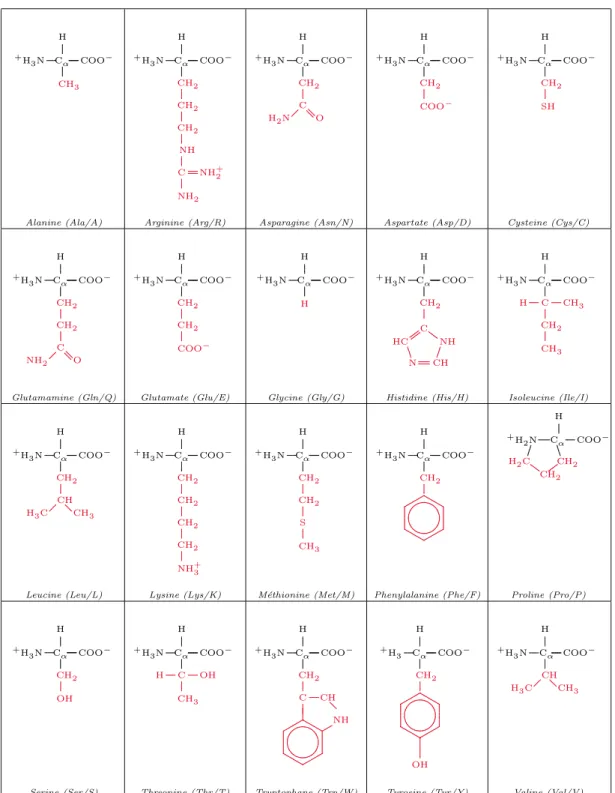
1.2 The 20 amino acids. The side chains are in red. . . 7
1.3 Peptide bond geometry in trans configuration . . . 12
1.4 Torsion angles between two peptide plans . . . 13
1.5 Ramachandran plot for the outer membrane protein A (PDB:1BXW) . . 14
1.6 Structure of collagen (PDB:1BKV) . . . 14
1.7 Structure of myoglobin (PDB:1A6M) . . . 15
1.8 Structure of insuline receptor (PDB:1GAG) . . . 15
1.9 Structure of an α-helix . . . 17
1.10 Antiparallel pairing (a) and parallel pairing (b) of β-strands . . . 17
1.11 Characteristics of a β-sheet. . . 18
1.12 Tertiary structure (a) and super-secondary structure (b) of the cystic fi-brosis transmembrane conductance regulator (PDB:1R0W) . . . 19
1.13 Quaternary structure of human hemoglobin (PDB:1MKO) . . . 19
1.14 Illustration of a biological membrane and embedded membrane proteins. . 21
1.15 Transmembrane proteins: (1) a single transmembrane hydrophobic α-helix - bitopic membrane protein, (2) several transmembrane hydrophobic α-helices, (3) transmembrane β-barrel protein. . . 22
1.16 Bacteriorhodopsin in purple membrane (PDB:2BRD) . . . 23
1.17 Outer membrane protein X (PDB:1QJ8) . . . 24
1.18 Hydrogen bonds represented in dash lines: (a) between water molecules and (b) between carboxylic and amino groups. δ+ and δ− are positive and negative partial charges, respectively. . . 27
2.1 The simplified geometry of a β-barrel, a schematic planar view for 6 strands (strand 1 is duplicated for clarity). Thick lines denote the peptide bonds that link consecutive amino acids along their strand. Thin lines denote the hydrogen bonds that link the amino acids of two adjacent strands. In this example, the shear number is S = 8, which is the ordinal distance between amino acids A and B. We note that all known β-barrels have a positive shear number [80] and are slanted “to the right”, as illustrated here. . . . 34
2.2 A schematic planar representation of 3 strands in a transmembrane β-barrel. The black residues direct their side chains toward the membrane and white ones toward the channel. The first and third strands are upward and the second one is downward. The first and second strands are odd
outward and the third one is odd inward. . . 36
2.3 The distribution of average hydrophobicity index of the hydrophilic side of the membrane spanning β-strands from PDBTM40 (see Section4.2) . . 37
2.4 The distribution of average hydrophobicity index of the hydrophobic side of the membrane spanning β-strands from PDBTM40 (see Section4.2) . . 37
2.5 A short example of the graph structure. Edge (v1, v2) is not allowed, since the two corresponding substrings overlap. Edges (v2, v3) or (v2, v6) are not allowed, since the substrings in between are respectively too short for a turn or too long for a loop, etc. . . 40
2.6 Different views of a β-barrel with a Greek key motif 3654, σ = 1 2 3 6 5 4 . 42 2.7 A permuted β-barrel with a Greek key motif 5436, σ = 1 2 5 4 3 6 . . . 44
2.8 Schema of sets confk corresponding to σ = {1, 2, 5, 4, 3, 6} . . . 46
2.9 Relation ∆k and its transitive closure ∆∗k on the kth substructure . . . 47
2.10 Illustration for property2.7 . . . 48
3.1 A graph and a tree decomposition of width 3 . . . 59
3.2 A path decomposition of width 3 of the graph in3.1 . . . 59
3.3 A graph and its modular decomposition are on the left. The quotient graph is on the right. . . 61
3.4 The β-barrel(a), Gc(b) and the tree/path decomposition(c) of σ = 1 4 3 2 5 6 7 8 . . . 63
3.5 Gc(a) and its tree decomposition(b) of σ = 3 2 1 4 5 6 7 8 . . . 69
3.6 Gc(a), its quotient graph(b) and its tree decomposition(c) of σ = 3 2 1 4 7 6 5 8 11 10 9 12 . . . 70
3.7 Gc(a), its quotient graph(b) and its tree decomposition(c) of σ = 1 4 3 2 5 6 7 8 . . . 70
3.8 Gc(a) and its tree decomposition(b) of σ = 1 2 3 4 5 8 7 6 . . . 71
3.9 Gc(a), its quotient graph(b) and its tree decomposition(c) of σ = 1 4 3 2 5 8 7 6 9 12 11 10 . . . 71
3.10 Gc(a), its quotient graph(b) and its tree decomposition(c) of σ = 1 2 5 4 3 6 9 8 7 10 11 12 . . . 72
3.11 Gc(a) and its tree decomposition(b) of σ = 1 2 3 4 5 6 7 10 9 8 . . . 72
3.12 Gc(a), its quotient graph(b) and its tree decomposition(c) of σ = 1 2 3 6 5 4 7 10 9 8 11 12 . . . 73
3.13 The reduced graph G+− for g+g−(a) and its tree decomposition of width 3(b) . . . 73
List of Figures
4.2 Energy distribution of setECOLI40, θ = arctanhSdn . . . 88 4.3 MCC of mutated setECOLI40 . . . 90 4.4 F-score of mutated setECOLI40 . . . 91 4.5 Distribution of 7! permutations on E. Coli OmpA 1BXW 8-strand barrel 93 4.6 Distribution of 7! permutations on E. Coli OmpX 1QJ8 8-strand barrel . 93
List of Tables
1.1 Hydrophobic scales . . . 8
1.2 Polarity, flexibility and other physicochemical parameters of amino acids . 9 1.3 Partial charges from the Gromos force field for standard amino acids. e is the absolute value of elementary charge unit. . . 26
1.4 The dielectric constant of selected mediums . . . 27
1.5 Typical values for van der Waals well depth and radius . . . 28
4.1 Transmembrane β-barrel proteins in setPDBTMB40 . . . 77
4.2 β-barrel proteins in setLIPOC . . . 79
4.3 Comparison of prediction accuracy on setTransFold. Q2 and M CC are measures on residues. Sensitivity and P P V are measures on β-strands. . 85
4.4 Comparison of prediction accuracy on setPREDTMBB. Q2 and M CC are measures on residues. T P , F P , T N are measures on β-strands. T OP is the number of proteins with correctly predicted topology, i.e. the pro-teins with correctly predicted number of β-strands. . . 85
4.5 Comparison of prediction accuracy on setPDBTMB40 . . . 86
4.6 Predicted optimal structures of transmembrane β-barrel proteins in setECOLI40. n is the number of β-strands, S is the shear number, the slant angles are expressed in degrees. . . 89
4.7 Comparison of prediction accuracy on setECOLI40 with different thresh-olds . . . 92
Acknowledgement
Acknowledgement
This thesis would not have been achieved without the aid of several people who, in one way or another, contributed their valuable assistance in the preparation and completion of my study; and it is my great pleasure to thank those who made it possible.
I owe my deepest gratitude to my supervisor, in fact more than a supervisor, Jean-Marc Steyaert, for his wise guidance throughout my research work as well as his warm support in my life. I have learned so much from his insight and personality.
I am heartily thankful to my co-advisor, Philippe Chassignet, who gave me many pieces of advice and helped me overcome various difficulties in all of the time of my research.
I would like to express my sincere gratitude to my Ph.D. committee. My heartiest thanks to Laurent Mouchard and Mikhail Roytberg for taking their time to thoroughly read my manuscript and giving me valuable assessments. My deepest appreciation to Gregory Kucherov and Mireille R´egnier for the reviews and comments on my thesis.
I gratefully acknowledge Saad Sheikh for all his help, enthusiasm and valuable hints during the time we worked together.
Many thanks go in particular to my group of Bioinformatics, especially Peter Clote, Thomas Simonson, Julie Bernauer, Yann Ponty, J´erˆome Waldisp¨uhl, Philippe Dessen, Mahsa Behzadi and Balaji Raman for their valuable suggestions and discussions, which helped me enrich my knowledge, broaden my view and develop my skills.
I would like to give special thanks to my colleagues, Morgan Barbier, Guillaume Quintin, J´erˆome Milan, Pierre Saint-Geours, with whom I have shared the same office. Thanks for the helpful discussions, the humors and, especially, the collection of soft drink cans for which I still owe a work of art. I would also acknowledge Thomas Clausen who pushed me to finish the writing of this manuscript.
I am grateful to the secretaries, Evelyne Rayssac and Catherine Bensoussan, for all their help in administrative works. Thanks to James Regis and Mathieu Guionnet for all technical assistances.
Thanks to LIX with all of its staff members for the warm environment. Thanks to Ecole Polytechnique where I have been staying since my undergraduate, for the fundings and a lot of beautiful memories.
I would like to show my gratitude to all my friends in Polytechnique, especially the Vietnamese ones, who are always beside me.
And last, but not least, I am greatly indebted to my family. I wish to thank my parents and my sister for their love and spiritual support. Words fail me to express my appreciation to my wife, whose love and dedication has brought more energy to me. Many thanks not only for her correction of my manuscripts, but also for our future son. To them I dedicate this thesis.
Abstract
The transmembrane β-barrel proteins (TMBs) are found in the outer membrane of Gram-negative bacteria, mitochondria and chloroplasts. They entirely span the biologi-cal membrane and perform a wide range of important functions. As the number of TMB structures known today is very limited, due to difficulties in experimental methods, it is arguable whether the learning-based prediction methods could work well for recognizing and folding TMBs which are not homologous to those currently known. We present a novel graph-theoretic model for classification and prediction of permuted super-secondary structures of TMBs from their amino acid sequence, based on energy minimization. The model does not essentially depend on learning. The algorithms are fast, robust with com-parable performance to the best currently known learning-based methods. This method can be thus a useful tool for the genome screening. Besides the performance on prediction and classification, this study gives an insight into TMB structures regarding the physic-ochemical constraints of biological membranes. The predicted permuted structures can also enhance the understanding on the folding mechanism of TMBs.
Keywords: transmembrane protein, β-barrel, super-secondary structure prediction, permuted structure, Greek key, ab initio modeling
R´esum´e
Les prot´eines transmembranaires canaux-β (TMBs) se trouvent dans les membranes externes des bact´eries `a Gram n´egatif, des mitochondries ainsi que des chloroplastes. Elles traversent enti`erement la membrane cellulaire et exercent diff´erentes fonctions im-portantes. Vu qu’il y a un petit nombre des structures des TMBs d´etermin´ees, en raison des difficult´es avec les m´ethodes exp´erimentales, il est douteux que ces approches puis-sent bien trouver et pr´edire les TMBs qui ne sont pas homologues avec celles connues. Nous construisons un mod`ele de graphe pour la classification et la pr´ediction de struc-tures super-secondaires permut´ees des TMBs `a partir de leur s´equence d’acides amin´es, en se basant sur la minimisation d’´energie. Le mod`ele ne d´epend essentiellement pas de l’apprentissage. Les algorithmes sont rapides, robustes avec des performances com-parables `a celles des meilleures m´ethodes actuelles qui utilisent l’apprentissage. Cette m´ethode peut ˆetre donc utile pour le screening des g´enomes. Outre la performance de pr´ediction et de classification, cette ´etude donne une vue plus profonde de la structure des TMBs en tenant compte des contraintes physicochimiques des membranes biologiques. Les structures permut´ees pr´edites peuvent aussi aider `a mieux comprendre le m´ecanisme du repliement des TMBs.
Mots-clefs: prot´eine transmembranaire, canaux-β, pr´ediction de structure super-secondaire, structure permut´ee, cl´e grecque, mod´elisation ab initio
Introduction
Motivation
Proteins can be considered as major elements and tools of life at the molecular scale as they carry out various functions in living organisms. These functions are expressed through their three-dimensional conformations, i.e. the way that amino acids are ar-ranged in the 3D space. Therefore, discovering the structures helps understand the functions associated to the proteins. Besides the experimental methods, the prediction of protein structure in silico from the amino acid sequence with high accuracy and reli-ability is one of the most important tasks, yet remains a challenge in bioinformatics and computational biology.
Transmembrane proteins play many important roles in the functioning of cells such as enzymes, receptors, transporters, and channels. They are also involved in many hu-man diseases including heart disease, cancer, Alzheimer’s, depression, migraine, retinitis pigmentosa, hereditary deafness, diabetes, cystis fibrosis, etc. [29, 42, 85]. As a result, they are the targets of a majority of current medicine and of an important research area. These proteins make up 20 − 30% of identified proteins in most whole genomes. How-ever, determining the structure of transmembrane proteins with experimental methods is difficult as they are totally destabilized by the change of environment after their removal from the membrane. Solved transmembrane protein structures constitute only about 1 − 2% of the RCSB Protein Data Bank (PDB) [6,13,23,40,118]. Therefore, structure prediction by computational methods for this class of proteins is of particular importance for both biological and medical sciences.
Transmembrane proteins are divided into two main types according to their conforma-tion: α-helical bundles and β-barrels, in which the transmembrane β-barrel (TMB) pro-teins are much less abundant than α-helical bundles in the PDB. These TMB propro-teins are found in the outer membrane of Gram-negative bacteria, mitochondria and chloroplasts. They entirely span the biological membrane and perform a wide range of functions, such as porins, passive or active transporters, enzymes, defense or structural support, multi-drug resistance [54,117]. Nevertheless, only a few non-homologous TMB structures have been experimentally determined due to difficulties in the experimental methods such as X-ray crystallography or nuclear magnetic resonance spectroscopy. Moreover, the folding mechanism of TMB proteins has not been well understood yet, though they are observed
in spontaneous folding process in certain experiments in vitro [17,117,131,132]. We particularly concentrate, in this thesis, on the super-secondary structure of TMB proteins, which describes the arrangement and interaction of the β-strands in the 3D space.
State of the art
Contrarily to the great progress in structure prediction on α-helical bundles [40], due to a tiny number of determined TMB structures, the learning-based predictions for these proteins are still far from being reliable, although various techniques have been recently developed for discriminating TMB proteins from globular and transmembrane α-helical proteins [41, 50, 51, 130], and for predicting TMB secondary structures [7, 50, 51, 96, 103, 130].
Gromiha et al. [50,51] used the amino acid compositions of both globular and outer membrane proteins (OMPs) to discriminate OMPs and developed a feed forward neural network-based method to predict the transmembrane segments. Bagos et al. [7] pro-duced a consensus prediction from different methods based on hidden Markov models, neural networks and support vector machines [1, 9, 16,51, 59, 86, 89, 94]. Waldisp¨uhl et al. [130] used a structural model and pairwise interstrand residue statistical potentials derived from globular proteins to predict the supersecondary structure of TMB proteins. Randall et al. [103] tried to predict the TMB secondary structure with 1D recursive neural network using alignment profiles. Ou et al. [96] proposed a method based on radial basis function networks to predict the number of β-strands and membrane spanning regions in β-barrel outer membrane proteins. Freeman et al. [41] introduced a statistical approach for recognition of TMB proteins based on known physicochemical properties. Most of these rely on the learning assumptions in the underlying models as well as the sampling of proteins in their training data set. As the number of TMB structures known today is very limited, it is arguable whether these approaches can work well for recognizing and folding TMB proteins which are not homologous to those currently known.
Moreover, the Greek key motifs are the topological signature of many β-barrel and β-sandwich structures [139]. This raises an open question whether the TMB structures are not merely a series of β-strands where each is bonded to the preceding and succeeding ones in the sequence order, but may contain Greek key or Jelly roll motifs as well: for instance, the C-terminal domain of the outer membrane usher protein PapC (PDB:3L48). This level of structure may be described as a permutation on the order of the bonded strands.
Contribution
We present a novel graph-theoretic model (see Chapter2and3) for predicting the super-secondary structure of transmembrane β-barrel proteins from their amino acid sequence.
Introduction
This structure is considered as a permuted arrangement or β-strands in a barrel, in which the β-strands are paired antiparallely or parallely. The problem consists in finding the thermodynamically most stable structure, i.e. the structure of minimum energy. This protein structure prediction problem can be modeled into finding the longest cycle-attached path in a graph with respect to a given permutation.
Each vertex in the graph represents an amino acid segment that satisfies the con-formational constraints, for instance, the length of β-strands, the hydrophobicity of side chains, the propensity for each segment to be a β-strand. . . A probabilistic model is built from the determined structures to calculate these propensities. It is applied as a filter for potential β-strands. Each edge presents a pair of segments whose loop in between satisfies the constraints on length, flexibility, polarity, etc. The energies are assigned to the vertices, the edges, as well as to the interaction between each pair of pairing segments. The amino acids are constructed in the three-dimensional space using the Dunbrack rotamer library. We then calculate the energies as the average on all rotamers. The hydrophobic interaction is computed on each pair of residue side chains using well-known hydrophobicity scales, while the electrostatic interactions between two amino acids are obtained thanks to the partial charges in the molecular mechanics force fields.
We prove the NP-completeness of the problem of finding the optimal permuted super-secondary structure. Then, a dynamic programming-based algorithm is proposed and implemented. This algorithm can find the optimum with a complexity in time of at most O(N4) for the structures containing disjoint Greek key motifs (see Chapter 2). This
complexity is improved to O(N3) with another algorithm that uses the concept of tree decomposition (see Chapter3).
To evaluate the performance of our method, we test the program on all TMB se-quences with known structures in the PDBTM database (see Chapter4). We show the accuracy of the approach with the F-score, sensitivity, specificity of more than 90% in the measure on β-strands and more than 74% in the measure on residues, which are compara-ble to the best learning-based methods. The ability of discrimination is also robust with 100% of α-helical transmembrane proteins and 97% β-barrel lipocalins being rejected. It also shows the ability to find the arrangement of β-strands with the “right permutation” locating in the zone of 0.7% - 1.5% of lowest-energy permutations. This method is thus potentially a useful tool for the genome screening. Beside the performance on prediction and classification, this study provides insight into TMB structures regarding the physic-ochemical constraints of biological membranes. The predicted permuted structures can also enhance the understanding on the folding mechanism of TMB proteins.
The program can be executed via the web-server BBP (Beta Barrel Predictor) (http://
Organization
The manuscript is organized as follows:
Introduction
This chapter presents the motivation of the work, the state of the art in this research area, the summary of our contribution and an outline of the manuscript.
Chapter 1
Fundamental review of proteins. We remind the fundamental notions in biology concern-ing the proteins and the methods of protein structure prediction.
Chapter 2
Folding β-barrels. We introduce our model and algorithm for determining the protein structure of minimal energy, then provide an analysis on the computational complexity with regard to different types of structures.
Chapter 3
Tree-decomposition based algorithm. We present an algorithmic improvement based on the tree decomposition technique, followed by an analysis on its computation complexity.
Chapter 4
Evaluation of performance of BBP (Beta-Barrel Predictor). We assess the performance of our prediction model on the experimentally determined structures.
Conclusion and perspectives
Chapter 1
Fundamental review of proteins
1.1
Introduction
This chapter provides the reader with fundamental notions in biology that are men-tioned throughout the manuscript and necessary for understanding the practical moti-vation of our work. The content is inspired from the Ecole Polytechnique text book of molecular and cellular biology by Yves Gaudin, Arnaud Echard and Sandrine Etienne-Manneville [44], the book on membrane structural biology by Mary Luckey [82], and J´erˆome Waldisp¨uhl’s PhD thesis [129].
We rapidly present the amino acids, constituent of proteins, before describing the properties and structures of the proteins themselves. Then, we focus on the class of transmembrane proteins, especially the β-barrels which are the subject of our whole work. We finally describe the problem of protein structure prediction and present the methods that have been developed to solve it.
1.2
Proteins
1.2.1 Amino acids
Amino acids have the general form:
... ... ... ... . . . . . . . . . . . . . . . . . . . . . . . . . . . . ... ... ... ... .... Cα COOH R H2N H
They contain an amine group NH2, a carboxylic group COOH and an organic
sub-stituent R. In aqueous solution at neutral pH, amino acids exist in the zwitterionic form where the amine functional group is protonated (NH+3) and the carboxylic functional group is deprotonated (COO−). The substituent R, also called side chain, varies
be-tween 20 different standard amino acids. The four groups attached to the α-Carbon are distinguished (except for Glycine in which the side chain R consists of a hydrogen atom). Therefore, there exists two reflection-symmetric isomers L and D (see Figure 1.1), of which only L isomers are present in proteins.
...... ... . . . . . . . . . . . . . . . . . . . . . . . . . . . . . . . . . . . . . . . . . . . . . . . . . . . . . . . . . . . . . . . . . . . . . . . . . . . . . . . . . . . . . . . . . . . . . . . . . . . . . . . . . . . . . . . . . . . . . . . . . . . . . . . . . . . . . . . . . . . . . . . . . . . . . . . . . . . . . . . . . . . . . . . . . . . . . . . . . . . . . . . . . . . . . . . . . . . . . . . . . . . . . . . . . . . . . . . . . . . . . . . . . . . . . . . . . . . . . . . . . . ... ... ... ... ... ... ... ... ... ... ... ... ... ... ... ... Cα R H H+3N COO− . . . . . . . . . . . . . . . . . . ... ... ... . . . . . . . . . . . . . . . . . . . . . . . . . . . . . . . . . . . . . . . . . . . . . . . . . . . . . . . . . . . . . . . . . . . . . . . . . . . . . . . . . . . . . . . . . . . . . . . . . . . . . . . . . . . . . . . . . . . . . . . . . . . . . . . . . . . . . . . . . . . . . . . . . . . . . . . . . . . . . . . . . . . . . . . . . . . . . . . . . . . . . . . . . . . . . . . . . . . . . . . . . . . . . . . . . . . . . . . . . . . . . . . . Cα R H H+3N COO−
Figure 1.1: Isomers L and D of amino acids
The 20 standard amino acids are shown in Figure1.2. Each amino acid is associated with a 3-letter abbreviation and a 1-letter code which we will use throughout our work.
1.2.2 Properties of amino acids
The individual properties of constituent amino acids play a major role in determining the conformation and function of the protein. They are determined by the amino acid side chains. We make use of certain particular properties in this work, such as electric charge, polarity and hydrophobicity which are able to be quantified.
Among these, the hydrophobicity is the most important factor. It measures the capacity of the amino acid to interact with water molecules or more generally its behavior in the solvent. Several hydrophobic scales have been developed [31,36, 37, 61,72,107, 108,131,133,134] (see Table1.1). They are clearly different due to the various methods that are used for measuring the hydrophobicity. Some methods examine proteins with known three-dimensional structures and define the hydrophobic character as the tendency for a residue to be found inside the protein rather than on its surface. Others result from the physiochemical properties of the amino acid side chains. The widely used Kyte-Doolittle scale [72] can help detect hydrophobic regions in proteins, in which regions with a positive value are considered hydrophobic. This scale can work for predicting surface-exposed regions as well as for finding transmembrane domains. The Engelman scale [37], or GES scale, is useful for prediction of transmembrane regions in proteins. Eisenberg et al. [36] proposed a normalized consensus scale which has many common features with
1.2. Proteins ... ... ... ... . . . . . . . . . . . . . . . ... ... .... Cα COO − CH3 +H 3N H Alanine (Ala/A) ... ... ... ... . . . . . . . . . . . . . . . ... ... .... Cα COO − +H 3N H ... ... CH2 ... ... CH2 ... ... CH2 ... ... NH ... ... ... ... C NH+2 NH2 Arginine (Arg/R) ... ... ... ... . . . . . . . . . . . . . . . ... ... .... Cα COO − +H 3N H ... ... CH2 . . . . . . . . . . . . . . . ... C O H2N Asparagine (Asn/N) ... ... ... ... . . . . . . . . . . . . . . . ... ... .... Cα COO − +H 3N H ... ... CH2 COO− Aspartate (Asp/D) ... ... ... ... . . . . . . . . . . . . . . . ... ... .... Cα COO − +H 3N H ... ... CH2 SH Cysteine (Cys/C) ... ... ... ... . . . . . . . . . . . . . . . ... ... .... Cα COO − +H 3N H ... ... CH2 ... ... CH2 . . . . . . . . . . . . . . . ... C O NH2 Glutamamine (Gln/Q) ... ... ... ... . . . . . . . . . . . . . . . ... ... .... Cα COO − +H 3N H ... ... CH2 ... ... CH2 COO− Glutamate (Glu/E) ... ... ... ... . . . . . . . . . . . . . . . ... ... .... Cα COO − H +H 3N H Glycine (Gly/G) ... ... ... ... . . . . . . . . . . . . . . . ... ... .... Cα COO − +H 3N H ... ... CH2 . . . . . . . . . . . . . . . . . ... . . ... . ... . . . . . . . . . . . . . . . . . . ... . . . . . . . . . . . . . . . . . . CH N HC C NH Histidine (His/H) ... ... ... ... . . . . . . . . . . . . . . . ... ... .... Cα COO − +H 3N H ... ... ... . . . . . . . . . . . . . . . C CH 3 H ... ... CH2 CH3 Isoleucine (Ile/I) ... ... ... ... . . . . . . . . . . . . . . . ... ... .... Cα COO− +H 3N H ... ... CH2 ... . . . . . . . . . . . . . . . CH CH3 H3C Leucine (Leu/L) ... ... ... ... . . . . . . . . . . . . . . . ... ... .... Cα COO− +H 3N H ... ... CH2 ... ... CH2 ... ... CH2 ... ... CH2 NH+3 Lysine (Lys/K) ... ... ... ... . . . . . . . . . . . . . . . ... ... .... Cα COO− +H 3N H ... ... CH2 ... ... CH2 ... ... S CH3 M´ethionine (Met/M) ... ... ... ... . . . . . . . . . . . . . . . ... ... .... Cα COO− +H 3N H ... ... CH2 ... ... ... ... . . . . . . . . . . . . . . . . . . . . . . . . . . . . . . . . .. . . . . . . . . . . . . . . . . . . . . . . . . . . . . . . . . . . . . ... ... ... ... ... ... ... ... ... ... .. . . . . . . . . . ... ... . . . . . . . . ... ... . . . . . . . . . . . . . . . . . . . . . . . . . . . . ... Phenylalanine (Phe/F) ... ...... ... ... + H2N Cα . . . . . . . . . . . . . . . . . . . . . . . . . . . . . . . . . . . . . . CH2 CH2 H2C COO− H ... ... ... ... Proline (Pro/P) ... ... ... ... . . . . . . . . . . . . . . . ... ... ... Cα COO− + H3N H ... ... CH2 OH Serine (Ser/S) ... ... ... ... . . . . . . . . . . . . . . . ... ... ... Cα COO− + H3N H ... ... ... . . . . . . . . . . . . . . . C OH CH3 H Threonine (Thr/T) ... ... ... ... . . . . . . . . . . . . . . . ... ... ... Cα COO− + H3N H ... ... CH2 ... ... ... C ... CH NH ... ... ... ... . . . . . . . . . . . . . . . . . . . . . . . . . . . . . . . . .. . . . . . . . . . . . . . . . . . . . . . . . . . . . . . . . . . . . . ... ... ... ... ... ... ... ... ... ... ... .. .. . . . . . . . ... ... ... . . . . . . . ... ... . . . . . . . . . . . . . . . . . . . . . . . . . . . . ... ... ... ... ... ... Tryptophane (Trp/W) ... ... ... ... . . . . . . . . . . . . . . . ... ... ... Cα COO− + H3 H ... ... CH2 ... ... ... ... . . . . . . . . . . . . . . . . . . . . . . . . . . . . . . . . . .. . . . . . . . . . . . . . . . . . . . . . . . . . . . . . . . . . . . . ... ... ... ... ... ... ... ... ... ... ... ... .. . . . . . . . . ... ... ... . . . . . . . . . ... ... . . . . . . . . . . . . . . . . . . . . . . . . . . . . ... ... ... OH ... ... .... Tyrosine (Tyr/Y) ... ... ... ... . . . . . . . . . . . . . . . ... ... ... Cα COO− + H3N H ... . . . . . . . . . . . . . . . CH CH3 H3C Valine (Val/V)
other hydrophobicity scales. Hopp-Woods scale [58] can be used for identification of putative antigenic sites in proteins. Cornette et al. [31] compared thirty-eight published hydrophobicity scales for their ability to identify the amphipathic α-helices and proposed an optimized scale using the eigenvector method. Janin scale [61] and Rose scale [107] evaluate the accessible and buried amino acid residues of globular proteins. Certain scales are calculated for specific classes of proteins: for instance, White & Wimley scale [131] evaluates the ability of amino acids to penetrate the hydrophobic membrane environment.
Amino acid Kyte-Doolittle Hopp-Woods
Cornette Eisenberg Rose Janin Engelman (GES) Wimley-White A 1.80 -0.50 0.20 0.62 0.74 0.30 1.60 0.50 R -4.50 3.00 1.40 -2.53 0.64 -1.40 -12.3 1.81 N -3.50 0.20 -0.50 -0.78 0.63 -0.50 -4.80 0.85 D -3.50 3.00 -3.10 -0.90 0.62 -0.60 -9.20 0.43 C 2.50 -1.00 4.10 0.29 0.91 0.90 2.00 -0.02 Q -3.50 0.20 -2.80 -0.85 0.62 -0.70 -4.10 0.77 E -3.50 3.00 -1.80 -0.74 0.62 -0.70 -8.20 0.11 G -0.40 0.00 0.00 0.48 0.72 0.30 1.00 1.15 H -3.20 -0.50 0.50 -0.40 0.78 -0.10 -3.00 0.11 I 4.50 -1.80 4.80 1.38 0.88 0.70 3.10 -1.12 L 3.80 -1.80 5.70 1.06 0.85 0.50 2.80 -1.25 K -3.90 3.00 -3.10 -1.50 0.52 -1.80 -8.80 2.80 M 1.90 -1.30 4.20 0.64 0.85 0.40 3.40 -0.67 F 2.80 -2.50 4.40 1.19 0.88 0.50 3.70 -1.71 P -1.60 0.00 -2.20 0.12 0.64 -0.30 -0.20 0.14 S -0.80 0.30 -0.50 -0.18 0.66 -0.10 0.60 0.46 T -0.70 -0.40 -1.90 -0.05 0.70 -0.20 1.20 0.25 W -0.90 -3.40 1.00 0.81 0.85 0.30 1.90 -2.09 Y -1.30 -2.30 3.20 0.26 0.76 -0.40 -0.70 -0.71 V 4.20 -1.50 4.70 1.08 0.86 0.60 2.60 -0.46
Table 1.1: Hydrophobic scales
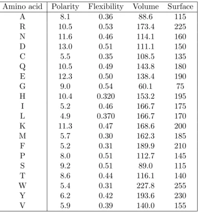
Table1.2shows other physicochemical properties, such as polarity [48], flexibility [15], volume [138] and surface area [27] associated to amino acids.
The 20 amino acids are classified into different categories regarding the properties of their side chain. The following is the most common classification.
• Glycine is the most simple amino acid with a hydrogen atom in the side chain. • Alanine, valine, leucine and isoleucine possess an aliphatic side chain that makes
1.2. Proteins
Amino acid Polarity Flexibility Volume Surface
A 8.1 0.36 88.6 115 R 10.5 0.53 173.4 225 N 11.6 0.46 114.1 160 D 13.0 0.51 111.1 150 C 5.5 0.35 108.5 135 Q 10.5 0.49 143.8 180 E 12.3 0.50 138.4 190 G 9.0 0.54 60.1 75 H 10.4 0.320 153.2 195 I 5.2 0.46 166.7 175 L 4.9 0.370 166.7 170 K 11.3 0.47 168.6 200 M 5.7 0.30 162.3 185 F 5.2 0.31 189.9 210 P 8.0 0.51 112.7 145 S 9.2 0.51 89.0 115 T 8.6 0.44 116.1 140 W 5.4 0.31 227.8 255 Y 6.2 0.42 193.6 230 V 5.9 0.39 140.0 155
Table 1.2: Polarity, flexibility and other physicochemical parameters of amino acids
• Serine and threonine have an aliphatic side chain with a polar hydroxyl group. • Phenylalanine, tyrosine and tryptophan contain an aromatic group. The hydroxyl
function of tyrosine is a weak acid with pKa ∼ 10. Tyrosine is then ionizable but
not ionized in physiological conditions.
• Lysine, arginine and histidine are basic. Lysine and arginine have a high pKa in
solution (10.5 and 12.5, respectively), and thus positively charged in physiological conditions. The low pKaof histidine (∼ 6) makes it neutral or protonated following
the pH of the solution.
• Aspartate and glutamate are acid (with low pKa of about 3.9 and 4.3, respectively)
and negatively charged at neutral pH (named also aspartic acid and glutamic acid). • Asparagine and glutamine are the amidated products of aspartate and glutamate,
and thus not ionisable.
• Cysteine and methionine possess a sulphur atom in their side chain. The sulfhydryl group in cysteine is a highly potent nucleophile and also a weak acid. It can be
easily oxidized to form with another cysteine a disulfide bond which stabilizes the tridimensional conformation of proteins.
• Proline has a formula that is different from other amino acids. The cyclic secondary amino function gives it a specific role in the establishment of the tridimensional structure of proteins.
1.2.3 Peptide bond
A peptide bond is a covalent bond formed between the α-carboxylic group of an amino acid and the α-amine group of the other one. This process combines two amino acids into an amide (dipeptide) and releases a molecule of water (H2O). It is thus called a dehydration reaction or a condensation reaction, which is written as:
... ... ... ... . . . . . . . . . . . . . . . . . . . . . . . . . . . . CH COO− R1 H+ 3N + ... ... ... ... . . . . . . . . . . . . . . . . . . . . . . . . . . . . CH COO− R2 H+ 3N −→←− ... ... ... ... . . . . . . . . . . . . . . . . . . . . . . . . . . . . CH R1 H+ 3N CO...NH... ... ... ... ... CH COO− R2 +H2O
Amino acids in a protein are covalently linked together by peptide bonds to form a non-branching polypeptide chain. A unit of amino acid is called a residue. A polypeptide possesses an amino-terminal extremity (N-terminus) and an carboxy-terminal extremity (C-terminus). The synthesis of a polypeptide is carried out in a so-called “translation” process, where residues are consecutively added from its N-terminus. The N-terminus is then considered as the beginning of the chain.
... ... ... ... . . . . . . . . . . . . . . . . . . . . . . . . . . . . CH R1 H+3N ... ... ... ... ... .... ... ... ... ... .... C O ... ... ... ... N H ... ... ... ... CH R2 ... ... ... ... ... .... ... ... ... ... .... C O ... ... ... ... N H ... ... ... ... CH R3 ... ... ... ... ... .... ... ... ... ... .... C O ... ... ... ... N H ... ... ... ... ... ... ... .... ... ... ... ... .... C O ... ... ... ... N H ... ... ... ... CH COO− Rn
A polypeptide chain is composed of a series of repetitive bonded atoms, namely backbone or main-chain,
1.2. Proteins ... ... ... ... . . . . . . . . . . . . . . . . . . . . . . . . . . . . Ni H ... ... ... ... ... ... ... ... .... Ci α H ... ... ... ... ... .... ... ... ... ... .... Ci O
and a variable part of amino acid side chains Ri, where i denotes the residue position
counting from the N-terminus. These side chains precisely determine the specific prop-erties and functions of each protein. The sequence of amino acids of a polypeptide chain is known as its primary structure.
The peptide bond has characteristics of a double bond due to the mesomeric (reso-nance) effect, thus the six atoms above are coplanar, making a peptide plan.
... . . . . . . . . . . . . . . . . . . . . . . . . . . . . . . . . . . . . . . . . . . . . . . . . . . . . . . . . . . . . . . . . . . . . . . . . . . . . . . . . . . . . Ci Ci α O ...... ... ... Ni+1 H Ci+1α −→ ←− ... . . . . . . . . . . . . . . . . . . . . . . . . . . . . ... ... Ci Ci α O− ...... ... ... Ni+1+ H Ci+1α
Two configurations, called trans and cis, occur according to whether the two α-carbons are on the same or opposite side, respectively.
... . . . . . . . . . . . . . . . . . . . . . . . . . . . . . . . . . . . . . . . . . . . . . . . . . . . . . . . . . . . . . . . . . . . . . . . . . . . . . . . . . . . . Ci Ciα O ...... ... ... Ni+1 H Ci+1α −→ ←− ... . . . . . . . . . . . . . . . . . . . . . . . . . . . . . . . . . . . . . . . . . . . . . . . . . . . . . . . . . . . . . . . . . . . . . . . . . . . . . . . . . . . . Ci Ciα O ...... ... ... Ni+1 Ci+1α H trans cis
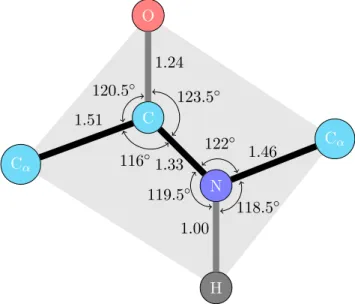
The trans configuration is energetically favored as it causes less repulsion between non-bonded atoms. The crystallographic studies showed almost constant values of distances and angles of the peptide bond for every polypeptide chain (see Figure1.3).
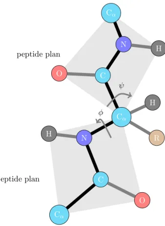
As the geometry of a peptide plane is fixed, the torsion angles φ and ψ are two degrees of freedom in determining the conformation of the polypeptide chain. φ is the dihedral angle around the N–Cα bond, determined by the two carbons CO. ψ is around C–Cα
1.51 1.24 1.33 1.00 1.46 120.5◦ 123.5◦ 116◦ 122 ◦ 119.5◦ 118.5◦ C O Cα Cα H N
Figure 1.3: Peptide bond geometry in trans configuration
bond, determined by the two nitrogens N (see Figure 1.4. There are strong constraints on the angles φ and ψ. Certain combinations are clearly impossible, while some others are energetically unfavorable. Ramachandran et al. [100,101] introduced Ramachandran diagram to visualize graphically the backbone dihedral angles φ and ψ in the polypeptide chain of proteins. Each amino acid in the protein is represented with the coordinate (φ, ψ) in the plot in the range of [−180◦, 180◦] [81]. The Ramachandran diagram of the constituent amino acids of the outer membrane protein A (PDB:1BXW) is presented in Figure 1.51. The limited regions of distribution of (φ, ψ) prove the restricted flexibility
of the polypeptide chain.
1.2.4 Protein
Proteins are macromolecules constituted by a large number of amino acids, from a few dozens to several hundred. This is one of the four important organic macromolecules in living organisms, along with nucleic acids, carbohydrates and lipids. Many proteins are composed of only one polypeptide chain (namely monomer). Others can be formed of more than one chains, and thus are called oligomers (e.g., dimer, trimer, tetramer. . . ). If these chains are identical, the protein is called homo-oligomer. Otherwise, it is a hetero-oligomer. Each constituent chain is a subunit, also known as a protomer.
Proteins are essential in organisms and take part in almost every process in the cells. They are usually classified into three major classes according to their overall
1.2. Proteins Cα C O Cα H N C O Cα H N R H peptide plan peptide plan φ ψ
Figure 1.4: Torsion angles between two peptide plans
dimensional structures and their functional roles: fibrous, globular and membrane pro-teins.
• Fibrous proteins (or scleroproteins), which tend to be elongated fibers, are generally inert and insoluble. These proteins are usually constructed of repetitive amino acid sequences. These characteristics make them appropriate to play structural roles in organisms for supportive and protective function. For example, keratin constructs hair, nails, and skin. . . ; collagen is abundantly found in connective tissues such as cartilage, tendons. . . ; elastin is important in ligaments, blood vessels. . . . An example of collagen is given in Figure1.62.
• Globular proteins, which comprise a large variety of proteins, are soluble and exist in an aqueous environment. Hence, these proteins generally have compact structures with polar residues on the surface and hydrophobic residues in the core. These 2Image generated by PyMOL [113]
Figure 1.5: Ramachandran plot for the outer membrane protein A (PDB:1BXW)
Figure 1.6: Structure of collagen (PDB:1BKV)
proteins are the most described in the Protein Data Bank (PDB) [13], since their structures are usually stable, and thus easy to determine experimentally. Two of the most known globular proteins, myoglobin and hemoglobin, are the first two experimentally determined structures by John Cowdery Kendrew [67] and Max Ferdinand Perutz [97], which led to them receiving a Nobel Prize in Chemistry in 1962. The structure of myoglobin is presented in Figure1.73.
• Membrane proteins exist in the cell membranes – a phospholipid bilayer with hy-drophobic core. They typically have hyhy-drophobic exposed regions in order to be stable in such an environment. Some proteins slightly adhere to the membrane, 3Image generated by PyMOL [113]
1.2. Proteins
Figure 1.7: Structure of myoglobin (PDB:1A6M)
while others are embedded in the lipid bilayer. Among the latter, some proteins, namely transmembrane proteins, entirely span the biological membrane one or sev-eral times (polytopic proteins). Figure 1.84 illustrates the structure of insulin
re-ceptor, a well known transmembrane protein which helps induce glucose uptake, thus causes diabetes in case of its insensitivity.
Figure 1.8: Structure of insuline receptor (PDB:1GAG)
1.2.5 Protein structure
The structure of a protein can be decomposed into different structural elements which allow to describe it in some level of precision. The standard classification proposed by Linderstrom-Lang [78,79] defined four structural levels: primary, secondary, tertiary and quaternary.
a. Primary structure
As mentioned in1.2.3, the primary structure is the sequence of amino acids constituting the polypeptide chain: R1R2. . . Rn.
b. Secondary structure
The secondary structure represents the local conformation of the polypeptide chain. Three main types of secondary structures are found: α-helices, β-sheets and loops.
α-helix
An α-helix is stabilized with hydrogen bonds between the C=O group in the main chain of residue i and the N−H group in the main chain of residue i+ 4. In such a regular structure, all residues are involved in hydrogen bonds. Generally, there are two other kinds of bonding though they are much less frequent. The 3.10 -helices and π-helices are characterized by hydrogen bonds between residues i and i + 3, and between residues i and i + 5, respectively.
An α-helix is geometrically considered as a chain of periodic tours which correspond to a 5.4˚A translation along the helix axis. Each tour contains, on average, 3.6 amino acids, thus the amino acids are translated 1.5˚A along the axis. The structure of an α-helix is illustrated in Figure1.9.
β-sheet
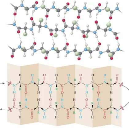
A β-sheet is composed of β-strand subunits. A β-strand can be considered as a degenerated helix with 2 amino acids per tour. Each strand interacts with its neighbors through hydrogen bonds between the C=O and N−H groups in the main chains. As in helices, all residues in a regular β-sheet are involved in hydrogen bonds. This bonding associates the β-strands to each other, making the β-sheet stable.
β-sheets are separated into two types regarding whether the constitutive β-strands are parallel or antiparallel, which is determined by the direction of the pairing β-strands (see Figure1.10). The β-sheet structure generated by antiparallel pairing is found more frequently than the one with parallel pairing, as the former is naturally more stable thanks to a better arrangement of residues.
The torsion angles φ and ψ are respectively around −119◦ and +113◦ for parallel
β-sheets, and around −139◦ and +135◦ for antiparallel ones. The distance between two consecutive residues in a strand is about 3.5˚A. In addition, the large β-sheets are not
1.2. Proteins
Figure 1.9: Structure of an α-helix
(a) (b)
plane, but rather make the curved surfaces. The residue side chains are alternatively located on the two sides of the β-sheet. Frequently, the β-sheets possess a hydrophobic surface oriented towards the protein interior and a hydrophilic surface oriented towards the solvent. An illustration of β-sheet characteristics is presented in Figure1.115.
Figure 1.11: Characteristics of a β-sheet.
c. Tertiary structure
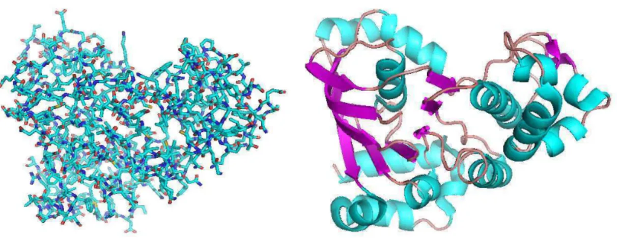
The tertiary structure is the tridimensional conformation of the polypeptide chain, i.e. the relative coordinates of all atoms constituting the protein. This level of structure is essentially stabilized by hydrophobic interaction. There is a considerable difference on the precision of description between secondary and tertiary structures. Hence, the super-secondary structure appears as an intermediary description level. This describes the secondary structure as well as its interactions. Figure 1.126 illustrates the tertiary and super-secondary structure of the cystic fibrosis transmembrane conductance regulator.
5Figure retrieved from
http://wps.prenhall.com/wps/media/objects/602/616516/Chapter 24. html
1.2. Proteins
Figure 1.12: Tertiary structure (a) and super-secondary structure (b) of the cystic fibrosis transmembrane conductance regulator (PDB:1R0W)
d. Quaternary structure
When the protein is a multi-subunit complex, i.e. a composition of several polypeptide chains, the quaternary structure describes the arrangement of these chains (stoichiometry, interaction interface, symmetry,. . . ). Figure 1.137 presents the quaternary structure of
human hemoglobin, which is a heterotetramer (α2β2) composed of two heterodimers
(αβ).
Figure 1.13: Quaternary structure of human hemoglobin (PDB:1MKO)
1.3
Transmembrane proteins
1.3.1 Biological membrane
Before introducing the transmembrane proteins, it is appropriate to start with biolog-ical membranes, the environment where those proteins are located. The constitutive molecules of living organisms are contained in cells − compartments that allow the exis-tence of a privileged environment in a restricted volume that differs from outside. This presents a thermodynamic advantage since it increases the probability of interaction of molecules, and thus the occurrence of chemical reactions. Such an enclosed space is defined by a plasma membrane (or cell membrane). This membrane separates the intra-cellular compartment, namely cytoplasm, and the extraintra-cellular environment. It not only determines the border of the cell, but it also helps maintain the difference of concentra-tions between the exterior and interior mediums, favor the entrance of nutrients into the cell, contribute to the elimination of waste of metabolism, and play an important role in intercellular communication.
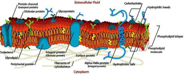
All the biological membranes have a common structure. This is a two-layered sheet (also bilayer) composed of two layers of lipid molecules [2,47,82] with embedded proteins (see an illustration in Figure1.14). The essential property of the membrane lipids, such as phospholipids, glycolipids and cholesterol, is their amphiphilic (or amphipathic) na-ture, i.e. they comprise both hydrophilic regions (dissolvable in water or “water-loving”) and hydrophobic regions (insoluble in water or “water-fearing”). The lipid bilayer is spontaneously formed as an assemblage of lipid molecules, thanks to such a character-istic, with hydrophobic portions pointing toward the interior of the sheet, making this region free from water. The two hydrophilic surfaces of the sheet are then exposed to the aqueous mediums (intra- and extra-cellular environments). This gives the lipid bilayer two important properties. On the one hand, with a hydrophobic core, the membrane is impermeable to most biological molecules, such as nucleic acids, amino acids, proteins, sugars or ions. Thus, the membrane acts as barrier between intra- and extra-cellular mediums. On the other hand, the lipid bilayer forms a two-dimensional liquid in which the constituent molecules can be rapidly laterally rearranged.
Membrane proteins are embedded in the lipid bilayer and ensure most of membrane functions. They constitute about 50% of the membrane mass [115]. We distinguish mem-brane proteins according to their interaction with the memmem-brane. These are illustrated in Figure 1.148.
• Transmembrane proteins are permanently attached to the membrane and span across the bilayer.
• Lipid-anchored proteins are attached to the lipid bilayer by a lipidated anchor. 8Figure retrieved from
http://commons.wikimedia.org/wiki/File:Cell membrane detailed diagram en.svg
1.3. Transmembrane proteins
• Peripheral proteins are located at the membrane surface. They are essentially bound to lipid bilayer or transmembrane proteins by electrostatic interaction.
Figure 1.14: Illustration of a biological membrane and embedded membrane proteins.
1.3.2 Transmembrane proteins
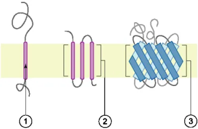
Transmembrane proteins entirely span across the biological membranes. The hydrophobic domains included in the proteins allow them to interact with the hydrophobic center of the lipid bilayer (see Figure1.159). They can possess one or more successive hydrophobic domains, and thus, can traverse the membrane one or several times. Certain proteins can also partially penetrate the bilayer. The extraction of these proteins is difficult and requires detergents, nonpolar solvents or denaturing agents, causing a denaturation.
Transmembrane proteins play several key roles in the human body including inter-cell communication, transportation of nutrients, and ion transport, etc. They also play key roles in human diseases like heart disease, cancer, Alzheimer’s, depression, migraine, retinitis pigmentosa, hereditary deafness, diabetes, cystis fibrosis, etc. [29,42, 85], and thus are targeted by a majority of pharmaceuticals being manufactured today.
The transmembrane proteins are divided into two main types according to their con-formation: α-helical bundles and β-barrels. These proteins make up 20−30% of identified proteins in most whole genomes. However, due to difficulties in determination of their structures, solved TMB structures constitute only a meagre 2% of the RCSB Protein Data Bank (PDB) [6, 13,23,118].
9Figure retrieved from
http://commons.wikimedia.org/wiki/File:Polytopic membrane protein. png
Figure 1.15: Transmembrane proteins: (1) a single transmembrane hydrophobic αhelix -bitopic membrane protein, (2) several transmembrane hydrophobic α-helices, (3) transmem-brane β-barrel protein.
a. α-helical bundles
Transmembrane α-helices dominate the picture of transmembrane proteins with early structural information on bacteriorhodopsin in 1970s [57, 68] and with the first X-ray structure solved for membrane proteins, that of the photosynthetic reaction center [34] (which led to authors receiving a Nobel Prize in Chemistry in 1988). The majority of transmembrane proteins with solved structures fall in this class. These α-helical bundles are found in all types of biological membranes. A bundle is composed of a certain number of helices arranging in such a way as to create a channel through the membrane. These membrane spanning helices are generally constituted by a large majority of hydropho-bic amino acids in order to adapt to the hydrophohydropho-bic characteristics of the biological membrane.
The folding process of α-helical bundles is assumed to be decomposed into two stages [98]. In stage 1, the transmembrane α-helical segments are formed (stabilized by hydrogen bonds along the backbone) and insert independently into the bilayer (driven by the hydrophobic effect), and in stage 2, they assemble by packing together (driven by intrinsic forces such as packing, electrostatic interactions, hydrogen bonds between side chains, interactions between the loops between helices and components at the surface of the membrane, etc.).
Bacteriorhodopsin, which is shown in Figure1.1610, is the well-known representative
of transmembrane α-helices.
1.3. Transmembrane proteins
Figure 1.16: Bacteriorhodopsin in purple membrane (PDB:2BRD)
b. β-barrels
This class is central to our concern in this thesis. The transmembrane β-barrel (TMB) proteins whose solved structures are much less abundant than those of helical bundles are found in the outer membrane of Gram-negative bacteria, mitochondria and chloroplasts. Gram-negative bacteria characteristically possess two membranes: an inner cytoplasmic membrane and an outer membrane facing the extracellular environment. The latter is an asymmetric bilayer with an outer leaflet composed of lipopolysaccharide and an inner leaflet composed of phospholipids [117]. Beside the important roles in the interaction of symbiotic or pathogenic bacteria with the host organisms, the outer membrane usually acts as a permeability barrier to prevent the penetration of noxious substances and to allow the influx of nutrient molecules [95]. This is similar to mitochondria and chloro-plasts. The TMB proteins located in those outer membranes perform diverse functions such as porins, passive or active transporters, enzymes, defense or structural support, multi-drug resistance [54,117]. The structure of TMB proteins is thus very important for both biological and medical sciences.
As the number of determined TMB structures are very limited [125], the principles governing their formation are still not thoroughly clear. The folding mechanism of TMB proteins is unlike that of α-helical bundles, because each helix can be formed indepen-dently thanks to hydrogen bonds along the backbone while β-barrels necessitate hydrogen bonds between neighboring strands. Certain experiments in vitro result in observations that the outer membrane proteins spontaneously fold into lipid bilayers [17,117,131,132]. TMB proteins are assumed to insert and fold into lipid bilayers in such a way that the transmembrane β-hairpins are concertedly translocated. The closure of β-barrels is syn-chronized to its formation, i.e. the hydrogen bonds between β-strands have to form along
with the translocation of the protein across the membrane [70,117].
The TMB proteins are usually created by a succession of antiparallely paired β-strands forming a channel. A β-barrel can be considered as a self-closed β-sheet. The observed structures are formed by 8 to 22 β-strands which incline at an angle of 20◦to 45◦with respect to the barrel axis. Each of these β-strands comprises about 9 to 11
residues. While 8 appears to be the lower bound on the number of necessary β-strands to form a channel [112], the upper bound of 22 is only obtained by experimental observa-tion [102]. The β-barrels are usually constituted by an even number of β-strands, which allows an antiparallel pairing at the barrel closure. An illustration of TMB protein is given in Figure1.1711.
Figure 1.17: Outer membrane protein X (PDB:1QJ8)
1.4
Folding energy
The function of a protein is determined by the arrangement of its atoms in the 3D space. This conformation is stabilized by non-covalent interactions (except for disulfide bonds) between protein atoms as well as between protein atoms and water molecules in the medium. These interactions induce an energy, namely folding energy. It is widely assumed that the most stable structure is the one possessing the minimal folding energy, yet we will not discuss the pertinency of this assumption in this thesis. The folding energy involves various components that are briefly described below.
1.4. Folding energy
1.4.1 Partial charges
A partial charge is a charge with a magnitude of less than one elementary charge unit (i.e. the charge of an electron). Partial charges of atoms are created due to the asymmetric distribution of electrons in chemical bonds. These charges are used to assess the energy of interactions. Their values are computed in various molecular mechanics force fields, such as AMBER [24], CHARMM [22], GROMOS [126], OPLS [63], etc. The values of partial charges from GROMOS force field (see Table 1.3) are used throughout our implementation.
1.4.2 Electrostatic interaction
Following Coulomb’s law, two charged particles interact to each other with a potential energy:
V = qiqj 4πǫ0ǫrrij
where qi and qj represent the charges of particles i and j, rij is the distance between
them, ǫ0≈ 8.85×10⁻¹²F.m−1is the vacuum permittivity and ǫris the dielectric constant
(or relative permittivity) of the medium (some examples are given in Table1.4).
The amino acid side chains can carry a ionized group (such as the ammonium (NH+3) cation of lysine, the guanidinium ([CH5N3]+) cation of arginine, carboxylate (COO−)
anion of aspartate and glutamate) or a polar group (such as the hydroxyl groups of serine, threonine and tyrosine). The polypeptide main chain also contains a positively charged amino-terminal extremity, a negatively charged carboxy-terminal extremity, as well as the polar groups C=O and N−H. These cause numerous electrostatic interactions between charged groups (potential ∼ O(1/r)), between a charge and a dipole (potential ∼ O(1/r2)) or between two dipoles (potential ∼ O(1/r3)), where r denotes their distance.
1.4.3 Hydrogen bond
The hydrogen bond is a particular type of electrostatic interaction, which can be con-sidered as an intermediary between covalent and ionic bonds. It is, intermolecularly or intramolecularly, formed by a dipole-dipole attraction between a hydrogen covalently at-tached to an electronegative atom (donor) and another electronegative atom (acceptor). The hydrogen atom has a positive partial charge, while the electronegative atom, usually oxygen, nitrogen or fluorine, has a negative partial charge. The hydrogen bond is viewed as an in-between state in the proton transfer from the donor D to the acceptor A:
D − H + A ⇄ Dδ−· · · Hδ+· · · A ⇄ D−· · · H − A+
The energy of a hydrogen bond depends on its bonding geometry. The optimal energy is obtained when H is aligned with D and A. Figure 1.18 illustrates the two popular examples of hydrogen bond.
Amino acid Atom type PDB codes Charge (e) D, E C CG (CD) 0.270 O ODi(OEi), i = 1,2 -0.635 N, Q N ND2 (NE2) -0.830 H HD2i (HE2i), i = 1,2 0.415 C CG (CD) 0.380 O OD1 (OE1) -0.380 C S SG -0.064 H HG 0.064 T C CB 0.150 O OG1 -0.548 H HG1 0.398 S C CB 0.150 O OG -0.548 H HG 0.398 R C CD 0.090 N NE -0.110 C CZ 0.340 N NHi, i = 1,2 -0.260 H HE, HHij, i, j = 1,2 0.240 K C CE 0.127 N NZ 0.129 H HZi, i =1,2,3 0.248 H (A/B)† C CD2/CG 0.130 N NE2/ND1 -0.580 C CE1 0.260 H HD1/HE2 0.190 F C CDi, CEi, i = 1,2, CZ -0.100 H HDi, HEi, i = 1,2, HZ 0.100 Y C CDi, CEi, i = 1,2 -0.100 H HDi, HEi, i = 1,2 0.100 C CZ 0.150 O OH -0.548 H HH 0.398 W C CG -0.140 C CD1, CE3, CZi, i = 2,3, CH2 -0.100 H HD1, HE3, HZi, i = 2,3, HH2 0.100 N NE1 -0.050 H HE1 0.190
† The partial charges for Histidine represent two possible ionized states which carry neutral charge.
1.4. Folding energy
Medium ǫr
Vacuum 1.0 (by definition)
Paraffin 2.0 − 2.5
Methanol 33.6
Water 20◦C 80.3 Water 0◦C 87.7
Table 1.4: The dielectric constant of selected mediums
O H H O H H δ+ δ+ δ− δ+ δ− δ+ (a) C O H N δ+ δ+ δ− δ+ (b)
Figure 1.18: Hydrogen bonds represented in dash lines: (a) between water molecules and (b) between carboxylic and amino groups. δ+
and δ− are positive and negative partial charges, respectively.
Only oxygen, nitrogen and sulfur take part in hydrogen bonding in protein structures. The groups OH, NH, SH are donors, while oxygen and non-protonated nitrogen play the role of acceptors. Each residue, except for proline, in the polypeptide chain possesses a donor (N-H) and an acceptor (C=O) that can take part in hydrogen bonds in the main chain. Moreover, the side chains of more than half of residues are also capable to hydrogen bond with other residues or water molecules.
1.4.4 Van der Waals forces and steric repulsion
When two atoms approach each other, the modification of the electron distribution in-duces a polarization. There appears an attractive interaction by van der Waals forces. These forces include forces between polar molecules (Keesom force), between a polar molecule and a corresponding induced dipole (Debye force), and between two instanta-neously induced dipoles (London dispersion force). Van der Waals forces have a potential of order 1/r6, where r is the distance between molecules.
Nevertheless, when two atoms are too close to each other, the steric repulsion becomes stronger and lead to a counterbalance to attractive forces. Its potential varies as O(1/r12). These two forces cause a Lennard-Jones potential, in the case of interaction between
two atoms of the same type, given by: V = E0 ! " 2r0 r #12 − 2" 2r0 r #6$
where E0is the van der Waals well depth and r0 is the van der Waals radius of the atom.
The interaction is quite weaker than normal chemical bonds, yet these forces play an important role in folding stability thanks to their abundance. Table 1.5 shows typical values for these parameters of common atoms.
Atom van der Waals well depth (kcal/mol) van der Waals radius (˚A)
H 0.02 1.00 C 0.12 1.85 N 0.16 1.75 O 0.20 1.60 S 0.20 2.00 P 0.20 2.10
Table 1.5: Typical values for van der Waals well depth and radius
1.4.5 Hydrophobic effect and interaction with the environment
The hydrophobic effect is the fact that a nonpolar molecule (or part of molecule) is inca-pable of hydrogen bonding with water molecules, thus agglomerate together in aqueous medium and exclude water molecules. It is not an attractive or repulsive force, but rather, it is entropically driven. Each water molecule is able to form four hydrogen bonds with its neighbors, thus in order that a nonpolar molecule dissolves into water, such hydrogen bonds have to be broken. The hydrogen bonding network of water disrupted by the nonpolar molecule will reform, by making a cage, around the molecule. This structure of cage is ordered, and thus is unfavored by the second law of thermodynamics which requires an increase in entropy. Hence, the corresponding free energy is unfavorable. The reorganization of water molecules is easier when the nonpolar surface exposed to the aqueous solution is reduced by aggregating the nonpolar molecules together. The hydrophobic effect plays the most important role in protein folding, compared to other non-covalent interactions. It helps polypeptide chains fold in a relatively compact form with a hydrophobic core.
Besides, due to the polarity of water molecules, amino acids with ionized or polar side chains have a tendency to interact with the aqueous medium through hydrogen bonds (see 1.4.3). This allows proteins to exist in water with a hydrophilic exterior.
1.5. Protein structure determination
1.4.6 Torsion energy around peptide bonds
The angles φ and ψ determining the polypeptide chain can differ from the theoretically optimal values which correspond to the equilibrium configuration. Such a deformation causes a energetic penalty, namely torsion energy.
1.4.7 Other interactions
Certain other interactions can also make an important contribution to the stability of a protein structure, such as salt bridge, cation-π interaction, π − π stacking. Salt bridge which often occurs between the carboxylate anion of aspartic acid (D) or glutamic acid (E) and the ammonium cation of lysine (K) or guanidinium cation of arginine (R) can be considered as a combination of hydrogen bonding and electrostatic interactions. Cation-π interaction arises from the face of an electron-rich π system and a cation. π−π stacking or aromatic-aromatic interaction consists of an attractive noncovalent interaction between aromatic rings. These interactions have an order of magnitude equivalent to hydrogen bonds.
The folding energy is finally defined as the sum of all the energies above.
1.5
Protein structure determination
The functions of proteins are performed through their conformations. Thus, it is crucial to determine the protein structures in order to understand the functions associated. Two different classes of methods have been used for protein structure determination: experimental methods which are based on physical measures and in silico prediction methods which used a wide range of computational tools.
1.5.1 Experimental methods
These methods are considered as providing the best approximation to real protein struc-tures as they are based on observations and physical measures on real proteins. There currently exists a number of methods for protein structure determination, in which the most popular ones are X-ray crystallography and NMR spectroscopy.
X-ray crystallography
Most structures archived in the PDB were determined using X-ray crystallography [73]. Starting with the first two proteins crystallized (myoglobin and hemoglobin) at the end of the 1950s, the number of entries determined with X-ray crystallography reached over 55000 in 2010, following the annual report of the PDB [13]. For this method, a beam of X-rays strikes a purified and crystallized protein, and thus is diffracted by the protein crystal. Measuring the diffraction pattern allows to determine the distribution of elec-trons in the protein crystal. This distribution, or the map of electron density, is then