Université de Montréal
A tumoral and invasive phenotype independent of c-Met mutation
By
Giuseppe Giannini
Physiology Department faculty of Medicine
This thesis is presented to la Faculté des Études Supérieures in the scope of obtaining a
Masters in Science (M.Sc.) in Physiology
June, 2003
Université
dI
de Montré al
Direction des bibliothèques
AVIS
L’auteur a autorisé l’Université de Montréal à reproduire et diffuser, en totalité ou en partie, par quelque moyen que ce soit et sur quelque support que ce soit, et exclusivement à des fins non lucratives d’enseignement et de recherche, des copies de ce mémoire ou de cette thèse.
L’auteur et les coauteurs le cas échéant conservent la propriété du droit d’auteur et des droits moraux qui protègent ce document. Ni la thèse ou le mémoire, ni des extraits substantiels de ce document, ne doivent être imprimés ou autrement reproduits sans l’autorisation de l’auteur.
Afin de se conformer à la Loi canadienne sur la protection des renseignements personnels, quelques formulaires secondaires, coordonnées ou signatures intégrées au texte ont pu être enlevés de ce document. Bien que cela ait pu affecter la pagination, il n’y a aucun contenu manquant.
NOTICE
The author of this thesis or dissertation has granted a nonexclusive license allowing Université de Montréal to reproduce and publish the document, in part or in whole, and in any format, solely for noncommercial educational and research purposes.
The author and co-authors if applicable retain copyright ownership and moral rights in this document. Neither the whole thesis or dissertation, nor substantial extracts from it, may be printed or otherwise reproduced without the author’s permission.
In compliance with the Canadian Privacy Act some supporting forms, contact information or signatures may have been removed from the document. While this may affect the document page count, it does not represent any loss of content from the document.
Université de Montréal
Faculté des Études Supéieures
This thesis entitled
A tumoral and invasive phenotype independent of c-Met mutation
Presented by
Giuseppe Giamiini
Has been evaluated by ajury consisting ofthe following individuals:
Jury president: Dr Angelino Calderone Member ofjury: Dr Lucie Parent
Study director: Dr Josette Noél
To my wtfe, !7v[arianne, ant[my
wfw[efamï1y, w[w provi[etf
support, &we
ant! encouragement
througfwut
rnj
academïcjourney...
ntfto Hie
[ovïng memonj
ofmygrandfatfwr
wFw
a[wags emp[iasizedt[tat
money
ant! wea[th can
Le [ost
at any Hme,
but your eéucation stays withyou untï[ Hie end ofyour dàys.
ACKNOWLEDGEMENTS
For the passion she expresses in her work and for the support, trust and ctre she endows in her students, I express my deepest thanks to my study director, Dr. Josette NoêÏ. Her generosity, constant encouragement, and valued advice throughout the period in attaining the Masters degree wiII aiways be greatly appreciated. Dr. No1 aiways made the impossible possible and for that I am forever grateful. Thank you Dr. Noêl!
I would like to thank my laboratory co-workers for their constant support and help.
Special thanks go out to Julie Vadnais and Yolaine Dodier for their direct contribution to Boyden chamber assays. In addition, I would like to thank David Germain for his valuable expertise and advice throughout my stay in the laboratory. Furthermore, I would like to thank Julie Verner from the tissue culture facility ofthc GRTM for lier valuable heÏp.
Additional thanks are intended for Dr. Ivan Robert Nabi in the Anatomy and Cellular Biology department of the Université de Montréal for the critical review of rny rnanuscript and for bis contribution (different MDCK celi unes) to my project.
Finally, I would like to take this opportunity to thank certain members of the department of physiology, namely Mrs. Hélène Klein, Mrs. Pierrette Fournel, Mrs. Joanne Payette, and Dr. Réjean Couture, for helping me make my sttidies botb a pleasurable and successfuï experience.
SUMMARY
The hepatocyte growth factor receptor (HGf-R) otherwise known as the c-Met proto oncogene is involved with various cellular responses such as morphogenesis, motogenesis, and mitogenesis. Phosphorylation and subsequent activation of the receptor’s tyrosine kinase domain, through bindïng of hepatocyte growth factor (HGF), has been shown to trigger various cellular responses via the binding of different src homology domain 2 (SH2) containing proteins to phosphorylated tyrosine residues of the multi-functional docking site found in the receptor’s C-terminal domain.
Various cancer colis express high levels of c-Met phosphorylation, which translates into high levels of c-Met activity. For this reason, much of the research performed today with regards to c-Met and its involvernent in tumorigenesis revolves around the possible mechanisms behind this constitutive autophosphorylation. Some groups point to the presence of point mutations within the tyrosine kinase domain of c-Met which leads to its increased activity. Others suggest the existence of an autocrine activation ioop. In fact, many of the cancers studied to date show evidence of these two possible phenomena.
In the present study, we wanted to analyze the tumorigenic character of an invasive variant of Moloney sarcoma virus-transformed MDCK ceils (MSV-MDCK-INV) and to assess c-Met’s role in this phenotype. Resu!ts dernonstrated that in fact MSV MDCK-INV cells have the capacity to form foci in soft agar similar in size and at a very similar efficiency to the turnorigenic NIH3T3/ras ce!! une. Although MDCK and MSV-MDCK colis formed foci in soft agar, the size and growth efficiency was small compared to MSV-MDCK-INV cells. Clones isolated from the soft agar were analyzed for their capacity to form colonies over monolayers of polarized MDCK ceils. Invasive clones along with NIH3T3/ras clones were the only ones capable of forming these superficia! colonies. Ce!!s from soft agar clones il!ustrated different expression levels of certain proteins involved with focal adhesion complexes and ceil-ceil contacts, nameiy E-cadherin, FAK and paxillin. CAMI-4, one ofthe invasive
soft agar clones studied, was found to express a more phosphorylated c-Met and a 142% invasive capability when cornpared to the invasive capability expressed by
MSV-MDCK-INV celis. I
The resuÏts presented above support the fact that the MSV-MDCK-INV ccli une expresses turnorigenic characteristics. Unfortunately though, no point mutations were observed in either the tyrosine kinase or C-terminal domains of c-Met that could explain the invasive and tumorigenic characteristics of these celis and the transformed phenotype observed for MSV-MDCK celis. Therefore, we assume the existence of an autocrine activation ioop among M$V-MDCK-INV celis that can, at least in part, explain the auto-phosphorylation of c-Met and the subsequent phenotype observed.
Key words: HGF -Rie-Met, auto-phosphorylation, invasive, tumorigenic, soft agar, MDCK, mutation, tyrosine kinase.
SOMMAIRE
Le récepteur du facteur de croissance hépatique (HGF-R), reconnu aussi comme le proto-oncogène c-Met, est impliqué dans plusieurs réponses cellulaires telles que la morphogenèse, la motogenèse et la mitogenèse. La liaison du facteur de croissance hépatique (HGf) à son récepteur mène à la phosphorylation et à l’activation du domaine tyrosine kinase du récepteur. Cette activation amorce ensuite de nombreuses réponses cellulaires via l’interaction de différentes protéines contenant une séquence homologue aux domaines de liaison 2 de la protéine src (SH2) avec les résidus tyrosine phosphorylés du site de liaison multi-fonctionnel du domaine C-terminal.
Certains cancers et métastases expriment un c-Met hautement phosphorylé qui se traduit en un récepteur c-Met plus actif que dans les cellules normales. Pour cette raison, de nombreuses études effectuées sur c-Met et son implication dans la formation de tumeur et de métastases consistent à comprendre les mécanismes pouvant expliquer cette auto-phosphorylation constitutive. Certains groupes de recherche ont montré la présence de mutations dans le domaine tyrosine kinase de c Met entraînant tine augmentation de l’activité kinase et l’activation des cascades dépendantes de cette activation. D’autres groupes font référence à une boucle autocrine. Ces deux modes d’action peuvent aussi être impliqués simultanément dans certains cancers dépendant de l’activation de c-Met.
Dans la présente étude, nous avons étudié les caractéristiques tumorales d’une lignée de cellules invasives (TNV) isolées de cellules MDCK transformées par le virus du sarcome de Moloney (MSV), les cellules MSV-MDCK-INV, ainsi que le rôle de c Met dans l’établissement de ce phénotype. Nos résultats démontrent que les cellules MSV-MDCK-INV ont la capacité de former des foyers indépendants, lorsqu’ensemencées dans un milieu semi-solide tel que l’agar mou, de la même grandeur et avec la même efficacité que les cellules tumorales NIH3T3 transformées par l’oncogère ras (N1H313/ras). Malgré la capacité des cellules MDCK et MSV MDCK à fonner des foyers dans l’agar mou, la grandeur de ceux-ci et l’efficacité de
croissance sont négligeables comparé à ce qui est observé pour les cellules MSV MDCK-INV. Des clones isolés à partir de l’agar mou ont été analysés pour leur capacité à former des colonies de cellules lorsqu’ensemencées sur une mondcouche de cellules épithéliales MDCK. Les cellules invasives et les cellules NIH3T3/ras étaient les seules capables de former des colonies superficielles sur la monocouche de cellules polarisées. Nous avons aussi montré que les cellules dérivées des colonies obtenues dans l’agar mou expriment à un degré différent, en relation avec leur phénotype, certaines protéines impliquées dans les plaques d’adhésion et les contacts cellules-cellules, telles la kinase présente dans les plaques d’adhésion (FAK), la paxilline une protéine de structure des plaques d’adhésion et la cadhérine-E une protéine nécessaire à l’établissement des contacts cellule-cellule. Les cellules CAMI
4, issues de l’une de ces colonies de cellules invasives tirées de l’agar mou, expriment
un c-Met hautement phosphorylé et une capacité d’invasion de 42% supérieure à celle observée pour les cellules invasives MSV-MDCK-INV. Ces observations suggèrent un lien direct entre le degré de phosphorylation de c-Met et la capacité motile des cellules MSV-MDCK-INV, en accord avec certaines observations récentes du laboratoire.
Les résultats présentés dans ce mémoire montrent pour la première fois que les cellules MSV-MDCK-INV possèdent des caractéristiques de cellules tumorales. Malheureusement, aucune mutation n’a été observée dans le domaine tyrosine kinase
ni le domaine C-terminal de c-Met qui pourrait expliqtier le phénotype invasif des
cellules MSV-MDCK-INV ainsi que le phénotype transformé des cellules MSV MDCK. Ces observations nous portent à croire que la présence d’une boucle d’activation autocrine du récepteur, via la sécrétion de HGF par ces cellules, expliquerait le haut degré de phosphorylation de c-Met ainsi que le phénotype particulier de ces cellules MSV-MDCK-INV. Nous n’excluons cependant pas la possibilité que d’autres facteurs puissent aussi influencer l’état de phosphorylation de c-Met dans ces cellules.
Mots clés: HGf-R!c-Met, auto-phosphorylation, invasive, tumeur, agar mou, MDCK, mutation, domaine tyrosine kinase.
TABLE 0F CONTENTS Dedication iii Acknowledgements iv Surnmary y Sommaire vii Table of Contents ix List of Figures xi
List of Tables xii
List of Abbreviations xiii
CHAPTER 1. INTRODUCTION
1.1. Cancer
.1 .1 Definition of cancer and its development 2 1 .1 .2 Oncogenes and their role in cancer developrnent 2 1.1.3 Turnor suppressor genes and their role in cancer development 5 1.1 .4 Neoplastic transformation of normal celis to cancer ceils 5 1 .1.5 Turnor progression and classification 7
1.1.6 Metastasis 8
1.2 Hepatocyte Growth Factor (HGF)
1.2.1 General aspects ofHGF 13
1.2.1.1 Historical overview ofHGF 13
1.2.1.2 HGF’s role in different cellular activities 14 1.2.1.3 Structural characteristics ofHGF and its activation 15
1.2.1.4 HGF’s role in ceil scattering 16
1.2.2 Receptor tyrosine kinases and their role in cell growth 19 1.2.2.1 Historical overview ofthe hepatocyte growth factor receptor
(HGF-R) 20
1.3 Mutations in the met gene and their role in carcinoma development
1.3.1 Met mutants in papillary renal carcinomas (PRC) 24
1.3.2 PRC Met mutants and tubulogenesis 27
1.3.3 PRC Met mutants and apoptosis inhibition 28 1.3.4 PRC Met mutants and anchorage-independent growth 30
1.4 The Madin Darby Canine Kidney (MDCK) ccli une
Ï .4.1 Historical Overview 30
1.4.2 The MDCK celi une and c-Met 30
1.4.3 HGF and c-Met’s role in MDCK ccli tubulogenesis 31
1.5 Objectives 34
CHAPTER2. A tumoral and ïnvasive phenotype independent on c-Met mutation
2.1 Abstract 38
2.2 Introduction 40
2.3 Material and rnethods 43
2.4 Results and Discussion 46
2.5 References 52
CHAPTER 3. DISCUSSION
3.1 Study overview 65
3.2 Growth properties ofMSV-MDCK-INV celis in soft agar and on a
monolayer ofepithelial celis 67
3.3 Absence of point mutations in the tyrosine kinase and C-terrninaldomains
of c-Met 70
Conclusion 77
LIST 0F FIGURES
CHAPTER 1
Figure 1.1 Life cycle ofa retrovirus
Figure 1.2 Replication and integration of the retroviral genome
Figure 1.3 Assays ofthe malignant phenotype
Figure 1.4 A model ofthe major steps ofmetastasis
Figure 1.5 Extravasation ofa tumor ce!!
Figure 1.6 Structure ofHGf/Sf
Figure 1.7 Integrin and growth factor signa!ling through paxillin and
interactions with the actin cytoske!eton.
Figure 1.8 Structure ofthe HGF Receptor
Figure 1.9 Loca!ization of Met mutations found inpapi!!ary renal
carcinornas (PRC) and their respective activity.
Figure 1.10 MetPI mutants and their characteristic anchorage-independent growth and protection from apoptosis
CHAPTER 2
Figure 2.1 Growth efficiency in soft agar
Figure2.2 Ability ofM$V-MDCK-INV like NIH 3T3/ras ceNs to grow on a monolayer ofepithe!ia! ceils.
Figure 2.3 Sequence comparison between the canine c-Met c-terminal domain and that of other species.
Figure 2.4 Si!ent mutations observed in the tyrosine kinase of c-Met in
LIST 0F TABLES
CHAPTER 1 I
LIST 0F ABBREVIATIONS
AIG Anchorage independent growth APC Adenomatous polyposis cou ATP Adenosine triphosphate bcr-abl Fusion protein
bFGF Basic fibroblast growth factor BSA Bovine serum alburnin
CaM Calmodulin
cDNA Complementary DNA
c-onc Cellular/proto-oncogene DNA Deoxyribonucelic acid ECM Extracellular matrix FAK Focal adhesion kinase
Fyn Kinase
Gabi Grb-2 associated binder 1
Grb-2 Growth factor receptor-bound protein 2 GTP Guanadine triphosphate
HGF Hepatocyte growth factor
HGF-R Hepatocyte growth factor receptor HIR Human insulin receptor
HOS Human osteogenic sarcoma HPRC Hurnan papillary renal carcinoma Jak Janus Kinases
kDa KiloDalton
LTR Long-terminal repeat MBP Myelin basic protein
MDCK Madin Darby canine kidney
MNNG N-methyl-N’-nitro-N-nitrosguanidine
mRNA Messenger RNA
NK ceils Natural killer celis
p85 Regulatory subunit of PI-3 kinase
Pi3 -kinase Phosphatidylinositol 3-kinase PKA Protein kinase A
PKC Protein kinase C
PRC Papiliary renal carcinoma
PRGF Plasminogen related growth factor RNA Ribonucleic acid
RTK Receptor tyrosine kinase Sf Scatter factor
SH2 src hornologydornain 2
SH3 src homoiogy domain 3
SHC Gene which encodes an adaptor protein for ccli signailing
SRC Family of kinases
STAT-3 Signal transducers and activators of transcription TGF-3 Transforming growth factor
TPR Translocated promoter region uPA Urokinase-type plasminogen
uPA-R Urokinase-type plasminogen receptor VEGF Vascular endotheiial growth factor
v-abt Protein tyrosine kinase encoded by Abelson murine leukemia virus
1.1 Cancer
1.1.1 Definition of cancer and its development
A cancer can be defined as a group of celis among a total population of ceils that have lost their normal control rnechanisms and thus experience unregulated growth. Cancer can develop from any tissue or organ and as cancerous ceils grow and rnultiply, they form a mass of cancerous tissue that invades adjacent tissues and spreads (metastasizes) throughout the body (Merck Manual, 1997). Cancer ceils develop from normal celis in a process refened to as transformation. During transformation, a normal ceil initialÏy experiences a genetic change which primes it to become cancerous afier exposure to agents referred to as carcinogens. Some examples of possible carcinogens include chemicals, virus, radiation, or sunlight. Not ail ceils are equally susceptible to carcinogens. A genetic flaw in the ceil, its promoter, or even chronic physical irritation may make celis more susceptible to develop cancer. Changes in a cell’s genetic composition are often hard to detect, but sometimes a change in the size or shape of one specific chromosome is a good indication of which type of cancer is involved. Moreover, these genetic changes may also lead to unscheduled cellular hyperproliferation, either through direct activation of celi cycle machinery or through signalling pathways that regulate the celi cycle (Merck Manual, 1997).
1.1.2 Oncogenes and their role in cancer development
Many genes involved in celÏular transformation leading to the development of a cancerous phenotype have been discovered over the past few years. Cancer ceils may possess oncogenes, otherwise known as tumour genes, which are involved in the induction of a cancer. At times, these oncogenes are abnormal versions of the genes responsible for growth and development before birth, which under normal circumstances are pemianently deactivated after birth. Their reactivation later in life may resuit in the development of a cancer. Oncogenes were originally identified in cancer causing viruses. These viruses may be divided into two groups: the DNA ttimor viruses that contain either linear or circular double-stranded DNA and the RNA-containing tumour viruses (also referred to as retroviruses). Stehelin and
collaborators, while studying the Rous Sarcoma virus (a retrovirus that causes sarcomas in chickens), were among the first to demonstrate that a viral transforming gene was derived from a normal cellular gene (Stehelin et al., 1976). Since then, many other retroviruses have been discovered and shown to contain different oncogenes derived from and closely related to their cellular counterparts (Bishop,
1985).
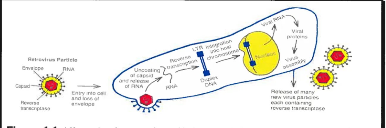
The normal cellular genes from which viral oncogenes (v-onc) are derived are referred to as proto-oncogenes (or c-onc). The way in which proto-oncogenes incorporate into the viral genome and are converted to viral oncogenes with a high degree of transforming ability involves the recombination between the retroviral and cellular genomes. This usually follows the integration of a retrovirus adjacent to a cellular proto-oncogene in a process known as transduction that is associated with alterations in the structure and regulation of oncogene sequences (Varmus, 1988). Figure 1.1 and Figure 1.2 demonstrate how viral RNA, after penetrating the cellular plasma membrane, is reverse-transcribed to a double-stranded DNA form. Once in the double-stranded form, it can then proceed to migrating to the nucleus and integrating itself into the chromosornal DNA.
Retravirus Partiale
EnveIoe RNA
Cap3vi —
EntryInto celi and Ioss ot Reverse envelope (ranscriptase
Figure 1 .1: Life cycle of a retrovirus. Under normal circumstances a virus contains two identical RNA strands. For clarity, only one is shown. After penetrating the plasma membrane, the single-stranded viral ANA genome is reverse transcribed to a double-stranded DNA form that contains a duplication at its ends referred to as the long terminal repeat (LTR). The viral DNA then migrates to the nucleus and integrates into the chromosomal DNA. The resultant single viral transcript can form the genome for progeny viruses or can be processed and translated to generate viral structural proteins.
5 -terminus 3 -terminus
RU UR
Viral Cap
•
AAAAA RNA structure gag pci env
Reverse I transcription (RT) UAU U, RU double-stranded ONA LTR LTR / Covatcntly closed doube-stranded vital DNA molecutes lntegration URU, U, RU. Cellular Cellular ONA
LTR gag pci env LTR DNA
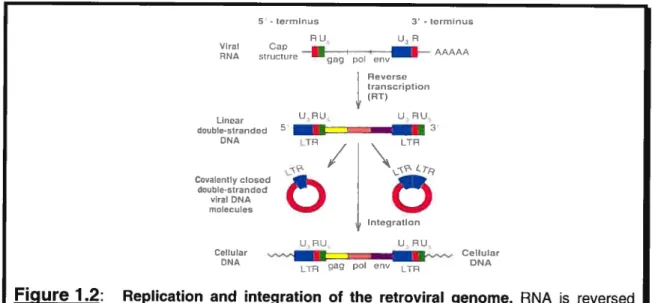
Figure 1.2: Replication and integration of the retroviral genome. RNA s reversed transcribed to form both linear and circular forms of viral DNA. It is the linear form of the DNA that integrates into cellular DNA, where t is referred to as a
provirus. Figure adapted from Tannock and Hill, 1998
A large majority of the oncogenes found in transforming retroviruses have also been identified in spontaneously arising tumors of nonviral origin where they are activated via other possible mechanisms. Some of these mechanisms are point mutations, gene amplification, and chromosomal transiocation. The activation of proto-oncogenes is associated with certain genetic alterations that result in either deregulation of (and an increase in) expression of the normal gene or alterations in the structure (and resulting function) of the encoded protein. In fact, ail celis which make up the human body contain a set of genes that can participate in malignancy following appropriate activation or deregulation. Proto-oncogenes encode various proteins involved in the control of ceil proliferation and differentiation, including many growth factors, growth factor receptors, components involved with signal transduction pathways and transcription factors that regulate the transcription of mRNA and therefore the expression of associated genes (Tannock and Hill, 1998).
1.1.3 Tumor suppressor genes and their role in cancer development
Tumor suppressor genes are also regarded as very important players in malignancy. They are genes that are likcly to play a role in negatively regulating celi growth. The loss or inactivation of these tumor suppressor genes, as their name implies, is associated with malignancy (Weinberg, 1991; Knudson, 1993). Tumor suppressor genes portray different characteristics in comparison to oncogenes. One main difference is that oncogenes act in a dominant manner and the activating event is usuaily acquired. Tumor suppressor genes on the other hand act in a recessive manner. Mutations within a tumor suppressor gene would resuit in its inactivation and mutated forms of the gene may be inherited. Therefore, turnor suppressor genes appear very often in the development of familial cancers, but the presence of mutated forrns of these genes in sporadic cancers is also very common (Tannock and Hill, 1998).
1.1.4 Neoplastic transformation of normal ceils to cancer celts
Neoplastic transformation of human ceils as well as ceils from other species in vitro is seen as a complex rnultistep process by which normal ceils acquire various
phenotypic abilities. four of these abilities are; the development of morphologicai transformation, growth in semisolid medium, immortality, and tumorogenicity. A large majorÏty of human turnor cells of mesenchymal origin do portray morphologicai transformations that aliow them to grow in conditions in which normal ceils cannot.
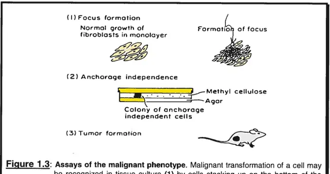
In vitro assays used to demonstrate the transformed phenotype of a ccli ail invoive
three main aspects as depicted in Figure 1.3. Initialiy, transformed celis are examined for their ability to forrn foci. Normal ceils will stop growing when coming into contact with nearby ceils due to a phenomenon known as contact inhibition. Transformed ceils are different because they are not contact-inhibited and continue to grow even when celis corne into contact with one another. In fact, transforrned or malignant cells grow by piling up over each other and forming bumps over the monolayer ofcells. These bumps are referred to as foci (Tannock and Hill, 199$).
Anchorage-independent growth, the ability to grow in a semi-solid medium, constitutes another very important property of malignant cells (Pavelic et al., 1980). Normal celis will flot grow unless they adhere to a supporting matrix or suhstrate such as glass or plastic. By contrast, transformed and malignant celis do flot require any adhesion or anchorage and are capable of growing when suspended in a semisolid medium such as soft agar (Tannock and Hill, 199$).
The last and seemingly most influential assay is the tumor formation assay following injection of tumor celis into nude animais. Normal ceils will flot form tumors when injected into syngeneic or immunologically deficient animals. Transformed cells on the other hand do possess this ability (Tannock and Hill, 1998). The phenomena and rnechanisms involved in induction and progression of a tumor will be discussed later in this chapter.
(I) Focus formation
(
Normal growth of Formoti of focus fibroblots in monoloyer
4.
(2) Anchoroge independence — -- 4_—Mefhyl cellulose ‘—Agar Colony of onchoroge independent cells (3) Tumor formationFigure 1 3: Assays of the malignant phenotype. Malignant transformation of a ceil may be recognized in tissue culture (1) by celis stacking up on the bottom of the culture dish to form a focus; (2) by growth under anchorage-independent conditions in semisolid media such soft agar; or (3) by tumor formation in syngeneic or immune-deprived animais. Adapted from Tannock and Hill, 1998
1.1.5 Tumor progression and classification
Due to the unpredictable nature of cancer, tumors derived from different cancers display different characteristics. In general, tumors tend to show or become increasingly malignant with tirne. Some tumors depict an orderly progression from a benign state to a noninvasive state, followed by the development of pre-malignant lesions and ultirnately malignancy. Highly malignant tumors on the other hand may arise de novo, without going through the different stages of progression. Thus, the degree of progression of any specific tumor is strongly dependent upon the type of tumor. In terms of classifying the severity of the tumors and deducing whether they are benign or malignant, it is common practice to assume that tumors that remain confined to a specific site with well differentiated celis are benign, whereas tumors composed of poorly differentiated ceils that have spread beyond the local site to seed metastases are malignant (Tannock and Hill, 1998).
Turnor progression has for a long time been associated with the acquisition of permanent, irreversible qualitative changes in one or more characteristics of a neoplasm resulting in the tumor becorning more autonomous and malignant at the sarne tirne (see Heppner and Miller, 1992; Klein, 1998 for an integrated view). Recently, rnuch more ernphasis has been placed on genetic changes and how they can explain what occurs during tumor initiation and progression (NoweIl, 1986). Genetic changes occur through various rnechanisms, namely point mutation, deletion, gene amplification, and translocation. A cell’s DNA can be damaged while exposed to various intracellular and extracellular agents, or through the errors made by polymerases whenever DNA is being replicated. In normal celis, this DNA damage
is repaired through DNA repair mechanisms found within the ceil or damaged ceils.
Those ceils whose DNA caimot be repaired undergo apoptosis. Unfortunately, these rnechanisms are flot 100% fool proof and therefore, this leads to a natural frequency of spontaneous mutations in ceils (Simpson, 1997). In effect, many cancer celis appear to possess an increased frequency of mutations due to their deficiency in the repair of DNA lesions and their decreased activation of apoptosis. Therefore, these
mutated celis forrn a sub-population that can survive and continue proliferating (Loeb, 199$).
1.1.6 Metastasis
As tumors progress, they acquire the final and most malignant stage, metastasis. Turnors that have reached this point in their development are usually more difficuit to treat than those that did not spread and invade other tissues. The cancerous ceils could spread via the Iymphatic vessels or via the blood vessels. It is for this reason that clinically, metastases can be divided into two categories: those found in the regional lymph nodes which have traveled through the lymphatic system, or those in more distant organs, which have traveled through the body’s blood vascular system (Taimock and Hill, 199$).
The metastatic process involves four main stcps. First, the metastatic celis must detach from the primary tumor mass and, depending upon the localization of the primary tumor mass, the celis will travel or spread via either the lymphatic or venous system. The inclusion of tumor ceÏÏs into blood vessels may occur as a resuit of a prior invasion of the tumor mass into the blood vessels or due to the fact that the vasculature ofsorne tumors may permit passage ofthe ceils into the blood circulation (Tannock and Hill, 199$). Figure 1.4 depicts an overview of the mechanism by which tumor celÏs from the primary tumor penetrate the blood vessels to enter the bloodstream and travel to distant organs to form metastases.
The initial detachment of the primary tumor celis from the primary tumor site is assumed to involve the decreased expression of adhesion molecules (e.g. cadherins) that are involved in the “homotypic” adhesion of celis to one another (Birchmeier and Behrens, 1994).
The second step in the metastasis process invotves the host’s immunotogic mechanisms and how they arc evaded during metastasis. There exists evidence in support of how some human tumors express weaker effects due to the presence of various cytokines and by nonspecific effector ceils such as natural killer celis (NK ceils) in the host. These two processes that represent the body’s defense mechanisms and are in turn inhibited in the presence of a cancer, could reduce the incidence of metastasis formation (Tannock and Hill, 199$). In effect Hanna, by using transplanted rodent ceils lias shown an inverse correlation between the activity of NK ceils in the host animal and the metastatic ability of injected tumor celis (Hanna,
PRIMARY CARCINOMA
D
BM ANGIOGENESIS ARRESI PRIMARY SAR COMA ANGIOGENESIS MIGRATION DISTANT METASTASES PROLI FERATI ONFigure 1 .4: A model of the major steps of metastasis. Anchorage-independent growth of epithelial ceils resuits in the formation of a primary carcinoma. The tumor
induces the growth cf blood vessels by angiogenesis. Some tumor celis separate f rom the primary tumor, invade through the basement membrane, enter the circulatory system, and eventually arrest in capillaries. At this point, they extravasate eut cf the blood vessels into the underlying connective tissue at the metastatic site; there, further ceil growth and angiogenesis resuit in the formation cf metastatic tumor growth. Angiogenesis is flot seen as being a crucial step for the invasion and metastasis cf sarcomas due te the fact that these tumors arise in the stroma in close vicinity to blood vessels.
1984). Therefore, a decrease in the number of effective NK celis may play an important role in the induction and development of metastases.
The third step in the development of a metastasis is rate-limiting and probably the most critical one. Tumor ceils that have successfully detached from the primary tumor enter the blood stream where they travel for sorne time before reaching their destination. In the blood stream, hemodynamic forces eliminate the majority of these celis and only a small percentage of them survive. It is this small population of tumor celis that can extravasate successfully and lodge themselves into different organs that
is responsible for the ongoing metastatic process. Before extravasation, tumor ceils
are trapped in the capillaries ofthe lung or liver because they are larger in comparison to the capillaries. The arrest of tumor ceils within smail blood vessels lias been associated for quite some time with thrombus formation and involves the interaction
of tumor ceils with platelets and leukocytes. The role of thrornbus formation remains unclear, but it lias been hypothesized that the adhesive interactions between tumor ceils and the endothelial ceils lining the capillaries, which occur after the arrest of tumor ceils, initiate a signal-transduction pathway that promotes the expression of various genes responsible for the next step of the metastatic process (Tannock and Hill, 1998).
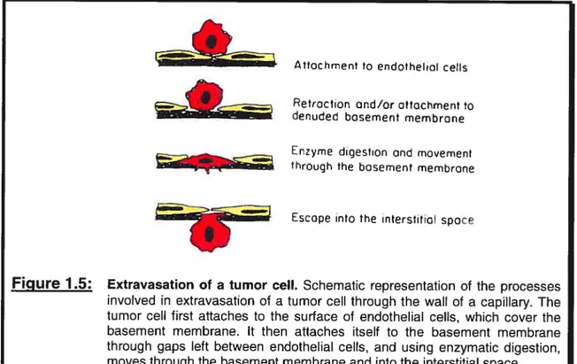
The step preceeding the arrest of cells in the circulation, as touched upon previously, involves the escape of cells from the circulation into the host tissue. The first step of this procedure, outlined in Figure 1.5, involves the extension of tumor celi
pseudopodia into the endothelial celi junctions or the induction of endothelial ccli
retraction that allows access to the basement membrane (Nicolson, 1982). These
pseudopodia are sometimes referred to as invadopodia and contain varying
concentrations of proteases and adhesive molecules that promote the extravasation and migration process. Direct attachrnent to an exposed portion of the basement membrane constitutes another route by which tumor ceils may adliere. In fact, the subendothelial basement membrane is a much better adhesive for tumor celis than is the endothelial celi surface (Nicolson, 1982). The adhesion of cells to the basement
Il
membrane involves the binding to certain membrane components such as larninin, fibronectin, vitronectin, type IV collagen, and proteoglycans (Liotta, 1986; Nicolson, 1988). The binding to these different components is mediated by certain celi-surface receptors, many of which are members of the integrin family. Following their adhesion to the basement membrane, tumor ceils then digest the basement membrane through the use of various proteolytic enzymes which they usually produce or which they acquire from normal ceils in the vicinity. Once tumor celis have managed to digest and penetrate the hasement membrane, they can then migrate through it and escape once again into the interstitial space (Stetier-Stevenson et aÏ., 1993; Mignatti and Rifldn, 1993; Sloaneet aÏ., 1994; MacDougall and Matrisian, 1995).
Aftachment ta endothelial ceils
Figure 1.5: Extravasation of a tumor celi. Schematic representation of the processes involved in extravasation of a tumor celi through the waII of a capillary. The tumor celi first attaches to the surface al endothelial celis, which cover the basement membrane. It then attaches itself to the basement membrane through gaps Ieft between endothelial celis, and using enzymatic digestion, moves through the basement membrane and into the interstitial space. Adapted from Tannock and Hill, 1998.
The last step in the progression of a metastasis is to stimulate proliferation among tumor ceils which have successfuily reached the target organs. One may wonder if there exists any specificity in terms of which organs are afflicted by the proliferating tumor ccli. In fact, ceils are known to require certain growth factors for successful
L Retroction ond/or ottachment to denuded bosement membrane
Enzyme digestion ond movement through the basement membrane
proliferation, and ceilular interactions with the extracellular matrix also play a pivotai role in proiiferation (Nicolson, 1988; Nicolson and Menter, 1995).
Aside from growth factors that encourage cellular proliferation among tumor ceiis, there also exist certain factors that inhibit metastasis sucli as TGF-j3, and these factors also play a role in organ-specific metastasis. In fact, there is evidence which shows that as tumor ceiis become more malignant, they may switch from being growth inhibited by TGf-f3 to being stimulated by TGF-E3 (Wright et al., 1993; Lu and Kerbei, 1994).
Another very important form of evidence that lias been weii documented over time is the fact that tumor celis do not require exogenous growth factors as cornpared to normal ceils. Ibis independence is in direct relation with their ability to form tumors in animais and to metastasize. In many instances, the independence of tumor celis for exogenous growth factors is reÏated to autocrine production of such factors and the modification ofthe response to such factors (Taimock and Hill, 1998).
An extremeiy important property pertaining to the growth of a metastasis is the ability of tumor ceils to induce angiogenesis. This is defined as the deveiopment of new blood vessels. Folkrnan was arnong the first to dernonstrate that when tumor fractions were implanted on the comea of the rabbit eye, new blood vessels formed (folkrnan, 1 976). There exist a number of growth factors such as the basic fibroblast growth factor (bFGF) (Folkman and Klagsbrun, 1987) and the vascular endothelial growth factor (VEGf) (Senger et ai., 1983) that are angiogenic and induce endothelial ceil growth as weii as morphologic differentiation. Without the development of new blood vessels to supply nutrients to the developing tumor celis, the tumor would not grow larger than a few miliimetersin diameter.
1.2 Hepatocyte Growth Factor (HGF)
1.2.1 General aspects ofHGF
The hepatocyte growth factor (HGf), also known as scatter factor, represents a typical example of how a regular growth factor critical during growth and development may trigger a change in celÏuÏar phenotype that in sorne instances could lead to the induction of a cancer. The hepatocyte growth factor is a cytokine that is ubiquitously expressed by ceils ofmesenchymal origin (Bottaro et al., 1991). In vitro HGF stimulates celi motility, invasion of surrounding extracellular matrices, proliferation, survival and morphogenesis, and induces the expression of specific genes by binding to its receptor, a transmembrane tyrosine kinase encoded by the MET proto-oncogene in epithelial ceils (Prat et al., 1998).
1.2.1.1 Historical overview ofHGF
Research groups working in the field of liver regeneration and epithelial/mesenchymal interactions were the first to identify the hepatocyte growth factor. Initially, HGf was identified as a potent mitogen of hepatocytes in culture. Studies based on whoÏe animaIs demonstrated the presence of a certain growth factor found in serum, which mediated rapid regeneration of liver parenchyma in vivo
(Moolten and Buchner, 1967; Fisher et al., 1971). Following these findings, investigators partially purified this growth factor from the serum of hepatectomized animais and showed it to be the rnost potent mitogen found in hepatocytes in culture. This new growth factor displayed characteristics that set it apart from other known growth factors such as epidermal growth factor, fibroblast growth factor, platelet derived growth factor, somatomedin, thrombin, and transferin. The newly discovered polypeptide was named HGF (Mïchalopoulos et al., 1984; Nakamura et ai., 1984). At approximately the same time that work was being performed on liver regeneration,
a different group of epithelial cell biologists investigating epithelial/mesenchymal
interactions identified a new factor which was found to be produced by fibroblasts and stimulated movement of cultured epithelial ceils in vitro. This factor derived from fibroblasts led to the disruption of celi-ceil junctions and Ïed to the scattering of
islands of non-confluent epithelial ceils in vitro (Stoker and Perryrnan, 1985; Stoker et al., 1987; Stoker, 1989). It was based upon these findings that the newly fibroblast derived growth factor was referred to as scatter factor (Sf). Initially, the biological activities of these two molecules set them as distinct from one another. Subsequent biochemical analysis and cDNA cloning demonstrated that HGF and Sf represented the sarne molecule (Nakamura et aï., 1986; Golida et al., 198$; Zarnegar and Michalopoulos, 1989). Furthermore, these resuits helped classify this growth factor as a member ofthe Plasminogen Related Growth Factors family (PRGF’s), due to its structural homology with enzymes of the bÏood-clotting cascade (Stella and Comoglio, 1999).
1.2.1.2 HGF’s role in different cellular activities
As seen previously, celis of mesenchymal origin are responsible for the production and release of HGF. The resulting biological activities of HGf are targeted mainly toward epithelial ceils that express its receptor, the HGF receptor otherwise known as c-Met. from this specific interaction existing between both ceil types, the HGF was c]assified as a paracrine mediator in the interaction that exists between mesenchymal and epithelial ce]ls in vivo (Stoker et aï., 1987; Birchrneier and Birchmeier, 1993).
Epithetial celis, as targets of HGF, are thought to influence the production of HGF by mesenchymal ceils in a paracrine fashion (Joseph et aÏ., 1995; Rosen et aÏ., 1994). The factors of epithelial nature, responsible for the stimulation of mesenchymal HGF production have not been well defined at this time. It is however known that HGF expression by fibroblasts is inhibited by several known peptide growth factors, such as transforming growth factor beta, epidermal growth factor, and transforming growth factor alpha (Seslar et al., 1993).
HGF/SF, in vivo, plays a role in the neural system (Streit et al., 1995; Ebens et ctl., 1996), kidney (Santos et al., 1994; Wolf et al., 1995) and marnmary gland (Niranjan et al., 1995; Soriano et aï., 1995; Yang et al., 1995) development, tissue regeneration (Matsumoto and Nakarnura, 1993), angiogenesis (Bussolino et al., 1992), turnor invasion and metastasis (Giordano et al., 1993; Rong et aï., 1994). HGF is also very
important during embryogenesis where its signailing is essential for liver and placenta development (Schmidt et al., 1995; Uehara et aÏ., 1995) as well as for migration ofmyoblast precursors from the limb buds (Biadt et al., 1995; Maina et al., 1996). In addition, this growth factor is involved flot only in the stimulation of hepatocytes, but also of other primary cuhured celis, including melanocytes and renal tubular ceils (Igawa et al., 1991; Kan et al., 1991; Matsumoto et aÏ., 1991; Rubin et al., 1991). Furthermore, serum HGF levels have been found to be elevated in patients suffering from chronic hepatitis and liver cirrhosis (Tomiya et aÏ., 1992) and in animais being treated with a hepato-promoter (Lindroos et al., 1992).
1.2.1.3 StructuraI characteristics ofHGf and its activation
In terms of its structural characteristics, the hepatocyte growth factor (HGF) is a heterodimeric protein containing a
cx
chain composed of an N-terminal hairpin loop and four kringle domains, as weii as a serine protease-like f3-chain (Stoker et aï., 1987; Mizuno and Nakamura, 1993; Nakarnura, 1991). It is synthesized and secreted as a biologicaily inactive single-chain 92 kDa precursor, which is converted into the bio-active dimer in the extracellular environment either by serine proteases activated during the coagulation process (Miyazawa et al., 1994), or by the urokinase type plasminogen activator (uPA) bound to its receptor (uPA-R) at the ceil surface (Naldini et al., 1992). Figure 1.6 illustrates how HGF goes from its inactive state where the cx and [3 chains are attached, to its active mature form where it is cleaved and the resulting cx and [3-chains are attached via a disulphide bond. Moreover, stimulation by HGF induces the expression of both uPA and its receptor (Boccaccio et al., 1994; Pepper et al., 1992). UPA is also a very important component for its ability to catalyze extraceliular matrix degradation (Blasi, 1993). Therefore, the activity generated by the coupiing of the HGF/HGF-R and uPA!uPA-R pairs is directly invoÏved in ccli associated proteolytic processes required for tissue remodelling, celI migration and celi invasion during wound healing, angiogenesis and tumor metastasis (Blasi, 1993).1.2.1.4 HGF’s foie iii celi scattering
HGf/SF’s role in ceil migration involves the initial recruiting of integrin receptors found on the ceil surface and in contact with the extracellular matrix, cytoskeletal proteins, and 125FAK into focal adhesions that are strongly dependent on tyrosine kinase activity (Matsurnoto et aÏ.1994). Unlike the growth factor receptors, integrin receptors lack intrinsic tyrosine kinase activity. It is for this reason that one of the initial steps during integrin signalling is the tyrosine phosphorylation of the non receptor tyrosine kinase, focal adhesion kinase (FAK), in response to ceil adhesion (Schaller et aÏ., 1994; Richardson and Parsons, 1995; Parsons 1996; Chandra Kurnar, 199$). AlI
13t
and Œ containing integrins share the ability to promote the assembly offocal adhesions and activate FAK. FAK is a non-receptor tyrosine kinase absent of any src homology (SH)2 and SH3 domains, and its phosphorylation is believed to resuit in the activation of a cascade of phosphorylation events and new protein interactions which are essential for adhesion-dependent signalling complexes (Figure 1.7). Pre-pro HGF Mtfl.A..%.f% Pro 29 26 amino amino acida acids cx chan (440 a.a.) s s So VaI_Arg___L’” j3 chainf234 amino f acids ) processing
‘1f
Mature HOP Pro N-terrnr,aI b s — p pFigure 1.6: Structure of HGF/SF. HGF/scatter factor in its pre-form where both X and
3-chains are attached and in its active form where it is cleaved and the Œ-chain is attached to the 13-chain via disuiphide bond (a). The a-subunit of HGF contains one hairpin Ioop and four kringle domains depicted in red that are triple-Iooped cysteine-rich motives involved in protein-protein interactions (b).
FIGURE 17: Integrin and growth-factor signalling through paxillin, and interactions with the actin cytoskeleton. A) Paxillin is involved with the recruitment of varfous signalling proteins to the plasma membrane, including the tyrosine kinases FAK and Src. The activation of these proteins is achieved through the interaction of integrins with the extracellular matrix, or after growth factor stimulation of receptor tyrosine kinases (RTK). The complex of proteins leads to the activation of the mitogen-activated protein kinase (MAPK) cascades, thereby stimulating gene expression. The signalling and cross-talk with integrins that is mediated by growth factor signalling, is enhanced by paxillins ability to bind a PKL-PIX-PAK-Nck complex. B) Paxillin is seen bound to various proteins that help in the organization of the actin cytoskeleton at contact sites between integrin receptors and the extracellular matrix (focal adhesion points). Vinculin and actopaxin physically bind paxillin to actin filaments. The binding of the adaptor protein Crk to paxillin, which is phosphorylation-dependent, is necessary for the regulation of celI motility in certain ceIl types. Microtubule binding to paxillin also contributes somewhat to the ability of this filament system to promote celI motility via the disruption of focal adhesions. Figure reproduced from Turner, 2000.
Talin for one, is a protein that binds directly to the carboxy-terrninal domain of fAK
tAAP kinasosuperr1yavar
PrsiHera,on
Cytcskeleta res,garszsLzn
et cii., 1996). Paxillin in turn binds to a distinct site in the carboxy terminus of FAK (Schaller et ai., 1995). Therefore, there is evidence to believe that the recruitment of
fAK to activated integrins is an indirect process mediated by talin, and that once
fAK lias been recruited to the focal adhesion complexes, it undergoes a
conformational change and interacts through its amino terminal dornain with the 3 subunit tau (Richardson and Parsons, 1996). This conformational transition is therefore a prerequisite for FAK’s catalytic activity because it leads to a trans autophosphorylation on residue 397 which in turn resuits in the interaction with Src and Fyn through their SH2 domain (Schaller et aÏ., 1994). Src and Fyn are then responsible for the pliosphorylation of a number of FAK-associated proteins including paxillin and tensin (Vuori et aï., 1996; Schlaepfer and Hunter, 1997; Schlaepfer et ai., 1997). Once in the phosphorylated state, FAK can then combine with and activate Pl3Kinase (Chen et aÏ., 1996). Hence, FAK is associated with various intracellular signalling pathways, namely those involved with cell adhesion and cell motility. Embryonïc fibroblasts derived from FAK knockout mice have shown to form numerous small focal contacts, but were unsuccessful in forming large peripheral focal adhesions and were found to migrate less efficiently than control celis (hic et ai., 1995). Another piece of evidence supporting FAK’s role in cell migration is that overexpression of FAK stimulates cell migration. A dominant negative forrn of FAK completefy inhibits celi migration (Cary et ai., 1996; Gilmore and Romer, 1996). These observations are contradicted by the report of Matsumoto and colleagues (Matsumoto et ai., 1994). He states, with regards to FAK, that a “GraduaI loss of tyrosine phosphorylation coincided with dismption of focal adhesions and conversion to a motile phenotype”. He also goes on to propose that “HGF/SF-induced phosphorylation of pl25’ may actually indicate the disassernbly of the focal adhesions, a process apparently required to ceil mobilization and locomotion” (Matsumoto et cii., 1994). Therefore, it would seern that according to Matsumoto, FAK would be over-expressed in motile ceils, but phosphorylation on FAK would be greatly diminished.
The presence of HGF bound to its tyrosine kinase receptor (RTK) as seen in Figure
1.7, is essential in triggering the aforementioned cascade leading to ceil scattering. Recruitment of integrin receptors on the ce!! surface as well as activation of different proteins invo!ved with cellular rnotility and scattering is strongly dependent upon the tyrosine kinase activity produced when HGF activates HGF-R (c-Met). It is for this reason that the expression of certain proteins such as FAK and paxillin are examined extensive!y when working with invasive or motile ccli unes.
1.2.2 Receptor tyrosine kinases and their role in ccli growth
The process of ccli growth and differentiation is triggered when extracellular substrates are recognized at the ce!! surface by a receptor, resuiting in the activation of associated cytop!asrnic and nuclear biochemical cascades. When deregulated, these normal growth-signalling pathways may be key to the maÏignant transformation of human ce!ls. Growth factors were initia!!y identified from culture medium for their abi!ity to independent!y sustain ce!l growth. Viraily transformed cel!s, unlike normal ceiis, depict !ower requirernents for exogenous growth factors. With the exception ofTGf-3, which stimulates the proliferation ofvarious mesenchymal ceils, inhibits the proliferation of many epithe!ia! cells and binds to a receptor with serine threonine kinase activity (Derynck, 1994). most growth factors bind to specific transmembrane receptors beionging to the family of receptor tyrosine kinases (RTK’s). These RTK’s have been invo!ved with human diseases such as deve!opmental disorders and even cancer. Signal transduction by this fami!y of RTK’s is achieved through a series of steps including: a) ligand binding to the receptor and subsequent receptor dimerization, b) receptor trans autophosphory!ation on tyrosine residues, c) recruitment to the receptor’s cytoplasmic tau and activation of cytoplasmic signaliing molecules that are mediators in the transmission of information to the nucleus (Longati et aï., 2001).
1.2.2.1 Historïcal overview of the hepatocyte growth factor (HGF-R)
HGF induced invasive growth is mediated by the HGF-R, or Met receptor. The HGF-R was identified as the c-met proto-oncogene (Bottaro et aÏ., 1991) and originally isolated as a transforming gene from a human osteogenic sarcoma ce!! !ine (HOS) that was treated in vitro with the chemical carcinogen N-methyl-N’-nitro-N nitrosguanidine (MNNG) (Blair et al., 1982; Cooper et aÏ., 1984). Its activation in these HOS ce!!s occurred via a DNA rearrangement that resulted in the fusion of sequences from the TPR (trans!ocated promoter region) !ocus on chromosome 1 to sequences from the met!ocus on chromosome 7 (Park et al., 1986; Dean et ai., 1987). Activation of the met oncogene via DNA rearrangement and fusion resemb!es to a great extent the activation process for several members of the tyrosine kinase family of oncogenes.
Nucleotide sequencing of a portion of the met oncogene showed that it was in fact a member of the tyrosine kinase fami!y of oncogenes. It resembles to various degrees the roii and sea oncogenes, and its kinase domain is most homologous to the human insu!in receptor (HIR) (Ulrich et al., 1985) and the murine v-abl oncogene (Reddy et al., 1983). Overexpression of the met proto-oncogene is evident in tumors of specific histotypes inc!uding thyroid (DiRenzo et ai., 1992), pancreatic carcinomas (DiRenzo et aÏ., 1995), and in hereditary papiiiary renai carcinomas (HPRC) (Schmidt et al.,
1997).
1.2.2.2 Structural characteristics of HGF-R
HGF-R is a heterodimer of 190 kDa comprising an Œ-chain of 50 kDa containing the HGF binding site. The Œ-chain is heavily glycosylated, found on the ccli surface, and which is linked via a disu!phide bond to a 13-chain. The 145 kDa f3-chain contains the kinase domain with the tyrosine autophosphory!ation sites and the multifunctional docking site that comprises a specific stretch of amino acids located in the carboxy terminal of the protein (Comog!io and Vigna, 1995). C-Met is synthesized as a giycosy!ated single chain precursor that undergoes proteolytic processing to form a mature heterodimer (Giordano et al., 1989a).
21
The Met cytoplasmic domain can be divided into three distinct and functional domains as depicted in Figure 1.8(A).
Figure 1.8: Juxtamembrane ®-s demain Y Catalytic p Y domain p Y C-terminal y domain j)
[1 H
Met A) B) P —SPhsphaseore Invasive-growth P —YPhophoIyroswe P’4et Receptor active HGF cx prHGF clen .sgeStructure of the HGF Receptor. A) Representation of the HGF/c-Met receptor. The receptor comprises an extraceliular transmembrane Œ-chain that forms the docking area for HGF. The f3-subunit contains three domains; the juxtamembrane domain, the catalytic domain and the C-terminai domain ail containing tyrosine residues which when phosphoryiated, are involved with HGF-Rs enzymatic activity and hence ceilular responses. B) The C-terminai domain contains a two-tyrosine multi-functional docking site which when phosphorylated (following activation by HGF), binds different SH2-containing factors involved with cellular responses such as celi motility. Figure 8(A) adapted from Bardellietat, 1997
The juxtamembrane domain, which immediately follows the transmembrane domain, negatively regulates Met activity. The phosphorylation of a serine in this domain, serine 975, by Protein Kinase C (PKC) or Ca27 Calmodulin-dependent kinase, andlor the recruitment of a cytoplasmic tyrosine phosphatase by the phosphorylated tyrosine 1003, inhibit the Met kinase activity (Gandino et aï., 1990; Gandino et al., 1994; Vilia-Moruzzi et al., 1993). Phosphorylation of two tyrosine residues in the kinase domain ofthe receptor (Y1234 and Y’235) positively regulates the enzymatic activity of the Met kinase domain (Ferracini et al., 1991; Longati et aÏ., 1994; Naldini et aï.,
1991). This increased c-Met activity can promote the metastatic spread of cancer due to its stimulatory effects on a variety of processes such as angiogenesis, cellular motiiity, and protease secretion (Jeffers et al., 1996). The C-terminal domain also contains a two-tyrosine region (Y’349 and Y1356) that is refened to as the multi functional docking site. Once both tyrosines become phosphorylated, this docking site, as depicted in Figure 1.8(B), binds multiple SH2-containing transducers, including p85 the catalytic subunit of the phosphatidylinositol 3-kinase (P13-kinase) (Graziani et al., 1991), phospholipase C-y, Src, the adaptors Grb2 (Growth factor receptor-bound protein 2) (Ponzetto et al., 1994) and SHC (Pelicci et al., 1995), the transcriptionai factor STAT-3 (Signal transducers and activators of transcription-3) (Boccaccio et al., 1998), and the docking protein Gabl (Grb-2 associated binder 1) (Weidner et al., 1996). This muiti-functional docking site is crucial for ail Met mediated responses. Substitution of both tyrosines (Y’349 and Y’356) with phenyialanine resulted in the abolishrnent of Met-mediated responses both in vitro and in vivo (Maina et al., 1996; Ponzetto et aÏ., 1994).
A study by Guai and collaborators bas recentiy demonstrated that the last 26 amino acids in the C-terminal domain positively regulate Met kinase activity and that their presence is critical for the transforming abiiity but not for Met-mediated invasiveness (Guai et ctl., 2001). By constructing mutants that contained various deletions of the C-terminal domain and transfecting these mutants into NIH3T3 ceils, they were able to establish that deletion ofthe last 47 arnino acids in the C-terminal domain (the A47 mutant) displayed a lower level ofphosphorylation. This was easiiy explained by the
fact that Met M? lacks three auto-phosphorylation sites (Y’349, Y’356 and Y’365) (Guai et aÏ., 2001).
Following these findings, they proceeded to look at the enzymatic activity of the various mutants in comparison to the wild type Met. With the use of an exogenous substrate, the myelin basic protein (MBP), they measured the phoshorylating ability of the different receptors. The deletion of the last 47 amino acids resulted in a 46% increase in Met’s ability to phosphorylate MBP in vitro as compared to the wild type. Thus, from these resuits it seems that this domain negatively regulates Met’s enzymatic activity. In agreement to this, work from Bardelli and collaborators showed that a celi-permeable peptide derived from the Met tau (Ile-Gly-Glu-His Tyr’ 349-Val-His-Val-Asn-Ala-Thr-Tyr’ 356-Vai-Asn-Val-Lys-Cys-Val-Ala) and containing the docking site inhibits receptor activity. Once internalized with the heip of the Antennapedia internalization domain at the amino terminus, the tau peptide blocked both ligand-dependent autophosphorylation and downstream Met signalling. One possible explanation for this inhibition as presented by Bardelli and his collaborators, us that the Met tau could impair receptor phosphorylation by interacting with another moiety of the catalytic domain. (Bardelli et aÏ.,1999). The A26 mutant was also of extreme interest. The deletion of the last 26 amino acids resulted in a 35% reduction in Met’s ability to phosphorylate MBP, compared to the wild type. These findings indicate that this portion of the receptor tail positively regulates the tyrosine kinase activity. Altogether, the findings by Guai and lis collaborators propose that the C-terminal domain of Met contains two regulatory regions displaying opposite effects on the receptor kinase activity.
In a study from Giordano and collaborators, Met-mediated transformation was examined in NIH3T3 fibroblasts transfected with the full-length met cDNA (Giordano et aÏ., 1993). Exposing these transfectants to HGF resuited in a stimulation of tyrosine kinase activity and induced changes in cell shape, migration in Boyden chambers, and invasion of collagen matrices in vitro. In addition to these findings, it was observed that their motile and invasive phenotypes were soiely
dependent upon the presence of the HGF/SF and that the factor promoted colony formation in soft agar in the presence of 5% calfserum.
Due to its seemingly influential role in cellular invasion and motility during early development, the possible role of c-Met in certain cellular transformations or even in certain cancers was exarnined. In fact, rnany research projects in the field of cancer research today aim to assess the peculiar ability of malignant tumor ceils to escape from the primary tumor and settie at distant organ sites. The HGFRhnet gene is physiologically expressed in many celi types, especially of epithelial nature (Tajima et al., 1992), and is frequently over-expressed in many types of cancer.
1.3 Mutations in the met gene and their role in carcinoma development.
1.3.1 Met mutants in papillary renal carcinomas (PRC)
The identification of germ-line and sornatic mutations in the Met gene in papillary renal carcinomas (Schmidt et al., 1997; Schrnidt et al., 1999) and chuldhood hepatocellular carcinornas (Park WS et al., 1999) bas established the connection between Met and human cancers. AÏ! of the mutations found to date are located within the tyrosine kinase domain, either in the N-terminal portion, which contains the ATP binding site, or in the C-terminal portion, which includes both the catalytic and activation loops. Studies have demonstrated that ail mutations reported so far seem to activate the catalytic activity of the receptor at different degrees (Jeffers et al., 1997; Bardelli et aÏ., 1998; Giordano et cii., 2000). Moreover, Jeffers and his collaborators illustrate that different Met mutations identified in both bereditary and sporadic cases of human papillary renal carcinomas resuÏted in different levels of enzymatic activity in c-Met whicb translated into different levels of tumorigenicity (Jeffers et al., 1997).
By stably transfecting NIH3T3 celis with some Met mutants, Jeffers and bis team observed higher levels of Met phosphorylation among certain mutants and lower levels among others (Jeffers et cii., 1997) (Table 1.1). In order to quantify the in vitro
transforming ability of the mutants, they performed a focus-formation assay among ail mutants. Their resuits demonstrated that the most active mutants (i.e M1268T, Y124$H, D1246H, D1246N and Y1248C) produced a higher number of foci per jig of DNA when compared to their less active counterparts that failed to produce any foci. Tumor formation in athymic mice is an additional assay which demonstrates the functional consequences of c-Met mutations. The results obtained illustrate that ceils expressing each of the mutant Met proteins form tumors, and that ceils expressing the wiid type protein failed to produce foci in soft agar as well as tumors in mice. As was observed with the focus—formation assay, a strong correiation exists between the Met mutants’ enzymatic activity and their resulting turnorigenicity. Therefore, from the gathered resuits, it is evident that the M1268T mutation found in the tyrosine kinase domain of c-Met among this and other cancers, remains one of the most influential and important mutation.
Table 1. Activitv of Met mutants
Tumoi formation
Met Focus # mice with
plios- formation tumors/# mice Mean turnor Met construct* phoryIation #foci/ig DNA injected size, mm
—Wild type — O 0/14v 01 M1268T(s) +++ >300 8/8 216 ± 77 Y1248H() ++ 156 ± 16 8/8 lOt) ± 40 D1246H(s) ++ t19 ± 16 8/8 60 ± 52 D1246N(g) ++ 147±5 9/9 50±25 Y1248C(g) ++ 115 ± Il 7/8 77 ± 89 V12381(g) ++ 0 5/8 3 ± 15 V1206L(g) + 0 6/6 50 ± 32 M1149T(g) - + 0 4/8 46 ±56
Table 7.1: Activity of Met mutations when transfected in NIH3T3 ceils. Table reproduced from Jefferset al., 7997
Other research groups sucli as that of Giordano and collaborators revealed the presence of mutations in the c-Met of papillary renal carcinomas (PRC) different from those analyzed by Jeffers and collaborators (Jeffers et aI., 1997). Giordano and her team also demonstrated the presence of mutations within the tyrosine kinase
domain of c-Met (Figure 1.9(A)) which supports the fact that mutation M1250T displays the highest transforming ability in relation to the focus formation assay (Figure 1.9(B) bottom portion) as well as the highest mitogen activity as measured by a luciferase activity assay (Figure 1.9(B) top portion) (Giordano et al., 2000). Figure 1.10(A) illustrates the precise localization of these mutations within the kinase domain of c-Met and the hornology with residues rnutated in the RET and KIT receptors. Both RET and KIT receptors are members of the receptor tyrosine kinase family (RTK). Ail members in this family of receptors prornote invasive growth signaling through their own distinct version of a docking site (Bardelli et al. 1997). Results obtained from their study illustrate that mutations that alter residues located in both the activation loop and in the N-terminus of the kinase domain are efficient enough at transforming mouse flbroblasts (Giordano et al., 2000). An example ofthis can be found in Figure 1.9(B), where it is clear that the MetLl 195V mutation found in the N-terminus of c-Met’s kinase dornain expresses the same trancriptional activity and produces the same number of foci as for instance, the MetYÏ23OC mutation found in the activation ioop.
The Met receptor as seen previously, is expressed in epithelial celis and elicits various biological responses such as ccli proliferation, protection against apoptosis, and invasion of the surrounding extraceÏÏular matrices. In order to examine the effect of some PRC mutations on these different biological properties, Giordano and ber team transfected human Met DNA containing the various mutations into MLP 29 murine celis which display many biological properties in response to HGf treatment (Medico et cii., 1996). The celis having been successfully transfected with the PRC mutations displayed a “scattered” phenotype consisting of the graduai disassembling of tightly packed islands typical of epithelial ceils. The introduction of exogenous HGF to these celis resulted in a higher degree of “scattering” in ceils expressing the PRC mutants as opposed to ceils expressing normal human Met (Giordano et al., 2000).
800 E1-Nl7Ras ±N17Ras <D c T 400 r 200- Œ
L1
M1131T Vil 88L L1195V V12201 D1228H (KIT) D1228N (KIT) Yl 230C Y1230H Ml 250T (PET) H C z O H H H H H z w w w w W D i00fE11Ï
t
Figure 7.9: Localization of Met mutations found in papillary renal carcinomas (PRC)
and their respective activity. A) Illustration of Met mutations found in
papillary renal carcinomas (PRC). Blue boxes represent the tyrosine kinase
domain (KD), which can be divided into N- and carboxyl-terminal lobes (N-L and C-L respectively) separated by a large ioop referred to as the activation
loop (AL). B) Upper portion: Transcriptional activity of Met mutants transfected in NIH3T3 celis in the presence (dashed bars) or the absence (open bars) of a
dominant negative Ras (Nl7Ras). Lower portion: Transforming ability ot
MET° mutants evaluated using the focus tormation assay with MLP 29
murine epithelial liver oval cells harbouring different PRC mutations. Adapted from Giordanoet al., 2000
1.3.2 PRC Met mutants and tubulogenesis
In addition to the characteristic scattering behaviour, ceils transfected with PRC
A
Y Y
B
collagen gels and form long and branched tubules (Khwaja et aL. 1998,). When HGf
is rernoved from the ceils’ environment, celis develop cystic structures with spikes
projecting outwards. This was not observed among normal non-transfected ceils. To further assess the capacity of these celis to display an invasive phenotype, Giordano and ber team tested their ability to invade reconstituted basement membrane (Matrigel®) in vitro. According to previous studies, Pl3Kinase plays a pivotai role in cellular invasion and tubulogenesis (Kliwaja et ut., 1998). for this reason, the interaction existing between the various PRC Met mutants and the p85 subunit of Pl3Kinase was elucidated. PulÏ-down experiments of the Met’ mutants with the GST protein fused to the SH2 domain of the amino-terminal in p85 illustrated that mutants expressing the better invasive and tubulogenic ability (i.e. MetLl 195V and MetYÏ23OC) interact efficientÏy with Pl3Kinase, whereas the mutants with decreased invasive and morphogenic ability (i.e. MetMI25OT and MetDl228H) interact at a lower level with Pl3Kinase. An imbalance between the activation of the Ras and PBKinase pathways couid explain this variation among mutants due to the fact that activation of the Ras pathway resuits in disorganized growth that translates into reduced invasion and tubulogenesis (Khwaja et aÏ., 1998).
1.3.3 PRC Met mutants and apoptosis inhibition
The appropriate progression of a turnor is also very dependent upon the inhibition of apoptosis. Cells expressing the PRC Met mutations were exposed to staurosporin (apoptosis-inducing drug) to analyze the mutant response during apoptosis. Ceils expressing mutants MetLl 195V and MetY123OC were found to be more resistant to apoptosis and this resistance was further enhanced by applying exogenous HGf
(Figure 1.10). Therefore, the resuits presented by Giordano and collaborators
dernonstrate that mutants with a Ïow transformation potential in vitro and a strong invasive and tubulogenic potential, are more resistant to apoptosis.
In fact, HGF1Met plays an important role in apoptosis. Experirnents performed on SK-LMS-1 leiomyosarcoma celis, demonstrate that the Akt kinase (kinase that protects celis from apoptosis) is activated by HGF in a time- and dose-dependent mariner by Pl3Kinase (Xiao et aÏ., 2001). Akt is also activated by tumorigenic forms of Met, truncated and constitutively dirnerized forms of Met, and mutationally activated versions of Met corresponding to what is found in human hereditary papillary renal carcinornas. furthermore, in NIH3T3 celis transfected with wild type Met, HGF was found to inhibit apoptosis induced by serum starvation and UV radiation (Xiao et cil., 2001). Thus, there is strong evidence that HGf could inhibit celi death through the Pi3-kinase/Akt signalling pathway.
400 S-HGF 300 •+HGF 200
J
100 -z o o (I) ci) C o o o o ci) D z > o x I— o o r’) Ç,.J o .- I— I.-f-o
w W W W W -HGF ÷HGFFigure 1.10: MetPRC mutants and their typical anchorage-independent growth and protection from apoptosis. Stable transfectants with Met mutants, and their ability to form colonies in soft agar either in the presence (blue bars) or the absence (red bars) of Baculovirus-produced human HGF.


![Correspondance : [carte du sous-sol de la Belgique]](data:image/gif;base64,R0lGODlhAQABAIAAAP///wAAACH5BAEAAAAALAAAAAABAAEAAAICRAEAOw==)