Reduced Global Cooperativity is a Common Feature
Underlying the Amyloidogenicity of Pathogenic
Lysozyme Mutations
Mireille Dumoulin
1†, Denis Canet
2†, Alexander M. Last
2, Els Pardon
3David B. Archer
4, Serge Muyldermans
3, Lode Wyns
3, Andre Matagne
5Carol V. Robinson
1, Christina Redfield
2and Christopher M. Dobson
1*
1Department of Chemistry
University of Cambridge Lensfield Road, Cambridge CB2 1EW, UK
2Oxford Centre for Molecular
Sciences, Chemistry Research Laboratory, University of Oxford, Mansfield Road, Oxford OX1 3TA, UK
3Laboratorium voor
Ultrastructuur, Department of Molecular and Cellular Interactions, Vlaams
Interuniversitair Instituut voor Biotechnologie, Vrije
Universiteit Brussel, Pleinlaan 2 1050 Brussel, Belgium
4School of Biology, University of
Nottingham, University Park Nottingham NG7 2RD, UK
5Laboratoire d’Enzymologie
Centre d’Inge´nerie des Prote´ines Institut de Chimie B6 Universite´
de Lie`ge, B-4000 Lie`ge (Sart Tilman), Belgium
One of the 20 or so human amyloid diseases is associated with the deposition in vital organs of full-length mutational variants of the antibacterial protein lysozyme. Here, we report experimental data that permit a detailed comparison to be made of the behaviour of two of these amyloidogenic variants, I56T and D67H, under identical conditions. Hydrogen/deuterium exchange experiments monitored by NMR and mass spectrometry reveal that, despite their different locations and the different effects of the two mutations on the structure of the native state of lysozyme, both mutations cause a cooperative destabilisation of a remarkably similar segment of the structure, comprising in both cases the b-domain and the adjacent C-helix. As a result, both variant proteins populate transiently a closely similar, partially unstructured intermediate in which the b-domain and the adjacent C-helix are substantially and simultaneously unfolded, whereas the three remaining a-helices that form the core of the a-domain still have their native-like structure. We show, in addition, that the binding of a camel antibody fragment, cAb-HuL6, which was raised against wild-type lysozyme, restores to both variant proteins the stability and cooperativity characteristic of the wild-type protein; as a consequence, it inhibits the formation of amyloid fibrils by both variants. These results indicate that the reduction in global cooperativity, and the associated ability to populate transiently a specific, partly unfolded intermediate state under physiologically relevant conditions, is a common feature underlying the behaviour of these two pathogenic mutations. The formation of intermolecular interactions between lysozyme molecules that are in this partially unfolded state is therefore likely to be the fundamental trigger of the aggregation process that ultimately leads to the formation and deposition in tissue of amyloid fibrils.
q2004 Elsevier Ltd. All rights reserved. Keywords: human lysozyme; amyloidogenic variants; camel antibody fragments; hydrogen/deuterium exchange; stabilization
*Corresponding author
0022-2836/$ - see front matter q 2004 Elsevier Ltd. All rights reserved.
†M.D. and D.C. contributed equally to this work.
Present address: D. Canet, GeneProt, Inc., 2, Pre´-de-la-Fontaine, 1217 Meyrin/GE, Switzerland.
Abbreviations used: ANS, 8-anilino-1-naphthalene-sulfonic acid; cAb-HuL6, a camelid antibody fragment raised against wild-type human lysozyme; ERAD, endoplasmic reticulum-associated degradation; H/D exchange, hydrogen/deuterium exchange; HSQC, heteronuclear single quantum coherence; MS, mass spectrometry; VHH, the
variable domain of a camelid heavy chain antibody; WT, wild-type. E-mail address of the corresponding author: [email protected]
Introduction
The amyloidoses constitute a group of more than 20 human disorders that are characterized by the pathological deposition in tissue of aggregated forms of proteins or protein fragments. These species include the Ab peptide (in Alzheimer’s disease), the islet amyloid polypeptide (in type II diabetes), transthyretin (in familial amyloidotic polyneuropathy) and the prion proteins (in the transmissible spongiform encephalopathies).1,2 Despite large variations in the amino acid sequences and native structures of the peptides and proteins associated with these diseases, the general features of the aggregated species appear to be closely similar to each other. They include fibrillar morphologies and a common cross-b structure in which the polypeptide chain forms b-strands that are perpendicular to the long axis of the fibril.3Recent evidence has suggested that the ability to form such structures, commonly known as amyloid (or amyloid-like) fibrils, is a generic property of peptides and proteins resulting from stable interactions involving the main-chain atoms that are common to all polypeptides.4–6 Thus, in order to form amyloid fibrils, globular proteins must first convert into precursor structures prone to self-association in which a significant segment of the polypeptide main chain is exposed to solvent. Such exposure can occur through partial or com-plete unfolding, which can be either at equilibrium or transient, under solution conditions in which stable intermolecular interactions can be formed.4,6,7 Exposure of the main chain and the hydrophobic regions can also originate from partial degradation as a result of, for example, proteolytic cleavage of the polypeptide chain.8–10
The antibacterial protein lysozyme has been found to be associated with a familial non-neuropathic systemic amyloidosis in which large quantities, sometimes kilograms, of aggregated protein accumulate in organs such as the liver, kidney, and spleen.11–13The affected individuals are heterozygotes with one of the single-point mutations I56T, F57I, W64R or D67H, or the double mutation F57I/T70N, in the gene coding for lysozyme.11–13The properties of the amyloidogenic I56T and D67H variant proteins have been studied extensively and compared with those of the wild-type protein in order to examine how these mutations affect properties such as activity, stability, structure, folding, dynamics and aggregation.14–20 These two variant proteins have been found to have native-state structures that are similar to that of wild-type lysozyme and both are enzymatically active.15 X-ray crystallography shows, however, that the D67H mutation disrupts a series of hydrogen bonds in the b-domain of the protein, resulting in a significant movement of the b-sheet structure and particularly the region containing residues 42–55 and 66–75 (Figure 1).15NMR studies show that the D67H variant has a region of substantially increased flexibility in the vicinity of
the mutation (residues 65–80).18Assuming that the variant proteins form fibrils through a similar mechanism, the structural changes and the greater flexibility of a region of the b-domain in the native state of the D67H variant do not, therefore, appear to play a fundamental role in the aggregation behaviour, because no change of similar magnitude is observed for the I56T amyloidogenic variant protein.15,18
Here, we report a detailed comparison of the effects of the I56T and D67H mutations on the stability, cooperativity and aggregation properties of human lysozyme using a variety of techniques including optical spectroscopy, and hydrogen/ deuterium (H/D) exchange experiments analyzed by mass spectrometry (MS) and nuclear magnetic resonance (NMR) spectroscopy. In addition, we have used a variable domain of a camelid heavy chain antibody (VHH)21 raised against the native
state of wild-type lysozyme22as a structural probe to establish further links between the perturbations of the molecular properties of the amyloidogenic variants and changes in their propensity to convert into amyloid fibrils. This study extends substan-tially earlier investigation of these two amyloido-genic variants by carrying out key experiments with the I56T variant that were previously applied only to the D67H variant,20 and by carrying out experiments under identical conditions on both variants in the presence and in the absence of the camelid antibody fragment. The results of this comparative study suggest that, despite their different locations and different effects on the native state of the lysozyme, the two mutations result in very similar reductions of stability and global Figure 1.Overlay of ribbon diagrams representing the structures of wild-type human lysozyme (grey) and the D67H (green) variant; the structure of the I56T variant is indistinguishable from that of the wild-type protein.15 The a-helices are labelled A–D and the three 310helices
are indicated. The four disulphide bonds are shown in red and the amyloidogenic mutations I56T and D67H are shown in blue. The black arrows indicate the relative movements in the positions of residues 42–55 and 66–75 in the D67H variant compared to those of the wild-type lysozyme. The lysozyme structures were generated from coordinates determined by X-ray diffraction15 (PDB 1LYY, 1LOZ and 1LZ1) and produced using MOLMOL.38
cooperativity, conferring upon the protein variants the capability of forming transiently closely similar intermediate species under physiologically relevant conditions. This specific reduction of global cooperativity appears, therefore, to be the origin of the in vivo amyloidogenicity of these two variants of human lysozyme.
Results
The I56T variant converts transiently into an intermediate state similar to that populated by the D67H protein
The effect of the I56T mutation on the stability and global cooperativity of lysozyme was first investigated by means of H/D exchange pulse-labelling experiments, analyzed by mass spec-trometry, using a procedure described previously and used to study the D67H variant in detail.20 Because experiments of this type are highly sensitive to small changes in solution conditions, the I56T variant and the wild-type protein were allowed to exchange simultaneously in the same solution to enable a direct comparison of their properties.23By using uniformly15N-labelled wild-type protein and unlabelled [14N]I56T variant, the molecular masses of the two proteins are suffi-ciently distinct for their peaks to be resolved in mass spectra during the H/D exchange experi-ments. Both wild-type lysozyme and the I56T variant were initially exposed to 2H2O (see
Materials and Methods) so as to replace all the labile hydrogen atoms with deuterium atoms; the exchange process therefore involved the replace-ment of these deuterium atoms with hydrogen atoms from the solvent H2O. The exchange was
carried out at pH 8.0 and 37 8C, and the reaction was quenched at various time-points after the exchange had been initiated by reducing the pH and temperature of the solution.24
The data obtained from representative experi-ments of this type are shown in Figure 2(a). The wild-type protein is visible as a single peak (coloured red on the right-hand side), whose mass decreases with the length of time the exchange was allowed to take place. This behaviour has been shown to result from amide hydrogen atoms undergoing exchange via an EX2 mechanism through localized independent fluctuations of the structure.20The data for the I56T variant protein, by contrast, show a clear bimodal distribution of masses as exchange takes place (the red and yellow peaks on the left-hand side of Figure 2(a)); this behaviour is remarkably similar to that observed for the D67H variant of lysozyme under similar conditions.20,24 The additional peak (coloured yellow on the left-hand side) results from amide hydrogen atoms undergoing exchange via an EX1 mechanism, and indicates that locally cooperative unfolding of a region of the protein occurs during the time-scale of the experiment.20The areas under
Figure 2.(a) Electrospray mass spectra of a mixture of [14N]I56T and 15N-labelled wild-type lysozyme. Mass spectra were recorded for an equimolar mixture of15 N-labelled wild-type lysozyme and the I56T variant follow-ing exposure to hydrogen exchange conditions at pH 8.0 and 37 8C. The peaks observed in spectra of control samples recorded after complete H/D exchange are shown in black. The peaks coloured yellow were observed in the spectra of the I56T variant but not in that of the wild-type lysozyme. They result from a locally cooperative unfolding event giving rise to exchange by an EX1 mechanism. The peaks coloured red arise from the gradual loss of deuterium during the course of exchange that occurs via an EX2 mechanism.20(b) Time-course of the relative intensity of the lower mass species (peaks coloured yellow in (a)) observed for the I56T variant (filled circles). Fitting these data to an exponential function (continuous line) indicates that the time constant of the unfolding process is 32.6(G2.5) seconds. Data obtained under similar conditions with the D67H variant24 are included for comparison (open squares); the time constant of the unfolding process for this variant is 15.3(G0.6) seconds.
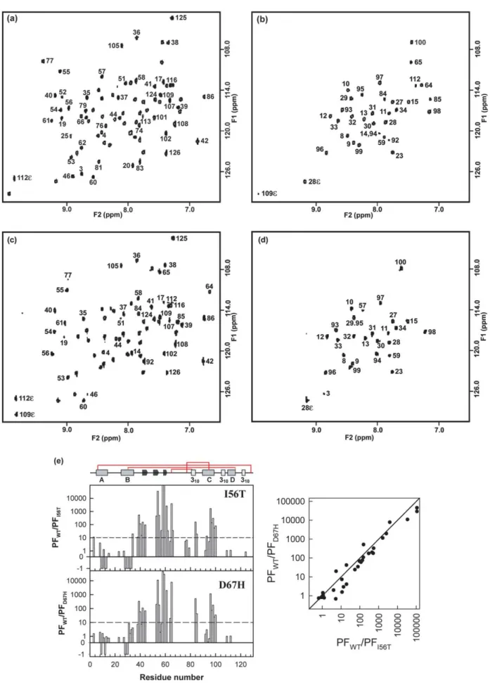
Figure 3.15N–1H HSQC NMR spectra of the I56T variant lysozyme recorded (a) immediately after exposure to2H 2O
and, (b) after 3.5 hours of exchange at pH 5.0 and 37 8C. For comparison, (c) and (d) show the previously published
15
N–1H HSQC NMR spectra of the D67H variant lysozyme recorded respectively immediately after exposure to2H2O
the two peaks in the mass spectra were integrated and the fraction of the lower mass species calcu-lated as a function of time; the time-course of the relative intensity of this peak then provides a direct measure of the opening rate of the cooperative fluctuations giving rise to the exchange. The time constant of the opening event for the I56T protein was determined in this manner to be 32.6(G2.5) seconds, a value of the same order of magnitude as that determined for the D67H protein under the same conditions (15.3(G0.6) seconds,Figure 2(b)).24 In order to obtain more residue-specific infor-mation about the structure of the intermediate state populated by the I56T variant, a series of 2D NMR experiments was carried out to probe the H/D exchange kinetics of individual amide groups. The experiments were carried out by dissolving the
15N-labelled proteins in 2H
2O at pH 5. This pH
value was chosen because under these conditions, the intrinsic exchange rates of amide hydrogen atoms are close to their minimum values. Therefore, it should allow the maximum number of amide hydrogen atoms to be observed in a series of15N–1H heteronuclear single quantum coherence (HSQC) NMR spectra recorded while hydrogen exchange is taking place. At the earliest acquisition time following the initiation of the H/D exchange reaction, cross-peaks from 78 amide hydrogen atoms are visible in the HSQC spectrum from the I56T variant (Figure 3(a)); this number is very similar to that observed for the wild-type protein (79 peaks),20 and somewhat higher than that observed for the D67H variant (66 peaks, Figure 3(c)).20 This number, however, depends critically on the exact length of time taken to record the first spectrum after the initiation of the exchange process, since amide protons with relatively low protection factors will exchange within the first half hour. The spectra recorded after 3.5 hours illustrate, however, qualitative differences in the H/D exchange behaviour of the I56T variant from that of wild-type lysozyme. The I56T variant has only 32 amide protons that have not exchanged signifi-cantly after this length of time (Figure 3(b)), whereas the wild-type protein has 50;20 for com-parison, the D67H variant has 27 (Figure 3(d)).20To obtain quantitative information on the exchange rates, we analyzed the height of individual cross-peaks in the spectra as a function of the time of exchange; the rates resulting from this analysis were then used to obtain individual protection factors.25The ratios of the protection factors of the wild-type protein to those of the I56T variant permit a residue-specific comparison of the H/D exchange
behaviour of the two proteins to be made (Figure 3(e)). The results show that the effects of the I56T mutation on the hydrogen exchange behaviour are predominantly on amide protons from residues in the b-domain (defined as contain-ing residues 41–89) and the followcontain-ing C-helix (residues 90–100). Remarkably, despite the different structural localization of the mutations, these results are almost identical to those obtained for the D67H protein,20which are shown for compari-son inFigure 3(c)–(e).
Binding of an antibody fragment stabilizes the native state of the I56T variant
We have shown that the binding of cAb-HuL6, a camelid antibody fragment21 raised against wild-type lysozyme,22 acts to inhibit dramatically the in vitro formation of amyloid fibrils by the D67H variant by stabilizing the variant protein so that it is no longer able to populate the transient inter-mediate to a detectable level under the conditions used in this study.24Here, we investigated whether the binding of the antibody fragment has a similar effect on the properties of the I56T amyloidogenic variant. Because experiments of this type are very sensitive to small changes in solution conditions, the behaviour of I56T and D67H variants was studied in parallel experiments under identical conditions.
We first measured the dissociation constant (KD)
for binding of the antibody fragment to the I56T variant. We found a KD of 0.6 nM, a value very
similar to those measured for the wild-type lyso-zyme (0.7 nM)22 and the D67H variant (1.2 nM).24 The site at which the antibody fragment binds to the I56T variant was then mapped using NMR experi-ments. Comparison of the 15N–1H HSQC NMR spectrum of the free I56T protein, uniformly labelled with 15N, with the spectrum of the 15 N-labelled protein complexed to the unN-labelled antibody fragment allows the binding region to be probed in a site-specific manner by analysis of the chemical shift perturbations of the resonances of the amide groups of individual residues (Figure 4(a)).24 Despite the relatively high molecular mass of the complex (about 28 kDa), a very well resolved spectrum was obtained, enabling essentially all the amide resonances to be identified (Figure 4(a)). Analysis of the perturbations of the chemical shifts reveals that the resonances of 30 residues exhibit a
significant chemical shift perturbation
(jDd1HjR0.1 ppm or jDd15NjR0.4 ppm) upon bind-ing to the antibody fragment (Figure 4(c)). This
amides that have not exchanged after 3.5 hours are labelled in (b) and (d). (e) The left-hand panel shows a comparison of protection factors of the I56T variant with those of the wild-type protein. The ratio of the protection factors is plotted as K1 for the residues that did not show significant exchange during the time-course of the experiment in both the wild-type protein and the variant. Amides whose exchange rates are accelerated by more than tenfold relative to the wild-wild-type protein are all located in the b-domain and the C-helix. Again, data for the D67H variant obtained under similar conditions are shown for comparison.20The right-hand panel shows a comparison of the protection factors of the I56T and D67H variants relative to that of the wild-type protein (WT).
number is closely similar to that found for the D67H protein (29,Figure 4(b)) and the wild-type protein (31).24The residues that are affected by the binding of the antibody fragment are located in the loop between the A and B helices in the a-domain, in the long loop within the b-domain and in the C-helix (Figure 4(d)) and include all the residues that have been found to be in direct contact with the antibody fragment in the crystal structure of the complex with the wild-type lysozyme.24The epitope for the
I56T variant protein is therefore closely similar to that for the D67H variant and for wild-type lysozyme.24 This finding is completely consistent with the very similar values of the dissociation constants for the three lysozyme species, and with the fact that the mutated residues are not located in the epitope region, as we discuss below.
We then investigated the ability of the antibody fragment to inhibit the formation of fibrils by the two lysozyme variants when incubated at pH 5.0 Figure 4.Overlaid15N–1H HSQC NMR spectra of (a) the free I56T variant (blue) and the I56T variant complexed to cAb-HuL6 (red), (b) the free D67H variant (blue) and the D67H variant complexed to cAb-HuL6 (red). The spectra were collected at pH 6.5 and 35 8C. Residues experiencing a chemical shift change Rj0.4j ppm for15N or Rj0.1j ppm for1H resonances are considered to be affected significantly by the binding of cAb-HuL6 and are labelled. (c) Chemical shift perturbations induced by the binding of cAb-HuL6 to I56T lysozyme. The pink and blue bars indicate the chemical shift perturbations of the1H and15N resonances, respectively. The peak corresponding to residue 78 in the I56T-cAb-HuL6 complex has not been assigned; therefore shift differences for this peak are not shown in the histogram. No unassigned peaks are visible in the spectra of the complex within 0.1 ppm (1H) and 0.4 ppm (15N) of the peak position of this residue
in the spectrum of the unbound protein. This finding suggests strongly that there are large chemical shift perturbations for this residue in the complex. (d) Ribbon diagram of the I56T variant showing the Ca
atoms of the residues affected significantly by the binding of cAb-HuL6 in space-filling representation. Residues affected only in the I56T variant are shown in pink, those affected only in the D67H variant are shown in blue, and residues affected in both variants are shown in green. The region of the molecule that unfolds transiently in a locally cooperative manner is coloured yellow. The lysozyme structure was generated from the X-ray coordinates (PDB entry 1LOZ)15and produced using MOLMOL.38
and 65 8C whilst the solutions were stirred. These conditions differ from those used previously,24and have the advantage of enabling aggregation to be monitored without the need for addition of a chemical denaturant. In the absence of the antibody fragment, both variant proteins aggregate within a few minutes under these conditions (Figure 5(a)). The kinetics of aggregation are similar for the two variants and follow a sigmoidal curve consistent with the nucleation-dependent growth model that has been suggested as a common mechanism of fibril formation.26 In contrast, when wild-type lysozyme was incubated under the same con-ditions, no significant increase in light-scattering was observed, indicating that no detectable
aggre-gation had taken place. The presence of large quantities of fibrillar material in the aggregates formed by both variant proteins was confirmed unambiguously by negative-stain transmission electron microscopy (Figure 5(b)). The structures observed include a proportion of well-resolved isolated fibrils, although the fibrils generally appear to be incorporated into large bundles. Such struc-tures have been observed with the D67H variant when incubated in different conditions (pH 5.5 in the presence of 3 M urea and at 48 8C).24When the variant proteins were incubated in the presence of an equimolar amount of the antibody fragment, no significant change in light-scattering was detectable during the first two hours of incubation. After Figure 5.(a) Time-course of the aggregation of the I56T and D67H variant lysozymes in the absence ( and for the I56T and D67H variants, respectively) and presence of an equimolar quantity of cAb-HuL6 ( and for the I56T and D67H variants, respectively) as monitored by light-scattering. Data are shown also for wild-type lysozyme in the absence of cAb-HuL6 ( ). Inset: expansion to demonstrate the lag phase at the onset of the aggregation process. The lysozyme concentration was 0.1 mg/ml and the data were recorded at 65 8C and pH 5.0. (b) Representative images of fibrils produced by transmission electron microscopy. Left-hand panel, I56T variant; right-hand panel, D67H variant. The scale bars represent 200 nm.
longer incubation times, however, a slow increase in light-scattering was observed for both variants. This increase is, however, likely to be due to the somewhat reduced stability of the antibody fragment under these conditions, as an increase in light-scattering was observed when the antibody fragment was incubated alone under the same conditions. Taken together, these results indicate clearly that the binding of the antibody fragment to the variant proteins decreases their rate of aggrega-tion significantly.
The effects on the equilibrium thermal unfolding behaviour of binding the antibody fragment to the variant proteins under the solution conditions (0.1 M sodium acetate buffer, pH 5.0) used to form fibrils were then investigated (Figure 6). Thermal denaturation experiments followed by far-UV
circular dichroism (CD) (Figure 6(a)) indicate that the temperatures of the mid-points of unfolding of the I56T and D67H variants are very similar (TmZ 68(G1) 8C in both cases). This value is about 10 deg. C lower than that of the wild-type protein, (TmZ80(G1) 8C) under similar conditions. In the presence of the antibody fragment, however, the stability of the variant proteins is raised by about 10 deg. C (TmZ78(G1) 8C), so that when
com-plexed to the antibody fragment the variant proteins are almost as stable as the wild-type
protein. Moreover, the thermally induced
unfolding of both the I56T and D67H variants was found to be associated with a substantial increase in the intensity of the fluorescence of 8-anilino-1-naphthalene-sulfonic acid (ANS) at 475 nm (Figure 6(b)) with a maximum at 66(G2) 8C, very close to the mid-point of unfolding. This finding indicates that a partially unfolded intermediate is populated significantly during unfolding, as 8-anilino-1-naphthalene-sulfonic acid (ANS) binds to accessible hydrophobic clusters in proteins. A much smaller enhancement of ANS fluorescence, at 75(G2) 8C, was observed when the variant proteins were heated in the presence of an equimolar amount of the antibody fragment, suggesting that partially unfolded structures at the mid-point of denaturation are populated only marginally in the presence of the antibody frag-ment. An increase of similar amplitude in the ANS fluorescence was observed at 78(G2) 8C with the wild-type protein in the absence of the antibody fragment, suggesting that it too could populate some form of partially structured state close to its denaturation temperature (Figure 6(b)).
The effects of the mutations on the dynamic properties of the variant proteins under much milder denaturing conditions were studied by means of H/D exchange experiments analyzed by MS and NMR. In a first set of experiments, real-time H/D exchange kinetics under EX2 conditions (pH 5.0, 37 8C) were monitored for the free amyloidogenic variants and for the proteins com-plexed with the antibody fragment (Figure 7(a) and (b)). Under EX2 conditions, the rate at which any unfolded regions of structure return to their protected state is much faster than the intrinsic exchange rate.27 This type of process results in the observation of a single peak whose mass decreases with the length of time the exchange is allowed to take place and allows the number of protected sites to be determined.15In the absence of the antibody fragment, about 31 and 32 residues for the I56T and D67H proteins, respectively, are observed by MS to be protected strongly after 3.5 hours; this number is consistent with data obtained from the NMR measurements under the same conditions (32 and 27 amide protons for the I56T and D67H variants, respectively;Figure 3(b) and (d)). In the presence of the antibody fragment, however, about 48 and 51 residues are observed by MS to be protected strongly in the I56T and D67H proteins, respectively (Figure 7(a) and (b)) a number in good agreement Figure 6.Thermal unfolding (a) monitored by far-UV
CD at 228 nm, (b) monitored by ANS fluorescence at 475 nm. ( ) Free I56T variant; ( ) free D67H variant; ( ) I56T variant complexed to cAb-HuL6; ( ) D67H variant complexed to cAb-HuL6; ( ) free wild-type lysozyme. The lysozyme concentration was 0.2 mg/ml and 0.035 mg/ml in 0.1 M sodium acetate buffer (pH 5.0) for CD and ANS fluorescence measurements, respectively. A.U., arbitrary units.
with that measured for wild-type lysozyme by NMR under similar conditions (50).20
In a second set of experiments, the effects of the mutations on the variant proteins was investigated by H/D exchange pulse-labelling experiments under conditions where an EX1 exchange mechan-ism has been observed for both variants (Figure 2(a)).20,24 Under these conditions (pH 8.0 and 37 8C), the refolding rate of any fluctuation enabling hydrogen exchange to take place is much slower than the intrinsic exchange rate; effectively all the labile hydrogen atoms that are exposed will undergo exchange each time such a fluctuation occurs. Thus, if a fluctuation results in the coopera-tive unfolding of a significant region of the protein molecule, a well-resolved bimodal distribution of masses should result; the two peaks represent the populations of molecules that have or have not experienced such a cooperative fluctuation (Figure 7(c)).20 An equimolar mixture of the I56T variant and15N-labelled wild-type protein was then incubated in the presence of a stoichiometric amount of the antibody fragment (i.e. I56T:WT:cAb-HuL6Z1:1:2). Exchange was carried out for 45 seconds and 120 seconds (i.e. the lengths of time necessary for about 50% and 75%, respectively, of the I56T protein molecules to access, at least once, the partially unfolded state in the absence of the antibody fragment;Figure 2(b)). The peak at lower mass that is observed for the I56T protein in the absence of the antibody fragment (the peak coloured yellow in the left-hand panel in Figure 7(c)), and which results from the transient unfolding of the b-domain and the C-helix, is not seen in the spectra obtained in its presence (the
right-hand panel in Figure 7(c)). Indeed, the I56T protein in the presence of the antibody fragment is visible as a single peak, whose mass decreases steadily as the length of time that exchange was allowed to take place increases; this situation indicates that the exchange process occurs through localized independent fluctuations of the structure through an EX2 mechanism. Thus, in the presence of the antibody fragment, virtually none of the lysozyme molecules transiently access the locally cooperative unfolded species within the duration of the exchange experiment. A similar inhibition of the transient partial unfolding on the binding of the antibody fragment has been reported for the D67H protein.24
Finally, a series of 2D NMR experiments was carried out to monitor in a site-specific manner the hydrogen exchange behaviour of both the amyloidogenic variants in the presence and in the absence of the antibody fragment. These experi-ments were carried out at pH 6.5 and 35 8C to ensure that the antibody binds tightly to the lysozyme molecules. Both variants of human lysozyme show much less protection against hydro-gen exchange than the wild-type protein; about ten amide protons are protected in both variant proteins compared to 23 in the wild-type protein after 13.2 hours of exchange (Figures 8(b), and 9(b) and (f)). Moreover, none of the b-domain or C-helix amide protons is observed to be protected in the I56T and D67H variant proteins under these conditions.The partial unfolding observed at pH 5.0 and 37 8C (Figure 3(b)) therefore occurs also at pH 6.5 and 35 8C. The formation of a complex between the variant proteins and the antibody Figure 7. Kinetic profiles of hydrogen exchange at pH 5.0, 37 8C monitored by mass spec-trometry: (a) for I56T lysozyme (C) free and (B) complexed to cAb-HuL6. (b) Equivalent data obtained for the D67H protein free (C) and (B) complexed to cAb-HuL6. (c) Pulse-labelling H/D exchange at pH 8 and 37 8C of a mixture of I56T variant and
15N-labelled wild-type lysozyme
in the absence (left-hand panel) and in the presence (right-hand panel) of cAb-HuL6. The peaks coloured yellow in the spectra of the I56T variant (left-hand panel) and that result from a locally cooperative unfolding event,20are not observed in the spectra of the I56T protein in the presence of the antibody fragment (right-hand panel).
fragment leads to dramatic perturbations to the exchange behaviour, with a significant retardation of the exchange rates of the b-domain and C-helix amide protons (Figures 8(d) and 9(h)). In the presence of an antibody fragment that does not interact with lysozyme, cAb-R2,22 the hydrogen exchange behaviour of the I56T variant protein is, however, nearly identical with that observed with the free protein (Figure 8(f)). This finding confirms that it is the specific binding of the cAb-HuL6 fragment that is responsible for the inhibition of the partial unfolding event. Taken together, these results demonstrate that in the presence of the antibody fragment, both amyloidogenic variants of lysozyme studied here display a behaviour vir-tually identical with that of the wild-type protein in its absence. The interaction of the variant proteins with the antibody fragment must, therefore, lead to a stabilization of the native conformation of the protein and, effectively, completely suppresses the partial unfolding event triggered by the mutational changes in the amino acid sequence.
Discussion
The amyloid diseases are characterised by the extracellular accumulation of protein aggregates that share a common fibrillar conformation. These disorders include Alzheimer’s and the prion diseases, and type II diabetes.1,2Moreover, diseases
are being added to the list each year, as tests reveal the presence of amyloid deposits in conditions such as amyotropic lateral sclerosis and some forms of arterial amyloidoses.2 Most of the diseases associated with protein aggregation are strongly age-dependent or related to modern life-styles; they are therefore becoming increasingly prevalent.28 Alzheimer’s disease and type II diabetes, for example, are rapidly becoming the most costly and socially disruptive diseases in the developed world.29 No effective treatment for this type of disease is available so far, although extensive research is being done to address the question of their prevention and therapy.30 Under-standing the mechanism and molecular details of the pathological conversions of amyloidogenic proteins from their soluble forms into fibrillar structures is of crucial importance for this objective.31
In this study, we have investigated and compared the effect of two natural amyloidogenic mutations on the properties of human lysozyme. The molecu-lar details that we have been able to gain in this study show that the two variant proteins behave in an astonishingly similar manner, despite the fact that the two mutations are sequentially and spatially distinct, and have different effects on the native-state structure and dynamics.15,18 Equi-librium thermal unfolding experiments show that both variants are less stable than the wild-type protein (by about 10 deg. C) as reported.15,24Most Figure 8.15N–1H HSQC NMR spectra of ((a) and (b)) free I56T variant lysozyme; ((c) and (d)) the I56T variant in the presence of a stoichiometric amount of cAb-HuL6; ((e) and (f)) the I56T variant in the presence of a stoichiometric amount of an unrelated antibody fragment (cAb-R2). The spectra were collected immediately after exposure to2H2O
(top) and after 13.2 hours (bottom) of exchange at pH 6.5 and 35 8C. Amide peaks are labelled in the last spectrum in which they are visible.
Figure 9.15N–1H HSQC NMR spectra of ((a) and (b)) free wild-type lysozyme; ((c) and (d)) wild-type lysozyme in the presence of a stoichiometric amount of cAb-HuL6; ((e) and (f)) free D67H variant protein; ((g) and (h)) the D67H variant in the presence of a stoichiometric amount of cAb-HuL6. The spectra were collected immediately after exposure to2H
2O
(top) and after 13.2 hours (bottom) of exchange at pH 6.5 and 35 8C. Amide peaks are labelled in the last spectrum in which they are visible.
importantly, their unfolding is substantially less cooperative than the wild-type protein under the conditions studied here. H/D pulse-labelling experiments reveal that the two variant lysozymes can populate an intermediate transiently under conditions that include physiologically relevant values of pH and temperature, where the proteins are otherwise stable. Interestingly, the present work demonstrates that the region of the protein struc-ture that is unfolded transiently is effectively identical in the two variant proteins. In this intermediate species, the b-domain and the adjacent C-helix are unfolded cooperatively, whereas the remainder of the a-domain appears to be largely native-like in structure (Figure 4(d)). The time-scale on which these fluctuations occur is very slow relative to most fluctuations occurring in protein structures, and is of the same order of magnitude in the two variants (32.6(G2.5) seconds and 15.3(G0.6) seconds for the I56T and D67H proteins, respectively, see above).
The remarkable similarity in the behaviour of the I56T and D67H variant proteins can be rationalized from structural data,15 which suggest that in both variants the crucial interface region between the a and b-domains is less constrained than in the wild-type protein, and that this perturbation of the interface is likely to be the origin of the reduction in global cooperativity. Interestingly, the inter-mediate that is sampled occasionally by the variant proteins resembles that populated in the normal refolding of the protein from a highly denatured state,20,32 emphasising the close link between normal and aberrant folding behaviour. A recent important observation is that for hen egg-white lysozyme, a protein homologous to human lyso-zyme with 60% sequence identity, fragments corre-sponding to part of the b-domain and all the residues of the C-helix, are cleaved readily from the protein by proteolysis when incubated at low pH and high temperature. Moreover, these frag-ments form amyloid fibrils rapidly,33in contrast to fragments corresponding to the remainder of the a-domain, which remain largely soluble under the same conditions.33 This result indicates that the region corresponding to the b-domain and C-helix has a high intrinsic propensity to aggregate. Thus, it is likely that, following exposure to the solvent as the result of partial unfolding, this region of the variant proteins readily initiates the aggrega-tion event that ultimately leads to the formaaggrega-tion of fibrils. This highly amyloidogenic character of the destabilized region is probably the reason why all the known amyloidogenic mutations are localised in the b-domain.
Furthermore, we have shown that binding a camelid antibody fragment to both the I56T and D67H variant proteins inhibits dramatically their ability to aggregate in vitro and to form amyloid fibrils. In addition, in the presence of the antibody fragment, virtually none of the molecules of the I56T (this work) and D67H22 variant proteins undergoes even a single locally cooperative
unfolding event on the time-scale of the experiment (up to one hour). These results indicate that the frequency of such fluctuations in both variants is reduced drastically as a result of binding to the antibody fragment. Real-time MS and NMR exper-iments have further confirmed that the binding of the antibody restores the high degree of global cooperativity that is characteristic of the wild-type protein. These results therefore provide further evidence that the formation of a partially unfolded species with a high propensity to aggregate, resulting from the locally cooperative unfolding of the b-domain and the C-helix, is the critical event that triggers the aggregation process in the absence of the antibody fragment. Our previous structural study of the complex between the wild-type protein and the antibody fragment revealed that the epitope includes neither the site of the mutation nor most of the residues in the region of the protein structure that is destabilized by the mutations.24 Thus, the effects of binding are not simply to mask the entire region of the protein destabilised by the mutation and hence to prevent its unfolding from the remainder of the structure. Rather, it appears that the binding of the antibody fragment restores the global cooperativity of the lysozyme structure that is disrupted by these two amyloidogenic mutations by a more subtle mechanism. It is of particular interest to note that perturbations to the NMR chemical shifts of the I56T protein upon the binding of the antibody fragment include the amide resonance of residue 57. The amide resonance of this residue, which is located at the interface of the two domains and is itself the location of an amyloidogenic mutation,13 is perturbed signifi-cantly as a result of the binding of the antibody fragment to the D67H protein.24Restoration of the global cooperativity therefore occurs, at least in part, through the transmission of long-range con-formational effects to the interface between the two structural domains of the protein in a process reminiscent of the final step in the normal folding process.16,32
Taken together, these results suggest that the disruption of the interface region between the a and b-domains is a crucial event in determining the amyloidogenicity of the variant proteins. Moreover, the observation that an antibody fragment can restore behaviour characteristic of the wild-type protein supports the idea that such species could be important leads in the search for therapeutic strategies.34 Although our results provide a clue as to the possible significance of partially folded species in amyloidogenicity, they raise the issue of the possible importance of the specific structure of the intermediate, which is similar for the two amyloidogenic proteins, in the formation and the structure of the resulting amyloid fibrils. The common, perhaps universal, ability of polypeptides to form amyloid fibrils with similar morphological properties indicates that the specific structure of the partially folded intermediate formed by amyloido-genic variants is unlikely to be critical in defining
the characteristic cross-b structure of the mature fibrillar assembly.20 The properties of the inter-mediate are, however, likely to be extremely important in determining the regions of the poly-peptide chain involved in the core of the cross-b structure and in defining the properties and rates of formation of the initial oligomers that ultimately form the mature fibrils. In particular, in the case of lysozyme, the high propensity of the most unfolded regions of the intermediate state to convert into amyloid fibrils is likely to be a crucial factor in the high amyloidogenicity of the protein. Thus, mutations causing amyloidosis are likely to be those that do not destabilize the protein sufficiently to trigger their destruction in endoplasmic reticu-lum-associated degradation (ERAD) process, but disrupt the global stability and cooperativity to such an extent that an intermediate is populated in which the partial unfolding event exposes a highly aggregation-prone region of the protein sequence.
These results demonstrate clearly the link between the ability of the amyloidogenic variant proteins to populate an intermediate that results from the perturbation of the structure of the interface between the two lysozyme domains and their propensity to convert into amyloid fibrils. In the native state of a globular protein, the higher cooperativity of the overall fold under physiological conditions locks the polypeptide chain within the folded structure and thus prevents the develop-ment of intermolecular interactions involving the majority of the main chain or of hydrophobic residues.5Fluctuations in the native structure that can give rise to hydrogen exchange but expose transiently only very local regions of the poly-peptide chain to the solvent are unlikely to generate conformations of a protein able to form strong intermolecular interactions. If such fluctuations expose significant regions of the polypeptide main chain and its hydrophobic core, however, they could represent an initial step in the development of intermolecular interactions whereby near-native like aggregates form prior to the formation of extensive region of b-sheet structure; such species have indeed been observed in other systems during the early stages of aggregation.35
In accord with this concept, conditions that readily stimulate formation of amyloid fibrils are generally those that at least partially denature the protein and expose substantial regions of the polypeptide backbone to the solvent. This unfold-ing event may in some cases arise from the global unfolding of the native structure, or from local cooperative unfolding as in the case with the amyloidogenic lysozyme studied here.1 Indeed, for the I56T and D67H variants, the population of a partially unfolded intermediate is much greater than for the wild-type protein under similar conditions and the local cooperativity of the unfolding process that generates the intermediates results in most of the b-domain and C-helix being unfolded simultaneously, under conditions where the native state is otherwise stable. Moreover, the
region exposed in this way is highly amyloidogenic, as shown by the study of the fragment of hen egg-white lysozyme.33This combination of factors could give rise to a sufficient concentration of soluble, partially folded species capable of forming signifi-cantly strong intermolecular interactions to initiate aggregation under much milder conditions than would be needed to generate a similar concen-tration of partially unfolded molecules for the wild-type protein. Such species may then convert into large aggregates with more extensive b-sheet structure characteristic of amyloid fibrils. This conclusion reinforces the view that the remarkable cooperativity of native protein structures is an essential evolutionary development to enable other-wise marginally stable structures to resist aggrega-tion under condiaggrega-tions in which they exert their biological function.5
Materials and Methods
Proteins
Wild-type human lysozyme and its I56T and D67H variants, including uniformly 15N-labelled proteins, cAb-R2 and the cAb-HuL6 were expressed and purified as described.22,36 The affinity of cAb-HuL6 for the lysozyme species was determined using surface plasmon resonance as described.22
Electron microscopy
Samples were applied to Formvar-coated nickel grids, stained with 2% (w/v) uranyl acetate solution and viewed in a Philips CEM100 transmission electron microscope operating at 80 kV.
Aggregation monitored by right-angle light-scattering Stock solutions of the proteins were prepared from lyophilized material, passed through a 0.22 mm pore size filter, diluted to the desired final concentration into 0.1 M sodium acetate buffer (pH 5.0) and incubated at 65 8C with stirring in a Cary Eclipse spectrofluorimeter. The excitation wavelength was 500 nm and changes in fluorescence emission were monitored at 500 nm with slit-widths of 5 nm.
Fluorescence measurements
ANS fluorescence was recorded on a Cary Eclipse spectrofluorimeter equipped with a four-cell Peltier holder with excitation and emission wavelengths of 350 nm and 475 nm, respectively. The excitation and emission slit-widths were both 5 nm. The protein con-centration was 2.4 mM in a 1 cm path-length cuvette and the final concentration of ANS was 315 mM as determined from the molar extinction coefficient of 4950 MK1cmK1. The buffer was 0.1 M sodium acetate (pH 5.0) and the data were corrected for the background fluorescence of the solution in the absence of the protein (bufferCANS). The temperature was increased monotonically from 5 8C to 97 8C at a rate of 0.5 deg. C/minute.
Circular dichroism
Heat-induced unfolding transitions were monitored by far-UV CD using a Jasco J-810 spectropolarimeter equipped with a six-cell Peltier holder with a 0.1 cm cell path-length. The protein concentration was 0.2 mg/ml in 0.1 M sodium acetate buffer (pH 5.0), based on A2801%Z2.5 and 2.1 for lysozyme and cAb-HuL6, respectively. The temperature was increased monotonically from 25 8C to 97 8C at a rate of 0.5 deg. C/minute. Data were acquired every 1 deg. C, with a four second integration time and a 2 nm bandwidth. The very low intensity of the far-UV CD signal of the antibody fragment compared to that of the human lysozyme, and the existence of only minor changes at 228 nm upon unfolding of the cAb-HuL6, enabled us to monitor specifically at that wavelength the unfolding of lysozyme within the complex with cAb-HuL6.
Pulse labelling hydrogen/deuterium exchange of the I56T variant analyzed by mass spectrometry
The [14N]I56T variant and uniformly15N labelled wild-type lysozyme were deuterated at exchangeable sites by unfolding in deuterated guanidinium hydrochloride followed by refolding in deuterated buffer (50 mM deuterated acetic acid, pH* 5.0). The proteins were subsequently concentrated and the buffer was changed to 10 mM deuterated acetic acid, pH* 5.0, using Centri-prep-3 concentrators (Amicon, Millipore) and mixed in equal proportions. For the experiments in the presence of cAb-HuL6, the deuterated lysozymes and protonated cAb-HuL6 were mixed immediately before the experi-ment was performed to give a stoichiometric ratio of I56T:WT:cAbHuL6Z1:1:2. The pulse-labelling hydrogen exchange experiments were carried out using a Bio-Logic QFM5 mixer connected to a circulating water-bath set at 37 8C as described.20One volume of protein solution was mixed with 15 volumes of 100 mM ammonia/formic acid buffer in H2O at pH 8.0, and the exchange was allowed to
proceed for various times between 0.4 second and 120 seconds. The resulting solutions were then mixed with seven volumes of a 1 M acetic acid solution in H2O,
to generate a pH of 3.5 for the solutions after the quench step. The samples were collected and placed on ice before analysis by mass spectrometry. A control sample corre-sponding to complete exchange was prepared by mixing one volume of protein solution with 22 volumes of 20 mM acetic acid/ammonia buffer in H2O at pH 5.0. This sample
was incubated for 15 minutes at 80 8C and subsequently placed on ice. The samples were electrosprayed at the base pressure of a Platform mass spectrometer (Micro-mass, UK) and at a cone voltage of 150V. Under these conditions, the antibody-lysozyme complexes dissociate in the mass spectrometer, enabling the mass distributions of the lysozyme molecules alone to be determined readily and directly. Mass spectra shown inFigures 2(a) and 7(c) represent the convolution of the C8, C9, and C10 charge states with minimal smoothing and converted to a mass scale. Data analysis was performed using Grams 32 V5.10 (Galactic Industries). The spectra were fitted to a linear baseline and a combination of Gaussian distributions to estimate the relative proportions of the different species. The calculated areas for each peak from the fits were analyzed as a function of time and used to extract kinetic parameters for the exchange reactions using Sigmaplot v5.0 (SPPS). No correction was made to the number of exchangeable amide protons for the w5% deuterium
present in the labelling pulse, since back exchange does not affect the relative proportions of each species. Real-time mass spectrometry
Stock solutions of 200 mM wild-type, I56T and D67H human lysozymes were prepared in MilliQ-prepared water adjusted to pH 5.0 with formic acid. Exchange was initiated by 20-fold dilution into 2H
2O at pH* 5.0
(uncorrected for the deuterium isotope effect), adjusted with deuterated formic acid at 37 8C. Spectra were recorded before the initiation of exchange and at various times during the exchange reaction. A correction was made to the number of exchangeable amide protons for the w5% deuterium present in the labelling pulse.
Real-time 2D NMR spectroscopy of the15N-labelled I56T variant
The I56T variant protein (5 mg) was dissolved in 575 ml of 20 mM acetic acid in 2H2O at pH 5.0* (value
uncorrected for the isotope effect). This pH value was chosen because the intrinsic exchange rates of amide hydrogen atoms are close to their minimum values. Therefore, it should allow the maximum number of amide hydrogen atoms to be observed in a series of
15
N–1H-HSQC NMR spectra recorded while hydrogen exchange is taking place. The exact pH values of the samples were determined at the end of each experiment and used in the calculation of protection factors.25Series of gradient-enhanced 15N–1H HSQC spectra were
collected at 37 8C in a 600 MHz NMR spectrometer. Each spectrum took w30 minutes to record, with 96 complex t1 increments of eight transients and 1024
complex data points. Sweep-widths of 8000 Hz and 1825 Hz were used in the F2 and F1 dimensions,
respectively. The data were processed with a Gaussian window function in F2and a 908 shifted sine bell in F1
using the program Felix 2.3 (Accelrys). After zero filling, the digital resolution was 3.9 Hz/point and 3.6 Hz/point in F2and F1, respectively. Hydrogen exchange rates were
monitored by measuring peak heights in the HSQC spectra as a function of time. Sigmaplot v5.0 (SPPS) was used to fit exponential functions to the data. The predicted exchange rates for the same sequence in a random coil conformation were divided by the experi-mentally measured rates to calculate protection factors. We used the published protection factors for the wild-type lysozyme.20 For the residues that did not show significant exchange during the time-course of the experiment in both the I56T variant and the wild-type protein, the ratio of the protection factors is plotted as K1 (Figure 3(e)). For the residues that did not show significant exchange during the time-course of the experiment for the wild-type protein but did for the variant, we have used previous data for the wild-type protein recorded at pH 5.5 and 35 8C for longer periods of time.37 Additional 2D correlated spectroscopy (COSY) and nuclear Overhauser effect spectroscopy (NOESY) experiments were recorded, at 20 8C and 37 8C, of the variant protein prepared in 2H2O or in H2O to obtain
assignments for the amide cross-peaks observed in the HSQC spectra.
Real-time experiments were carried out at pH 6.5 and 35 8C for both variant proteins and wild-type lysozyme in the absence and in the presence of the antibody fragment in a home-built 750 MHz NMR spectrometer belonging to the Oxford Centre for
Molecular Sciences. H/D exchange was monitored using a series of eight HSQC spectra using samples (0.6 mM for the I56T variant and the wild-type protein and 0.33 mM for the D67H variant) dissolved in 20 mM phosphate buffer (pH 6.5) in 2H2O (value
uncorrected for the isotope effect). When present the antibody was added in 15% excess. The excess of antibody ensured that all lysozyme molecules were bound to the antibody molecule in the complex. Each spectrum took w1.8 hours to record, with 128 complex t1 increments of 16 transients and 1024
complex data points. Sweep-widths of 10,582 Hz and 2273 Hz were used in the F2 and F1 dimensions,
respectively. The data were processed with a Gaussian window function in F2 and a 908 shifted sine bell in
F1. After zero filling, the digital resolution was
5.2 Hz/point and 4.4 Hz/point in F2 and F1,
respectively.
NMR studies of the I56T:cAb-HuL6 complex
An NMR sample of the complex was made containing w0.7 mM unlabelled cAb-HuL6 and w0.6 mM 15 N-labelled I56T variant at pH 6.5 in 20 mM sodium phosphate buffer made with 95% (v/v) H2O/5% (v/v) 2
H2O. The excess of antibody concentration ensured that
all the lysozyme molecules were bound to an antibody molecule in the complex. A 2D15N–1H HSQC spectrum of the I56T variant bound to the antibody fragment was collected at 35 8C on the 750 MHz spectrometer. The spectrum was collected with 128 complex t1increments of
64 transients and 1024 complex data points. Sweep-widths of 10,582 Hz and 2273 Hz were used in the F2and
F1 dimensions, respectively. The data were processed
with a Gaussian window function in F2and a 908 shifted
sine bell in F1, using the program FELIX (Accelrys). After
zero filling the digital resolution was 5.2 Hz/point and 4.4 Hz/point in F2and F1, respectively. The resonances
arising from the I56T protein in the complex were assigned by comparison with the assigned spectrum of the unbound protein18and that of the wild-type protein in the complex.24
Acknowledgements
We thank Jean-Marie Fre`re for many helpful discussions, Janet Kumita and Russell Johnson for critical reading of the manuscript, Leon G.J. Frenken and Cees van Vliet for providing the cAb-R2, and Andrew Spencer for assistance in expression and purification of human lysozymes. M.D. and D.C. were supported by fellowships from the European Community. C.R. was supported by a BBSRC Advanced Research Fellowship, A.M. is a Research Associate of the FNRS and was supported, in part, by a grant from the Fonds de la Recherche Fondamentale et Collective (contract number 2.4545.01). C.V.R. is a Royal Society University Research Fellow and the research of C.M.D. is supported, in part, by a Programme Grant from the Wellcome Trust. This work was supported also by a BBSRC grant (to C.M.D., C.V.R. and D.B.A.) and by the Belgian Government under the
framework of the Interuniversity Attraction Poles (I.A.P. P5/33).
References
1. Dobson, C. M. (2001). The structural basis of protein folding and its links with human disease. Phil. Trans. Roy. Soc. ser. B, 356, 133–145.
2. Selkoe, D. J. (2003). Folding proteins in fatal ways. Nature, 426, 900–904.
3. Sunde, M. & Blake, C. C. F. (1998). From the globular to the fibrous state: protein structure and structural conversion in amyloid formation. Quart. Rev. Biophys. 31, 1–39.
4. Chiti, F., Webster, P., Taddei, N., Clark, A., Stefani, M., Ramponi, G. & Dobson, C. M. (1999). Designing conditions for in vitro formation of amyloid proto-filaments and fibrils. Proc. Natl Acad. Sci. USA, 96, 3590–3594.
5. Dobson, C. M. (1999). Protein misfolding, evolution and disease. Trends Biochem. Sci. 24, 329–332.
6. Fandrich, M., Fletcher, M. A. & Dobson, C. M. (2001). Amyloid fibrils from muscle myoglobin. Nature, 410, 165–166.
7. Guijarro, J. I., Sunde, M., Jones, J. A., Campbell, I. D. & Dobson, C. M. (1998). Amyloid fibril formation by an SH3 domain. Proc. Natl Acad. Sci. USA, 95, 4224–4228. 8. Schenk, D. (2002). Amyloid-beta immunotherapy for Alzheimer’s disease: the end of the beginning. Nature Rev. Neurosci. 3, 824–828.
9. Sacchettini, J. C. & Kelly, J. W. (2002). Therapeutic strategies for human amyloid diseases. Nature Rev. Drug Discov. 1, 267–275.
10. Dobson, C. M. (2003). Protein folding and disease: a view from the first Horizon symposium. Nature Rev. Drug Discov. 2, 154–160.
11. Pepys, M. B., Hawkins, P. N., Booth, D. R., Vigushin, D. M., Tennent, G. A., Soutar, A. K. et al. (1993). Human lysozyme gene mutations cause hereditary systemic amyloidosis. Nature, 362, 553–557.
12. Valleix, S., Drunat, S., Philit, J. B., Adoue, D., Piette, J. C., Droz, D. et al. (2002). Hereditary renal amyloidosis caused by a new variant lysozyme W64R in a French family. Kidney Int. 61, 907–912. 13. Yazaki, M., Farrell, S. A. & Benson, M. D. (2003).
A novel lysozyme mutation Phe57Ile associated with hereditary renal amyloidosis. Kidney Int. 63, 1652–1657.
14. Funahashi, J., Takano, K., Ogasahara, K., Yamagata, Y. & Yutani, K. (1996). The structure, stability, and folding process of amyloidogenic mutant human lysozyme. J. Biochem. (Tokyo), 120, 1216–1223. 15. Booth, D. R., Sunde, M., Bellotti, V., Robinson, C. V.,
Hutchinson, W. L., Fraser, P. E. et al. (1997). Instability, unfolding and aggregation of human lysozyme variants underlying amyloid fibrillogenesis. Nature, 385, 787–793.
16. Canet, D., Sunde, M., Last, A. M., Miranker, A., Spencer, A., Robinson, C. V. & Dobson, C. M. (1999). Mechanistic studies of the folding of human lysozyme and the origin of amyloidogenic behavior in its disease-related variants. Biochemistry, 38, 6419–6427. 17. Morozova-Roche, L. A., Zurdo, J., Spencer, A., Noppe,
W., Receveur, V., Archer, D. B. et al. (2000). Amyloid fibril formation and seeding by wild-type human lysozyme and its disease-related mutational variants. J. Struct. Biol. 130, 339–351.
Redfield, C. & Dobson, C. M. (2001). Characterization of the structure and dynamics of amyloidogenic variants of human lysozyme by NMR spectroscopy. Protein Sci. 10, 2525–2530.
19. Takano, K., Funahashi, J. & Yutani, K. (2001). The stability and folding process of amyloidogenic mutant human lysozymes. Eur. J. Biochem. 268, 155–159. 20. Canet, D., Last, A. M., Tito, P., Sunde, M., Spencer, A.,
Archer, D. B. et al. (2002). Local cooperativity in the unfolding of an amyloidogenic variant of human lysozyme. Nature Struct. Biol. 9, 308–315.
21. Muyldermans, S. (2001). Single domain camel anti-bodies: current status. J. Biotechnol. 74, 277–302. 22. Dumoulin, M., Conrath, K., Van Meirhaeghe, A.,
Meersman, F., Heremans, K., Frenken, L. G. et al. (2002). Single-domain antibody fragments with high conformational stability. Protein Sci. 11, 500–515. 23. Hooke, S., Eyles, S. J., Miranker, A., Radford, S. E.,
Robinson, C. V. & Dobson, C. M. (1995). Cooperative elements in protein folding monitored by electrospray ionization mass spectrometry. J. Am. Chem. Soc. 117, 7548–7549.
24. Dumoulin, M., Last, A. M., Desmyter, A., Decanniere, K., Canet, D., Larsson, G. et al. (2003). A camelid antibody fragment inhibits the formation of amyloid fibrils by human lysozyme. Nature, 424, 783–788. 25. Bai, Y., Milne, J. S., Mayne, L. & Englander, S. W.
(1993). Primary structure effects on peptide group hydrogen exchange. Proteins: Struct. Funct. Genet. 17, 75–86.
26. Harper, J. D. & Lansbury, P. T., Jr (1997). Models of amyloid seeding in Alzheimer’s disease and scrapie: mechanistic truths and physiological consequences of the time-dependent solubility of amyloid proteins. Annu. Rev. Biochem. 66, 385–407.
27. Englander, S. W. (2000). Protein folding intermediates and pathways studied by hydrogen exchange. Annu. Rev. Biophys. Biomol. Struct. 29, 213–238.
28. Dobson, C. M. (2003). Protein folding and misfolding. Nature, 426, 884–890.
29. DeLaGarza, V. W. (2003). Pharmacologic treatment of Alzheimer’s disease: an update. Am. Fam. Physician, 68, 1365–1372.
30. Cohen, F. E. & Kelly, J. W. (2003). Therapeutic approaches to protein-misfolding diseases. Nature, 426, 905–909.
31. Dobson, C. M. (2004). In the footsteps of alchemists. Science, 304, 1259–1262.
32. Hooke, S. D., Radford, S. E. & Dobson, C. M. (1994). The refolding of human lysozyme: a comparison with the structurally homologous hen lysozyme. Biochemistry, 33, 5867–5876.
33. Frare, E., Polverino de Laureto, P., Zurdo, J., Dobson, C. M. & Fontana, A. (2004). A highly amyloidogenic region of hen lysozyme. J. Mol. Biol. 340, 1153–1165. 34. Dumoulin, M. & Dobson, C. M. (2004). Probing the
origins, diagnosis and treatment of amyloid diseases using antibodies. Biochimie, 86, 589–600.
35. Bouchard, M., Zurdo, J., Nettleton, E. J., Dobson, C. M. & Robinson, C. V. (2000). Formation of insulin amyloid fibrils followed by FTIR simultaneously with CD and electron microscopy. Protein Sci. 9, 1960–1967.
36. Spencer, A., Morozov-Roche, L. A., Noppe, W., MacKenzie, D. A., Jeenes, D. J., Joniau, M. et al. (1999). Expression, purification, and characterization of the recombinant calcium-binding equine lysozyme secreted by the filamentous fungus Aspergillus niger: comparisons with the production of hen and human lysozymes. Protein Expr. Purif. 16, 171–180.
37. Woodruff, N. D. (1998). Investigation of protein structure and folding by NMR spectroscopy. PhD thesis, University of Oxford.
38. Koradi, R., Billeter, M. & Wuthrich, K. (1996). MOLMOL: a program for display and analysis of macromolecular structures. J. Mol. Graph. 14, 29–32.
Edited by P. T. Lansbury Jr