O
pen
A
rchive
T
OULOUSE
A
rchive
O
uverte (
OATAO
)
OATAO is an open access repository that collects the work of Toulouse researchers and
makes it freely available over the web where possible.
This is an author-deposited version published in :
http://oatao.univ-toulouse.fr/
Eprints ID : 10069
To link to this article : doi:10.1039/c3ra43348j
URL :
http://dx.doi.org/10.1039/c3ra43348j
To cite this version : Hameau, Aurélien and Colliere, Vincent and
Grimoud, Julien and Fau, Pierre and Roques, Christine and Caminade,
Anne-Marie and Turrin, Cédric-Olivier PPH dendrimers grafted on
silica nanoparticles: surface chemistry, characterization, silver colloids
hosting and antibacterial activity. (2013) RSC Advances, vol. 3 (n° 41).
pp. 19015-19026. ISSN 2046-2069
Any correspondance concerning this service should be sent to the repository
administrator:
staff-oatao@listes-diff.inp-toulouse.fr
PPH dendrimers grafted on silica nanoparticles: surface
chemistry, characterization, silver colloids hosting and
antibacterial activity3
DOI: 10.1039/c3ra43348j
Aure´lien Hameau,abVincent Collie`re,abJulien Grimoud,cPierre Fau,ab
Christine Roques,cAnne-Marie Caminadeaband Ce´dric-Olivier Turrin*ab
Polyphosphorhydrazone (PPH) dendrimers have been grafted on silica nanoparticles, and the surface functions of the dendrimers have been derivatized to phosphonates with lateral poly(ethyleneglycol) (PEG) chains. All materials have been thoroughly characterized by MAS NMR, FT-IR, electron microscopy, TGA and elemental analysis. These materials successfully hosted silver and silver oxide nanoparticles. The resulting composites exhibit antibacterial activity.
Introduction
The manipulation and the accurate functionalization of nano-objects is undoubtedly a promising and challenging field of research. In this regard, the association of soft and hard nano-objects, like dendrimers1 and metal or metal oxide nanopar-ticles2 (NPs) is offering unprecedented possibilities to reach high degrees of multifunctionalization, allowing to envision a large scope of applications. In this wide field of research, many efforts have been devoted to the production of dendrimer encapsulated metal NPs3or dendrimer-stabilized metal NPs.4 Another active field of investigation encompasses dendrimer– silica hybrid mesoporous materials obtained by sol–gel procedures.5,6 The interaction of small-sized, discrete silica particles, with dendrimers is more scarcely reported, and most of the work is dedicated to surface modification of silica NPs through a stepwise growth of dendrimer-like structures from reactive or initiator centres. For example, Tsubokawa et al. reported in a seminal work the stepwise outgrowth of hyperbranched polyamidoamine (PAMAM)-like structures on the surface of silica nanoparticles7having a mean diameter of 16 nm on which the amine initiator group was introduced by means of aminopropyltriethoxysilane (APTES). This strategy
opened new perspectives for the design of dendrimer-modified silica-based stationary phases for chromatography purposes8 or for the development of easily recyclable catalytic systems. In the latter case, the catalytic properties can be brought by metal complexes on the surface of the dendrimers,9,10 or by
transition metal NPs embedded in the dendrimeric architec-tures.11Alternatively, dendrimers can be covalently grafted on the surface of silica NPs by a direct post-synthesis silica modification through reactive or hooking centres. This strategy allows a higher control on the structural definition of the organic content of the final hybrid material, but invariably leads to a certain degree of reticulation of the inorganic building blocks due to dendrimer multivalency, even though it can be prevented by the use of dendrons instead of dendrimers. It should be noted that despite its attractiveness, this strategy is much less documented than the stepwise approach evoked above. In this respect, large silica NPs derived from MCM-41 mesoporous silica with a mean diameter of 250 nm have been loaded with a fluorescent dye, covalently modified with generation 2 PAMAM dendrimers through isocyanate hooking centres and then successfully assayed as transfection reagents.12 Another example also
involves large silica microspheres (100–400 nm) which have been covalently modified with generation 4 PAMAM dendri-mers. For this purpose, epoxide hooking centres were generated on the silica microspheres in a two-step procedure by means of vinyltriethoxysilane. The silica-hooked dendri-mers were further loaded with Pd(II) ion complexes. The reduction of the hybrid systems led to silica–dendrimer core– shell structures with dendrimer encapsulated NPs which were efficiently assayed and recycled in catalytic hydrogenation reactions.13In this example, the dendrimer scaffold affords a suitable environment for Pd NPs hosting. The hosting of metallic NPs on the surface or within silica networks can be
aLaboratoire de Chimie de Coordination du CNRS, BP 44099, 205 route de
Narbonne, 31077 Toulouse cedex 4, France. E-mail: turrin@lcc-toulouse.fr; Fax: 33 5615 5003
bUniversite´ de Toulouse, UPS, INPT, F-31077 Toulouse Cedex 4, France
cLaboratoire de Ge´nie Chimique, UMR 5503, UPS, Faculte´ de Pharmacie, 35 chemin
des Maraıˆchers, 31062 Toulouse cedex 9
3Electronic supplementary information (ESI) available: Multinucleus MAS NMR spectra (CP-MAS and HPDEC experiments), FT-IR spectra, TGA and electronic microscopy and EDX data of the silica Si-1 to Si-5, NMR data and mass spectrometry data of compound 2a to 2d, FT-IR and CP-MAS NMR data of starting dendrimer 1, pictures of MBC cultures and data related to the adsorption test experiment. See DOI: 10.1039/c3ra43348j
achieved according to a large variety of processes. In the case of silver NPs containing silica, catalytic,14 antibacterial15 or sensing properties can be obtained.16 Nevertheless, most of these synthetic techniques are not compatible with the incorporation of organic materials which can offer comple-mentary functionalities to the final material, in addition to the NP-stabilizing ones. It is the case of one-pot sol–gel processes which require silica-densifying steps under harsh conditions17 or co-precipitation under strong acidic conditions.15 Alternative strategies involve surface decoration of preformed silica networks with stabilizing functions.15To the best of our
knowledge, hyperbranched macromolecules, which have proved to offer excellent silver NPs stabilising properties in homogeneous conditions,18 have not been used to achieve such purposes on silica surfaces.
Phosphorus-containing dendrimers, also coined as poly (phosphorhydrazone) (PPH) dendrimers,19have also been used
to prepare silica-based materials incorporating covalently bonded PPH architectures. We have described the synthesis of mesostructured silica bulk materials according to sol–gel procedures involving siloxane-cored PPH dendrons.6 Alternatively, the covalent grafting of PPH dendrimers on silica can be envisaged through a multivalent attachment involving the surface groups of symmetrical dendrimers. In this regard, our group has developed a strong expertise on the covalent immobilization of PPH dendrimers onto glass slides.20 This
technique, which was made possible by preliminary experi-ments on quartz surfaces modified with APTES,21has led to the development of sensors, namely dendrichipsTM. Nevertheless, we have never explored in-depth structural and spectroscopic investigations on the nature of the hybrid interface related to the use of bulk support (glass, mica, quartz plates).
Herein we describe a stepwise process to modify the surface of finely divided silica NPs with a simple surface derivatization approach using PPH dendrimers which offers post-synthesis possibilities for surface modification of the final hybrid nanocomposites. The work described in this report is based on a straightforward transfer of the APTES-mediated grafting strategy to finely divided material, namely silica NPs, which allows better spectroscopic description of the hybrid interface thanks to FT-IR and MAS-NMR analyses. Additionally, the functional versatility of our nano onto nano approach, offered by the highly reactive PPH surface, is illustrated with the preparation of PEG–aminophosphonate surface functions which have never been described on dendrimeric architec-tures. The applicability of these new highly functional nanocomposites is illustrated by the efficient hosting of tiny silvers NPs and the preliminary evaluation of the bacteriostatic and bactericidal properties of these materials.
Results and discussion
Chemistry
We have chosen to work on a simple water-releasing condensation reaction between aldehyde terminated PPH
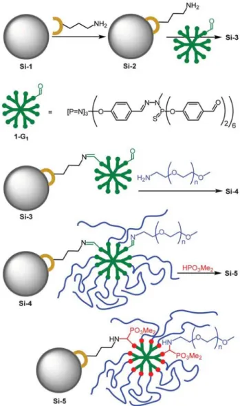
dendrimers and amine-bearing NPs. This reaction allows the creation of a reliable covalent bonding of the PPH dendrimers onto the surface of the nanoparticles according to routine-like procedures, in the view of creating chemical modifications which can be easily monitored by readily accessible spectro-scopic methods. This grafting strategy (Fig. 1), as mentioned above, has been successfully developed to immobilize high generation PPH onto macroscopic glass substrates21 for the large-scale production of dendrislidesTMand dendrichipsTM.20
The grafted dendrimer layer can further undergo chemical transformations and the overall process can be easily monitored by MAS NMR and FTIR techniques to follow the chemical modifications that are made on the nanocomposites. Commercially available silica nanoparticles (mean diameter 12 nm) were activated by thermal treatment (Si-1), and the creation of active Si–OH moieties was unambiguously detected on FTIR spectra by a sharp band centred on 3747 cm21 (see
Fig. 1 Schematic representation of the strategy for dendrimer grafting and post-synthesis functionalization. The drawing is not scaled for clarity purposes, NPs are in reality ca. 5 times bigger than dendrimers and can be attached by several dendrimers.
Fig. 2 and ESI
3
). Activated Si-1 was then derivatized with (3-aminopropyl)triethoxysilane (APTES) according to standard procedures.22 The resulting amino-silanized silica NPs Si-2 were treated with a large excess of first generation aldehyde-terminated PPH dendrimer191-G1in a THF–methanol mixtureat 55uC.
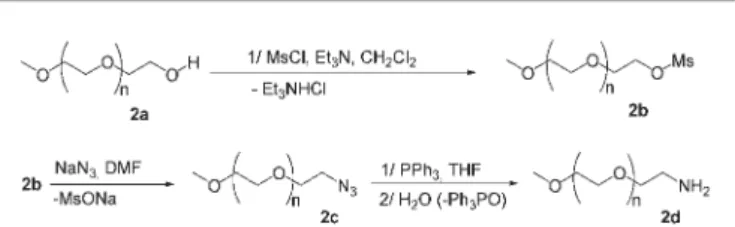
Having in hands these dendrimer-capped silica NPs Si-3, the unreacted aldehyde functions were further condensed with an amine-functionalized poly(ethyleneglycol) (PEG) 2d (Scheme 1) having an average weight of 560 g mol21, containing roughly 11 ethylene glycol moieties. The latter was prepared from commercial methoxy–PEG (MWy550 g mol21) 2a on a 10 g
scale according to a routine procedure23 involving the corresponding Mesyl-PEG 2b and azido-PEG 2c as intermedi-ates (Scheme 1). This intermediate was reduced in a one-pot, two-step procedure involving a Staudinger reaction with
triphenylphosphine and subsequent hydrolysis of the result-ing iminophosphorane. This sequence was preferred to the alternative catalytic hydrogenolysis of the azide which is always accompanied by entrapment of transition metal residues in the PEG moieties, easily detectable by the darkening of the PEG compound. The weight distribution of the resulting amino-PEG 2d was found to be similar to the one of the starting commercial PEG 2a, as determined by mass spectrometry (see ESI
3
). The length of the PEG was elicited in order to provide efficient PEG capping to the dendrimer-modified NPs without preventing complete condensation of the aldehyde functions, which might be most difficult to achieve with long-chained PEGs.During the grafting of dendrimer 3 onto the surface of Si-2, the information provided by FTIR and13C CP-MAS NMR (see next sections) showed a significant decrease of the intensity of the aldehyde signals. It was then assumed that the covalent immobilization of dendrimer 1-G1 involved several of the
twelve aldehyde functions of each dendrimer. To evaluate the number of remaining reactive aldehyde functions, we have reacted a small amount of Si-3 with incremented quantities of amino-PEG 2d (0.5 eq. per increment). The consumption of the amine was followed on TLC plates. After addition of 5 equivalents of amino-PEG 2d per grafted dendrimer, traces of unreacted 2d were detected in the supernatant. This procedure allowed us to assume that dendrimers immobilized on Si-3 are connected to the silica surface through ca. 7 imine bonds.
The small quantity of unreacted PEG was not removed after the imination step to prevent the risk of hydrolysis of the imine, and also to favour the completion of the next step reaction. The hydrophosphorylation of Schiff bases with dialkyl phosphite, which can be considered as a Pudovik reaction applied to azomethines, or a Kabachnik-Fields reaction, can be performed in two separate steps with isolation of the intermediate imine, or in one pot,24 all components being added at once, and in both cases the mechanism of this reaction remains unclear.25,26This hydrophosphorylation step was run in the presence of an excess of dimethyl phosphite used as solvent, as previously described for other PPH dendrimers in homogeneous conditions.27All materials were fully characterized by FTIR and13C, 31P and29Si MAS NMR spectroscopies, which gave complementary information. Proton MAS NMR spectroscopy resulted in very broad signals (see ESI
3
). The organic loading was studied by TGA and confirmed by elemental analysis. Transmission Electronic Microscopy was also used to image the modified silica NPs. FTIR analysisAfter the first chemical modification of the silica NPs, the signature of the alkylamine on normalized FTIR spectrum was found to be rather weak on Si-2 (Fig. 2). Actually, only a weak C–H asymmetrical stretching band at 2933 cm21and a weak C–H bending band at 1472 cm21were detectable, along with a strong Si–O stretching band at 1055 cm21and a Si–O bending band at 800 cm21(not shown).
The presence of imine bonds on the surface of Si-3 was evidenced by a typical stretching band at 1645 cm21 (Fig. 2) located between the aldehyde stretching band at 1705 cm21
Fig. 2 FT-IR monitoring of the synthesis of modified silica. Spectra are normal-ized and data have been offset for clarity (reference at 1046 cm21for Si-1 and
Si-2 and 1500 cm21for Si-3, Si-4 and Si-5).
and an aromatic stretching band at 1600 cm21, this imine band being not observed on the starting material 1-G1 (see
ESI
3
). It should be noted that no trace of adsorbed dendrimer 1-G1was detected by NMR or IR spectroscopies, proving theefficiency of the washing sequence with a THF/dichloro-methane mixture on sintered glass filter plate. This finding was confirmed by an adsorption test reaction run under routine conditions. Typically the reaction between dendrimer 1-G1(100 mg) and the activated, but not functionalized silica
Si-1 (150 mg) in a methanol/THF (5 : 2, v/v) at 55uC overnight showed that no trace of adsorbed dendrimer was detected after the washing sequence (see ESI
3
). Following this step, the unreacted aldehyde functions were reacted with PEG derivative 2d to afford Si-4. The aldehyde stretching band (1705 cm21)dramatically vanishes during this step while the imine stretching band at 1645 cm21 increases. Nevertheless a very weak aldehyde stretching band was found to remain, tradu-cing the presence of traces of unreacted aldehydes, contrarily to what is observed when the reaction is performed with the free dendrimer 1-G1 in homogeneous conditions.28 This
condensation reaction was assumed to be almost complete on the basis of FTIR and CP-MAS NMR analysis (see next section), and the traces of unreacted aldehyde could not be determined.
The following hydrophosphorylation of Si-4 was also monitored by FTIR analysis (Fig. 2), but this technique did not allow to ascertain the complete transformation of the imine functions as the stretching band of the imine at 1648 cm21overlays with the bending band of the secondary amine which is generated during the reaction. The signature of residual aldehyde function is still noticeable with presence of a very weak band at 1700 cm21, confirming the existence of a few unreactive sites. Nevertheless it must be noted that during this step the intensity of the residual aldehyde fades. This observation can be related to a possible condensation completion thanks to the presence of remaining PEG derivative 2d which was not removed at the previous step. NMR analysis
MAS NMR analysis proved to be a valuable tool for structural analysis of the modified silica NPs. On Si-2 material, the presence of signals at 259 and 267 ppm on29Si CP-MAS NMR
spectra, corresponding to T2and T3silicon atoms respectively,
confirmed the presence of covalently bonded aminopropyl residues.29Comparison of the29Si CP-MAS NMR spectra of the grafted silica NPs showed that the inorganic component of the hybrid systems is not affected by the complete reaction sequence (Fig. 3).
Accurate information was provided by 13C CP-MAS NMR
analysis. On the spectrum of Si-2 three signals at 9.7, 26.9 and 43.3 ppm were detected, corresponding to the methylene groups of the aminopropyl linker (Fig. 4). The next chemical modification involving the covalent grafting of dendrimer 1-G1
on the amine-bearing silica NPs Si-2 was easily monitored by this technique. The rise of a signal at 160 ppm attributed to the CHLN linkage was observed along with a fair decrease in the intensity of the signal at 190 ppm attributed to the unreacted aldehydes. Other typical signals of the grafted dendrimer 1-G1 were not significantly affected by the
immobilization. Signals corresponding to the unreacted aminopropyl linker were still remarkably detectable on Si-3, indicating incomplete reaction of the amine groups, as expected if one considers the presence of strongly interacting amine groups with the silica surface30 (Fig. 4). In detail, a signal at 43 ppm confirmed the presence of unreacted –CH2–
NH2, this signal being already present on the13C–{1H} CP-MAS
NMR spectrum of Si-2. After condensation, a new signal at 62.6 ppm rises, and can unambiguously be attributed to the same methylene group neighbouring the imine formed during this grafting step (CH2–NLCH). As detailed in the chemistry
section, it was deduced that dendrimers immobilized on Si-3 are connected to the silica surface through ca. 7 imine bonds (Fig. 4). This can be related to the significant decrease of the aldehyde signal on13C–{1H} CP-MAS NMR and FTIR spectra (see previous section). This multiple attachment is allowed by the flexibility of the PPH dendrimer scaffold, as previously observed with high generation dendrimers which can flatten onto silica surfaces.31
The 13C{1H} CP-MAS NMR spectrum of material Si-4 also confirmed the condensation of aldehyde function by almost complete disappearance of the CHO signal at 189.9 ppm, and the signal at 161.1 ppm was unambiguously attributed to the imine bond formed during this step (Fig. 5). The fact that residual aldehyde functions were still detectable after longer times of reaction (up to 2 days) and despite the use of non-sterically impaired, short-chained PEG 2d, was attributed to
Fig. 329Si CP-MAS NMR monitoring of the synthesis of modified silica.
Fig. 4 Schematic representation of the surface of Si-2 with unreacted aldehyde functions (red dots).
the presence of unreactive and confined aldehyde function most possibly facing the silica surface.
In the case of Si-5, the complete disappearance of the imine group at 160 ppm is observed, while typical signals of the amino-methylenephosphonate are detected at 53 and 61 ppm for the P–OMe and the CH–P groups respectively. As discussed above, the FTIR analysis of materials Si-4 and Si-5 revealed the presence of traces of aldehyde functions which were not transformed into imines even in the presence of an excess of amino-PEG 2d. The aldehyde signature at ca. 190 ppm is not easy to detect on 13C CP-MAS NMR spectra of Si-4 and Si-5. This is due to the fact that amorphous samples usually give broadened CP-MAS NMR signals, and in our case the observation of such small proportions of unreacted aldehyde functions is less obvious.
Complementary information was provided by 31P{1H} CP-MAS NMR analysis of Si-5, which shows the presence of a typical signal at 25.8 ppm for the aminophosphonate moieties (Fig. 6). Other signals at ca. 60 and 8 ppm are also observable on the31P{1H} CP-MAS NMR spectra of Si-3 and Si-4, and are attributed to the internal thiophosphorus and cyclotripho-sphazene moieties respectively.
Thermogravimetric and elemental analysis
The weak spectroscopic signature of the aminopropyl groups grafted on Si-2 can be correlated to a low organic content of
the amino-silanized silica NPs. Si-2 silica NPs exhibit a 6.7% weight loss between 200uC and 1000 uC by thermogravimetric analysis (TGA, Fig. 7 and ESI
3
). This value roughly corresponds to a 1.15 mmol g21content in reactive amines.The organic weight fraction of Si-2 deduced from TGA corresponds to a 6.5% weight fraction and a 6.7% organic molar fraction, hence the formula of Si-2 would be (SiO2)93.3(C3H8N)6.7. For Si-3, the incremented organic content
can also be rapidly deduced from TGA analysis. The TGA weight loss of Si-3 over the same temperature range is 15.5%. The relative contribution of the aminopropyl residue deduced above being constant, the residual incremented contribution for dendrimer 1-G1is 9%. Taking into account that dendrimer
1-G1 loses only 51.2% of its weight under the same thermal
conditions (between 200uC and 1000 uC under nitrogen), the relative weight fraction of 1-G1to Si-3 is 17.6%, leading to the
following weight fractions: [(SiO2)93.5(C3H8N)6.5]82.4(1-G1)17.6,
or (SiO2)77.04(C3H8N)5.36(1-G1)17.6. From this expression, the
crude formula of Si-3 can be expressed as (SiO2)92.87(C3H8N)6.68(1-G1)0.45. This formula indicates that
the aminopropyl anchor/dendrimer ratio is close to 15. Taking into account that 7 aldehyde functions of 1-G1 are
involved (out of twelve) in the covalent immobilization on silica, this finding indicates that ca. half of the aminopropyl
Fig. 513C CP-MAS NMR monitoring of the synthesis of modified silica.
Fig. 631P CP-MAS NMR monitoring of the synthesis of modified silica (# indicate
rotation bands).
groups are not involved in the dendrimer immobilization step. This point is in agreement with 13C CP-MAS NMR analysis which show that a significant amount of aminopropyl groups is not affected by the immobilization step.
The obtaining of an approximate formula for Si-5 is much less direct. Considering the FTIR and CP-MAS NMR results (almost complete disappearance of the aldehyde signals), it can be assumed that almost all the aldehyde functions are transformed to aminophosphonates. If one considers that 5 aldehyde functions per dendrimer are condensed with amino-PEG 2d, and assuming a complete phosphorylation reaction, each dendrimer grafted on Si-5 would bear, in average, 5 amino-PEG-phosphonate groups, roughly 6 silica-connecting amino-phosphonate groups, and 1 unreacted aldehyde group (Fig. 8).
From these hypotheses supported by experimental data, the weight fraction of amino-PEG 2d (C25H51NO12, MWy558 g
mol21) in Si-5 would be 13%, and then the weight fraction of each incremented component would lead to the following expression: [(SiO2)77.04%(C3H8N)5.36%(19-G1)17.6%]87(2d)13,
where 19-G1 stands for a theoretical dendrimer having 9
aminophosphonate groups (without the weight contribution of both the aminopropyl groups and the amino-PEG component) and 3 residual insaturations CHLN and CHO (C150
H173N15O47P18S6 MWy3688 g mol21). From this expression,
the crude formula of Si-5 can be expressed as (SiO2)91.183
(C3H8N)6.573(C150H173N15O54P21S6)0.339(C25H51NO12)1.905. It
must be noted that the Si-5 weight loss measured by TGA cannot be directly correlated to its composition or organic content, mostly because it is impossible to predict the weight loss of a theoretical dendrimer having on the surface the connecting distribution deduced above (Fig. 8), the thermal degradation of PPH dendrimer being highly dependent on the nature of their surface functions.32 The data obtained from
TGA have been compared to elemental analysis obtained for Si-2, Si-3 and Si-5 (Table 1). For all materials, the Si/C and Si/P or (Si/N) ratios have been compared, and in most cases the deviation is lower than 10%, except for Si-2 which exhibits a higher carbon content than the predicted content obtained from TGA analysis. Taking into account the necessary approximations that have been made to operate the TGA data, the fact that both techniques give comparable results significantly confirms the possible surface functions distribu-tion of the final material Si-5 and the fashion in which these organic functions are incremented on the surface of silica NPs. Imaging techniques
TEM imaging of the NPs showed the absence of significant geometrical modification (Fig. 9), confirming the observations of29Si CP-MAS NMR monitoring. Typically, the starting naked silica NPs were found to be already significantly aggregated, and aggregation was observed for Si-1, Si-2 and Si-3. One could expect reduction of the aggregation thanks to the organic coating,33 but during the preparation of Si-3, the aggregation
via dendrimer mediated reticulation cannot be excluded, despite careful dilution of the silica nanoparticles and relatively smooth reaction conditions. Finally, the presence of isolated NPs was scarcely detected in all cases, and the mean diameter of NPs was found to be rather constant and close to the data provided by the commercial source of the naked native silica, that is roughly 10 to 15 nm.
Energy-dispersive X-ray (EDX) spectroscopy confirmed the presence of organic materials on the silica NPs (see ESI
3
). The typical signature of the PPH dendrimer can be detected on Si-3Fig. 8 Possible connecting distribution of dendrimers grafted on the surface of Si-5.
Fig. 9 TEM imaging of isolated silica Si-1, Si-2, Si-3 and Si-5.
Table 1 TGA and elemental analysis
Si-X Chemical composition from TGA analysis Si/C ratio Si/P or Si/N ratio from TGA
from elemental
analysis from TGA
from elemental analysis
Si-2 (SiO2)93.3(C3H8N)6.7 4.65 3.45 13.92 (Si/N) 13.37 (Si/N)
Si-3 (SiO2)92.87(C3H8N)6.68(1-G1)0.45 1.16 1.01 22.97 (Si/P) 22.12 (Si/P)
and Si-5 materials at 2.01 eV (phosphorus), whereas the presence of aminopropyl group is hardly detectable on Si-2 with a weak signal at 0.39 eV (nitrogen) shouldering the oxygen absorption signal located at 0.523 eV.
Ag NPs hosted on dendrimer-modified SiO2NPs
Addition of silver acetate in a water suspension of modified silica Si-X (X = 2, 3 or 5) with a typical metal/silica ratio close to 1 : 10 (w/w) led to the darkening of silica suspensions, whereas the liquid phase, upon decantation, remained colorless. This color change, traducing the formation of silver colloids, was observable after a few minutes and increased with time. TEM and HRTEM imaging of the suspensions confirmed the presence of silver colloids on the grafted silicas, whereas no nano-object could be found away from silica. Same observa-tions were made in the absence of light. When the colloidal growth is run in the absence of reducing agent, it appears that the degree of functionalization of the hosting silica clearly influences the size and size distribution of condensed silver colloids onto the silica (Fig. 10). Actually, the Ag particles are rather homogeneously condensed on silica with grafted amines (Si-2), and display a very low mean size with a narrow size distribution (Table 2). When more bulky molecules bearing new chemical functions (Si-3 and Si-5) are grafted on the silica, the deposition mechanism generally evolves towards the deposition of larger silver particles, with broader size distributions (Fig. 10 and Table 2).
EDX analysis also confirmed that the silver NPs were present only in the vicinity of modified silica, X-ray emissions of phosphorus, silicon and silver atoms being systematically co-located (see ESI
3
). UV-vis analysis of both the transparent liquid phases (after decantation) and the suspensions revealed the absence of plasmonic absorption in all cases (Fig. 12), even after 2 weeks, excluding the formation of metallic silver colloids under these conditions. In addition, as revealed by HRTEM analysis, the crystallinity of the condensed silver species is low at that stage. Nevertheless, on selected areas of the particles we applied a Fourier transformation in order to evidence the corresponding crystal patterns (Fig. 11). The results indicated the presence of various silver oxide structures (AgO, Ag2O, Ag3O…) on the different analyzed NPs, although insome cases the presence of metallic silver cannot be fully ruled out (Table 2, ESI
3
). The absence of UV-Vis absorption in such cases must be related either in the very low quantity of metallic silver compared to the oxide phase, and/or to very small metallic cluster displaying no plasmon resonance due to size effects.The growth of silver NPs with these modified silica was then repeated with the same metal/silica ratio (1 : 10, w/w) in the presence of sodium borohydride as a reducing agent which was added after one hour of stirring (Table 2, entries 4–6). Under these conditions, the darkening of the silica
suspen-Fig. 10 HRTEM imaging of silver species condensed on Si-2 (left), Si-3 (middle) and Si-5 (right), which appear as black dots in the silica matrix (grey).
Fig. 11 HRTEM image of a single Ag oxide nanoparticle on sample Si-2 (left) and the corresponding Fourier transformation attributed to Ag2O structure
(right) (the red square corresponds to the analysis zone).
Table 2 Silver NPs data
Entry Compound Size distributionamin-maxa l plasmon band (nm) Crystalline networkb
1 AgII@Si-2 1.9 (0.9) —c silver oxided
1–7
2 AgII@Si-3 6 (6.8) —c silver oxide
1–40
3 AgII@Si-5 4.1 (3.5) —c silver oxided
1–18 4 Ag0@Si-2 8.5 (5.6) 416 silver 3–30 5 Ag0@Si-3 5.5 (5.0) 394 silvere 1–38 6 Ag0@Si-5 13.4 (10.9) 412 silver 3–60
aMean size and standard deviation calculated from TEM and HRTEM images are given in nanometers, minimum and maximum diameters
are also indicated.bFrom HRTEM.cNo plasmon absorption band observed.dThe presence of metallic silver NPs cannot be ruled out.eThe sample was also found to contain silver oxide.
sions was also rapidly observed. The resulting Ag0@Si-X (X = 2, 3, 5) were found to be located only on silica. Again, the influence of the grafted dendrimer on the silver NPs morphology was hard to rationalize as the colloids were found to have a rather large size distribution, between 2 and 30 nm for Ag0@Si-2 and Ag0@Si-3 and 2 to 70 nm for Ag0@Si-5 (Table 2). The mean size of the silver particles is higher after reduction in the case of Si-2 and Si-5, indicating that after 1 h the condensation process is not achieved and once the reduction has started, more Ag species are still available for particle growth on the silica surface. Such phenomenon is not
observed for Si-3 since the mean size of the particles is similar before and after oxidation. This indicates that the silver species are poorly condensed on the moieties bearing the aldehydes functions compared to the two other cases (amines or PEG/phosphonate functions), a hypothesis which is supported by the good stabilizing properties of amines, phosphonates and PEGs, in comparison to those of aldehydes. Contrarily to what was observed in the absence of reducing agent, the UV-vis analysis of an aliquot of the Ag0@Si-X (X = 2, 3, 5) suspensions revealed a typical plasmon absorption band centred between 394 and 416 nm (Fig. 12), indicating the presence of metallic silver NPs. The absorption band for Ag0@Si-3 presents a much lower intensity correlated with a broad shape, in comparison with the absorption spectrum of Ag0@Si-2. This observation is in accordance with the TEM characterization of this sample which displays both low particle mean size (5.5 nm) and the highest dispersion level. Additionally, one should notice the presence of a strong absorption band centred on 290–300 nm for Si-3 and Si-4 sample which can be unambiguously attributed to the dendrimer absorption.
Upon decantation, the UV spectra vanished almost to zero in all cases, confirming the fact that the silver NPs are located only onto the grafted silicas. The Fourier transform diffraction patterns obtained from HRTEM images confirmed the face-centered cubic structure of the Ag NPs obtained after reduction, as shown on a typical sample obtained with Si-5 (Fig. 13).
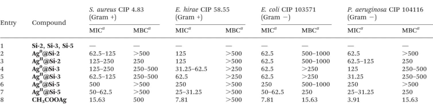
Antibacterial activity
The antibacterial activity of the silver-loaded nanoparticles was estimated by determining the Minimal Inhibitory Concentration (MIC) and the Minimal Bactericidal Concentration (MCB) on 4 typical bacterial strains34 (Table 3), two Gram-negative bacteria (E. coli CIP 103571 and P. aeruginosa CIP 1041161), and two Gram-positive bacteria (S. aureus CIP 4.83 and E. hirae CIP 58.55).35 All samples were carefully decanted and centrifuged prior to bactericidal evaluation. As expected in the absence of silver (entry 1), no antibacterial activity was observed. In all cases the silver-loaded silica NPs exhibited bacteriostatic activities against all bacterial strains in the 50–500 ppm range (silver equivalents).
Fig. 12 UV-vis absorption spectra. a: AgII@Si-X, b: AgII@Si-X after reduction at
day14, c: Ag0@Si-X, d: Ag0@Si-X after decantation. X = 2 (left), 3 (middle), 5
(right).
Fig. 13 HRTEM imaging of a Ag NP on Si-5 (left) and Fourier transform diffraction pattern (right).
Table 3 MIC and MBC determined on 4 typical bacterial strains
Entry Compound S. aureus CIP 4.83 (Gram +) E. hirae CIP 58.55 (Gram +) E. coli CIP 103571 (Gram 2) P. aeruginosa CIP 104116 (Gram 2)
MICa MBCa MICa MBCa MICa MBCa MICa MBCa
1 Si-2, Si-3, Si-5 — — — — — — — —
2 Ag0@Si-2 62.5–125 .500 125 .500 62.5 500–1000 62.5 .500 3 AgII@Si-2 125–250 250 125 .500 62.5 500–1000 62.5–125 250 4 Ag0@Si-3 125–250 250–500 31.25–62.5 .250 62.5 .250 125 250–500 5 AgII@Si-3 62.5–125 250–500 62.5 .250 62.5 .250 31.25 250–500 6 Ag0@Si-5 500 .500 250 .500 250 500–1000 250 .500 7 AgII@Si-5 50–62.5 .500 25–31.25 .500 50–62.5 250 25–31.25 250 8 CH3COOAg 15.63 500 7.81 .500 7.81 15.63 3.91 15.63
aIn ppm (silver equivalents). Experiments have been performed once or twice, each time in duplicates: when two values are given, the
Bactericidal effects have also been observed in some cases, but one should note that all activities were found to be lower than the ones measured for silver acetate. This finding can be related to the fact that in the case of colloidal silver, only a tiny portion of the overall metal content is potentially interacting with the microorganisms.
Interestingly, a small difference was observed between Gram-positive and Gram-negative strains for all materials and for silver acetate. In this regard, Gram-positive bacteria were found to be slightly less susceptible than Gram-negative bacteria which is in agreement with previous reports.36 The antimicrobial property of silver is due to the ability of its ionized form to cause damage to cells by interacting with thiol-containing proteins and DNA.37In this respect, the antibacter-ial activity of silver colloids is also related to their ability to release ionized silver. Nevertheless the antibacterial property of silver NPs is highly related to their size,38the smaller ones being more active.39This latter finding is related to the higher
percentage of surface atoms, which are prone to release silver ions, in small NPs. Despite the fact that we have observed by TEM and HRTEM that all silica-grafted silver NPs have quite large size distribution, between 1 and 20 nm, the size could be more critical than oxidation state. In the case of Si-3 system, Ag0 and AgII NPs have roughly the same average size, and AgII@Si-3 is slightly more active than Ag0@Si-3. In this case the difference of activity can putatively be related to ion release ability. In the case of Si-2 and Si-5 systems, AgIIcolloids are much smaller than Ag0colloids and significantly exhibit better
activity.
Conclusions
Our strategy leads to highly functional PEG-phosphonate-dendrimer coated silica NPs. The covalent hybrid interface has been fully described by means of routine spectroscopic techniques such as FT-IR and MAS NMR. This straightforward strategy is easily amenable to other functional surfaces and other dendrimeric species, and the versatility of dendrimer surface chemistry could open exciting perspectives for applications in the fields of nanomaterials, catalysis and drug-delivery. In order to illustrate these possibilities, silver nanoparticles have been grown on these dendrimer modified silicas in a very simple fashion, and the resulting materials were found to exhibit preserved antibacterial activities. We are currently investigating the versatility of this strategy with other supporting nanoparticles and other colloidal systems.
Experimental section
General procedures and instrumentation
All reactions were carried out in the absence of air using standard Schlenk techniques and vacuum-line manipulations when carried out in organic solvents. Commercial samples (Acros, Fluka, Aldrich) were used as received. Solvents were dried and distilled according to routine procedure before use. Commercial silica was activated by thermal treatment (250uC
for 24 h) to afford Si-1.1H,13C,31P NMR, HMQC and HMBC measurements were performed on Bruker AV 300, DPX 300, AV 400, Avance 400 WB (solid state) and Avance 500. Coupling constants are reported in Hz and chemical shifts in ppm/TMS for1H and13C. Chemical shifts for31P spectra are calibrated with phosphoric acid as an external reference. The first-order peak patterns are indicated as s (singlet), d (doublet), t (triplet), q (quadruplet), qn (quintuplet). Complex non-first-order signals are indicated as m (multiplet).13C NMR signals
were assigned using HMQC and HMBC sequences when required. UV-Vis spectra were recorded on a Perkin Elmer Lambda 35. FTIR spectra were recorded on a Perkin Elmer GX 2000. Spectra were normalized on signals that are not affected by the chemical modifications: 1046 cm21for Si-1 and Si-2 and 1500 cm21 for Si-3, Si-4 and Si-5. Mass spectroscopy was
performed on a Waters MALDI micro MX and Xevo Q Tof spectrometers. A small drop of colloidal solution was deposited on a grid to perform the TEM, HRTEM, STEM and EDX analyses. The grid was home-made from a commercial copper grid (diameter 3.05 mm) which was covered by a thin collodion membrane, on which carbon was evaporated (approximately 50 nm thickness). Low resolution TEM analyses were performed on a JEOL JEM 1011 (100 kV, resolution 4 Å). A wide angle Megaview III (SIS) camera was used for routine imaging. High resolution TEM analyses and EDX analyses were performed on the TEM-FEG (Field Emission Gun) JEOL JEM 2100F (200 kV, resolution 2.3 Å), Analyses X PGT (resolution 135 eV). Images were acquired with a CDD Gatan 2 K 6 2 K camera. Particle size was measured with UTHSCSA ImageTool software Version 3.0 Final. TGA measurements were recorded on a TGA7 Perkin-Elmer or Setaram 92.16.18 apparatus operating between 20 and 1500uC. Curves were recorded between 20 and 1000uC (20 uC min21)
using an Al2O3sample boat, and nitrogen as a vector (1 L h21).
Synthesis of the amino-PEG550
Synthesis of O-methanesulfonyl-O9-methylpolyethylene gly-col 550 (2b). Commercial O-methylpolyethylene glygly-col 550 2a (27.9 g, 48 mmol, 1 eq.) was dried by heating at 80uC overnight under vacuum. The material was then dissolved in dry dichloromethane (100 mL), and freshly distilled triethylamine (62.5 mmol, 8.7 mL, 1.3 eq.) was added. The mixture was cooled at 0uC and methanesulfonylchloride (62.5 mmol, 4.9 mL, 1.3 eq.) was added dropwise under stirring over 15 min. After addition of all the reagents, the mixture was stirred at the same temperature for an additional 15 min., then at room temperature overnight. The resulting heterogeneous solution was filtered and the filtrate concentrated to dryness. The oil was dissolved in dichloromethane (200 mL) and washed twice with water (30 mL). The combined aqueous layers were extracted 3 times with dichloromethane (100 mL). The combined organic layers were then dried over MgSO4, filtered
and the solvent was evaporated to obtain 26.5 g of compound 1 (yield 98%). 1H NMR (CDCl
3, 300.13 MHz): d = 3.08 (3H,
CH3SO3, s); 3.38 (3H, CH3O, s); 3.48–3.58, (2H, CH2OCH3, m);
3.59–3.71 (43H, CH2O, m); 3.72–3.82 (2H, CH2O, m); 4.23–4.55
(2H, CH2OSO2, m). 13C {1H} NMR (CDCl3, 75.47 MHz): d =
37.66 (CH3SO3); 58.95 (CH3O); 68.95 (CH2O); 69.31 (CH2OSO2);
dithranol, NaI): m/z range for [MNa+] from 397.2 (5 CH2CH2O
units) to 1019.6 (19 CH2CH2O units) separated by 44 Da
(CH2CH2O); the most intense peak was at 661.4 (11 CH2CH2O
units).
Synthesis of O-(2-ethylazido)-O9-methylpolyethylene glycol 550 (2c). 2b (14 g, 21.90 mmol, 1 eq.) was dissolved in N,N-dimethylformamide (50 mL) and sodium azide (5.7 g, 87.7 mmol, 4 eq.) was added. The schlenk was flushed with argon and the mixture stirred in a pre-warmed oil bath at 80uC. After 10 h the resulting mixture was diluted with water (20 mL) and stirred for a further 30 min. The mixture was extracted 6 times with dichloromethane (125 mL). The combined organic layers were then dried over MgSO4, filtered and the solvent was
evaporated. To perform the drying the oil was lyophilized to obtain 12.6 g of compound 2 (yield 98%).1H NMR (CDCl3,
400.13 MHz): d = 3.31–3.43 (5H, CH3O and CH2N3, m); 3.49–
3.58, (2H, CH2OCH3, m); 3.58–3.78 (47H, CH2O, m).13C {1H}
NMR (CDCl3, 75.47 MHz): d = 50.67 (CH2N3); 59.02 (CH3O);
70.02, 70.51, 70.56, 70.62, 70.66, 70.69 (CH2O); 71.92
(CH2OCH3). MS (Maldi TOF, dithranol, NaI): m/z range for
[MNa+] from 344.2 (5 CH2CH2O units) to1004.6 (20 CH2CH2O
units) separated by 44 Da (CH2CH2O); the most intense peak
was at 608.4 (11 CH2CH2O units).
Synthesis of O-(2-ethylamino)-O9-methylpolyethylene glycol 550 (2d). Azidoethyl-polyethyleneglycol mono methyl ether 2c (12.1 g, 20.66 mmol, 1 eq.) was dissolved in dry tetrahydrofur-ane (200 mL) and triphenylphosphine (8.13 g, 31 mmol, 1.5 eq.) was added. The homogeneous solution was stirred 4 days at room temperature, then hydrolysed with water (50 mL) and stirred for a further 5 h. The solution was evaporated at 50uC under 60 mBar to obtain a mixture of oil and white solid (triphenylphosphine oxide). The mixture was solubilised in water (200 mL) and filtrated to partially eliminate the phosphine oxide. The aqueous layer was extracted 5 times with benzene (50 mL). The aqueous layer was evaporated at 50 uC under 60 mBar and lyophilized to perform the drying. The procedure gave 10.9 g of (2d) as a light yellow oil (yield 94%).
1H NMR (CDCl 3, 300.13 MHz): d = 1.76 (2H, NH2, bs); 2.87 (2H, CH2NH2, t, 3JH–H = 5.1 Hz); 3.38 (3H, CH3O, s); 3.52 (2H, CH2CH2NH2, t,3JH–H= 5.1 Hz); 3.54–3.62, (2H, CH2OCH3, m); 3.62–3.82 (44H, CH2O, m).13C {1H} NMR (CDCl3, 100.62 MHz): d = 41.75 (CH2NH2); 59.02 (CH3O); 70.28, 70.51, 70.57, 70.60
(CH2O); 71.93 (CH2OCH3), 73.30 (CH2CH2NH2). MS
(pos-ESI-TOF): m/z range for [MH+] from 340.2 (6 CH2CH2O units) to
824.5 (17 CH2CH2O units) separated by 44 Da (CH2CH2O); the
most intense peak was at 560.4 (11 CH2CH2O units).
Synthesis of the silica-dendrimer core–shell particle
Si-2. A suspension of Si-1 (1.95 g) in absolute ethanol (90 mL) was added to 50 mL of a 2% weight solution of c-aminopropyltriethoxysilane in distillated water. The suspen-sion was heated at 85uC during one day. After cooling, the new silica was filtered, washed with ethanol (4 6 50 mL), dichloromethane (4 6 50 mL) and diethyl ether (4 6 50 mL), in that order. The resulting material was dried under vacuum at 100uC during 2 days to obtain 2.1 g of finely divided particle.13C CP-MAS NMR: d = 9.73 (CH2–Si); 26.88 (CH2–CH2–
CH2); 43.34 (CH2–NH2).29Si CP-MAS NMR: d = 2109.25 (Q4
site); 2100.22 (Q3site); 267.54 (T3site); 259.48 (T2site).
Si-3. To a solution of dendrimer 1-G1(215 mg, 75 mmol) in
140 mL of a mixture MeOH/THF (7 : 3, v/v) was added 300 mg of silica Si-2. The suspension was heated at 55uC overnight. After cooling, the silica was separated by filtration on a sintered glass filter, washed with THF (4 6 50 mL) and dichloromethane (4 6 25 mL) and dried under vacuum at 100 uC to obtain 350 mg of Si-3. The filtrate was evaporated and the unreacted dendrimer was precipitated to afford 153 mg of a white powder.31P CP-MAS NMR: d = 9.59 (P
0); 60.70 (P1).13C
CP-MAS NMR: d = 10.80 (CH2–Si); 23.39 (CH2–CH2–CH2); 32.88
(CH3–N); 43.34 (CH2–NH2); 50.7 (residual methanol); 62.63
(CH2–NL); 121.67 (CHar); 132.98 (CHar); 139.90 (HCLN–NMe);
151.76 (CHar); 160.30 (HCLN); 189.90 (CHO)29Si CP-MAS NMR:
d = 2109.81 (Q4 site); 2101.32 (Q3 site); 266.42 (T3 site);
259.04 (T2site).
Si-4. To a suspension of Si-3 (205 mg) in methanol (2 mL) was added a solution of 2d (35 mg; 62 mmol) in methanol (3 mL). After one day at room temperature the solvent was evaporated to dryness.31P CP-MAS NMR: d = 9.10 (P
0); 61.25
(P1).13C CP-MAS NMR: d = 10.38 (CH2–Si); 22.81 (CH2–CH2–
CH2); 32.80 (CH3–N); 42.77 (CH2–NH2); 50.7 (residual
metha-nol); 58.47 (CH3–O); 60.96 (CH2–NL); 70.56 (O–CH2–CH2–O);
121.72 (CHar); 130.07 (CHar); 133.09 (CHar); 139.31 (HCLN–
NMe); 151.97 (CHar); 161.09 (HCLN).29Si CP-MAS NMR: d =
2110.08 (Q4site); 2101.34 (Q3site); 265.94 (T3site); 256.67
(T2site).
Si-5. To 140 mg of silica Si-3 was added 1.5 mL of dimethylphosphite used as solvent and reactant. After one day at room temperature 20 mL of dichloromethane was added to the mixture. The silica was separated by filtration on a sintered glass filter, washed twice with 25 mL of EtOH/H2O
(7 : 3, v/v), ethanol (4 6 25 mL) and dichloromethane (4 6 25 mL). The resulting silica was dried under vacuum at 65 uC during 2 days to obtain 150 mg of Si-5.31P CP-MAS NMR: d = 8.99 (P0); 25.78 (PO3Me2); 63.37 (P1).13C CP-MAS NMR: d =
10.50 (CH2–Si); 22.19 (CH2–CH2–CH2); 33.11 (CH3–N); 42.73
(CH2–NH2); 52.91 (POCH3); 58.80 (CH3–O) 60.88 (CH–PO3Me2);
70.88 (O–CH2–CH2–O); 121.81 (CHar); 129.89 (CHar); 132.29
(CHar); 139.50 (HCLN–NMe); 151.19 (CHar).29Si CP-MAS NMR:
d = 2109.81 (Q4site); 2101.39 (Q3site); 266.42 (T3site).
Synthesis of Ag NPs on dendrimer-modified SiO2NPs
To a suspension of 50 mg of grafted silica (Si-2, Si-3 or Si-5) in milli-Q water (2.5 mL) was added 2.5 mL of a solution of silver acetate in milli-Q water (1 g L21) corresponding to 15 mmol. The resulting mixture was left under stirring for one hour and then separated in two aliquots. One aliquot was left under stirring for two additional weeks (AgII@Si-X samples). The other aliquot was reacted with 600 mL of a solution of sodium borohydride in milli-Q water (2 g L21) corresponding to 16 mmol and left under stirring for 18 additional hours (Ag0@Si-X samples). Electron microscopy samples were prepared by deposition of a small drop of crude suspensions onto carbon coated grids. After the appropriate time of reaction the different samples of silica were purified by 10 successive runs of centrifugation (2 min at 5000 rpm) and washing with milli-Q water (7 ml). Then the silica samples were freeze-dried.
Bacterial strains and culture conditions
The 4 tested strains belong to 4 potent pathogenic species obtained from the Institute Pasteur Collection (Paris, France): Staphylococcus aureus CIP 4.83, Escherichia coli CIP 103571, Enterococcus hirae CIP 58.55, Pseudomonas aeruginosa CIP 104116. The strains were stored at 280 uC in Eugon broth (AES, Rennes, France) with 20% (v/v) glycerol (Fluka, Butch, Switzerland) and reactivated at 37uC under aerobic conditions on trypcase soy agar medium agar plates (Biome´rieux, Craponne, France).
Antibacterial activities, MIC and MBC determination
The tested compounds were dissolved in sterile distilled water to obtain an initial concentration of 1000 ppm (silver equivalents) for the tested samples. Owing to compounds availability the initial concentrations were 500 ppm and 100 ppm respectively for AgII@Si-3 and AgII@Si-5. Samples without silver were diluted to obtain the same concentration of SiO2as
for the samples with silver. 100 mL of the resulting solutions were then diluted in the first wells of microtiter plates in 100 mL trypcase soy broth (Biome´rieux, Craponne, France). Twofold serial dilutions were then performed from well 1 to well 10. Bacteria were grown overnight in trypcase soy broth at 37 uC until reaching stationary phase culture. Bacterial suspensions were then washed by centrifugation (3500 rpm, 5 min) and optical density adjusted in sterile distilled water to obtain 108 bacteria/mL and the microplate was then inocu-lated to obtain a final concentration of 106 bacteria/ml (Denley multipoint inoculator). After 24 h of incubation at 37uC, the MICs were defined as the lowest concentrations with no visible growth. Rows 11 and 12 were used for growth negative control and positive control, respectively. The Minimal Bactericidal Concentrations (MBC) were defined as the concentrations that cause bacterial death (>99% of the population) and were checked by lack of macroscopic sign of cellular growth after subcultivating the MICs cultures on trypcase soy agar plates for 24 h at 37uC. All experiments were carried out in duplicate at each concentration.
Acknowledgements
We acknowledge financial support from the CNRS. This work has been partially supported by the French National Agency (ANR; COPPERTREE, project Nu ANR-11-BS07-018).
Notes and references
1 A. M. Caminade, C. O. Turrin, R. Laurent, A. Ouali and B. Delavaux-Nicot, Dendrimers. Towards catalytic, material and biomedical uses., Wiley & Sons Ltd, Chichester, 2011. 2 G. Schmid, in Nanoparticles: From theory to applications
(second edition), ed. G. Schmid, Wiley VCH, Weinheim, 2010.
3 R. W. J. Scott, O. M. Wilson and R. M. Crooks, J. Phys. Chem. B, 2005, 109, 692–704.
4 M. Q. Zhao, L. Sun and R. M. Crooks, J. Am. Chem. Soc., 1998, 120, 4877–4878.
5 A. El Kadib, N. Katir, M. Bousmina and J. P. Majoral, New J. Chem., 2012, 36, 241–255.
6 C. O. Turrin, V. Maraval, A. M. Caminade, J. P. Majoral, A. Mehdi and C. Reye, Chem. Mater., 2000, 12, 3848–3856. 7 N. Tsubokawa, H. Ichioka, T. Satoh, S. Hayashi and
K. Fujiki, React. Funct. Polym., 1998, 37, 75–82.
8 K. Sakai, T. C. Teng, A. Katada, T. Harada, K. Yoshida, K. Yamanaka, Y. Asami, M. Sakata, C. Hirayama and M. Kunitake, Chem. Mater., 2003, 15, 4091–4097.
9 J. P. K. Reynhardt and H. Alper, J. Org. Chem., 2003, 68, 8353–8360.
10 S. Antebi, P. Arya, L. E. Manzer and H. Alper, J. Org. Chem., 2002, 67, 6623–6631.
11 G. Larsen and S. Noriega, Appl. Catal., A, 2004, 278, 73–81. 12 D. R. Radu, C. Y. Lai, K. Jeftinija, E. W. Rowe, S. Jeftinija and V. S. Y. Lin, J. Am. Chem. Soc., 2004, 126, 13216–13217. 13 A. V. Biradar, A. A. Biradar and T. Asefa, Langmuir, 2011, 27,
14408–14418.
14 S. P. Ramnani, S. Sabharwal, J. V. Kumar, K. H. P. Reddy, K. S. R. Rao and P. S. S. Prasad, Catal. Commun., 2008, 9, 756–761.
15 Q. D. Viet, P. B. Sarawade, A. Hilonga, J.-K. Kim, Y. G. Chai, S. H. Kim, J.-Y. Ryu and H. T. Kim, Appl. Surf. Sci., 2011, 257, 6963–6970.
16 J.-S. Hwang, K.-Y. Chen, S.-J. Hong, S.-W. Chen, W.-S. Syu, C.-W. Kuo, C.-W.-Y. Syu, T. Y. Lin, H.-P. Chiang, S. Chattopadhyay, K.-H. Chen and L.-C. Chen, Nanotechnology, 2010, 21, 025502.
17 M. A. Villegas, M. A. Garcia, S. E. Paje and J. Llopis, Mater. Res. Bull., 2005, 40, 1210–1222.
18 Y. W. Zhang, H. S. Peng, W. Huang, Y. F. Zhou and D. Y. Yan, J. Colloid Interface Sci., 2008, 325, 371–376. 19 N. Launay, A. M. Caminade, R. Lahana and J. P. Majoral,
Angew. Chem., Int. Ed. Engl., 1994, 33, 1589–1592.
20 E. Trevisiol, V. Le Berre-Anton, J. Leclaire, G. Pratviel, A. M. Caminade, J. P. Majoral, J. M. Francois and B. Meunier, New J. Chem., 2003, 27, 1713–1719.
21 B. Miksa, S. Slomkowski, M. M. Chehimi, M. Delamar, J. P. Majoral and A. M. Caminade, Colloid Polym. Sci., 1999, 277, 58–65.
22 M. Avella, F. Bondioli, V. Cannillo, E. D. Pace, M. E. Errico, A. M. Ferrari, B. Focher and M. Malinconico, Compos. Sci. Technol., 2006, 886–894.
23 N. Mejı´as, R. Pleixats, A. Shafir, M. Medio-Simo´n and G. Asensio, Eur. J. Org. Chem., 2010, 5090–5099.
24 T. Akiyama, M. Sanada and K. Fuchibe, Synlett, 2003, 1463–1464.
25 A. A. Sobanov, A. V. Zolotukhin, V. I. Galkin, O. A. Mostovaya, R. A. Cherkasov and A. N. Pudovik, Russ. J. Gen. Chem., 2003, 73, 871–876.
26 R. A. Cherkasov and V. I. Galkin, Russ. Chem. Rev., 1998, 67, 857–882.
27 L. Griffe, M. Poupot, P. Marchand, A. Maraval, C. O. Turrin, O. Rolland, P. Me´tivier, G. Bacquet, J. J. Fournie´, A. M. Caminade, R. Poupot and J. P. Majoral, Angew. Chem., Int. Ed., 2007, 46, 2523–2526.
28 A. Hameau, C.-O. Turrin and A.-M. Caminade, unpublished results.
29 G. S. Caravajal, D. E. Leyden, G. R. Quinting and G. E. Maciel, Anal. Chem., 1988, 60, 1776–1786.
30 C. H. Chiang, N. I. Liu and J. L. Koenig, J. Colloid Interface Sci., 1982, 86, 26–34.
31 G. Schmid, E. Emmrich, J. P. Majoral and A. M. Caminade, Small, 2005, 1, 73–75.
32 C. O. Turrin, V. Maraval, J. Leclaire, E. Dantras, C. Lacabanne, A. M. Caminade and J. P. Majoral, Tetrahedron, 2003, 59, 3965–3973.
33 H. Xu, F. Yan, E. E. Monson and R. Kopelman, J. Biomed. Mater. Res., 2003, 66A, 870–879.
34 H. Ibrahim, A. Furiga, E. Najahi, C. P. Henocq, J. P. Nallet, C. Roques, A. Aubouy, M. Sauvain, P. Constant, M. Daffe and F. Nepveu, J. Antibiot., 2012, 65, 499–504.
35 J. Aze´ma, B. Guidetti, A. Korolyov, R. Kiss, C. Roques, P. Constant, M. Daffe´ and M. Malet-Martino, Eur. J. Med. Chem., 2011, 46, 6025–6038.
36 K. Kawahara, K. Tsuruda, M. Morishita and M. Uchida, Dent. Mater., 2000, 16, 452–455.
37 Q. L. Feng, J. Wu, G. Q. Chen, F. Z. Cui, T. N. Kim and J. O. Kim, J. Biomed. Mater. Res., 2000, 52, 662–668.
38 V. Dal Lago, L. F. de Oliveira, K. D. Goncalves, J. Kobarg and M. B. Cardoso, J. Mater. Chem., 2011, 21, 12267–12273. 39 J. R. Morones, J. L. Elechiguerra, A. Camacho, K. Holt, J. B. Kouri, J. T. Ramirez and M. J. Yacaman, Nanotechnology, 2005, 16, 2346–2353.