HAL Id: inserm-00667533
https://www.hal.inserm.fr/inserm-00667533
Submitted on 7 Feb 2012HAL is a multi-disciplinary open access archive for the deposit and dissemination of sci-entific research documents, whether they are pub-lished or not. The documents may come from teaching and research institutions in France or abroad, or from public or private research centers.
L’archive ouverte pluridisciplinaire HAL, est destinée au dépôt et à la diffusion de documents scientifiques de niveau recherche, publiés ou non, émanant des établissements d’enseignement et de recherche français ou étrangers, des laboratoires publics ou privés.
Glycosaminoglycans inhibit the adherence and the
spreading of osteoclasts and their precursors: role in
osteoclastogenesis and bone resorption.
Marc Baud’Huin, Carmen Ruiz-Velasco, Gaëtan Jego, Céline Charrier, Nijole
Gasiunas, John Gallagher, Mike Maillasson, Annamaria Naggi, Marc
Padrines, Françoise Redini, et al.
To cite this version:
Marc Baud’Huin, Carmen Ruiz-Velasco, Gaëtan Jego, Céline Charrier, Nijole Gasiunas, et al.. Gly-cosaminoglycans inhibit the adherence and the spreading of osteoclasts and their precursors: role in osteoclastogenesis and bone resorption.: glycosaminoglycans and osteoclastogenesis. Eur J Cell Biol, 2011, 90 (1), pp.49-57. �10.1016/j.ejcb.2010.08.001�. �inserm-00667533�
Glycosaminoglycans inhibit the adherence and the spreading of osteoclasts and their precursors: role in osteoclastogenesis and bone resorption
Marc BAUD’HUIN1,2, Carmen RUIZ-VELASCO1,2, Gaëtan JEGO3,Céline CHARRIER1,2,
Nijole GASIUNAS4, John GALLAGHER4, Mike MAILLASSON5, Annamaria NAGGI6,
Marc PADRINES1,2, Françoise REDINI1,2, Laurence DUPLOMB1,2,7 and Dominique
HEYMANN1,2,7
1
INSERM, UMR-S 957, Nantes, F-44035 France
2
Université de Nantes, Nantes atlantique universités, Laboratoire de Physiopathologie de la Résorption Osseuse et Thérapie des Tumeurs Osseuses Primitives, EA3822, Nantes, F-44035, France
3
INSERM U892, Nantes, F-44093 France
4
Paterson Institute for Cancer Research, University of Manchester, England M20 4BX
5
INSERM U892 and IFR26- Ouest genopole, F-44035 France
6
Institute for Chemical and Biochemical Research G. Ronzoni, Milan, Italy
7
CHU, Hôtel Dieu, Nantes, France
Running title: glycosaminoglycans and osteoclastogenesis Correspondence and reprint request to
Pr. D. HEYMANN
dominique.heymann@univ-nantes.fr
INSERM UMR-S 957, Physiopathologie de la Résorption Osseuse et Thérapie des Tumeurs Osseuses Primitives
1 rue Gaston Veil 44035 Nantes Cedex 1 Fax: (011) 33 2 40 41 28 60
Abstract
The bone microenvironment (e.g. glycosaminoglycans (GAGs), growth factors) plays a major
role in bone resoption, especially in the formation of osteoclast which differentiate from the
hematopoietic lineage in the presence of RANKL. Previous studies revealed that GAGs may influence osteoclastogenesis, but data are very controversial, some studies showing an inhibitory effect of GAGs on osteoclastic differentiation whereas others demonstrated a stimulatory effect. To clarify their activities, we investigated the effect of 5 families of GAGs in three different models of human/mouse osteoclastogenesis. The present data revealed that heparin inhibited osteoclastogenesis in these 3 models, which was confirmed by a decrease in mRNA expression of osteoclastic markers and by an inhibition of the bone resorption
capacity. We also demonstrated in RAW 264.7 cells that other families of GAGs different
from heparin inhibited RANKL-induced osteoclastogenesis, and that this inhibition was
dependent on the length and the level of sulfation of GAGs. In the present work, heparin did
not bind to RANKL and did not modulate RANKL signaling. Heparin acted at 2 distinct steps
of osteoclastogenesis from human CD14+ cells: first, heparin strongly decreased the
adherence of osteoclast precursors, and secondly inhibited osteoclasts to spread and to be
active. Furthermore, the second action of heparin was reversible as the removal of heparin at the end of the culture time allowed the condensed cells to spread out and showed the formation of morphological active osteoclasts. The present work clearly evidences that GAGs inhibit osteoclastogenesis in vitro and strengthens the therapeutic interest of defined GAGs in osteolytic diseases.
Key Words: osteoclasts, oligosaccharides, RANKL, cell adherence, bone metabolism, bone remodelling
Introduction
Bone metabolism is regulated by a functional balance between catabolic and anabolic activities of bone cells. Thus, osteoclasts are multinucleated cells specialized in bone
catabolism which participate to phosphocalcic homeostasis together with cells playing
anabolic functions named osteoblasts. Osteoclasts originate from the monocyte/macrophage lineage through a series of events associating membranous, soluble and extracellular matrix compounds (Bruzzaniti et al, 2006). Among these factors, some are required for proliferation and differentiation of osteoclast progenitors such as macrophage-colony stimulating factor (M-CSF) (Biskobing et al, 1995; Felix et al, 1990; Witkor-Jedrzejcak et al, 1990) while other factors such as receptor activator of nuclear factor kB ligand (RANKL) are more specifically involved in the commitment of mononuclear precursors to the fusion and formation of multinucleated resorbing osteoclasts (Baud’huin et al, 2007). In this system, RANKL expressed by osteoblasts and stromal cells binds to its receptor RANK expressed at the surface of osteoclast precursors and consequently activates specific signal pathways leading to the formation, maturation and survival of osteoclasts (Boyle et al, 2003; Wittrant et al, 2004). Furthermore, the RANKL/RANK activities are controlled by osteoprotegerin (OPG) which acts as a soluble decoy receptor blocking the binding of RANKL to RANK, subsequently reducing osteoclastogenesis and bone resorption (Simonet et al, 1997; Yasuda et al, 1998).
Extracellular matrix components such as glycosaminoglycans (GAGs) also participate to bone metabolism (Lamoureux et al, 2007). GAGs are linear polymers which are bound to a core protein to form proteoglycans (Lamoureux et al, 2007). GAGs are composed of repeated
disaccharidic units of hexosamine and hexuronic acid, except for keratan sulfate in which
hexuronic acid is replaced by galactose. According to the epimerization of hexosamine and uronic acid, several families of GAGs have been established. Proteoglycans and GAGs
contribute to the maintenance of bone mass through their involvement in collagen organization (Corsi et al, 2002). They could exert several activities on bone cells as co-factors in cell-to-cell adhesion, or to modulate the binding and activation of several growth factors or receptors such as OPG by syndecan-1 (Bernfield et al., 1999; Hildebrand et al., 1994; Robinson et al., 2005; Mosheimer et al., 2005; Standal et al., 2002). Unfortunately, the data available on the effects of GAGs on osteoclastogenesis are very limited and controversial. For example, Ariyoshi et al. (2005) showed that hyaluronic acid, the most abundant GAG in mammalian tissues, enhances osteoclast formation and function, whereas in 2007, Chang et al. described an opposite effect on osteoclastogenesis. Irie et al. (2007) recently demonstrated
that heparin in combination with 1,25(OH)2D3/PGE2 enhances the pit-forming activity of
osteoclasts obtained from the coculture of mouse osteoblasts with bone marrow cells. However, they did not observe any direct effect of heparin on osteoclastogenesis. On the other hand, Shinmyouzu et al. (2007), demonstrated that high concentrations of dermatan sulfate inhibit osteoclastogenesis, and the same group showed similar activities with heparin (Ariyoshi et al, 2008). The authors suggested that GAGs act through an inhibition of RANKL signaling (inhibition of p38 and ERK phosphorylation following RANKL stimulation) to achieve their inhibitory effect on osteoclastogenesis. Moreover, these controversial findings on GAG effects on osteoclastogenesis are strengthened by the study of Folwarczna et al. (2005) who pointed out species differences in the sensitivity of bone marrow cells to standard
and low-molecular weight heparins. For instance, in a rat model, low concentrations of
heparin increased the formation of osteoclasts, whereas it decreased with the highest
concentrations. In mouse bone marrow cell cultures, heparin suppressed the formation of osteoclasts, with the exception of low concentrations of standard heparin which intensified this process (Folwarczna et al, 2005).
In this high controversial context, the aim of our study was to clarify the direct effect of GAGs on osteoclastogenesis using three different cellular models: murine RAW 264.7
monocytic cell line, murine purified CD11b+ cells and human purified CD14+ monocytes.
These models are characterized by the absence of osteoblastic/stromal cells, allowing us to investigate the direct effect of GAGs on osteoclast precursors. GAGs from various origins (bovine and porcine heparins with different sulfation levels, heparan-, chondroitin- and dermatan-sulfate, hyaluronic acid and oligosaccharides of different lengths) were assessed in in vitro osteoclastogenesis models.
Materials and methods
Materials
Human M-CSF (hM-CSF), mouse M-CSF (mM-CSF) and human OPG (hOPG) were obtained from R&D Systems (Abington, UK). Human RANKL (hRANKL) was kindly provided by Amgen Inc. (Thousand Oaks, USA). Heparin sodium salt, heparan sulfate from bovine kidney (bHS), heparan sulfate from porcine intestinal mucosa (pHS), chondroitin sulfate from shark cartilage (CS), dermatan sulfate from porcine intestinal mucosa (DS) and hyaluronic acid were purchased from Sigma (St Quentin Fallavier, France). Heparin-derived oligosaccharides of defined size were prepared by digestion of porcine mucosal heparin with heparinase I followed by gel filtration chromatography on a Bio-Gel P-10 column [24].
Heparin initially contained 97.7% of N-sulfate groups, 89.3% of 2-O-sulfate groups, and
92.4% of 6-O-sulfate groups. De-N-sulfated/re-N-acetylated heparin contained 90.5% of
2-O-sulfate groups, 85.3% of6-O-sulfates, and a very low amount of remaining N-sulfategroups
(2.4%). De-2-O-sulfated heparin contained 80.2% of 6-O-sulfate groups, 91.4% of N-sulfate groups, and a residual 2.2% of the 2-O-sulfates. De-6-O-sulfated heparin contained 98.2% of
N-sulfate groups, 54.7% of 2-O-sulfate groups,and a residual 4.2% of 6-O-sulfates (Lyon et
al, 2000).
Osteoclastogenesis assays
Differentiation from the murine RAW 264.7 monocytic cell line
Murine RAW 264.7 monocytic cells (ATCC, Promochem, Molsheim, France) were
cultured in phenol red-free α-Minimal Essential Medium (α-MEM) (Invitrogen, Eragny,
France) supplemented with 10% fetal calf serum (FCS) (Perbio, Logan, USA), 1% non essential amino acids (Invitrogen). To induce osteoclast formation, RAW 264.7 cells were
scrapped then incubated at 37°C for 2 minutes to allow adherence of the more differentiated
cells. Non adherent cells were then seeded in fresh medium, at a density of 3 x 10³ cells/well
in a 96-well plate. After 2 hours of culture, the medium was changed for a fresh one containing 100 ng/ml hRANKL and various forms of glycosaminoglycans at different concentrations (see result section and figure legends). Multinucleated cells (>3 nuclei) were counted under a light microscope (Leica DM IRB, Nanterre, France; Camera: Olympus D70, Analysis software: Olympus DP Controller/Manager, Hamburg, Germany) after TRAP staining (Sigma, Saint Quentin-Fallavier, France).
Differentiation from murine CD11b+ monocytes
CD11b+ monocytes were purified from murine bone marrow cells, obtained by
flushing the bone marrow from femora and tibiae of 4 week-old C57BL6 male mice. CD11b+
cells were magnetically labelled with CD11b Microbeads and positively selected by MACS
technology (Miltenyi Biotec, Bergisch Gladbach, Germany). CD11b+ cells were seeded in
24-well plates (500 x 103 cells / well) in α-MEM without phenol red, containing 10% FCS and 25
ng/ml mM-CSF. After 3 days of culture, medium was replaced by fresh medium containing
10% FCS, 25 ng/ml mM-CSF, with or without 100 ng/ml hRANKL, and with or without 5 µM heparin. Thereafter, medium was changed every 4 days. The formation of osteoclasts occurred after around 15 days of culture and was observed by TRAP staining.
Osteoclastogenesis and dendritic cell formation from purified human CD14+ cells
Human peripheral blood mononuclear cells (PBMCs) were isolated by centrifugation
over Ficoll gradient (Sigma). CD14+ cells were magnetically labeled with CD14 Microbeads
and positively selected by MACS technology. For osteoclast differentiation, CD14+ cells were
α-MEM supplemented with 10% FCS and 25 ng/ml hM-CSF (Duplomb et al, 2008). At day 3 of the culture, medium was changed for fresh medium containing 10% FCS, 25 ng/ml hM-CSF and 100 ng/ml hRANKL, with or without heparin (5 µM). Then medium was changed every 4 days. The formation of osteoclasts occurred after around 15 days and was observed by TRAP
staining. In some experiments, heparin was added at different time points of the culture
period, as indicated. To test the capacity of osteoclasts to resorb bone, CD14+ cells were
cultured on dentine slices in the conditions previously described. At the end of the culture period, osteoclasts were removed by bleach; dentin slices were fixed with 4% glutaraldehyde in 0.2 M sodium cacodylate solution for 30 minutes, followed by staining with 1% toluidine blue in 0.5% sodium tetraborate solution for 3 minutes (Chu et al, 2006). Resorption lacunae were identified by light stereomicroscopy (Zeiss, STEMI 2000-C, Göttingen, Germany) and resorbed surfaces were measured using QWin software (Leica, France). To determine the effect of heparin of mature osteoclasts, we used a technique established by Fuller et al (2006).
Briefly, after formation of osteoclasts as described above, the medium was removed and the
cell layer washed three times with PBS without calcium and magnesium. Six hundred
microliters of 0.02% EDTA were added per well (6-well plate) and cells were incubated for
20 min at room temperature.EDTA was then removed from the dish and replaced with 600 µl of calcium/magnesium-free PBS. A cell scraper was used to scrape the cells in PBS, and the
resulting cell suspension was mixed using a pipette to ensure uniform cell dispersal. Two
hundred and fifty microliters of this cell suspension were added to each well (24-well plate)
on a dentin slice in 250 µl αMEM, 10% FCS. Cells were allowed to sediment for 20 min at
37°C before dentin slices were washed. Cells were incubated in 300 µl αMEM, 10% FCS in
the presence or the absence of heparin. After incubation, bone resorption was assessed as described above.
Adherence of CD14+ cells was analyzed by counting the adherent cells after 3 days of culture in the presence of hM-CSF (25 ng/ml) with or without heparin (5 µM). Briefly, cells were washed 3 times with PBS (Lonza, Verviers, Belgium) and adherent cells were detached with trypsin solution (Lonza); cells were counted using trypan blue exclusion.
Dendritic differentiation was obtained upon stimulation with 5 ng/ml hIL-4 (Invitrogen)
+ 100 ng/ml human GM-CSF (kindly provided by UTCG, CHU Nantes) (Sallusto and
Lanzavecchia, 1994). Briefly, 1 x 10⁶ CD14+ cells or PBMCs were cultured in 6-well plates in
3 ml of RPMI 1640 (Lonza) supplemented with 10% FCS, in the presence or the absence of
glycosaminoglycans. Medium was replaced after 3 days of culture. After 2 more days, cells
were harvested and double stained for 15 min at 4°C with antibodies against CD1a-APC
(Becton Dickinson, Le Pont de Claix, France) and CD14-PE (Immunotech, Marseille, France)
in PBS and then washed and fixed in PBS containing 1% formaldehyde. Irrelevant
isotype-matched antibodies were used to determine levels of nonspecific binding. Flow cytometry
analysis was carried out on a FACScan using the CELLQuest software (both from Becton
Dickinson).
Surface plasmon resonance-binding assay
Experiments were carried out on a BIAcore 3000 instrument (Biacore, Sweden).
RANKL (2 µg/mL) in 5 mM maleate, pH 5.75 was covalently immobilized to the dextran matrix
of a CM5 sensor chip (BIAcore) at a flow rate of 5 µl/min. Immobilization levels in the range of? 4000 RU were obtained. Binding assays were performed at 25°C in 10 mM Hepes buffer, pH 7.4, containing 0.15 M NaCl and 0.005% P20 surfactant (HBS-P buffer, BIAcore) at a flow rate of 30 µl/min for heparin (1 to 20 nM) and 20 µl/min for hOPG (25nM). The resulting sensorgrams were fitted using BiaEval 4.1 software (Biacore).
Western Blot Analysis
After 5 hours of culture in serum-free medium, undifferentiated CD14+ cells were
stimulated with 100 ng/ml of hRANKL for 15 minutes at 37°C in the presence or absence of 125 µg/ml heparin. Cell lysates were obtained and protein concentrations were determined as described previously (Duplomb et al., 2008). Proteins were run on 10% SDS-PAGE gels and transferred to Immobilon-P membranes (Millipore, USA) which were then incubated with
antibodies to Phospho-ERK1/2, Phospho-p38, Phospho-p105, Total-ERK1/2, Total-p38 and
Total-p105 (Cell Signaling Technologies, USA). Bands were visualized using ECL reagent
(Roche, Germany).
Statistical analysis
Each experiment was repeated in triplicate three times independently. The mean + SD was calculated for all conditions and compared by ANOVA. Differences relative to a probability of two-tailed p < 0.05 were considered significant.
Results
Glycosaminoglycans inhibit RANKL-induced osteoclastogenesis in murine and human models
RAW 264.7 cells are murine monocyte/macrophage cells which can differentiate into TRAP-positive multinucleated cells in 5 days upon RANKL stimulation. As shown in figures 1A and 1B, addition of heparin inhibited RANKL-induced osteoclastogenesis by 83% (p<0.01). This result was confirmed by the analysis of osteoclastic markers using real-time PCR. Indeed, after 5 days of culture, mRNA expression of osteoclastic markers such as TRAP and Cathepsin K was strongly increased in the presence of RANKL, while addition of heparin diminished the expression of these markers by around 50% (Figure 1C). These results were
confirmed in a second model of murine osteoclastogenesis involving CD11b+ monocytes
purified from bone marrow and cultured in the presence of RANKL and heparin. As shown in
figure 1D, heparin totally inhibited RANKL-induced osteoclastogenesis of CD11b+ cells.
Similar effects were observed in a human model of osteoclastogenesis, using CD14+ purified
monocytes. As shown in figures 2A and 2B, RANKL stimulation of CD14+ cells induced
their differentiation into TRAP+ multinucleated cells. Again, the addition of heparin strongly
inhibited RANKL-induced osteoclastogenesis (76%, p<0.01). Heparin had no effect on the
proliferation of CD14+ cells cultured in the presence or absence of M-CSF for 3 or 6 days
(aditional data 1). Furthermore, when cultured on dentine slices, TRAP+ osteoclasts showed a
strong capacity of resorption (Figure 2C) which was significantly reduced in the presence of
heparin (Figure 2D, p<0.05). Similar experiments were performed using mature osteoclasts
isolated from differentiated RANKL-CD14+ cell cultures, and in this context heparin had no
To investigate the effect of other GAG families on RANKL-induced osteoclastogenesis, heparan sulfate (bovine and porcine origin), chondroitin sulfate and
dermatan sulfate at a concentration of 5 µM were added to the culture of RAW 264.7 cells.
After 5 days, all the GAGs tested inhibited RANKL-induced osteoclastogenesis between 65 and 80% (Figure 3A) while GAGs alone had no effect on the RAW 264.7 cell line (at the proliferation or apoptosis levels) and did not reveal any cell toxicity (data not shown).
Furthermore, addition of heparin-derived oligosaccharides [4, 14 and 24 (corresponding respectively to 4, 14 and 24 disaccharide-unit length)] to the culture inhibited RANKL-induced osteoclastogenesis in a dose- and size-dependent manner (Figure 3B). Indeed, 1.56 µM of oligosaccharide 4 inhibited osteoclastogenesis by 6%, whereas at the same concentration oligosaccharides 14 and 24 inhibited osteoclastogenesis by respectively 24% and 53%. In the same way, inhibition of RANKL-induced osteoclastogenesis using 12.5 µM oligosaccharide was around 17% with oligosaccharide 4, 44% with oligosaccharide 14, and 72% with oligosaccharide 24. At 100 µM, osteoclastogenesis was almost totally abolished with all oligosaccharides, whatever their size. These oligosaccharides had no effect on RAW 264.7 cells cultured in medium without RANKL. We then analyzed the effect of hyaluronic acid which is a huge non-sulfated molecule. As shown in figure 3C, hyaluronic acid also inhibited osteoclastogenesis in a dose dependent manner.
Sulfation of oligosaccharides is a key parameter for the inhibition of RANKL-induced osteoclastogenesis
To decipher the mechanisms involved in the inhibition of osteoclastogenesis observed in the presence of oligosaccharides and GAGs, we analyzed the importance of quantitative and qualitative sulfation by comparing the effects of normal standard heparin with several
heparin, completely desulfated heparin, de-2O-sulfated heparin, de-6O-sulfated heparin, and de-N-sulfated heparin. As shown in figure 4, RANKL-induced osteoclastogenesis was inhibited by normal heparin (83% of inhibition, p<0.01), by de-N-sulfated re-N-acetylated and de-N-sulfated heparins (around 50% of inhibition), and by de-2O- and de-6O-sulfated
heparins (around 75% of inhibition, p<0.01). However, totally desulfated heparin poorly
inhibited RANKL-induced osteoclastogenesis (around 14%; p<0.05).
GAGs induce differentiation of human monocytes into dendritic cells
To determine whether the inhibitory activity of GAGs was specific of osteoclastogenesis, the effects of heparin-derived oligosaccharide 16 (32-mer), dermatan sulfate and heparin were investigated during the differentiation process of human monocytes
into dendritic cells (non adherent cells) using two different approaches: isolated CD14+
monocytes (Figure 5A) or monocytes obtained after 2h of adherence of total PBMCs (Figure 5B). As shown in figure 5, oligosaccharide 16, dermatan sulfate and heparin potentiated the effect of the GM-CSF/IL-4 cocktail to induce dendritic cell differentiation from both isolated
CD14+ cells and total PBMCs. For example in the model of CD14+ monocytes, GM-CSF/IL-4
induced around 20% of CD14- / CD1a+ dendritic cells after 5 days of culture, whereas
oligosaccharide 16 or heparin addition significantly enhanced this differentiation process by 10 to 20% (Figure 5A). The same effect was observed using total PBMCs: oligosaccharide 16, dermatan sulfate and heparin significantly increased the dendritic cell differentiation with a mean average of about 12.5% (Figure 5B) whereas these GAGs/oligosaccharides have no effect alone. Thus the inhibitory effect of GAGs observed on osteoclast differentiation is specific to this commitment because no inhibition could be shown with other differentiation systems such as dendritic cell differentiation.
Heparin does not bind to RANKL and does not modulate RANKL signaling
Shinmyouzu et al. demonstrated that dermatan sulfate inhibits RANKL-induced osteoclastogenesis in a mouse model of osteoclast differentiation from bone marrow (Shinmyouzu et al., 2007). They showed that this inhibition occurred through the binding of dermatan sulfate to RANKL leading to the inhibition of RANKL interaction to its receptor
RANK, and thus to the inhibition of RANKL signaling. Based on these observations, the
authors suggested that the same phenomenon could be involved in the inhibition of RANKL-induced osteoclastogenesis which they also observed with heparin or chondroitin sulfate E. In the present work, surface plasmon resonance experiments demonstrated that heparin did not bind to RANKL (Figure 6A). Similarly, chondrotin sulphate, dermatan sulphate, heparin sulphate and oligosaccharides did not bind immobilized RANKL in contrast to immobilized OPG (Théoleyre et al, 2006; Lamoureux et al, 2009). However, soluble OPG and soluble RANK bound to immobilized RANKL with high affinity (data not shown). In agreement with
these data, heparin did not inhibit RANKL signaling in RAW 264.7 cells nor in CD14+ human
monocytes (Figure 6B).
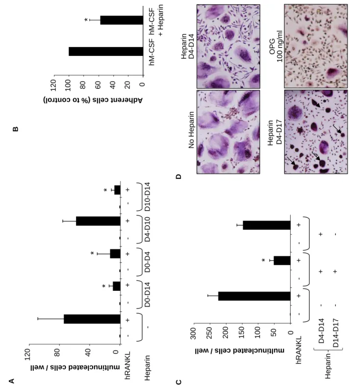
Heparin inhibits the adherence and spreading of osteoclasts and thus their functionality
To better characterize the mechanisms by which heparin inhibits osteoclast formation,
especially in the early adherence and spreading phases, the effect of heparin was assessed at
different times during the culture period. When heparin was added during the first 4 days
(D0-D4) of culture in the presence of M-CSF only very few osteoclasts were generated after 14
days (Figure 7A). Moreover, when heparin was added during the first 4 days, it inhibited by
around 40% the number of adherent CD14+ osteoclast precursors compared to the control
condition (Figure 7B). Thus in this culture condition, fewer osteoclast precursors adhered to the plastic surface and less osteoclasts were generated in the presence of RANKL at D14. The
addition of heparin during intermediate culture step (D4-D10) did not inhibit osteoclast formation, and the number of osteoclasts formed was similar to the control group in the presence of RANKL (Figure 7A). In contrast, when heparin was added during the fusion step of osteoclastogenesis (D10-D14), heparin inhibited significantly the number of osteoclast formed (Figure 7A).
In the presence of heparin and RANKL, CD14+ cells, as well as RAW 264.7 cells,
developed two different morphological shapes: large multinucleated TRAP+ cells and
condensed cells (see arrows on Figures 1A and 2A). As this latter group of cells were TRAP+
and seemed multinucleated, we suggested that heparin inhibited the spreading of RANKL-generated osteoclasts. Such modifications of cell morphology were not observed in the presence of OPG (Figure 7D). Thus, after the formation of large osteoclasts in the control medium containing RANKL (after 14 days), heparin was maintained (D4-D17) or removed (D4-D14) and the cells cultured for 3 more days. Osteoclasts did not die during this additive period and surprisingly, the number of condensed cells decreased whereas the number of large osteoclasts increased, suggesting a recovery of the spreading of the initially condensed cells during these 3 days (Figures 7C and 7D). We confirmed this phenomenon using time laps experiment. The same protocol was performed and a picture was taken every 10 min during
11 hours to create a time lapse movie (aditional data 2-4). These movies showed that
condensed cells observed in the presence of RANKL and heparin recovered spreading ability when heparin was removed of the culture.
These result clearly demonstrated a sequential effect of heparin on RANKL-induced osteoclastogenesis. Heparin acted at two distinct levels of osteoclastogenesis: i) at the early steps of the process by affecting and decreasing cell adherence and ii) at the end of the osteoclastogenesis process by inhibiting the spreading of the preformed osteoclasts, which were no more functional as shown by the inhibition of their ability to resorb dentine substrate.
Furthermore, as shown in figures 7C and 7D, this second effect is reversible as condensed osteoclasts are able to spread again when heparin was removed only 3 days after the predicted end of the culture. In summary, these results suggest that heparin did not inhibit the fusion of
osteoclast precursors (CD14+ monocytes), but induced a morphological change of the
Discussion
Bone is a connective tissue composed of cells and mineralized extracellular matrix. Its normal remodeling and volume are maintained through the balance of bone formation by osteoblasts and resorption by osteoclasts. Although this equilibrium between osteoblast and osteoclast activities is controlled by a plethoric number of cytokines and growth factors, components of the extracellular matrix including proteoglycans and glycosaminoglycans (GAGs) are also involved in this phenomenon (Lamoureux et al, 2007). Indeed, heparan sulfate proteoglycans are found ubiquitously on both the surface of cells as well as within the
extracellular matrix where they bind and modify the function of numerous ligands
(Lamoureux et al, 2007). The influence of GAGs on bone metabolism has been revealed many years ago by long-term administration of heparin which can lead to the development of osteoporosis. Rats treated once daily by subcutaneous injections of heparin exhibited decreased trabecular bone volume both by decreasing the rate of bone formation and increasing the rate of bone resorption (Muir et al, 1996). Similarly, Barbour et al. (1994) showed that 36% of pregnant women undergoing long-term treatment with heparin had a 10% reduction in femoral bone mineral density. However, the mechanism sustaining this osteoporosis was unclear and it was difficult to determine if these effects on bone resorption were due to the direct effect of heparin on osteoclasts or indirectly via its osteoblast activity. This study takes place in this context and analyzes the influence of GAGs on osteoclastogenesis and resorption activity.
The effect of GAGs on osteoclastogenesis is controversial, as some studies showed a stimulation of osteoclastogenesis (Irie et al, 2007; Fuller et al, 1991) and others an inhibition (Shinmyouzu et al, 2007; Ariyoshi et al, 2008). It has been suggested that the mechanisms of action of GAGs on osteoclasts involved the inhibition of OPG, the decoy receptor for
RANKL (Irie et al, 2007), or a direct interaction with RANKL leading to its inactivation (Shinmyouzu et al, 2007; Ariyoshi et al, 2008). It is worth noting that there are differences in the models used by these two teams. Indeed, Irie et al., used a system of coculture of mouse
bone marrow with calvarial osteoblasts in a medium supplemented with 1,25(OH)2D3/PGE2
(Irie et al, 2007) whereas Shinmyouzu et al. (2007) performed their studies on mouse bone marrow cells alone. To better understand the precise role of GAGs on osteoclastogenesis, we
used three different models of osteoclastogenesis (murine or human) and various tested
GAGs. Our results clearly demonstrated that all GAGs inhibited osteoclastogenesis in all systems tested. Furthermore, we demonstrated the importance of the length and the sulfation of the GAGs in their inhibitory effect. Such structural significance has been already shown in other biological models (Hallak et al, 2000; McDonnell et al, 2004; Rajgopal et al, 2008). The influence of GAG length on osteoclastogenesis has been also suggested by in vivo study. Indeed, in contrast to unfractioned heparin which seems to decrease bone formation and increase bone resorption, low molecular weight heparins cause less bone loss because they only decrease bone formation and have no effect on bone resorption (Rajgopal et al, 2008). The sulfation also plays a key role in GAGs biological activities, as revealed by the present work. The sulfation has clearly been shown to participate to the control of cell biology. For example, Hallak et al. demonstrated that efficient infection of cells by the Respiratory Syncycial Virus requires an interaction of the virus to GAGs containing N-sulfation and a minimum saccharide chain length of 10 (McDonnell et al, 2004). McDonnell et al. showed that reduced GAG chain sulfation by chlorate treatment decreases the frequency of spontaneous acetylcholine receptor clustering in skeletal muscle cells (McDonnell et al, 2004). Furthermore, sulfation strongly modulates the interaction of GAGs with proteins such as growth factors or enzymes (Lamoureux et al, 2007; Gallagher, 2006). Similarly, osteoclast differentiation and activity are regulated by GAGs at different levels, as revealed in previous
studies. For instance, in an in vitro model of osteoclastogenesis, FGF-2 upregulated the expression of RANKL on rheumatoid arthritis synovial fibroblasts which was diminished by the removal of heparan sulfate with heparatinase (Nakano et al, 2004). Heparan sulfate can also participate in bone resorption regulation through the inhibition of cathepsin K activity, as demonstrated by the study of Li et al. (2004). Cathepsin K is a lysosomal papain-like cysteine protease mainly involved in bone matrix destruction that forms complexes with chondroitin sulphate. If sulfation clearly modulates GAG activities on osteoclastogenesis, their length appears to be another key parameter in their biological functions. Indeed, although hyaluronic acid is not sulfated (Lamoureux et al, 2007), its large size can explain its inhibitory activity on osteoclastogenesis. These data then revealed a complementary influence of length and
sulfation of GAGs on osteoclastogenesis. We also have obtained some evidence that heparin
and other GAGs inhibit osteoblast differentiation of bone marrow mesenchymal stem cells in vitro (unpublished data), demonstrating that GAGs exert their activities on osteoclasts as well
as on osteoblasts. Overall, our data are in favour of a direct inhibitory activity of GAGs on
osteoclastogenesis, and the effect of unfractionated heparin observed in vivo may be explained
by its effects on the bone osteoblast compartment and subsequently by the dysregulation of
the balance between osteoblasts and osteoclasts or by a slow down of bone remodeling.
However, how can we explain the strong discrepancies between previous published data? First, the models used were different and the present work is the first comparing simultaneously the GAG effects on human and murine cells (purified primary culture cells and cell lines). Ariyoshi et al. (2008) previously had shown that hyaluronic acid upmodulates osteoclastogenesis through activation of CD44 signaling pathway whereas Chang et al. (2007) demonstrated opposite effects revealing an activation of TLR4 signaling pathway without any involvement of CD44. However, the present work did not show any evidence for alteration in
has been observed after GAG treatments. Ariyoshi et al. (2008) demonstrated the binding of heparin to RANKL, but our experiments using surface plasmon resonance methodology did not confirm such binding, in contrast for instance to OPG, a heparin binding protein [data not shown, (Theoleyre et al, 2006; Lamoureux et al, 2009)]. Moreover, Shinmyouzu et al. (2007) published that dermatan sulfate inhibits osteoclast formation by binding to RANKL. However, these authors used non relevant physiological concentrations of dermatan sulfate (300 µg/ml) and non purified osteoclast precursors to study osteoclastogenesis. Such effects may be due to the activation of Toll-like receptors as shown by Chang et al. (2007). In this
context, i.e. the absence of RANKL-GAG binding and signalisation, we analyzed the effects
of GAGs on adhesion and fusion of osteoclast precursors. Thus, the second major finding of this study is the morphological changes induced by heparin on the cells obtained in the presence of RANKL. First, these cells can not be counted as active osteoclasts because the number of nuclei is less than 3, and second, these cells are not able to resorb dentine, indicating that they can be considered as immature osteoclasts or as non resorbing-osteoclasts. However, this effect is reversible by removing heparin from the culture medium and few
hours only are needed to get normal osteoclasts. We clearly showed that GAGs inhibit the
osteoclast precursor adhesion, as well as the step of cell fusion. The alteration of cell adhesion and morphology avoids the cell fusion of osteoclast precursors and blocks osteoclast resorption, which is particularly sensitive to cell morphology to develop their brush border (Rousselle and Heymann, 2002).
The present work evidences a novel mechanism of action of GAGs on osteoclasts and their precursors. However, although a direct activity of GAGs on osteoclasts has been demonstrated, the mechanisms of action of low molecular weight heparin which have gradually replaced the use of unfractionated heparin in part due a lower risk of inducing osteoporosis, are not yet totally elucidated (Rajgopal et al, 2008). Indeed, if short
oligosaccharides are less efficient to inhibit osteoclastogenesis, the effect of low molecular weight heparins on osteoblasts and on osteoblast-osteoclast communications needs to be investigated. Moreover, complementary studies to determine whether the effects of heparin on bone are reversible are needed.
Acknowledgments
This work was supported by the Région des Pays de la Loire [Program entitled “Ciblage Moléculaire et Applications Thérapeutique” (CIMATH)] and by the ANR 2007 INSERM Pathophysiology of Human Diseases project N° R07196NS. Marc Baud’huin received a fellowship from the Région des Pays de la Loire. We thank Régis Brion for the technical assistance for the preparation of mature osteoclasts isolated from the cultures of
References
Ariyoshi, W., Takahashi, T., Kanno, T., Ichimiya, H., Shinmyouzu, K., Takano, H., Koseki, T., Nishihara, T., 2008. Heparin inhibits osteoclastic differentiation and function. J. Cell. Biochem. 103, 1707-1717.
Ariyoshi, W., Takahashi, T., Kanno, T., Ichimiya, H., Takano, H., Koseki, T., Nishihara, T., 2005. Mechanisms involved in enhancement of osteoclast formation and function by low molecular weight hyaluronic acid. J. Biol. Chem. 280, 18967-18972.
Barbour, L. A., Kick, S. D., Steiner, J. F., LoVerde, M. E., Heddleston, L. N., Lear, J. L., Baron, A. E., Barton, P. L., 1994.A prospective study of heparin-induced osteoporosis in pregnancy using bone densitometry. Am. J. Obstet. Gynecol. 170, 862-869.
Baud'huin, M., Lamoureux, F., Duplomb, L., Redini, F., Heymann, D., 2007. RANKL, RANK, osteoprotegerin: key partners of osteoimmunology and vascular diseases. Cell. Mol. Life Sci. 64: 2334-2350
Bernfield, M., Gotte, M., Park, P. W., Reizes, O., Fitzgerald, M. L., Lincecum, J., Zako, M., 1999. Functions of cell surface heparan sulfate proteoglycans. Annu. Rev. Biochem. 68:729-777.
Biskobing, D. M., Fan, X., Rubin, J. 1995. Characterization of MCSF-induced proliferation and subsequent osteoclast formation in murine marrow culture. J. Bone Miner. Res. 10,1025-1032.
Boyle, W. J., Simonet, W. S., Lacey, D. L., 2003.Osteoclast differentiation and activation. Nature 423, 337-342.
Bruzzaniti, A., Baron, R., 2006. Molecular regulation of osteoclast activity. Rev. Endocr. Metab. Disord. 7, 123-139.
Chang, E. J., Kim, H. J., Ha, J., Kim, H. J., Ryu, J., Park, K. H., Kim, U. H., Lee, Z. H., Kim, H. M., Fisher, D. E., Kim, H. H., 2007. Hyaluronan inhibits osteoclast differentiation via Toll-like receptor 4. J. Cell. Sci. 120, 166-176.
Chu, K., Snyder, R., Econs, M.J., Facp, M.D., 2006. Disease Status in Autosomal Dominant Osteopetrosis Type 2 Is Determined by Osteoclastic Properties. J. Bone Miner. Res. 21, 1089 - 1097.
Corsi, A., Xu, T., Chen, X. D., Boyde, A., Liang, J., Mankani, M., Sommer, B., Iozzo, R. V., Eichstetter, I., Robey, P. G., Bianco, P., Young, M. F., 2002. Phenotypic effects of biglycan deficiency are linked to collagen fibril abnormalities, are synergized by decorin deficiency, and mimic Ehlers-Danlos-like changes in bone and other connective tissues. J. Bone Miner. Res. 17, 1180-1189.
Duplomb L., Baud'huin M., Charrier C., Berreur M., Trichet V., Blanchard F., Heymann D., 2008. Interleukin-6 inhibits receptor activator of nuclear factor kappaB ligand-induced osteoclastogenesis by diverting cells into the macrophage lineage: key role of Serine727
phosphorylation of signal transducer and activator of transcription 3. Endocrinology 149, 3688-3697.
Felix, R., Cecchini, M. G., Fleisch, H., 1990. Macrophage colony stimulating factor restores in vivo bone resorption in the op/op osteopetrotic mouse. Endocrinology 127, 2592-2594. Folwarczna, J., Sliwinski, L., Janiec, W., Pikul, M., 2005. Effects of standard heparin and low-molecular-weight heparins on the formation of murine osteoclasts in vitro. Pharmacol. Rep. 57, 635-645.
Fuller, K., Chambers, T. J., Gallagher, A. C., 1991. Heparin augments osteoclast resorption-stimulating activity in serum. J. Cell. Physiol. 147, 208-214.
Fuller, K., Kirstein, B., Chambers, T.J.., 2006. Murine osteoclast formation and function: differential regulation by humoral agents. Endocrinology 147, 1979-1985.
Gallagher, J. T., 2006. Multiprotein signalling complexes: regional assembly on heparan sulphate. Biochem. Soc. Trans. 34438-41.
Goger, B., Halden, Y., Rek, A., Mosl, R., Pye, D., Gallagher, J., Kungl, A. J., 2002. Different affinities of glycosaminoglycan oligosaccharides for monomeric and dimeric interleukin-8: a model for chemokine regulation at inflammatory sites. Biochemistry 41:1640-1646.
Hallak, L. K., Spillmann, D., Collins, P. L., Peeples, M. E., 2000. Glycosaminoglycan sulfation requirements for respiratory syncytial virus infection. J. Virol. 74, 10508-10513. Hildebrand, A., Romaris, M., Rasmussen, L. M., Heinegard, D., Twardzik, D. R., Border, W. A., Ruoslahti, E., 1994. Interaction of the small interstitial proteoglycans biglycan, decorin and fibromodulin with transforming growth factor beta. Biochem. J. 302, 527-34.
Irie, A., Takami, M., Kubo, H., Sekino-Suzuki, N., Kasahara, K., Sanai, Y., 2007. Heparin enhances osteoclastic bone resorption by inhibiting osteoprotegerin activity. Bone 41, 165-174.
Lamoureux, F., Baud'huin, M., Duplomb, L., Heymann, D., Redini, F., 2007. Proteoglycans: key partners in bone cell biology. Bioessays 29, 758-771.
Lamoureux, F., Picarda, G., Garrigue-Antar, L., Baud'huin, M., Trichet, V., Vidal, A., Miot-Noirault, E., Pitard, B., Heymann, D., Redini, F., 2009. Glycosaminoglycans as potential regulators of osteoprotegerin therapeutic activity in osteosarcoma. Cancer Res. 69, 526-536. Li, Z., Yasuda, Y., Li, W., Bogyo, M., Katz, N., Gordon, R. E., Fields, G. B., Bromme, D., 2004. Regulation of collagenase activities of human cathepsins by glycosaminoglycans. J. Biol.Chem. 279, 5470-5479.
Lyon, M., Rushton, G., Askari, J. A., Humphries, M. J., Gallagher, J. T., 2000. Elucidation of the structural features of heparan sulfate important for interaction with the Hep-2 domain of fibronectin. J. Biol. Chem. 275, 4599-4606.
McDonnell, K. M., Grow, W. A., 2004. Reduced glycosaminoglycan sulfation diminishes the agrin signal transduction pathway. Dev. Neurosci. 26, 1-10.
Mosheimer, B. A., Kaneider, N. C., Feistritzer, C., Djanani, A. M., Sturn, D. H., Patsch, J. R., Wiedermann, C. J., 2005. Syndecan-1 is involved in osteoprotegerin-induced chemotaxis in human peripheral blood monocytes. J. Clin. Endocrinol. Metab. 90, 2964-2971.
Muir, J. M., Andrew, M., Hirsh, J., Weitz, J. I., Young, E., Deschamps, P., Shaughnessy, S. G., 1996. Histomorphometric analysis of the effects of standard heparin on trabecular bone in vivo. Blood 88:1314-1320.
Nakano, K., Okada, Y., Saito, K., Tanaka, Y., 2004. Induction of RANKL expression and osteoclast maturation by the binding of fibroblast growth factor 2 to heparan sulfate proteoglycan on rheumatoid synovial fibroblasts. Arthritis Rheum. 50, 2450-2458.
Rajgopal, R., Bear, M., Butcher, M. K., Shaughnessy, S. G., 2008. The effects of heparin and low molecular weight heparins on bone. Thromb. Res. 122, 293-298.
Robinson, C. J., Harmer, N. J., Goodger, S. J., Blundell, T. L., Gallagher, J. T., 2005. Cooperative dimerization of fibroblast growth factor 1 (FGF1) upon a single heparin saccharide may drive the formation of 2:2:1 FGF1.FGFR2c.heparin ternary complexes. J. Biol. Chem. 280, 42274-42282.
Rousselle, A. V., Heymann, D., 2002. Osteoclastic acidification pathways during bone resorption. Bone 30, 533-540.
Sallusto, F., Lanzavecchia, A., 1994. Efficient presentation of soluble antigen by cultured human dendritic cells is maintained by granulocyte/macrophage colony-stimulating factor plus interleukin 4 and downregulated by tumor necrosis factor alpha.J Exp Med 179, 1109-1118.
Shinmyouzu, K., Takahashi, T., Ariyoshi, W., Ichimiya, H., Kanzaki, S., Nishihara, T., 2007. Dermatan sulfate inhibits osteoclast formation by binding to receptor activator of NF-kappa B ligand. Biochem. Biophys. Res. Commun. 354, 447-452.
Simonet, W. S., Lacey, D. L., Dunstan, C. R., Kelley, M., Chang, M. S., Luthy, R., Nguyen, H. Q., Wooden, S., Bennett, L., Boone, T., Shimamoto, G., DeRose, M., Elliott, R., Colombero, A., Tan, H. L., Trail, G., Sullivan, J., Davy, E., Bucay, N., Renshaw-Gegg, L., Hughes, T. M., Hill, D., Pattison, W., Campbell, P., Sander, S., Van, G., Tarpley, J., Derby, P., Lee, R., Boyle, W. J., 1997. Osteoprotegerin: a novel secreted protein involved in the regulation of bone density. Cell 89, 309-319.
Standal, T., Seidel, C., Hjertner, O., Plesner, T., Sanderson, R. D., Waage, A., Borset, M., Sundan, A., 2002. Osteoprotegerin is bound, internalized, and degraded by multiple myeloma cells. Blood 100, 3002-3007.
Theoleyre, S., Kwan Tat, S., Vusio, P., Blanchard, F., Gallagher, J., Ricard-Blum, S., Fortun, Y., Padrines, M., Redini, F., Heymann, D., 2006. Characterization of osteoprotegerin binding to glycosaminoglycans by surface plasmon resonance: role in the interactions with receptor
activator of nuclear factor kappaB ligand (RANKL) and RANK. Biochem. Biophys. Res. Commun. 347, 460-467.
Wiktor-Jedrzejczak, W., Bartocci, A., Ferrante, A. W., Jr., Ahmed-Ansari, A., Sell, K. W., Pollard, J. W., Stanley, E. R., 1990. Total absence of colony-stimulating factor 1 in the macrophage-deficient osteopetrotic (op/op) mouse. Proc. Natl. Acad. Sci U.S.A. 87, 4828-4832.
Wittrant, Y., Theoleyre, S., Couillaud, S., Dunstan, C., Heymann, D., Redini, F., 2004. Relevance of an in vitro osteoclastogenesis system to study receptor activator of NF-kB ligand and osteoprotegerin biological activities. Exp. Cell. Res. 293:292-301.
Yasuda, H., Shima, N., Nakagawa, N., Mochizuki, S. I., Yano, K., Fujise, N., Sato, Y., Goto, M., Yamaguchi, K., Kuriyama, M., Kanno, T., Murakami, A., Tsuda, E., Morinaga, T., Higashio, K., 2004. Identity of osteoclastogenesis inhibitory factor (OCIF) and osteoprotegerin (OPG): a mechanism by which OPG/OCIF inhibits osteoclastogenesis in vitro. Endocrinology 139, 1329-1337.
Figure legends
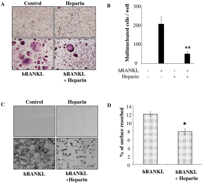
Figure 1: Heparin inhibits RANKL-induced osteoclastogenesis in two murine models of osteoclastogenesis. RAW 264.7 cells were cultured in the presence of hRANKL (100 ng/ml) and heparin (5 µM). After 5 days, cells were stained for TRAP expression. Small osteoclasts (arrowheads) and condensed cells (arrows) formed in the presence of heparin are shown. (A)
TRAP+ multinucleated cells (more than 3 nuclei) were counted under a light microscope
(original magnification x 40). Each value represents the mean (± SD) of multinucleated cells per well of a triplicate experiment (B). mRNA expressions of osteoclastic markers (RANK, TRAP, Ctsk) were analyzed by real-time PCR after 5 days of culture. Cyc1 and Hprt1 were used as internal control. Results are expressed as fold increase compared to the control (C).
Experiments were performed independently at least 3 times in triplicate. CD11b+ purified
cells from mouse bone marrow were cultured for 15 days in presence of 25 ng/ml mM-CSF with or without hRANKL (100 ng/ml) and heparin (5µM). At the end of the culture period,
TRAP staining was performed and TRAP+ multinucleated cells (more than 3 nuclei) were
counted under a light microscope (D) (** p<0.01).
Figure 2: Heparin inhibits RANKL-induced osteoclastogenesis in human CD14+ monocytes. CD14+ purified monocytes were cultured for 15 days in the presence of 25 ng/ml hM-CSF with or without hRANKL (100 ng/ml) and heparin (5 µM). At the end of the culture
period, TRAP staining was performed (original magnification x 40) (A) and TRAP+
multinucleated cells (more than 3 nuclei) were counted under a light microscope (B). In some
conditions, CD14+ monocytes were cultured on dentine slices for 15 days. Resorption areas
were visualised on dentine slices after the culture of CD14+ cells in the presence or absence of
hRANKL, heparin or hRANKL+heparin (C) and were measured using Qwin software (Leica) (D). (* p<0.05).
Figure 3: Various GAG families are able to inhibit RANKL-induced osteoclastogenesis. RAW 264.7 cells were cultured in presence of hRANKL (100 ng/ml) and 5µM of heparan sulfate (bHS: heparan sulfate from bovine kidney, pHS: heparan sulfate from porcine intestinal mucosae), chondroitin sulfate C (CS-C) and dermatan sulfate (DS) (A) or increasing concentrations of heparin oligosaccharides: 4 (8-mer, grey bar), 14 (28-mer, dark bar) and 24 (48-mer, hatched bar) (B) or increasing concentrations of hyaluronic acid (C). After 5 days, RAW 264.7 cells were stained for TRAP expression and multinucleated cells (more than 3 nuclei) were counted under a light microscope. Results are expressed as number of multinucleated cells per well: each value represents the mean (± SD) of multinucleated cells per well of a triplicate. Experiments were performed at least 3 times in triplicate (* p<0.05, ** p<0.01, *** p<0.001).
Figure 4: Sulfation is a key parameter in the inhibition of RANKL-induced osteoclastogenesis by heparin. RAW 264.7 cells were cultured in the presence of RANKL (100 ng/ml) and different forms of heparin (5µM). After 5 days, RAW 264.7 cells were stained for TRAP expression and multinucleated cells (more than 3 nuclei) were counted under a light microscope. Results are expressed as number of multinucleated cells per well: each value represents the mean (± SD) of multinucleated cells per well of a triplicate. Experiments were performed at least 3 times in triplicate (* p<0.05, ** p<0.01).
Figure 5: Oligosaccharides promote dendritic differentiation. Human CD14+ cells (A) or
total PBMCs (B) were cultured in cell media supplemented with 100 ng/ml GM-CSF + 5
ng/ml IL-4, in the presence or absence of heparin oligosaccharide 16 (32-mer, Oligo 16),
CD14, and analyzed by flow cytometry. Percentages of CD1a+ cells (dendritic cells) were plotted.
Figure 6: Heparin does not bind to hRANKL and does not modulate RANKL signalization. (A) Heparin (1 to 20 nM) or hOPG (25nM) were injected at a flow rate of 30
µl/min and 20 µl/min, respectively, over the immobilized-RANKL sensorchip for 6 min. In a pH
7.4 buffer, hOPG but not heparin binds to hRANKL. (B) Human CD14+ monocytes were incubated for 15 min at 37°C with 100 ng/ml hRANKL in the presence or the absence of 5 µM heparin. Protein lysates were prepared and expression of Phospho-ERK1/2, total-ERK1/2, Phospho-p38, total-p38, Phospho-p105, total-p105 was analyzed by western blotting. All experiments were repeated three times, and a representative blot is shown.
Figure 7: Heparin acts at two different levels of RANKL-induced osteoclastogenesis of human CD14+ cells. CD14+ cells were cultured in the presence of hM-CSF and hRANKL with or without heparin (5 µM) or OPG (100 ng/ml) added at different time points of the culture. After TRAP staining, osteoclasts were counted under a light microscope. (A) Heparin was added during the first 4 days (D0-D4) of the culture, added after 4 days of culture to days 10 (D4-D10), or added during the last 4 days of the culture (D10-D14) or maintained all along
the culture (D0-D14). (B) Heparin inhibits the adherence of CD14+ cells when added only
during the first 4 days of the culture, in the presence of hM-CSF only. (C and D) Heparin
inhibits osteoclast spreading: CD14+ monocytes were cultured with hM-CSF and hRANKL,
with or without heparin (5 µM) until the formation of osteoclasts in the control condition
(D4-D14). Heparin was then removed from the medium or maintained and culture was extended
for 3 more days (D14-D17). TRAP staining was then performed and osteoclasts were counted under a light microscope (original magnification x 40). Black arrows show the condensed
cells obtained in heparin conditions after the normal culture period + 4 additional days (* p<0.05)
Aditional data 1
GAGs and heparin do not modulate the proliferation of osteoclast precursors.
Osteoclast precursors (CD14+ cells) were treated from the day of cell plating by 25 ng/ml M-CSF and increased doses of heparin. The number of viable cells was measured by XTT assays 72h later. In the second set of experiments, CD14+ cells were first incubated with 25 ng/ml M-CSF for 3 days before addition of increased doses of heparin for 11 days. The number of viable cells was determined by using XTT three days later. The results revealed that heparin and other GAGs (only experiment performed with heparin and DS after 3 days of culture in the presence of M-CSF has been shown, similar results have been obtained with chondroitin sulfate and oligosaccharides, after 6 days of culture or in the absence of M-CSF) did not affect the proliferation of osteoclast precursors.
Aditional data 2-4
CD14+ monocytes were cultured in a 24-well plate with hM-CSF and hRANKL as described
in the present paper, and with our without heparin (5 µM). At the end of the culture, heparin was removed or not from the medium and the culture was extended for 11 more hours. A picture was taken every 10 minutes during 11 hours, and the movies were reconstituted using the ImageJ software.
0 20 40 60 M u lt in u cl ea te d c el ls / w el l Control hRANKL Heparin hRANKL + Heparin
**
0 20 40 60 80 M u lt in u cl ea te d c el ls / w el l A B C D Figure 1 F o ld i n cr ea se (c o m p a r e to c o n tr o l) hRANKL 0 10 20 30 RANK TRAP CtsK -+ -+ + + Heparin hRANKL -+ -+ + + Heparin hRANKL -+ -+ + + HeparinFigure 2 Control hRANKL Heparin hRANKL + Heparin A Control hRANKL Heparin hRANKL +Heparin C M u lt in u cl ea te d ce ll s / w el l 0 100 200 300 B
**
hRANKL -+ -+ + + Heparin % o f su rf a ce r es o rb ed 0 2 4 6 8 10 12 14 hRANKL hRANKL + Heparin*
D0 20 40 60 80
C
M u lt in u cl ea te d c el ls / w el l 0 20 40 60 80 100A
M u lt in u cl ea te d c el ls / w el lB
Figure 3 M u lt in u cl ea te d c el ls / w el l 0 20 40 60 80 100*
**
**
**
*
*
*
**
**
*
**
**
*
**
** **
*
**
**
**
**
**
* *
***
**
***
***
***
*** **
hRANKL 100 ng/ml -+ - bHS + pHS GAGs 5µM + + + CS-C DS hRANKL 100 ng/ml -+ - 1.56 + 3.1 Oligosaccharides µM + + + 6.25 12.5 + 100 + + 25 50 hRANKL 100 ng/ml -+ - 5 + 20 Hyaluronic acid (µg/ml) + + 100To tal D e-s ulf ate d De -N -su lfa ted re- N-ac ety lat ed 0 2 0 4 0 6 0 no rm al De -2-sul fat ed -6- De sul fat ed -N De -su lfa ted Mu lti nu cle ate d c ell s / w ell F ig u re 4
**
**
**
*
*
*
h R A N K L 1 0 0 n g /m l -+ + F o rm s o f p o ly m er ic h ep a ri n (5 µ M ) + + + + +F ig u re 5 O li g o 1 6 H ep a ri n D S h G M -C S F + h IL -4 h G M -C S F + h IL -4 + G A G % o f C D1 a + cel ls 0 1 0 2 0 3 0 4 0
A
B
0 2 0 4 0 6 0 8 0 h G M -C S F + h IL -4 h G M -C S F + h IL -4 + G A G % o f C D1 a + cel ls O li g o 1 6 H ep a ri nto ta l E rk P -E rk 1 /2 to ta l p 3 8 P -p 3 8 to ta l p 1 0 5 P -p 1 0 5 h R a n k L 1 0 0 n g /m l H e p a ri n 1 2 5 µ g /m l - -+ -+ + - + C h ip R A N K L H e p a ri n 0 1 0 0 2 0 0 3 0 0 0 1 0 0 2 0 0 3 0 0 4 0 0 h O P G Re sp on se (R U) T im e ( s ) A B F ig u re 6
F ig u re 7 B 0 2 0 4 0 6 0 8 0 1 0 0 1 2 0 h M -C S F h M -C S F + H e p a ri n Ad he re nt ce lls (% to c on tro l) * A H e p a ri n h R A N K L -+ -+ -+ -+ -+ 0 4 0 8 0 1 2 0 mu lti nu cle ate d c ell s / w ell D 0 -D 1 4 D 0 -D 4 D 1 0 -D 1 4 D 4 -D 1 0 * * D H e p a ri n D 4 -D 1 4 H e p a ri n D 4 -D 1 7 N o H e p a ri n O P G 1 0 0 n g /m l * 0 5 0 1 0 0 1 5 0 2 0 0 2 5 0 3 0 0 mu lti nu cle ate d c ell s / w ell h R A N K L C * D 4 -D 1 4 D 1 4 -D 1 7 -+ -+ -+ -+ + -+ -H e p a ri n
-H e p a ri n D e rm a ta n S u lf a te 1 6 0 1 4 0 1 2 0 1 0 0 8 0 6 0 4 0 2 0 0 OD (% c om pa re dt o t he co ntr ol) H e p a ri n D e rm a ta n S u lf a te 1 6 0 1 4 0 1 2 0 1 0 0 8 0 6 0 4 0 2 0 0 H e p a ri n D e rm a ta n S u lf a te ( D S ) 1 6 0 1 4 0 1 2 0 1 0 0 8 0 6 0 4 0 2 0 0 OD (% c om pa re dt o t he co ntr ol)