Gene expression pattern
Differential expression of two somatostatin genes during zebrafish
embryonic development
Nathalie Devos
a, Gianluca Deflorian
b, Fre´de´ric Biemar
a, Marino Bortolussi
b, Joseph A. Martial
a,
Bernard Peers
a,*, Francesco Argenton
ba
Laboratoire de Biologie Mole´culaire et de Ge´nie Ge´ne´tique, Institut de Chimie, Baˆtiment B6, Universite´ de Lie`ge, B-4000 Lie`ge (Sart-Tilman), Belgium
b
Dipartimento di Biologia, Universita` di Padova, Via U. Bassi 58/B, I-35131 Padova, Italy Received 22 February 2002; received in revised form 15 March 2002; accepted 18 March 2002
Abstract
We have identified the cDNAs of two new zebrafish preprosomatostatins, PPSS1 and PPSS3, in addition to the previously cloned PPSS2
(Argenton et al., 1999). PPSS1 is the orthologue of mammalian PPSSs, with a conserved C-terminal SS-14 sequence, PPSS2 is a divergent
SS precursor and PPSS3 is a cortistatin-like prohormone. Using whole-mount in situ hybridisation, we have analysed the expression of
PPSS1 and PPSS2 in zebrafish embryos up to 5 days post fertilisation. PPSS1 was expressed in the developing pancreas and central nervous
system (CNS), whereas PPSS2 expression was exclusively pancreatic. In the CNS, PPSS1 was detected in several areas, in particular in the
vagal motor nucleus and in cells that pioneer the tract of the postoptic commissure. PPSS1 was also expressed transiently in the telencephalon
and spinal motor neurons. In all areas but the telencephalon PPSS1 was coexpressed with islet-1. q 2002 Elsevier Science Ireland Ltd. All
rights reserved.
Keywords: Somatostatin; Pancreas; Central nervous system; Zebrafish; Development; In situ hybridisation; Masterblind; islet-1
1. Results and discussion
Somatostatin (SS), synthesised as preprosomatostatin
(PPSS), undergoes a tissue-specific proteolytic cleavage
generating two bioactive peptides (SS-14 and SS-28),
corre-sponding to its 14- and 28-aminoacid C-termini,
respec-tively. SS is produced throughout the central and
peripheral nervous systems, where it affects cognitive,
loco-motor, sensory and autonomic functions, and in some
peripheral organs, where it inhibits cell proliferation and
secretion of a wide range of endocrine and exocrine cells
(Patel, 1999; Zeyda et al., 2001). SS is also involved in
developmental processes as evidenced by the early onset
of PPSS transcription and transient expression in mouse
embryos (Bendotti et al., 1990) and its capacity to affect
neuronal migration in the developing rodent nervous system
(Yacubova and Komuro, 2002).
We previously cloned a zebrafish cDNA encoding a SS
precursor, hereafter called PPSS2 (Argenton et al., 1999).
Herein, we report the sequences of two additional zebrafish
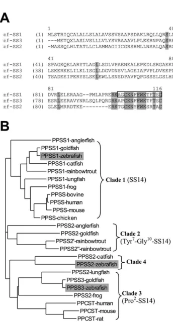
SS-like precursors (Fig. 1A), designated PPSS1 and PPSS3.
Several vertebrate genes encoding SS-like precursors have
been identified and grouped in four clades (Fig. 1B; Lin et
al., 2000). PPSS1 belongs to the first group, comprising the
genes encoding the precursor of a strictly conserved SS-14,
while PPSS2 is grouped with catfish PPSS2 (Fig. 1B).
PPSS3 (Fig. 1A), belongs to the third group together with
the preprocortistatin genes. This mammalian peptide
precursor gives rise to cleavage product comparable to
SS-14 and SS-28 and seems to play a role in neuronal
depression and sleep modulation (Spier and de Lecea,
2000).
Here we have analysed the expression of PPSS1 and
PPSS2 in zebrafish embryos at various stages up to 5 days
post fertilisation (dpf) by whole-mount in situ hybridisation.
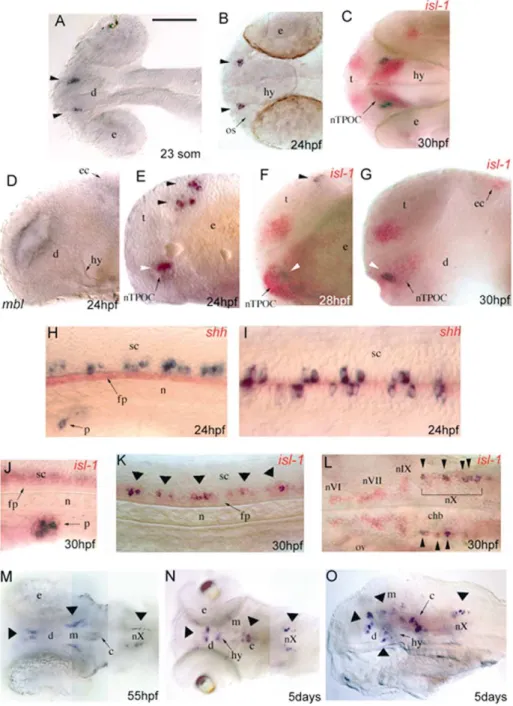
At all investigated stages, PPSS2 expression was
exclu-sively pancreatic, while PPSS1 was expressed both in the
pancreas and central nervous system (CNS). In the
pancrea-tic primordium, the first PPSS2-expressing cells appeared at
the 16-somite stage (17 hours post fertilisation (hpf)),
whereas PPSS1 expression was first detected in few cells
at 24 hpf (Fig. 2A–D). From 24 to 77 hpf, PPSS1- and
PPSS2-expressing cell number increased and they gathered
in a single islet (Fig. 2). PPSS2 was expressed in a larger
0925-4773/02/$ - see front matter q 2002 Elsevier Science Ireland Ltd. All rights reserved. PII: S 0 9 2 5 - 4 7 7 3 ( 0 2 ) 0 0 0 8 2 - 5
www.elsevier.com/locate/modo
* Corresponding author. Tel.: 132-4-366-33-74; fax: 132-4-366-29-68. E-mail address: [email protected] (B. Peers).
number of cells than PPSS1 (Fig. 2F, H) and double staining
showed that most PPSS1-expressing cells also expressed
PPSS2 (data not shown). In the CNS, PPSS1 expression
was first detected at the 23-somite stage (19 hpf) in two
symmetrical clusters of cells in the rostro-ventral
dience-phalon (Fig. 3A) and in isolated cells distributed along the
ventral spinal cord. At 24 hpf, these expression domains
were maintained (Fig. 3B, E, H, I) and, in addition,
PPSS1-expression was also observed in two bilateral cell
clusters in the dorsal telencephalon (Fig. 3E) and in one or
two cells on both sides of the caudalmost hindbrain. The
telencephalic PPSS1 labelling, that was reduced at 28 hpf
and no longer observed at 30 hpf (Fig. 3F, G), was the only
site in which PPSS1 did not colocalise with islet-1
expres-sion. At 30 hpf, all PPSS1-expressing cells also expressed
islet-1 (Fig. 3C, G, J–L), allowing their precise
identifica-tion since islet-1-expressing cells in zebrafish CNS are well
characterised (Korzh et al., 1993; Inoue et al., 1994;
Higa-shijima et al., 2000). In the diencephalon, the PPSS1 signal
was included in the islet-1 expression domain of the nucleus
of the tract of the postoptic commissure (nTPOC) (Fig. 3C,
F, G). These results prompted us to check PPSS1 expression
in masterblind/axin1 embryos, in which the telencephalon
and part of the diencephalon are lacking, and the
islet-1-expressing cells of the nTPOC are absent (Heisenberg et al.,
1996, 2001; van de Water et al., 2001). At 24 hpf,
master-blind/axin1 mutants lacked both the telencephalic and
dien-cephalic PPSS1 staining (Fig. 3D), indicating that the
PPSS1 gene is indeed expressed in a islet-1-expressing
subpopulation of cells of the nTPOC, responsible for
pioneering the postoptic commissure (Heisenberg et al.,
1996). In the caudalmost hindbrain, starting from 24 hpf,
PPSS1 was expressed in a subset of neurons of the vagal
motor nucleus (nX) (Fig. 3L–O). Expression of the PPSS1
orthologue in nuclei of the vagus motor nerve has been
reported in adult lungfish (Trabucchi et al., 1999). In the
spinal cord, PPSS1 expression was detected, from the
23-somite stage, in several islet-1 expressing primary motor
neurons located bilaterally just above the floor plate (Fig.
3H, I, K). Our results support the hypothesis of a role (direct
or not) of this LIM homeobox protein on the SS gene
expression (Leonard et al., 1992; Vallejo et al., 1992).
From 55 hpf to 5 dpf, the expression of the PPSS1 gene
was maintained in the pancreas (Fig. 2F), diencephalon
and motor neurons of the nX (Fig. 3M–O), but not in the
spinal cord. From 55 hpf, PPSS1 expression was also
detected in the mesencephalon and cerebellum (Fig. 3M–
O), and an additional signal in the hypothalamus was
observed at 5 dpf (Fig. 3O).
The dynamic, specific and transient pattern of PPSS1
expression in the embryonic zebrafish CNS is in
accor-dance with the situation observed in other vertebrates
where SS peptide is detected transiently in various regions
of the developing CNS including forebrain, diencephalic
area and spinal cord (Senba et al., 1982; Shiosaka et al.,
1982).
Fig. 1. (A) Comparison of zebrafish PPSS1, PPSS2 and PPSS3. Strictly conserved aminoacids are boxed with gray background. SS-14 mature peptide is boxed. The PPSS1 cDNA is 711 bp long and contains an open reading frame of 114 aminoacids with the conserved SS-14 in its C-termi-nus. The PPSS2 corresponds to the previously reported PPSS (Argenton et al., 1999). (B) Full-length aminoacid sequences from all vertebrate SS-related peptide precursors available to date were used to calculate a phylo-genetic tree with ClustalX (Page, 1996). PPSS, preprosomatostatin; PPCST, preprocortistatin. Zebrafish PPSSs are boxed. SS-like precursors are grouped according to Lin’s description (Lin et al., 2000). The first group is composed of precursors of a strictly conserved SS-14. The three other groups can be characterised by common substitutions found in the deduced mature SS-14. The Tyr7–Gly10–SS-14 variants constitute group 2. The third group is composed of frog, goldfish and lungfish genes encoding Pro2 -variants of SS-14 but also of the mammalian cortistatin genes. In our phylogenetic analysis, the previously described zebrafish SS cDNA is grouped with the catfish PPSS2 gene (clade 4). These two genes potentially encode divergent SS-14 with the common substitutions Tyr6, Ser10, Arg11 and Ala13.
2. Materials and methods
The zf-PPSS1 cDNA was obtained from the RZPD
(http:www.rzpd.de) as EST in the zebrafish databank,
sequenced on both strand on an ABI 310 Genetic Analyser
and submitted to Genbank (Accession number: AF435965).
The zf-PPSS3 nucleotide sequence (EST fr91b10) was
obtained by using the BLAST program with zf-PPSS1
sequence
on
the
NCBI
EST
databases
(http://
www.ncbi.nlm.nih.gov/blast/). Then, the zf-PPSS3 amino
acid sequence was deduced from the fr91b10 (Accession
number: BI472739 and BI473045) complete sequence.
2.1. Whole-mount in situ hybridisation
Antisense RNA probes were prepared by transcribing
linearised cDNA clones either with SP6 or T7 polymerase
and digoxigenin or fluorescein labelling mix (Roche).
Single and double whole-mount RNA in situ hybridisations
and detection were carried out as previously described
(Hauptmann and Gerster, 1994), with a 658C overnight
hybridisation step. Embryos were mounted in 87% glycerol
in phosphate buffered saline (PBS) and photographed with
Nomarski optics using a Leica digital camera. Digital
images were processed using Adobe Photoshop software.
Acknowledgements
This work was supported by a grant from the EC, Fifth
Framework (QLRT-1999-00149 to N.D., B.P. and M.B.).
F.A. is supported by a Telethon grant (E0941). F.B. holds
a doctoral fellowship from the ‘Fonds pour la Formation a` la
Recherche dans l’Industrie et dans l’Agriculture (F.R.I.A)’.
B.P. is ‘Chercheur Qualifie´’ from the ‘Fonds National pour
la Recherche Scientifique (F.N.R.S.)’. G.D. holds a
fellow-ship from cofin2000 (MM05A97553-003). We thank Paolo
Sordino for providing masterblind/axin1 embryos.
References
Argenton, F., Zecchin, E., Bortolussi, M., 1999. Early appearance of pancreatic hormone-expressing cells in the zebrafish embryo. Mech. Dev. 87, 217–221.
Bendotti, C., Hohmann, C., Forloni, G., Reeves, R., Coyle, J.T., Oster-Granite, M.L., 1990. Developmental expression of somatostatin in mouse brain. II. In situ hybridization. Brain Res. Dev. Brain Res. 53, 26–39.
Hauptmann, G., Gerster, T., 1994. Two-color whole-mount in situ hybri-dization to vertebrate and Drosophila embryos. Trends Genet. 10, 266. Heisenberg, C.P., Brand, M., Jiang, Y.J., Warga, R.M., Beuchle, D., van Eeden, F.J., Furutani-Seiki, M., Granato, M., Haffter, P., Hammersch-midt, M., Kane, D.A., Kelsh, R.N., Mullins, M.C., Odenthal, J., Nusslein-Volhard, C., 1996. Genes involved in forebrain development in the zebrafish, Danio rerio. Development 123, 191–203.
Heisenberg, C.P., Houart, C., Take-Uchi, M., Rauch, G.J., Young, N., Coutinho, P., Masai, I., Caneparo, L., Concha, M.L., Geisler, R., Dale, T.C., Wilson, S.W., Stemple, D.L., 2001. A mutation in the Gsk3-binding domain of zebrafish Masterblind/Axin1 leads to a fate transformation of telencephalon and eyes to diencephalon. Genes Dev. 15, 1427–1434.
Higashijima, S., Hotta, Y., Okamoto, H., 2000. Visualization of cranial motor neurons in live transgenic zebrafish expressing green fluorescent protein under the control of the islet-1 promoter/enhancer. J. Neurosci. 20, 206–218.
Inoue, A., Takahashi, M., Hatta, K., Hotta, Y., Okamoto, H., 1994. Devel-Fig. 2. Expression of PPSS1 and PPSS2 in the pancreas of wildtype zebrafish embryos. Ventral views of whole-mount in situ hybridisation with the complete PPSS1 (A,C,E,F) or PPSS2 (B,D,G,H) antisense mRNA probes. Embryos were at the 16-somite stage (17 hpf) (A,B), 24 hpf (C,D), 48 hpf (E,G) or 77 hpf (F,H). Scale bar represents 50 mm.
Fig. 3. Whole-mount in situ staining of zebrafish embryos, visualizing the expression pattern of PPSS1 (blue reaction product), islet-1 (isl-1) and sonic hedgehog (shh) (red reaction product) mRNAs. The yolk was removed and embryos were mounted between glass coverslips. (A,B) Dorsal views of PPSS1 expression (arrowheads) in the head at 23-somite stage (19 hpf) and 24 hpf, respectively. (E) Lateral view, at 24 hpf, reveals PPSS1 expression in the ventral diencephalon (white arrowhead) and dorsal telencephalon (black arrowheads). (D) Lateral view of a 24-hpf masterblind/axin1 (mbl) mutant showing the lack of PPSS1 staining in the head. (C) Dorsal view of a double staining with islet-1 at 30 hpf shows that the PPSS1 signal is found in a subset of islet-1-expressing cells in the nTPOC (nucleus of the tract of the postoptic commissure). Lateral views at 28 hpf (F) and 30 hpf (G) show that the telencephalic PPSS1 labelling does not colocalise with islet-1 at 28 hpf and is absent at 30 hpf. Lateral (H) and dorsal (I) views of 24-hpf embryos double stained with the floor plate marker sonic hedgehog show that PPSS1 expression is distributed bilaterally along the ventral spinal cord. (J–L) Double staining with islet-1 at 30 hpf: (J) lateral view, in the pancreatic primordium PPSS1-expressing cells are a subpopulation of islet-1-positive cells; (K) lateral view, along the ventral spinal cord PPSS1 expression colocalises with islet-1-positive primary motor neurons (arrowheads); (L) dorsal view, in the hindbrain PPSS1 signal overlaps with islet-1-positive neurons of the vagal motor nucleus (nX), but not with those of the abducens (nVI), facial (nVII) and glossopharyngeal (nIX) motor nuclei. (M) Dorsal view at 55 hpf, PPSS1 expression (arrowheads) is observed in the diencephalon, mesencephalon, cerebellum and vagal motor nucleus. (N) Dorsal and (O) lateral (eyes removed) views at 5 days, PPSS1 mRNA (arrowheads) is expressed in the same regions of the CNS as observed at 55 hpf and, in addition, also in the hypothalamus. c, cerebellum; chb, caudalmost hindbrain; d, diencephalon; e, eye; ec, epiphysial cluster; fp, floor plate; hy, hypothalamus; m, mesencephalon; n, notochord; nTPOC, nucleus of post optic commisure; os, optic stalk; ov, otic vesicle; p, pancreas; sc, spinal cord; t, telencephalon. In all pictures anterior is to the left; in lateral views dorsal is to the top. Scale bar: A–G and J–L 100 mm, H and I 50 mm, M–O 150 mm.
opmental regulation of islet-1 mRNA expression during neuronal differ-entiation in embryonic zebrafish. Dev. Dyn. 199, 1–11.
Korzh, V., Edlund, T., Thor, S., 1993. Zebrafish primary neurons initiate expression of the LIM homeodomain protein Isl-1 at the end of gastru-lation. Development 118, 417–425.
Leonard, J., Serup, P., Gonzalez, G., Edlund, T., Montminy, M., 1992. The LIM family transcription factor Isl-1 requires cAMP response element binding protein to promote somatostatin expression in pancreatic islet cells. Proc. Natl Acad. Sci. USA 89, 6247–6251.
Lin, X., Otto, C.J., Cardenas, R., Peter, R.E., 2000. Somatostatin family of peptides and its receptors in fish. Can. J. Physiol. Pharmacol. 78, 1053– 1066.
Page, R.D., 1996. TreeView: an application to display phylogenetic trees on personal computers. Comput. Appl. Biosci. 12, 357–358.
Patel, Y.C., 1999. Somatostatin and its receptor family. Front. Neuroendo-crinol. 20, 157–198.
Senba, E., Shiosaka, S., Hara, Y., Inagaki, S., Sakanaka, M., Takatsuki, K., Kawai, Y., Tohyama, M., 1982. Ontogeny of the peptidergic system in the rat spinal cord: immunohistochemical analysis. J. Comp. Neurol. 208, 54–66.
Shiosaka, S., Takatsuki, K., Sakanaka, M., Inagaki, S., Takagi, H., Senba, E., Kawai, Y., Iida, H., Minagawa, H., Hara, Y., Matsuzaki, T., Tohyama, M., 1982. Ontogeny of somatostatin-containing neuron
system of the rat: immunohistochemical analysis. II. Forebrain and diencephalon. J. Comp. Neurol. 204, 211–224.
Spier, A.D., de Lecea, L., 2000. Cortistatin: a member of the somatostatin neuropeptide family with distinct physiological functions. Brain Res. Brain Res. Rev. 33, 228–241.
Trabucchi, M., Tostivint, H., Lihrmann, I., Jegou, S., Vallarino, M., Vaudry, H., 1999. Molecular cloning of the cDNAs and distribution of the mRNAs encoding two somatostatin precursors in the African lungfish Protopterus annectens. J. Comp. Neurol. 410, 643–652. Vallejo, M., Penchuk, L., Habener, J.F., 1992. Somatostatin gene upstream
enhancer element activated by a protein complex consisting of CREB, Isl-1-like, and alpha-CBF-like transcription factors. J. Biol. Chem. 267, 12876–12884.
van de Water, S., van de Wetering, M., Joore, J., Esseling, J., Bink, R., Clevers, H., Zivkovic, D., 2001. Ectopic Wnt signal determines the eyeless phenotype of zebrafish masterblind mutant. Development 128, 3877–3888.
Yacubova, E., Komuro, H., 2002. Stage-specific control of neuronal migra-tion by somatostatin. Nature 415, 77–81.
Zeyda, T., Diehl, N., Paylor, R., Brennan, M.B., Hochgeschwender, U., 2001. Impairment in motor learning of somatostatin null mutant mice. Brain Res. 906, 107–114.