HAL Id: hal-01632799
https://hal.archives-ouvertes.fr/hal-01632799
Submitted on 8 Nov 2019
HAL is a multi-disciplinary open access archive for the deposit and dissemination of sci-entific research documents, whether they are pub-lished or not. The documents may come from teaching and research institutions in France or abroad, or from public or private research centers.
L’archive ouverte pluridisciplinaire HAL, est destinée au dépôt et à la diffusion de documents scientifiques de niveau recherche, publiés ou non, émanant des établissements d’enseignement et de recherche français ou étrangers, des laboratoires publics ou privés.
Electron paramagnetic resonance of radicals induced in
drugs and excipients by radiation or mechanical
treatments
J Raffi, S Gelly, L Barral, F Burger, P Piccerelle, P Prinderre, Michel Baron,
Alain Chamayou
To cite this version:
J Raffi, S Gelly, L Barral, F Burger, P Piccerelle, et al.. Electron paramagnetic resonance of radicals induced in drugs and excipients by radiation or mechanical treatments. Spectrochimica Acta Part A: Molecular and Biomolecular Spectroscopy, Elsevier, 2002, 58 (6, SI), p.1313-1320. �10.1016/S1386-1425(01)00720-X�. �hal-01632799�
Electron paramagnetic resonance of radicals induced in
drugs and excipients by radiation or mechanical treatments
J. Raffi
a,*, S. Gelly
a, L. Barral
a, F. Burger
b, P. Piccerelle
c, P. Prinderre
c,
M. Baron
d, A. Chamayou
daLARQUA/LRMO, Faculte´ des Sciences de Saint-Je´roˆme, case D52, 13397 Marseille cedex 20, France bLMRE, Faculte´ des Sciences de Saint-Je´roˆme, case D52, 13397 Marseille cedex 20, France
cLaboratoire de Pharmacie Gale´nique Industrielle et Cosme´tologie,27 Boule!ard Jean Moulin, 13385 Marseille cedex 5, France dLaboratoire de Ge´nie des Proce´de´s des Solides Di!ise´s, UMR CNRS 2392, Ecole des Mines d’Albi-Carmaux,
81013 Albi CT cedex 09, France
Abstract
Radiation as well as mechanical treatments induced in drugs and excipients radicals, which can be studied by electron paramagnetic resonance. A special attention is pointed about the use of electron paramagnetic resonance (EPR) to bring the proof whether or not a drug has been irradiated or not. We also discuss of other methods (thermoluminescence (TL), gas phase chromatography (GPC)) which can be used to bring the same proof in case of irradiated drugs, excipients and cosmetic products. © 2002 Elsevier Science B.V. All rights reserved.
Keywords: Electron paramagnetic resonance; Thermoluminescence; Drug; Radiation; Mechanical treatment
1. Introduction
Gamma rays and electron beams have been used for a long time in order to sterilise medical devices. This treatment will be more and more applied for sterilisation of drugs [1,2]. As for foodstuffs [3,4], it could be interesting to be able to prove whether or not a drug has been irradiated.
Active ingredients may be irradiated alone but also in mixtures with a lot of products; thus we
can find natural vegetal products (starch, cellu-lose,…) more or less modified and simple sugars used as excipients. For active ingredients we took the example of some antibiotics but also the one of simple products such as amino acids; for in-stance leucine can be used, due to its ability to stimulate or modulate translation initiation fac-tors in rat liver [5,6], translation initiation in skeletal muscles [7], arginine produces beneficial effects on the heart rate and tissue oxygen extrac-tion in haemorrhaged rabbits [8], tyrosine trans-port pathway may be used to improve nasal absorption of poorly permeable drugs [9], dimethylglycine leads to a significant reduction in
* Corresponding author. Tel./fax: +33-4-91-985055.
glutathione levels [10], and a lot of amino acids are given as nutritional complements for some patients [11].
One opportunity is to detect by electron para-magnetic resonance (EPR) the radicals induced in solid and relatively dry state; in these conditions the induced and trapped radicals may have a long life time (up to several months or years). In the first part of this paper, we shall give examples of such radicals; but we also show that the only presence of radicals is not a proof of irradiation, as radicals may also be induced by grinding, UV-radiation…. Thus we must take precautions before to ascertain any conclusion
In a second part we will see how thermolu-minescence (TL) and gas phase chromatography (GPC) can also be used.
2. Experimental
2.1. Grinding experiments
Grinding experiments were performed at room temperature in a vibro-activator (one-ball vibra-tion mill of type Dangoumau, Fig. 1). Dangoumau grinder has be used [12] to study phase transfor-mations of minerals, and recently to increase bio-availability of drugs by co-grinding [13]. The theoretical prediction of dynamic regimes in the vibro-activator was published lately [14]. The quantity of induced radicals depends on the grind-ing time; a too long time destroys the radicals, probably by heating of the medium and/or recom-bination of radicals [15], in most cases a time of 10– 15 min leads to a maximal quantity of radicals.
2.2. Irradiations
There were performed at room temperature in the ‘CIGAL’ cell of Cadarache (50 000 Ci of cobalt 60) supplying a dose rate of 6.1 kGy h− 1.
Storage of samples was always at room tempera-ture and in absence of light.
2.3. EPR and TL measurements
The EPR spectra were recorded at room tem-perature with a Bruker EMS104 spectrometer with the following average conditions: Microwave power, 0.79 mW; sweep width, 20 mT (200 G); modulation, 0.4 mT (4 G); number of sweeps, 3– 55, respectively, for radiation or mechanical induced radicals.
The TL spectra were recorded with a Harshaw 4000 A apparatus with the following average con-ditions: initial temperature, 50 °C, during 10 s; final temperature, 350 °C; increasing temperature rate, 8 °C s− 1.
3. Results and discussion
3.1. ESR study of gamma induced radicals in drugs and excipients
A lot of drugs and excipients can be irradiated
Fig. 2. EPR spectra of radicals induced in anhydrous ampi-cilline acid just after a 50 kGy irradiation (left) and 6 months later (right).
…) show an evolution of the shape of their EPR spectra but the signal is always very important, 2 years after irradiation, allowing an easy detection of the treatment. But in some other cases the life time can be very short (several weeks for cysteine and valine to only 2– 3 days for leucine); more-over, due to the distance between the irradiator and the laboratory (about a 2 h car drive), we have observed no signal after a 5 kGy room temperature irradiation of methionine! It was also the same in case of irradiation of a lyophilised collagen dressing whatever the radio-induced ef-in solid state; generally they show no EPR signal
in the unirradiated sample which is consequently very easy to be detected.
On another hand, the irradiated drugs present complex spectra; these drugs being dry, the life time of the signals induced by radiation is very long, several months or years; Fig. 2 shows the example of an irradiated antibiotic, anhydrous ampicilline acid [16,17] which presents a large and very stable signal (more than 1 year).
Consequently, ESR detection of irradiated drugs must be very easy. However, in case of natural products used in some drugs, the unirradi-ated sample may also present a single line very often found in vegetal products, probably due to a quinone radical [4,18]; but the single line is easy to be distinguished from the complex signal in-duced in irradiated drugs and is not, conse-quently, a limit to the detection of an irradiated sample.
The case of amino-acids (Fig. 3) seems also simple: the unirradiated samples show no ESR signal, and the irradiated ones show large signals, more or less complex, depending on the chemical nature of the amino-acid. Generally, the storage time is very important (several months, even years) and even used, in case of alanine, for dosimetry [19,20]; for some of them a storage time of 2 years does not show any important evolution in the shape of the EPR signal (alanine, arginine, glycine); to see such an evolution, it is necessary to heat the sample during several hours at 70 °C (for example, glycine). The great majority of amino-acids (histidine, lysine, proline, tyrosine,
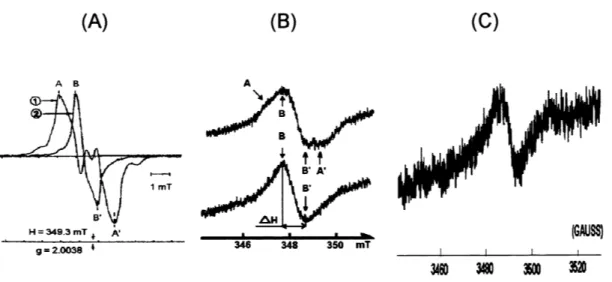
Fig. 3. EPR spectra of some irradiated amino-acids (H= his-tidine, P= proline, C= cysteine, V= valine, L= leucine; ini= just after irradiation, y= year, m= month, w= week, d= day; other recording conditions in the text).
Fig. 4. EPR signals of radicals induced in starches by gamma radiation (A), UV radiation (B) or grinding (C). (A) Curve 1 recorded just after gamma irradiation, curve 2 recorded 6 months later (gain multiplied by 20). (B) Upper curve recorded just after UV irradiation, curve 2 recorded several hours later.
fects observed by other techniques such as mi-croelectrophoresis or electronic microscopy [21]. Thus the fundamental studies [22– 25] must be reoriented to find what would be the life time of the ESR signals, product per product: the pres-ence of an EPR signal will be a proof of irradia-tion but its absence will not be a proof of no irradiation, at least for some amino acids.
In excipients such as starch [26] (Fig. 4(A)), cellulose, mannitol… there is no signal or a simple singulet in the unirradiated sample and a complex EPR signal, relatively stable with time (at least in crystalline parts) in the irradiated samples. Here too the detection of an irradiation treatment will be easy, under the condition that the signal shape is characteristic of irradiation, which was not always the case (the storage time of cereals may be greater than 1 year) [27].
3.2. ESR study of radicals induced by other treatments
We have only to consider here the radicals induced in solid state; it is well known that ther-mal or ultrasound [28– 31] treatments induced radicals but they disappear very quickly as they are always induced in ‘liquid state’.
However, we should be note that radicals de-tected by EPR can also be induced by other
treatments such as UV radiation [32,33] or by mechanistic action such as grinding even if radical were induced in very small quantities [34– 38]. For instance, Fig. 4 shows that the main radicals induced by gamma (Fig. 4(A)) [26] or UV radia-tion (Figs. 1– 4) [39,40], or by grinding (Fig. 4(C)) are identical; other excipients such as lactose (Fig. 5(A)), carboxymethylcellulose (Fig. 5(B)), cellu-lose, mannitol,… [15] may also present ESR sig-nals due to grinding but these sigsig-nals are always very weak with regard to the ones radio-induced in drugs. Thus we can consider that, for a syn-thetic drug, there is no signal at all present in the unirradiated sample.
Let us point out another fact. The fact that we observe the same radicals whatever is the treat-ment (gamma or UV radiation, or grinding) only means that we observe at room temperature the same more stable and, consequently, the more probable radicals, the primary mechanisms are different! This also means that the presence of radicals in a solid such as a drug is not a proof of an irradiation treatment; the proof is the fact that we observe relatively large quantities of radicals inside the whole sample, UV only induces radicals at the outside of the product; thermal radicals are destroyed in the same time they are produced (bimolecular reactions in liquid phase)….
3.3. Thermoluminescence (TL) measurements Here too this method must be applied to drugs irradiated in powder and dry state, allowing the detection of radio-induced ions trapped in the solid matrix. On the opposite of the protocol used for detection of irradiated foodstuffs where the TL signal is due to silicate impurities [41,42], there is no problem of extraction as the TL signal is recorded on the whole sample [16,43].
But the study may be more complex. First at all, the unirradiated sample presents as the unirra-diated one a TL spectra, for instance two peaks in case of anhydrous ampicilline acid (Fig. 6(A)); in this very favourable case, the two peaks vary on different ways with regard to the irradiation dose (Fig. 6(B)) and thus lead to a proof of irradiation; moreover, we can distinct the products irradiated at 25– 30 kGy (usual dose range for sterilisation) from those only irradiated at 10 kGy (final ‘bar-rier treatment’). But in other close antibiotics, the peaks may have very different variations [43]: – in case of the three-hydrated amoxicilline acid,
the first peak increases with regard to the sec-ond with increasing doses, i.e. just the opposite of anhydrous ampicilline acid! It is approxi-mately the same for the monohydrated sodium salt of cloxacilline;
– in case of pentahydrated ceftazidime, there are two peaks which seems to have a constant ratio with regard to the dose.
Consequently, each drug is a different case and requires a specific study. There will not be a general detection protocol as in case of foodstuffs [41].
Moreover, in case of mixtures, which is often the case, it will be impossible to attribute the observed TL signal to a special product; thus the method will be limited to pure products. On an-other hand, in case of vegetal ingredients, the foodstuffs protocol [41] may be applied.
3.4. A comment on the use of gas phase chromatography
In case of drugs not irradiated in solid state, we can make the hypothesis that the protocol devoted to foodstuffs containing lipids [44,45] may be applied. Let us remember that, when such foodstuffs are irradiated, the percentage of radio-induced hydrocarbons is directly linked to the chemical composition of lipids; this is due to the fact that, when fatty acids are exposed to high energy radiation, they undergo preferential cleav-ages, unlike other treatments, in the ester car-bonyl region; the two main hydrocarbons, derived from each fatty acid of structure C(n:m) are those of structure C(n−1:m), with one carbon less than the parent fatty acid and of structure C(n− 2:m+1), with two carbons less and one extra double bond. The method was successfully
Fig. 6. TL recording (left) of unirradiated anhydrous ampicilline acid and variation of peak intensities (right) with irradiation dose (kGy).
Fig. 7. Compositions (in %) of lipids and radio-induced hydrocarbons in a mixture (1/1/1) of peanut, sunflower and extra virgin olive oils.
plied to foodstuffs with a maximum of three or four main lipids, that it to say representing more than 95% of the total lipids composition [46], Fig. 7 shows the example of a synthetic mixture (1/1/1) of peanut, sunflower and olive oils irradiated at 5 kGy.
In fact the problem will be that main cosmetic products, or creams used as vectors in pharmaco-poeia to transfer active ingredients through the skin, have complex compositions which probably does not allow us to reach, in each case, a proof of the irradiation treatment. Moreover, these products also contain phospholipids, which may do not follow the same radiolysis rules than the other lipids.
4. Conclusions
Thus, whatever is the studied drug or excipient irradiated in solid state, there will be no problem to detect its irradiation treatment by EPR. How-ever, on a fundamental point of view, there will be a lot of experiments to carry out in order to improve the knowledge of the structure radio-in-duced radicals, at least in case of drugs; the case of excipients is generally relatively well known. Let us just notice that an absence of an EPR signal will be a proof of non irradiation treatment in the only case where the products are crys-tallised without water; in case of amorphous structures, initial researches must be carried out in
order to study the stability of radicals with regard to the storage time and parameters.
On another hand, the presence of an EPR signal in a complex mixture (excipients+drugs) will probably bring the proof of an irradiation treatment at least on one of the components but it will be very difficult to find the nature of this irradiated component… except if it is the only one treated by gamma rays. This ambiguity comes from the following facts:
– all organic radicals show EPR signals approxi-mately centred in the same area, 350 mT for 9.5 GHz spectrometers;
– in solid phase, the EPR spectra are quite al-ways poorly resolved, due to the overlapping of the different spectra relative to the different directions of initial spins with regard to the magnetic field.
More generally, we can ascertain that, for drugs and cosmetic products, we can write in the future some protocols of detection of an irradiation treatment if:
– the product is irradiated in solid and dry state, by ESR in quite every case, and by TL at least in some cases;
– the product is rich in lipids, by GPC, at least in some cases.
But, before writing these protocols, there will be a lot of fundamental researches to carry out. Acknowledgements
We have to thank Jose´ Vicente (CEA/DSV/ DEVM, Cadarache) for carrying out the irradiations.
References
[1] P. Piccerelle, J.-P. Reynier, J. Joachim, B. Tilquin, J. Raffi, Radio-ste´rilisation de me´dicaments: inte´reˆt, le´gisla-tion et travaux a` entreprendre, J. Pharma. Belg. 55 (2000) 131– 136.
[2] H. Ambroz, E. Kornacka, B. Marciniec, M. Ogrodowczyk, G. Przybytniak, EPR study of free radicals in some drugs gamma-irradiated in the solid state, Radiat. Phys. Chem. 58 (2000) 357– 366.
[3] L. Douifi, J. Raffi, P. Stocker, F. Dole, A point about
EPR detection of irradiated foodstuffs, Spectrochim. Acta (A) 54 (1998) 2403– 2412.
[4] J. Raffi, Identifying irradiated foods, Trends Anal. Chem. 17 (1998) 226– 233.
[5] O.J. Shah, D.A. Antonetti, S.R. Kimball, L.S. Jefferson, Leucine, glutamine and tyrosine reciprocally modulate the translation initiation factors eIF4F and eIF2B in perfused rat liver, J. Biol. Chem. 274 (1999) 36168– 36175. [6] T.G. Anthony, J.C. Anthony, F. Yoshizawa, S.R.
Kim-ball, L.S. Jefferson, Oral administration of leucine stimu-lates ribosomal protein mRNA translation but not global rates of protein synthesis in the liver of rats, J. Nutrition 131 (2001) 1171– 1176.
[7] J.C. Anthony, F. Yoshizawa, T. Gautsch-Anthony, T.C. Vary, L.S. Jefferson, S.R. Kimball, Leucine stimulates translation initiation in skeletal muscle of post-absorptive rats via a rapamycin-sensitive pathway, J. Nutrition 130 (2000) 2413– 2419.
[8] Z. Todorovic, M. Prostran, S. Vuckocic, The influence of
L-arginine on heart rate and tissue oxygen extraction in
haemorrhaged rabbits, Pharmacol. Res. 43 (2001) 321– 327.
[9] C. Yang, A.K. Mitra, Nasal absorption of tyrosine-linked model compounds, J. Pharm. Sci. 90 (2001) 340– 347. [10] K. Hariganesh, J. Prathiba, Effect of dimethylglycine on
gastric ulcers in rats, J. Pharm. Pharmacol. 52 (2000) 1519– 1522.
[11] Anonymous, Nutrineal PD4 (Baxter), BIAM (Banque de Donne´es Automatise´e sur les me´dicaments;
www.BIAM2.org) 31 August 1998.
[12] J.L. Dandurant, Contribution a` l’e´tude des effets du broyage sur les transformations physicochimiques des ma-te´riaux, Doctorat d’Etat N°820, Universite´ Paul Sabatier, Toulouse (1978).
[13] M. Baron, J. Dodds, A. Chamayou, S. Pakkanen, J.O. Larsen, G. Nachon, L. Oudet, P. Tchoreloff, G. Couar-raze, Co-broyage de principes actifs pharmaceutiques pour ame´liorer leur biodisponibilite´, Colloque CNRS, Toulouse 26– 27 Novembre 1998.
[14] V.E. Mizonov, A.A. Kurbatov, M. Baron, A. Chamayou, Z.P. Zhukov, Calculation of dynamic regimes of vibrator activation of fine powders (in Russian), Chem. Chem. Technology 1 (2001).
[15] M. Kuzuya, S.-I. Kondo, A novel collision-induced solid state radical recombination, Mol. Cryst. Liq. Cryst. 277 (1996) 343– 349.
[16] M. Gibella, A.-S. Crucq, B. Tilquin, P. Stocker, J. Raffi, Utilisation de la RPE et de la TL pour de´tecter l’irradia-tion de me´dicaments, Ann. Experts Chim. 92 (1999) 435– 441.
[17] M. Gibella, A.-S. Crucq, B. Tilquin, P. Stocker, G. Lesgards, J. Raffi, Electron spin resonance studies of some irradiated pharmaceuticals, Radiat. Phys. Chem. 58 (2000) 69– 76.
[18] J. Raffi, J.-P. Agnel, Electron spin resonance identifica-tion of irradiated fruits, Radiat. Phys. Chem. 34 (1989) 891– 894.
[19] J.-M. Dolo, E. Pichot, V. Feaugas, Evaluation of some parameters which influence the ESR measurements for the fading study of alanine dosimeters, Appl. Magn. Reson. 15 (1998) 269– 277.
[20] N. Yordanov, V. Gancheva, Selfcalibrated alanine/EPR dosimeters: a new generation of solid state/EPR dosime-ters, J. Radioanal. Nucl. Chem. 240 (1999) 215– 217. [21] F. Burger, I. Pouliquen-Sonaglia, S. d’Avino, J. Raffi,
Etude de l’irradiation gamma du collage`ne en phase solide et en phase liquide, J. Pharm. Belg. 55 (2000) 145– 154.
[22] H. Shields, P.-J. Hamrick Jr, The influence of crystal structure on radical formation in X-irradiated amino acids, J. Chem. Phys. 64 (1976) 263– 270.
[23] A. Minegishi, R. Bergene, P. Riesz, ESR of spin-trapped radicals in gamma-irradiated polycrystalline amino-acids,
N-acetyl amino acids and dipeptides, Int. J. Radiat. Biol.
38 (1980) 627– 650.
[24] D. Hill, R. Garrett, S. Ho, J. O’Donnell, P. O’Sullivan, P. Pomery, Radiolysis of model polypeptiides in the solid state: radiation sensitivity and side chain structure radiat, Phys. Chem. 17 (1981) 163– 171.
[25] J. Pilbrow, D. Hutton, Y. Zhong, C. Noble, R. Song, Pulsed EPR investigation of hyperfine structure in gamma-irradiated alanine, Appl. Radiat. Isot. 47 (1996) 1257– 1261.
[26] J. Raffi, J.-P. Agnel, Influence of the physical structure of irradiated starches on their ESR spectra kinetics, J. Phys. Chem. 87 (1983) 2369– 2373.
[27] J. Raffi, J.-P. Agnel, S.-R. Kassis, Identification par re´-soance paramagne´tique e´lectronique de ce´re´ales irradie´es, Sci. Aliments 4 (1987) 657– 663.
[28] T. Kudo, H. Heusinger, ESR-spin-trap and UV investiga-tions of soluinvestiga-tions ofD-glucose and lactose irradiated with
ultrasound, Carbohydr. Res. 123 (1983) 41– 52.
[29] T. Matsuyama, H. Menhofer, H. Heusinger, ESR spin trap investigations on aqueous glucose solutions irradi-ated by ultrasound and gamma-rays, Radiat. Phys. Chem. 32 (1988) 735– 739.
[30] H. Heusinger, comparison of the reactions induced by ultrasound and gamma rays in aqueous lactose solutions, Ultrasonics 28 (1990) 30– 36.
[31] V. Misil, P. Riesz, EPR characterization of free radicals intermediates formed during ultrasound exposure of cell culture media, Free Radic. Biol. Med. 26 (1999) 936– 943. [32] S.-P. Zhang, J. Xie, J.-P. Zhang, J.-Q. Zhao, L.-J. Jiang, Electron spin resonance studies on photosensitized
forma-tion of hydroxyl radical by C-phycocyanin from Spirulina
platensis, Biochim. Biophys. Acta 1426 (1999) 205– 211.
[33] K.J. Reszka, P. Bilski, C.-F. Chignell, J. Dillon, Free radical reactions photosensitized by the human lens com-poent, kynurenine: an EPR and spin trapping investiga-tion, Free Radic. Biol. Med. 20 (1996) 23– 34.
[34] S.-I. Kondo, S. Hosaka, M. Kuzuya, Mechano-chemical solid state polymerization. VII. Novel composite poly-meric prodrugs prepared by mechano-chemical polymer-ization in the presence of pharmaceutical acids, Chem. Pharm. Bull. 46 (1998) 669– 675.
[35] A. Marino, R. Becker, Mechanically induced free radicals in bone, Nature 218 (1968) 466– 467.
[36] T. Kudo, H. Heusinger, Formation of mechano-radicals in D-glucose and lactose, Carbohydr. Res. 103 (1982)
7– 14.
[37] M. Desrosiers, M. Simic, F. Eichmiller, A. Johnston, R. Bowen, Mechanically-induced generation of radicals in tooth enamel, Appl. Radiat. Isot. 40 (1989) 1195– 1197. [38] C.J. Kay, Mechanochemical mechanism for peptidyl free
radical generation by amyloid fibrils, FEBS Lett. 403 (1997) 230– 235.
[39] A. Bertolini, C. Mestres, P. Colonna, J. Raffi, Free radi-cals formation in the UV and gamma irradiated cassava starch, Carbohydr. Polymers. 44 (2001) 269– 271. [40] A. Bertolini, C. Mestres, J. Raffi, A. Bule´on, D. Lerner,
P. Colonna, Photodegradation of cassava and corn starches, J. Agric. Food. Chem. 49 (2001) 675– 682. [41] CEN, Detection of irradiated food from which silicate
minerals can be isolated, method by thermoluminescence, EN 1788 (1996).
[42] S. Chabane, I. Pouliquen-Sonaglia, J. Raffi, Detection of irradiated spices by different physical techniques, Can. J. Physiol. Pharm. 79 (2001) 1– 6.
[43] P. Stocker, M. Gibella, A.-S. Crucq, B. Tilquin, G. Lesgards, J. Raffi, Thermoluminescence de quelques me´dicaments irradie´s, J. Chim. Phys. 96 (1999) 174– 177. [44] J. Raffi, R. Siadous, 1997– 1998: a turn in food irradia-tion, Agro-Food-Industry Hi-Tech, July/August (1998) 31– 35.
[45] CEN, Detection of irradiated food containing fat, gas chromatographic analysis of hydrocarbons, EN 1784 (1996).
[46] G. Lesgards, J. Raffi, I. Pouliquen, A.A. Chaouch, P. Giamarchi, M. Prost, Use of radiation-induced alkanes and alkenes to detect irradiated food containing lipids, J. Am. Oil. Chem. Soc. 70 (1993) 179– 185.