Direction des bibliothèques
AVIS
Ce document a été numérisé par la Division de la gestion des documents et des archives de l’Université de Montréal.
L’auteur a autorisé l’Université de Montréal à reproduire et diffuser, en totalité ou en partie, par quelque moyen que ce soit et sur quelque support que ce soit, et exclusivement à des fins non lucratives d’enseignement et de recherche, des copies de ce mémoire ou de cette thèse.
L’auteur et les coauteurs le cas échéant conservent la propriété du droit d’auteur et des droits moraux qui protègent ce document. Ni la thèse ou le mémoire, ni des extraits substantiels de ce document, ne doivent être imprimés ou autrement reproduits sans l’autorisation de l’auteur.
Afin de se conformer à la Loi canadienne sur la protection des renseignements personnels, quelques formulaires secondaires, coordonnées ou signatures intégrées au texte ont pu être enlevés de ce document. Bien que cela ait pu affecter la pagination, il n’y a aucun contenu manquant.
NOTICE
This document was digitized by the Records Management & Archives Division of Université de Montréal.
The author of this thesis or dissertation has granted a nonexclusive license allowing Université de Montréal to reproduce and publish the document, in part or in whole, and in any format, solely for noncommercial educational and research purposes.
The author and co-authors if applicable retain copyright ownership and moral rights in this document. Neither the whole thesis or dissertation, nor substantial extracts from it, may be printed or otherwise reproduced without the author’s permission.
In compliance with the Canadian Privacy Act some supporting forms, contact information or signatures may have been removed from the document. While this may affect the document page count, it does not represent any loss of content from the document.
Sleep-disordered breathing in the child and adolescent orthodontie patient
par Paul Morton
Département de santé buccale Faculté de médecine dentaire
Mémoire présenté à la Faculté des études supérieures et postdoctorales en vue de l'obtention du grade de Maîtrise en sciences
en médecine dentaire option orthodontie
mai 2008
Faculté des études supérieures et postdoctorales
Ce mémoire intitulé :
Sleep-disordered breathing in the child and adolescent orthodontie patient
présenté par : Paul Morton
a été évalué par un jury composé des personnes suivantes:
Rr_J.~wkJ)Jrtç~.w~ç_?:,_pxQf~~~_~_l)x_~gr~g~,_S~f!jg.I:1
__
9.~QnlWgQmj_~ président-rapporteur Rr~_A!h~m~_P_~p~g~kj_~,_p.rgJ~~_~~1-!r_~_~giJ9_i_lJ:t~,_~~~t!çm_~r.Q!1_l)._9_9.çmt!~ directrice de recherche Rr_Çlm-!g~_R~}JÜ~~-,_pxQf~_~~_~_l)X_tjtl)J<:l.~r~_~t_gh:~_ç~_~:t:l_~_g~_l~_~~ft!Qp._(rQ!1h9.9.Qntj~ co-directeur Rr_J.~~.I).:J~hwl~~_L~tQwn~<:l.lJ,_Q!1bg_9.9.nt!~t~ membre du juryRÉSUMÉ
Introduction : Les troubles respiratoires du sommeil représentent un continuum de symptômes allant du ronflement primaire jusqu'à l'apnée obstructive du sommeil (AOS). Ces troubles ont des effets significatifs sur la santé globale des enfants, ainsi que sur leur comportement et sur leur performance scolaire. Bien que plus souvent associées avec une hypertrophie adénoïdienne et amygdalienne, des malformations craniofaciales contribuent autant au désordre. Objectif: L'objectif de cette étude transversale est double: 1. de déterminer la prévalence des troubles respiratoires du sommeil chez une population orthodontique et d'évaluer les facteurs morphologiques qui leurs sont associés, et 2. de déterminer les relations statistiques entre les caractéristiques du patient impliquées dans les dimensions réduites des voies aériennes supérieures et les symptômes d'AOS. Matériels et méthodes: 604 sujets âgés de 7 à 17 ans se présentant pour une évaluation orthodontique à une clinique universitaire. Les parents ont complété un questionnaire de santé et de sommeil sur leur enfant avant l'évaluation à la clinique d'orthodontie. Résultats: La prévalence des facteurs morphologiques, l'état de santé et les symptômes d'AOS pédiatriques signalés reflètent ceux trouvés dans la population pédiatrique en général. Des relations significatives ont été démontrées entre les facteurs morphologiques et les symptômes d'AOS. Le tableau clinique de ces caractéristiques a été celui d'un patient avec le syndrome long-face : dolichofacial, plan mandibulaire ouvert, palais étroit, chevauchements sévères au maxillaire et à la mandibule, allergies, rhumes fréquents et respiration buccale. Les facteurs associés à une déficience mandibulaire ont démontré un manque de relations positives. Conclusion: En tant que spécialiste de la santé examinant les caractéristiques morphologiques des jeunes patients en croissance, un orthodontiste devrait toujours évaluer la possibilité de troubles respiratoires du sommeil.
Mots clés: Apnée du sommeil, malocclusion, obstruction, prévalence, respiration buccale, ronflement, voies aériennes
ABSTRACT
Introduction: Childhood sleep-disordered· breathing (SDB) represents a continuum of disorders ranging from primary snoring to obstructive sleep apnea (OSA). SDB has significant effects on a child's health, behaviour, and performance. Though most frequently associated with adenotonsillar hypertrophy, craniofacial malformations also contribute to the disorder. Objective: The aim ofthis cross-sectional study is twofold: 1.
to determine the prevalence of sleep disordered breathing and associated morphological and health-related factors in an orthodontic population, and 2. to determine statistical relationships between patient characteristics implicated in reduced upper airway dimensions and OSA symptoms reported from a pediatric sleep questionnaire. Materials and Methods: Subjects were 604 patients aged 7 to 17 years presenting for orthodontic screening in a university clinic. The parents completed a health and sleep behaviour questionnaire prior to the child being evaluated in the orthodontic clinic. Results: The prevalence of morphological factors, reported health-features, and reported pediatric OSA symptoms mirrored those found in the general pediatric population. Positive relationships were found between morphological factors and pediatric OSA symptoms. The clinical picture of those characteristics found to be statistically significant was that of a long-face syndrome patient: dolichofacial, high mandibular plane angle (MPA), narrow palate, severe crowding of the maxilla and mandible, allergies, frequent colds, and habituaI mouth breathing. Factors associated with reduced antero-posterior dimensions were not related with SDB. Conclusion: As a health specialist who examines the morphological characteristics of patients, an orthodontist should always be screening for possible SDB in growing patients.
Key words: Malocclusion, mouth breathing, obstruction, prevalence, sleep apnea, snoring, upper airway
TABLE OF CONTENTS
RÉSUMÉ ... iii
ABSTRACT ... .iv
TABLE OF CONTENTS ... V LIST OF TABLES ... .ix
LIST OF FIGURES ... xii
LIST OF ABBREVIA TIONS ... xiii
ACKNOWLEDGEMENTS ... xvii
1. INTRODUCTION ... 2
2. LITERATURE REVIEW ... 6
2.1 Sleep-Disordered Breathing ... · ... 6
2.1.1 Obstructive Sleep Apnea ... 7
2.2 Pediatric Obstructive Sleep Apnea ... 8
2.2.1 Diagnosis ... 8
2.3 Diagnostic Toois for SDB ... 9
2.3.1 Sleep Questionnaires ... 9
2.3.2 Clinical Evaluation ... 10
2.3.3 Radiological Imaging ... ll 2.3.4 Polysomnography ... 12
2.4 Factors Associated with SDB ... 13
2.4.1 Symptoms of SDB ... 13
2.4.2 Medications ... 15
2.4.3 Leaming and Behaviour Problems ... 15
2.4.4 Allergies and Asthma ... 16
2.4.5 Medical Disorders and Syndromes ... 16
2.4.6 Obesity ... 17
2.4.7 Upper Airway Obstruction ... 20
2.4.9 Craniofacial Morphology ... 21 2.4.10 Skeletal Morphology ... 21 2.4.1 0.1 Sagittal ... 21 2.4.10.2 Vertical ... 22 2.4.10.3 Transverse ... 22 2.4.11 Soft Tissue ... 23 2.4.11.1 Adenotonsillar hypertrophy ... 23
2.5 Consequences of Pediatrie Obstructive Sleep Apnea ... 24
2.6 Treatment of Sleep-Disordered Breathing ... 25
2.6.1 Behavioural Modification ... 26
2.6.2 Continuous Positive Airway Pressure (CPAP) ... 26
2.6.3 Oral Appliances ... 27
2.6.3.1 Mandibular Repositioning Appliances ... 27
2.6.3.2 Rapid Palatal Expansion ... 28
2.6.4 Surgical ... 28
2.6.4.1 Soft Tissue Surgery ... 28
2.6.4.2 Adenotonsillectomy ... 29
2.6.4.3 Osseous Surgery ... 30
2.7 The Child and Adolescent Orthodontie Patient ... 30
2.8 The Orthodontist's Role in Pediatrie OSA ... 32
2.8.1 2.9 Diagnosis ... 32
Aims and Hypotheses ... 33
3. MATERIALS AND METHODS ... 35
3.1 3.2 3.2.1 3.2.2 3.2.3 3.2.4 Study Group ... 35 Questionnaires ... 36
Medical and Dental History ... 36
Bruxism and TMD Habits ... 36
Sleep and Daytime Behaviour ... 37
3.3 Clinical Evaluation ... 37 3.4 Statistics ... 3 8 4. RESULTS ... 40 4.1 Patient Characteristics ... 40 4.1.1 Population ... 40 4.1.2 Orthodontie Evaluation ... .42
4.1.3 Medical and Dental History ... .44
4.1.4 Bruxism and TMD Habits ... ~ ... 47
4.1.5 Sleep and Daytime Behaviour ... .4 7 4.1.6 Sleep Quantity and Quality ... 50
4.2 Statistical Associations of SDB Symptoms ... 51
4.2.1 Craniofacial Morphology ... 54
4.2.1.1 Vertical Excess ... 54
4.2.1.2 Transverse Maxillary Deficiency ... 55
4.2.1.3 Sagittal Mandibular Deficiency ... 57
4.2.1.4 Dental Arch Deficiency ... 59
4.2.2 Soft Tissue Morphology ... 61
4.2.2.1 Tonsillar Hypertrophy ... 61
4.2.2.2 Macroglossia ... 61
4.2.3 Mouth Breathing ... 62
4.2.4 Obesity ... 64
4.2.5 Allergies, Co Ids, and Pulmonary Problems ... 65
4.2.6 Treatments and Medications ... 67
4.2.7 Heàdaches, Bruxism, and Thumb/Finger Sucking Habits ... 68
4.2.8 Sleep Quantity and Quality ... 70
5. DISCUSSION ... 74
5.1 Epidemiology ... 74
5.2 Orthodontie Evaluation and Pediatrie OSA Symptom Associations ... 78
6. CONCLUSIONS ... 87 7. BIBLIOGRAPHY ... 90 8. ApPENDIX ... II 8.1 Appendix 1: Patient Questionnaires (Medical, Dental, Habits, and Sleep) ... II 8.2 Appendix II: Orthodontie Evaluation Forrn ... VII
LIST OF TABLES
Table 1. Factors Implicated in Sleep-Disordered Breathing in Children and Adolescents
... 11
Table II. Symptoms of Sleep-Disordered Breathing in Children and Adolescents ... 14
Table III. BMI-for-Age Weight Status Categories and Corresponding Percentiles ... 20
Table IV. Demographic Data of Study Group ... .41
Table V. Morphological Data from the Orthodontic Evaluation ... .43
Table VI. Reported Medical and Dental History Data from the Patient Questionnaire .. 46
Table VII. Reported Bruxism and TMD Habits from the Patient Questionnaire ... 47
Table VIII. Reported Sleep and Daytime Behaviour Data from the Patient Questionnaire ... 49
Table IX. Reported Sleep Quantity and Quality During the Past Month from the Patient Questionnaire ... 50
Table X. Summary of Statistical Associations Between Evaluated Patient Morphological and Functional Features and Pediatric OSA Symptoms ... 52
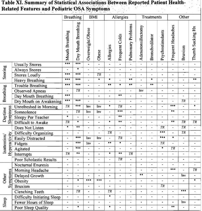
Table XI. Summary of Statistical Associations Between Reported Patient Health-Related Features and Pediatric OSA Symptoms ... 53
Table XII. Statistical Associations Between Evaluated Facial Morphology and Reported Pediatric OSA Symptoms ... 54
Table XIII. Statistical Associations Between Evaluated Long Face Morphology and Reported Pediatric OSA Symptoms ... 55
Table XIV. Statistical Associations Between Evaluated MP A and Reported Pediatric OSA Symptoms ... 55
Table XV. Statistical Associations Between Evaluated Open Bites and Reported Pediatric OSA Symptoms ... 55
Table XVI. Statistical Associations Between Evaluated Maxillary Width and Reported Pediatric OSA Symptoms ... 56
Table XVII. Statistical Associations Between Evaluated Palatal Morphology and Reported Pediatric OSA Symptoms ... 57 Table XVIII. Statistical Associations Between Evaluated Maxillary Posterior Crossbites and Reported Pediatric OSA Symptoms ... 57 Table XIX. Statistical Associations Between Evaluated Mandibular Deficiency and Reported Pediatric OSA Symptoms ... 58 Table XX. Statistical Associations Between Evaluated Skeletal Classification and Reported Pediatric OSA Symptoms ... 58 Table XXI. Statistical Associations Between Evaluated Dental Classification and Reported Pediatric OSA Symptoms ... 59 Table XXII. Statistical Associations Between Evaluated Incisor Overjet and Reported Pediatric OSA Symptoms ... 59 Table XXIII. Statistical Associations Between Evaluated Maxillary Dental Crowding and Reported Pediatric OSA Symptoms ... 60 Table XXIV. Statistical Associations Between Evaluated Mandibular Dental Crowding and Reported Pediatric OSA Symptoms ... 60 Table XXV. Statistical Associations Between Evaluated Tonsillar Morphology and Reported Pediatric OSA Symptoms ... 61 Table XXVI. Statistical Associations Between Evaluated Macroglossia and Reported
Pediatric OSA Symptoms ... 62 Table XXVII. Statistical Associations Between Evaluated Mouth Breathing and Reported Pediatric OSA Symptoms ... 63 Table XXVIII. Statistical Associations Between Reported Mouth Breathing and Reported Pediatric OSA Symptoms ... 63 Table XXIX. Statistical Associations Between Reported Daytime Mouth Breathing and Reported Pediatric OSA Symptoms ... 64 Table XXX. Statistical Associations Between Ca1culated Overweight and Obese BMI Categories and Reported Pediatric OSA Symptoms ... 65
Table XXXI. Statistical Associations Between Calculated Obese BMI Category and Reported Pediatric OSA Symptoms ... 65 Table XXXII. Statistical Associations Between Reported Allergies and Reported Pediatric OSA Symptoms ... 66 Table XXXIII. Statistical Associations Between Reported Colds and Reported Pediatric OSA Symptoms ... 66 Table XXXIV. Statistical Associations Between Reported Pulmonary Problems and Reported Pediatric OSA Symptoms ... 67 Table XXXV. Statistical Associations Between Reported Adenotonsillectomy Procedure and Reported Pediatric OSA Symptoms ... 67 Table XXXVI. Statistical Associations Between Reported Bronchodilator Medication Use and Reported Pediatric OSA Symptoms ... 68 Table XXXVII. Statistical Associations Between Reported Psychostimulant Medication Use and Reported Pediatric OSA Symptoms ... 68 Table XXXVIII. Statistical Associations Between Reported Frequent Headaches and Reported Pediatric OSA Symptoms ... 69 Table XXXIX. Statistical Associations Between Reported Bruxism and Reported Pediatric OSA Symptoms ... 69 Table XL. Statistical Associations Between Reported History of Finger/Thumb Sucking Habit and Reported Pediatric OSA Symptoms ... 70 Table XLI. Statistically Significant Associations Between Reported Sleep Quantity and Selected Reported Pediatric OSA Symptoms ... 72
LIST OF FIGURES
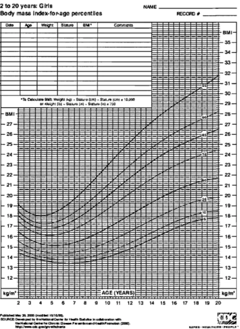
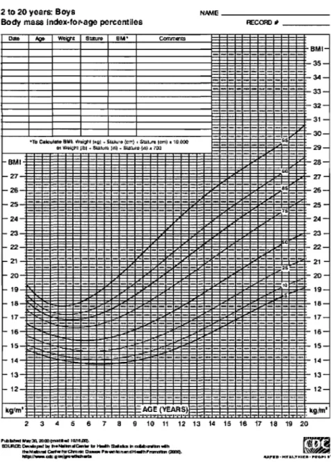
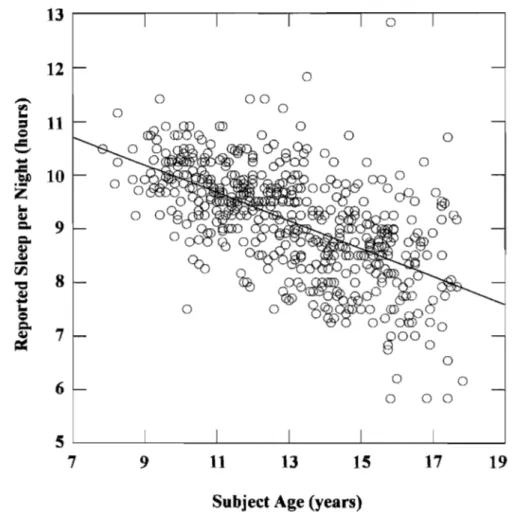
Figure 1. BMI-for-age girls 2 to 20 years ... 18 Figure 2. B1-H-for-age boys 2 to 20 years ... 19 Figure 3. Tonsillar hypertrophy and pharyngeal airway obstruction in an adolescent orthodontie patient ... 24 Figure 4. Average ho urs of reported subject sleep per night ... 71
LIST OF ABBREVIATIONS
% ADHD AI AHI ANOVA BMI CI CNS CPAP EEG h kg LAUPm
minmm
MMA MPA NHANES III OB OJ OSA OR PSG RDI REM RPE Percent Attention-DeficitIHyperactivity Disorder Apnea Index Apnea-Hypopnea Index Analysis of Variance Body Mass Index Confidence Interval Central Nervous SystemContinuous Positive Airway Pressure Electroencephalogram Hour(s) Kilogram( s) Laser-Assisted Uvuloplasty Meter(s) Minute(s) Millimeter(s) Maxillomandibular Advancement Mandibular Plane Angle
National Health and Nutrition Estimates Survey III Overbite
Overjet
Obstructive Sleep Apnea Odds Ratio
Polysomnography
Respiratory Disturbance Index Rapid Eye Movement
RVTR SD SDB TMD UARS UPPP
Radiofrequency Volumetric Tissue Reduction Standard Deviation
Sleep-Disordered Breathing Temporomandibular Disorder Upper Airway Resistance Syndrome Uvulo-Palatopharyngoplasty
ACKNOWLEDGEMENTS
From planting the first seeds to its completion, many have supported me in my master's specialty pro gram in orthodontics. 1 have been fortunate to have been encouraged by educators in dentistry from both renowned institutions on the slopes of Mont Royal. First, and foremost, 1 would like to thank my wife Amy. The support of my best friend and partner is precious to me. Though being separated in order to follow our common goal, you continue to bring out the best in me. You are my love.
Thank you to Dr. Athena Papadakis for having mentored and guided me throughout this research project. Thanks for having approached me with an interesting proposaI for a project. Your dedication, support and encouragement for this project, as weIl as in teaching orthodontics, have made my studies the accomplishment that they are. 1 wish you continued success in orthodontics, education, research, and, most importantly, in life. 1 wish you best of luck with your new family.
1 would like to thank Dr. Claude Remise for taking a chance on me from our first meeting in the admissions interview. 1 hope 1 have eamed your trust and that 1 will develop into an orthodontist that you will be proud of. 1 would like to thank you for being such a dedicated educator and mentor.
Thank you to Dr. Jack Turkewicz and Dr. Jean-Charles Létoumeau for accepting to be on my jury panel for this project. Your meticulous corrections and attention to detail are appreciated. 1 would also like to thank aIl of my teachers, orthodontists and professors, who have dedicated themselves to giving back to our profession in order to guide the following generations. Without you, there would be no future in the dental profession. 1 would like to thank Dr. Gilles Lavigne for his assistance in focusing this endeavour. 1 have been fortunate to have had such an eminent sleep medicine specialist to guide me with this project. 1 wish you best ofluck as Dean of the Faculty of Dentistry.
The statistical portion of this study would not have been possible without Mr. Pierre Rompré. Thank you for your patience with a statistical novice. 1 did not have an appreciation of the amount work going into the statistical analysis until 1 attempted to decipher the 2000-plus pages of data outputs! Thank you for helping guide me in focusing the data on our significant results.
Those who organized the orthodontie screening c1inic were essential to the success of this project. Thank you to Sophie Fournier, Anne Saumure, and Arun Un for having administered the patient questionnaires so efficiently, and for your patience in running the sometimes chaotic c1inics.
Thanks to my coIleagues and friends at the Université de Montréal. 1 wish future happiness and success to my friends and c1assmates Normand and Serge. We went through a lot of enjoyable and stressful times over three years together. 1 reaIly appreciate you warmly welcoming me to your school. 1 also wish the best to aIl of the residents in the Orthodontie Section at the Université de Montréal.
A final thanks to my parents, and also to my sisters Anne and Carole. Your unending encouragement is extremely important to me. 1 am so fortunate to have such a loving and supportive family.
1.
INTRODUCTION
Sleep-disordered breathing (SDB) encompasses a spectrum of disorders. These range from a partial obstruction of the upper airway, producing primary snoring, increasing in upper airway resistance to complete obstruction, which results in obstructive sleep apnea (OSA). SDB is characterized by recurring episodes of partial or complete obstruction of the upper airway during sleep, often in association with loud snoring. The upper airway obstruction is often associated with arousals, sleep fragmentation, non-restorative sleep, intermittent hypoxemia and hypercapnia, and noctumal hypertension (American Thoracic Society 1996). Even simple chronic snoring is considered abnormal in a pediatric population (O'Brien 2004).
The pharynx is a single conduit which must be compliant for food propulsion and vocalization, and also be firm enough for air flow. The basis of the obstruction in OSA is a decrease in nasopharyngeal and oropharyngeal dimensions.
Obstructive sleep apnea (OSA) is a multifactorial disease. It is related to upper airway skeletal deformities, soft tissue anatomy changes, the size of lymphoid structures, height, increased weight, age, and medical conditions (Nixon 2005).
Difficulties with sleep onset occur in about Il % of children (Paavonen 2000). Primary snoring is thought to occur in 3.2% to 12.1 % of the pediatric population, and OSA has been estimated to affect 0.7% to 10.3% of children (Gislason 1995, Ali 1993, Redline 1999). Adenotonsillar hypertrophy has long been identified as the primary cause of reduced airway dimensions in children. However, the obesity epidemic in the industrialized world may now be adding to the problem.
Sleep apnea has major health and social consequences. Growth may be inhibited due to OSA. Severe cardiovascular complications may result from both OSA and childhood
obesity. Leaming and behaviour problems in children, such as attention-deficit hyperactivity disorder (ADHD), have shown a three-fold increase in children with SDB (American Academy of Pediatrics 2002).
Despite their impact on public health, craniofacial and orthodontic anomalies related to OSA are often ignored. Sorne of the abnormalities associated with OSA are related to maxillo-mandibular development. A rarely investigated, complex interaction exists between facial growth and nasal breathing (Guilleminault 2001). Pediatricians and orthodontists should therefore consider orthodontic problems as part of a growing child's overall health (Guilleminault 2005).
In an attempt to prevent the consequences of OSA, young patients with SDB should be carefully examined to rule out physical abnormalities. Clinical examination of the upper airway anatomy may identify anatomical risk factors predisposing a patient to the development of ab normal breathing during sleep. Though airway obstruction is aggravated by muscle tone changes during sleep, underlying anatomical factors, such as adenotonsillar hypertrophy, obesity, and craniofacial abnormalities predispose a patient to OSA (Primhak 2005).
Pediatric sleep questionnaires are designed to obtain maximal information from parents and children about his/her sleep behaviour. However, clinical history alone is not sufficiently reliable to distinguish OSA from primary snoring (Carroll 1995). Therefore a complete sleep evaluation is required for the final diagnosis of OSA. Though ovemight polysomnography (PSG) may be the gold standard diagnostic sleep study for OSA, it is both expensive and of limited accessibility. A detailed questionnaire lS
extremely useful for screening prior to the diagnostic polysomnographic examination. It is important that children and adolescents with OSA be identified and treated as early as possible, not only for their respiratory problems, but also for their dentofacial development. This requires an integrated diagnosis and management of pediatric sleep apnea. The orthodontist is well positioned in this respect as a majority of patients
presenting for orthodontie evaluations are growing children and adolescents. Many of the craniofacial structures evaluated by an orthodontist in the planning of orthodontie treatment may be related to upper airway obstruction. Other symptoms may be elucidated from medical and sleep questionnaires. The orthodontist should identify patients at-risk and refer them to sleep specialists for further study and diagnostic confirmation. Integrated within the treatment team, the orthodontist may also participate in the evaluation of the patient's growth post-adenotonsillectomy, assist in the treatment of mild to moderate OSA cases with anterior mandibular repositioning devices or palatal expansion, or prepare the pre-surgical orthodontie treatment of severe cases.
The purpose of this pilot study is to assess a child and adolescent population which presents for evaluation at a university orthodontie clinic. This epidemiological study will identify the prevalence of different skeletal and dental aspects of malocclusion and SDB/OSA parameters in this orthodontie population. It will also attempt to demonstrate statistically significant correlations between clinical signs of malocclusion and reported symptoms of SDB. It is hypothesized that deficiencies in maxillo-mandibular transverse and antero-posterior dimensions, as weIl as increased vertical facial height will be related to reported symptoms of SDB. However, the most important objective of this study is to increase awareness of the importance of evaluating aIl orthodontie patients for sleep and breathing problems in order to prevent serious long-term health and quality of life consequences.
6
2.
LITERATURE REVIEW
2.1 Sleep-Disordered Breathing
Sleep is a delicate balance among three behavioural states: wakefulness, non-rapid eye movement sleep, and rapid eye movement (REM) sleep. The balance that exists among the se states can be easily disrupted by medications, physiological and psychological factors, as weIl as environmental causes (Waite 1998).
At the onset of sleep, there is an increase in upper airway resistance due to a reduction in pharyngeal muscle activity (Worsnop 2000). A slight decrease in tidal volume also occurs. An increased breathing frequency normally compensates for these decreases, keeping the minute ventilation normal. Increased upper airway resistance due to physical obstructions may lead to a collapse ofthe upper airway. Apneic events occur in these circumstances (Guilleminault 2005).
A wide range of dyssomnias exist. Sleep disorders range from insomnia and primary snoring to OSA and narcolepsy (Waite 1998). SDB is described as a continuum of severity from primary snoring to OSA.
Primary snoring prevalence among children and adolescents has been reported to be between 3.2% and 12.1 % (Ersu 2004, Gislason 1995, Ali 1993). Snoring sounds are produced by vibrations ofthe pharyngeal tissues due to air turbulence during inspiration. Reduced airway dimensions are associated with snoring. The prevalence of snoring increases with age, reaching between 40% and 50% of the general population above the age of 65 (Lavigne 1999). Snoring is an important indicator of apnea. Chronic snoring, though common in adulthood, should be considered abnormal in a pediatric population (O'Brien 2004).
Upper airway resistance syndrome (UARS) is a disease that presents in slim people complaining of excessive snoring and daytime fatigue, but without apnea, hypopnea, or oxygen desaturation. Sleep fragmentation and REM-sleep depravation may be related to this syndrome (Waite 1998).
Sleep apnea can be classified as obstructive, central, or mixed (Lavigne 1999). Central sleep apnea is a neurological condition resulting from a depression of the motor respiratory system. Mixed sleep apnea is a combination of obstructive and central apneas.
2.1.1 Obstructive Sleep Apnea
The American Thoracic Society (1996) defines OSA as "a disorder of breathing during sleep characterized by prolonged partial upper airway obstruction and/or intermittent complete obstruction (obstructive apnea) that disrupts normal ventilation during sleep and normal sleep patterns." Repeated episodes of partial or complete upper airway obstruction during sleep characterize OSA. This is usually accompanied by a reduction in oxygen saturation. Partial (hypopnea) or complete (apnea) obstruction results in brief awakenings from sleep, or transitions to lighter stages of sleep. Sleep fragmentation and unrefreshing sleep thus ensue.
The estimated prevalence in the general adult population is 2% to 4%, mostly affecting overweight, middle-aged males (Young 1993). Post-menopausal women are also affected. A large number of anatomic and physiological factors render OSA a complex multifactorial condition.
8
2.2 Pediatrie Obstructive Sleep Apnea
Sleep apnea in children has a different etiology, clinical presentation and treatment. Upper airway obstruction in children is more likely to involve a reduction in airflow (hypopnea) than a complete obstruction (apnea) (Nixon 2005). Though pediatric populations experience sleep apnea, no large population based studies have evaluated pediatric OSA prevalence (Guilleminault 2005). Studies of pediatric OSA with limited sample sizes have estimated its prevalence to be between 0.7% and 10.3% (Gislason 1995, Ali 1993, Redline 1999). OSA has become recognized as one of the most common respiratory disorders of childhood, with similar rates for girls and boys (Redline 1999).
2.2.1 Diagnosis
Child and adolescent sleep-disordered breathing is often first suspected based on parental concems. A thorough interview about sleep behaviour and SDB-associated factors should be undertaken using questionnaires. Though clinical history-taking and questioning have demonstrated low sensitivity and specificity regarding the diagnosis of SDB, it remains the primary screening tool leading to further assessment by ovemight polysomnography (PSG) (American Academy of Pediatrics 2002).
Systematic parental questioning regarding their child's symptoms is essential for a chi Id suspected of SDB. Questionnaires can give an initial indication of the problem. A variety of pediatric questionnaires exist for signs and symptoms, and quality of life implications of OSA (Chervin 2000, de Serres 2000, Owens 2000, Brouillette 1984). It is essential to evaluate a patient' s medical history including: reports of snoring, daytime sleepiness, unrefreshing sleep, moming headaches, choking during sleep, memory impairments, depressive reactions, enuresis, and behavioural problems.
Much can be leamed from a clinical examination. Abnormal narrowing of the nose, nasopharynx, oropharynx, or hypopharynx may lead to abnormal air flow during sleep and to clinical symptoms of SDB (Guilleminault 2005).
The gold standard diagnostic tool is the ovemight sleep study. Various sleep parameters are measured by PSG and it distinguishes between the various forms of SDB to confirm the presence or absence of OSA.
2.3 Diagnostic Tools for SDB
The diagnostic tools used to evaluate pediatric patients for SDB include questionnaires, a clinical exam, diagnostic imaging, and final confirmation with the ovemight PSG laboratory exam.
2.3.1 Sleep Questionnaires
Many questionnaires exist for the assessment of OSA. Standardization and validation of these questionnaires has helped in the screening of SDB. Questionnaires such as the Berlin Questionnaire, the Pittsburgh Sleep Quality Index, the Epworth Sleepiness Scale, and the Sleep Apnoea Quality of Life Index are aIl validated screening instruments used for adults (Netzer 1999, Buysse 1989, Chervin 1997, Lacasse 2002). In children, the Pediatric Sleep Questionnaire and the Children's Sleep Habits Questionnaire are two instruments which have recently gained in popularity (Chervin 2000, Owens 2000). Other questionnaires have also been developed using common signs and symptoms of SDB in order to attempt to identify OSA in children. Pediatric sleep questionnaires have been available for nearly 25 years (Brouillette 1984). The difficulty of low sensitivity and specificity relating to OSA diagnosis cornes from the difficulty of differentiating primary snoring from OSA (American Academy of Pediatrics 2002, Carroll 1995).
Parental reporting of sleep behaviour may be another confounding factor in reporting SDB. Sorne studies have demonstrated good correlation, while others question whether a parent who sleeps in another room can accurately report the sleep behaviour of a child (Lumeng 2008, Chervin 2007, Johnson 2006). One study by Chervin et al. (2000) has demonstrated high sensitivity and specificity between pediatrie OSA-related questions and diagnosis on polysomnography.
Questionnaires are important to aid In the detection of possible SDB. However,
diagnosis of OSA through clinical history and physical examination has been shown to correlate poorly with polysomnographic confirmation of the diagnosis in children. (Brietzke 2004, American Academy of Pediatries 2002, Carroll 1995) Sleep questionnaires have difficulty in differentiating between the different severities of SDB. Given the expense and limited availability of ovemight PSG, questionnaires and a clinical exam remain the most important diagnostic tools for SDB.
2.3.2 Clinical Evaluation
Since OSA may be due to upper airway obstruction, a pediatrie exam as weIl as a thorough upper airway evaluation is required. Abnormal anatomy and/or poor pharyngeal muscle tone may result in inadequate airway patency. A multitude of clinical features are related to OSA. Risk factors for SDB are found in Table 1.
Tabié I.
Faétorslmpli~at~~'jil~'leep-Disordered
BreathinginChiÙi;~:ri:~hd ~t:~i,;
"'J
Adoléscents ' ': ',. ",8., 4,' ' " , ' " , ' , ;';,.'.,,'
:",1
,~,' :', .'7>','.'> ;'<' ! ' " C ' . . >< : . , , \ ' ' 1
Craniofacial morphology Other
Soft Tissue
•
Muscular hypotonia•
Adenotonsillar hypertrophy•
Mouth breathing•
Narrowed pharyngeal dimensions•
Asthma•
Nasal obstruction•
Allergies•
Elongated soft palate•
Allergie rhinitis•
Macroglossia•
ObesitySkeletal
•
Hypertension•
N arrow palate•
Congestive heart failure•
Posterior crossbite•
CNS depressant medications•
Long face (Adenoid facies)•
Alcohol•
Severe dental crowding•
Smoking•
Mandi bular retro gnathism•
Developmental delay•
Severe overjet•
Short stature•
Inferiorly placed hyoid bone•
Craniofacial syndromes•
Maxillary hypoplasia(Halbower 2006, Guilleminault 2005, Primhak 2005, Crabtree 2004, Goldstein 2004, Shin 2003, Schechter 2002, Chervin 2000, Cistulli 1996)
2.3.3 Radiological Imaging
Three dimensional imaging using magnetic resonance or computed tomography gives precise information about the soft and hard tissue anatomical structures. Imaging studies have also shown that anatomical obstruction in OSA patients occurs primarily in retropalatal and retroglossal areas (Schwab 2001).
Lateral cephalometric radiographs assess the skeletal and dental maxillary and mandibular relationships. Soft tissue relationships of the palate, tonsils and adenoids and the posterior pharynx can also be assessed. Decreased airway dimensions due to soft tissue and skeletal causes have been demonstrated by cephalometry (Lowe 1997). Cephalometric evaluation may aid in the screening of the posterior airway and in the longitudinal evaluation of treatment, but has the disadvantage of being a static two-dimensional image (Pracharktam 1996). It is, however, a useful tool in planning orthognathic surgery.
2.3.4 Polysomnography
An accurate diagnosis of respiratory pauses during nighttime sleep is based on a comprehensive monitoring of various sleep parameters. Polysomnography (PSG) recordings during sleep are the gold standard used to confirm the presence of SDB. Various physiological parameters of sleep are measured in an overnight sleep study. Monitoring of sleep/wake states through electroencephalography (EEG), electro-oculography, electrocardiography, electromyography of the chin and leg, body position, and appropriate monitoring of breathing must be included in the overnight study. Other monitoring may include nasal cannula-pressure transducer, oral thermistor, chest and abdominal belts, a neck microphone, pulse oximetry, and videotaping (Guilleminault 2005).
Recordings may be made in a sleep laboratory or in an ambulant setting. The home setting has been shown to be more acceptable to younger patients and their parents. It helps to maintain an environment that does not disrupt the child's normal sleeppattern (Nixon 2005). However, if performed in a home setting, the validity of these results is still uncertain when compared to the gold standard of overnight laboratory PSG (American Academy of Pediatries 2002). If performed in a sleep laboratory, a parent should stay with the child throughout the night.
Diagnostic criteria for pediatrie PSG differ from adult norms. Consensus on these criteria has been difficult to achieve (Sheldon 2001). Recent guidelines from the American Academy of Pediatries (2002) and the American Thoracic Society (1996) have clarified the thresholds used for pediatrie OSA. Apnea is considered as an absence of airflow at the nose and mouth lasting longer than 2 respiratory efforts. If, according to the apnea index (AI), more than 1 apneic event occurs per hour of sleep, pediatrie OSA is confirmed. Hypopnea is a reduction in airflow of at least 50% in nasal flow amplitude longer than 2 breaths and may be associated with an arterial oxygen desaturation and an EEG arousal. If the apnea-hypopnea index (AHI) records at least 5 events per hour, OSA is also confirmed. The respiratory disturbance index (RDI) allows for the inclusion of other abnormal breathing events such as snoring and a corresponding EEG arousal to be included. A eut-off of at least 5 events per hour is also used to demonstrate pathological sleep breathing (Guilleminault 2005).
Following the complete medical exam and PSG testing, the diagnosis of OSA should be made by a sleep medicine or respiratory disorder specialist.
2.4 Factors Associated with SDB
Pediatrie OSA has been recognized as a multifactorial disease. There is no pathognomonic sign or symptom that is clinically predictive of OSA, rendering a clinical diagnosis difficult.
2.4.1 Symptoms of SDB
Sleep-disordered breathing In children has been associated with a wide variety of
clinical findings. Patients often report excessive daytime fatigue, morning headaches, insomnia, loud and abnormal snoring, restless sleep, impaired intellectual function,
mood disturbance, aggressive behaviour, and hyperactivity (Mitchell 2006). Symptoms of SDB can be divided into nighttime and daytime observations. (See Table II)
Nighttime
• Chronic, heavy snoring
• Difficulty breathing during sleep • Witnessed breathing pauses during
sleep
• Mouth breathing • RestIess sleep
• Periodic limb movement • Delayed onset of sleep • Insomnia
• Frequent awakenings • Nocturnal migraine
• Abnormal sleeping positions • Drooling • Sleep talking • Sleepwalking • Sleep terror • Nocturnal sweating • Enuresis
• Difficulty waking up in the morning • Confused arousal
Daytime
• Morning tension-type headache • Mouth breathing
• Excessive morning thirst
• Excessive fatigue and sleepiness • Abnormal shyness, withdrawn and
depressive presentation • Behavioural problems
• Pattern of attention-deficit/ hyperactivity disorder (ADHD) • Aggressiveness
• Irritability • Poor concentration • Leaming difficulties • Memory impairment
• Poor academic performance
(Beebe 2006, Halbower 2006, Guilleminault 2005, Primhak 2005, Crabtree 2004, Goldstein 2004, Shin 2003, Chervin 2002, Cistul/i 1996)
2.4.2 Medications
Medications affecting dopamine, acetylcholine, serotonin, and other neurotransmitter levels in the central nervous system (CNS) also induce alterations in sleep (Pagel 2001). Central nervous system depressant medications, sedative-hypnotic drugs, antidepressants, alcohol, and smoking may also adversely affect the patient's sleep. Though uncommon in younger children, these substances may be used by older adolescents.
Alerting or psychostimulant medications such as methylphenidate (Rital in) used in the treatment of ADHD are increasing in prevalence in the pediatric population. These medications may also adversely affect sleep quality and quantity (Sangal 2006).
2.4.3 Learning and Behaviour Problems
Childhood is a period of rapid neurological development. Sleep disruption throughout this period may lead to significant neurocognitive deficits (Kennedy 2004). Leaming performance, attention and daytime behavioural problems have been demonstrated in children where SDB is present (Goldstein 2000, Gozal 1998). Hyperactivity and behaviour problems have long been described by parents of children with OSA (Ali 1993). Memory, leaming and problem solving are also reduced in children with SDB (Owens 2000). Cognitive deficits and the possibility of permanent neuronal injury as sequelae to OSA have also been demonstrated (Halbower 2006).
An association exists between the presence of SDB and leaming and behavioural problems; however a link between the severity of the OSA and the severity of neurocognitive scores has not been shown (Mitchell 2007, Beebe 2006, Friedman 2003).
2.4.4 Allergies and Asthma
Nasal obstruction due to allergic and infectious enlargement of tonsils and adenoids reduces the upper airway lumen and increases the symptoms of SDB. Increased nasal resistance due to occlusion inevitably leads to mouth breathing (Salem 2004). Allergic edematous conditions such as rhinitis and rhinosinusitis should be evaluated. Nasal occlusion has been related to an increase in OSA symptoms in children and adolescents. Patients that present with increased habituaI snoring have an increased risk for pediatric OSA (McColley 1997, Millman 1996).
2.4.5 Medical Disorders and Syndromes
High risk groups for OSA are those children with hypothyroidism and premature birth. Syndromes with severe craniofacial malformations which result in a narrowing of the naso-pharyngeal airway are also associated with OSA (Sheldon 2001). Syndromes with severe mandibular deficiencies, such as Pierre-Robin sequence, Treacher-Collins syndrome, or those with hypoplasia of the maxilla such as Apert's and Crouzon's demonstrate SDB (Primhak 2005).
The following syndromes, which demonstrate pharyngeal muscle hypotonia, are strongly associated with OSA: Duchenne muscular dystrophy, Down syndrome, Prader-Willi syndrome, Marfan's syndrome, achondroplasia, mucopolysaccharoidosis, spina bifida, and cerebral paIsy (Primhak 2005, Nixon 2005, de Miguel-Diez 2003, Cistulli 1996). Though OSA may predominate, central and mixed apneas may also occur in children with neurological abnormalities (Sheldon 2001).
2.4.6 Obesity
Over the past 25 years, the prevalence of childhood obesity has more than doubled (Whitlock 2005). A recent study of Canadian schoo1children and adolescents has shown that approximately 26% of boys and 17% of girls are either overweight or obese (Boyce 2008). The same study found a rise in obesity from 4% to 6% between 2002 and 2006. Being overweight in childhood is associated with an increased risk for early bone maturation, type 2 diabetes mellitus, glucose intolerance, hyperlipidemia, hypertension, and other cardiovascular diseases (Dietz 1998). Significant short-term consequences are psychosocial in origin. Decreased self-esteem, quality of life and social marginalization are common (Strauss 2003). Being overweight and obese in childhood and adolescence tends to carry on into adulthood.
With the increasing prevalence of obesity, a greater percentage of the pediatric population is at-risk for OSA (Redline 1999). The deposition of adipose tissue in the upper airway may play an important role in the development of pediatric OSA (Yu 2003). SDB has been shown to be common in populations of overweight children (Verhulst 2007). However, though obesity is often related to OSA, it is not an essential factor as thin people may also have OSA.
The body mass index (BMI) is a screening tool which is used to calculate healthy weights related to the height and sex of an individual. BMI has been shown to correlate with direct measures of body fat in children and adolescents (Mei 2002). Elevated childhood BMI also predicts future adiposity, as weIl as future morbidity and premature mortality (Must 1999). In growing children and adolescent patients, BMI is age-specific and sex-specific ~n order to compensate for differences in body fat at different ages and between the sexes. This differs significantly from adult BMI which does not take age or sex into account (Kuczmarski 2000).
BMI-for-age curves have been developed by the Centers for Disease Control in the United States to evaluate the size and growth of children (Figures 1 and 2) (Kuczmarski
2000). Using percentiles, four categories have been delineated to classify children according to their weight status (Table III).
210 20 years: GIrls
Body mass Index-fo",ago percentlles
BMI -27 -26 -2'5 -24 23
-Tb C,ak'\J.rrto eWli: Wri;iU "QI ... flw.utù «(!fft ~ S"ilt_~ IUft) " "iO,.tXJ:t
Of tH~:n1 fb1 ,.. SUlM\l fn) ~ SID':I.itu Çot)J JI ".'00
21 , -20 -19 111 r 17 r 16 r 15 r 14 r 13 '- 12 NMIE _ _ _ _ _ _ _ _ _ _ _ RECORD (J _ _ _ _ _ BMI 35- 34- 33- 32- 31- 30- 29- 26- 27- -.;;~=;...26-- -.;;~=;...26-- -.;;~=;...26-- -.;;~=;...26-- -.;;~=;...26-- -.;;~=;...26-- , .;;~=;...26-- 25-7si=== :""24- 23- 2221 - 20- 19- 18- 17- 16- 15- 14- 13- 12-kglml c - - - " " . , . . . . :--:AG,;,~~RS) :---~---:, kg,m' 2 3 4 5 6 7 8 9 10 11 12 13 14 15 16 17 18 19 20
l'lti~ MIr ~:zo:JJ ))raI1Ia 1~·It1~ ..
m:II.IU:Z: o.v.6:J.-t b( .,.NItm .. c;., .. b ... Q.lab. h cd"nnüœl",h
thlNIIIIu,. c-..I.rgam..-: 1J . . . 1' ... tJn .. d .. ' . . flflrm"dh'I ama; •
• /I ... c:ra:.g1M'gv'llCl'l:fl..m
2 to 20 years: Boys NMIE _ _ _ _ _ _ _ _ _ _ _ Body mass lnd&x·tol'age percCntl108 RECORD Il
aMI -'Zr -26 -25 -24 -21 20 19 la 17 16 15 14 13 12 Bill'
-To ~Q10 ElWl. w'i;f1f ' • .QI .. SU.h.llQ ~~) l :s.~t.;tQ Ifffl) Il 'a),iK'JO' Ot w.ttcfll fbl + amA:fil t·nl .. 6Uî.W1> 1:f~) .II 1'00
',s-25 la ~ AGE (VEARS} _ .. _ .. _ .. .... -.,.. • "'t/" ""'" "I!'" 6,.,1- 35- 34- 33- 32- 31- 30- 29- 25- 27- _1:-26- 25- 24- 23- 2221 - 20- 19- 18- 17- 16- 15- 14- 13- 12-kg,tn' :5 6 7 8 9 10 11 12 13 14 15 1617 HI 19 20
,t.tIbtnt ., ... :11, ZlJh)TO:tI.t 1Q,1a~
S:Il.ltœ c. .... JB! brt h_NIi'b'lJlc... il" ... ~d:a" ~~b1"h
tNNlllbl1llll c..twr.arar*l D _ " . . . . lIDn.,dH.."PI'ln'db'l ~
. . # .... ·ai::IIlIft'gv"llll!tdl . .
Weight Status Category Underweight Healthy weight Overweight Obese (Adaptedfrom Barlow 2007) Percentile Range < 5th percentile 5th to 84th percentile 85th to 94th percentile 2: 95th percentile
An additional tool to evaluate obesity is neck circumference. It has been a useful predictor in adults for obesity correlations with apnea, but no appropriate scale has been developed for growing children (Guilleminault 2005).
2.4.7 Upper Airway Obstruction
Any anatomical narrowing in the various parts of the upper airway will have additive effects contributing to SDB. Abnormalities that decrease the radius of the nasal, oral, or pharyngeal airway result in increased airway resistance. Though enlarged tonsils and adenoids contribute greatly to SDB, multiple anatomical obstructions should also be considered (Guilleminault 2005).
2.4.8 Nasal Morphology
Upper respiratory tract pathosis has been associated with OSA. AlI aspects of the nose should be evaluated for obstruction. Asymmetry of the nares, colIapse of the nasal valves during inspiration, a large septal base, septal deviations, turbinate hypertrophy, enlarged adenoids, masses, and polyps are evaluated (Guilleminault 2005). HabituaI
mouth breathing may also be a result of nasal occlusion. This is a factor which has been implicated in OSA (Salem 2004).
2.4.9 Craniofacial Morphology
Malformations of the maxilla, mandible and associated structures can also result in upper airway obstruction during sleep. A micrognathic or retrognathic mandible will likely cause the tongue to reduce the pharynge al airway space and decrease the airflow during sleep. Cleft palate and other craniofacial syndromes have also shown that severe abnormal positioning of the maxillae increases the risk for OSA (Muntz 2008).
2.4.10 Skeletal Morphology
Dimensions of the orofacial skeleton are implicated in pediatric OSA. Extremely reduced airway dimensions due to craniofacial syndromes demonstrate that the maxillae have a significant role to play in the etiology of SDB (Primhak 2005). A high narrow palate, narrow maxilla, posterior crossbites, increased overjet, and dental crowding aIl demonstrate abnormal maxillomandibular development.
2.4.10.1 Sagittal
Mandibular retrusion has long been a factor associated with OSA (Triplett 1989). Mandibular deficiency impinges on the pharyngeal airway space, thereby increasing posterior airway resistance. Advancing the mandible in children with removable functional appliances decreases the incidence of OSA (Villa 2002). Angle's molar classification as weIl as the severity of overjet (OJ) will indicate problems relative to maxillary and mandibular length.
Cephalometrics in OSA patients has shown that decreased mandibular length, decreased maxillary length, skeletal retrusion, increased mandibular plane angle (MPA), and low hyoid position have implications in SDB (Kulnis 2000, Lowe 1997). A decreased mandibular body length has a clinically significant association with OSA (Miles 1996). Other studies in children with mild to moderate OSA have demonstrated no association between mandibular dimensions and OSA (Schiffman 2004).
2.4.10.2 Vertical
An increased lower facial third in dolichofacial or long~face patients has been implicated as a risk factor in OSA (Contencin 2003). The mandible is located in a more retruded position in these patients. AIso, the mandibular plane is often steeper. Children with long faces, retropositioned mandibles and associated lip incompetence have been shown to have increased sleep-disordered breathing symptoms (Zucconi 1999, Guilleminault 1996). Increased facial dimensions can be measured by facial height proportions (long-face), relative height-to-width proportions (dolichofacial), and increased MP A. Patients are also more prone to having anterior open bites (Salem 2004).
2.4.10.3 Transverse
Maxillary constriction is a sign of reduced transverse dimension of the upper airways. Patients with constricted maxillae have increased nasal resistance which results in increased mouth breathing. An associated low tongue posture may also be present, resulting in posterior airway narrowing (Cistulli 1996). Transverse maxillary deficiency can be clinically assessed. A high narrow palate can be observed. Severe crowding of the maxilla and mandible may also be present. Teeth may be tipped buccally or lingually (Betts 1995). Posterior dental crossbites of two or more permanent teeth can be considered a skeletal crossbite (Jacobs 1980).
2.4.11 Soft Tissue
The intra-oral exarn should evaluate the tongue, soft palate, uvula, tonsils and pharyngeal walls as possible causes of obstruction. The evaluation of the oropharynx begins with the relationship between the soft palate, uvula and tongue. The Mallampati (1985) classification, originally developed for evaluation prior to endotracheal intubation, may be a useful scale to assess posterior airway opening. Low lying, thickened palates are a significant finding. The modified Mallampati classification is graded from l to IV depending on the arnount of pharyngeal space and soft palate visible when the mouth is opened wide (Friedman 1999).
The tongue should be evaluated relative to its size. Macroglossia is an extrusion of the tongue above the plane of occlusion of the mandible (Waite 1998). An enlarged tongue may impinge on the posterior airway space and be implicated in OSA.
Neuromuscular disease and decreased tonicity of the pharyngeal musculature also predisposes a patient to pharyngeal collapse during inspiration (Amin 2006).
2.4.11.1 Adenotonsillar hypertrophy
Adenoid or tonsillar enlargement is the primary cause of anatomical obstruction in SDB in children (Gozai 1998). The enlargement of these tissues decreases the radius of the nasal and oral airway, thereby increasing airway resistance. This increase in resistance may become clinically significant during nighttime sleep (Sheldon 2001).
The peak of pediatrie OSA is thought to occur in the preschool years due to adenotonsillar hypertrophy (Gislasen 1995). Tonsils and adenoids progressively enlarge during childhood and adolescence, followed by a decrease in size during adult life (Vogler 2000, Jeans 1981). Studies have demonstrated that enlarged tonsils and
24
adenoids are correlated with increased prevalence of OSA, but are not correlated with the severity of OSA (Brooks 1998).
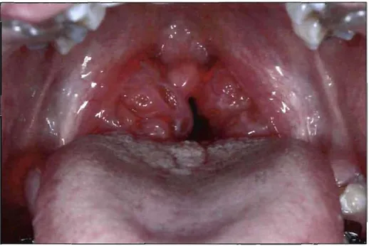
Tonsillar size is graded in relation to the obstruction of the airway (Figure 3). A commonly used scale scores the tonsils, from 0 to 4+, based on the amount of tonsillar pharyngeal obstruction visible from the widest mouth opening (Friedman 1999).
Figure 3. Tonsillar hypertrophy and pharyngeal airway obstruction in an adolescent orthodontie patient
2.5 Consequences of Pediatrie Obstructive Sleep Apnea
Overlooking a diagnosis or mismanaging a patient with OSA can have senous consequences (Capdevila 2008). Hypoxemia and increased intrathoracic pressure are associated with cardiopuJmonary changes in OSA. Cardiopulmonary dysfunction and excessive daytime somnolence may lead to the most severe consequences of pulmonary
dysfunction and premature death (Waite 1998). Fortunately, mortality due to OSA during childhood is unusual (Sheldon 2001).
Functional and neurocognitive dysfunctions contribute to the significant morbidity of OSA. Excessive daytime sleepiness, hyperactivity, personality changes, and deficits of attention, concentration, psychomotor skills, memory and higher cognitive functions contribute to poor intellectual and social achievement. Developmental delays and depression may also occur (Crabtree 2004, Sheldon 2001). Health-related quality of life is decreased in SDB patients (Rosen 2002). Recent studies also indicate that neuronal brain injury may also occur (Halbower 2006).
If OSA progresses into adulthood, workplace or motor vehicle accidents related to daytime hypersomnolence may lead to severe injury and mortality (Masa 2000). The natural history of OSA eventually results in myocardial ischemia, infarcts, arrhythmias, hypertension, and cerebrovascular accidents. Mortality is increased as the severity of OSA is increased (Partinen 1988).
2.6 Treatment of Sleep-Disordered Breathing
Treatment ofpediatric OSA targets the cause of the airway obstruction. The treatment of choice is tonsillectomy and adenoidectomy, as the hypertrophy of the se structures is the most common cause of childhood OSA (American Academy of Pediatrics 2002). However, other causes of obstruction may also exist. Treatment options range from behavioural changes to surgical treatment of mandibular advancement, bariatric surgery and tracheostomy.
Multidisciplinary management of OSA should involve respiratory physicians, sleep laboratory technicians, otorhinolaryngologists, oral and maxillofacial surgeons, and orthodontists (Sherring 2001).
26
The consequences of non-treatment of OSA in childhood may be severe due to the serious long term cardiovascular complications. Developmental processes may also be affected with lasting effects (Nixon 2005).
2.6.1 Behavioural Modification
Changes in sleep position, cessation of depressant medications and decreasing alcohol or drug use aids in preventing OSA. A voiding sleeping on the back may prevent snoring and anatomical narrowing of the airway. Alcohol and sedatives relax the pharynge al musculature, reducing airway patency. Their avoidance prior to sleep in adolescents may help decrease SDB symptoms. Allergies can be treated using intranasal steroids, and oral or topical decongestants (Lampasso 2004).
Though difficult to maintain long term, the prevention of obesity and weight control management are also essential during childhood. Not only does it help prevent OSA, but also a host of cardiovascular complications (Barlow 2007).
2.6.2 Continuous Positive Airway Pressure (CPAP)
The gold standard in treatment of OSA in adults is continuous positive airway pressure (CP AP). Nasal CP AP for children has been shown to be an effective and non-invasive treatment. However cooperation and training of the child and family remains the most common impediment to its use (Guilleminault 2005). The benefits of CPAP are decreased daytime sleepiness, fewer arousals, less oxygen desaturation, fewer apneic episodes, reduced hypertension, and improved cognitive function (Marcus 1995).
CP AP has poor compliance due to its si de effects which include: mask rash, conjunctivitis, rhinorrhea, sinusitis, congestion, epistaxis, tympanic rupture, pneumothorax, aerophagia, and chest pain (Hoffstein 1992).
Regular reassessments are required in growing children undergoing CP AP treatment. Mask and headgear fit should be evaluated, and attention placed on assessment of any possible maxillary growth restraint (Li 2000).
2.6.3 Oral Appliances
Functional readaptation may be attempted as a part of the treatment of SDB patients. These treatments may aid in enlarging the upper airway. These treatments are amenable to those who have had adenotonsillectomy and present with snoring or mild to moderate OSA, and those who are non-compliant with their CPAP.
2.6.3.1 Mandibular Repositioning Appliances
An increase in posterior airway space is found when using mandibular repositioning appliances (Schmidt-Nowara 1995). Functional appliances which reposition the mandible anteriorly have demonstrated improvement in OSA symptoms (Cozza 2004, Villa 2002, Eveloff 1994). These appliances may be of modified Herbst, monoblock or twin block design. Though these dental and orthodontie procedures may help in the treatment of pediatric OSA, the se interventions have not been systematically tested in this population (Sheldon 2001). Questions remain regarding their effectiveness in children (Carvalho 2007).
Side effects of mandibular appliances are changes in occlusion over long periods of time. These appliances require regular monitoring in the growing child. Other reported side effects of the se appliances include temporomandibular disorder (TMD) symptoms, extremes of dry mouth or increased salivation, dental pain, and gingival irritation (Pantin 1999).
28
2.6.3.2 Rapid Palatal Expansion
Rapid palatal expansion (RPE) is a distraction technique which splits the maxilla at its mid-palatal suture. Bone formation then occurs from the cartilage borders to the center of the palate. It is a procedure which is performed in children and adolescents prior to the fusion of the mid-palatal suture. A surgical distraction approach is required post-fusion (Proffit 2007).
Since palatal distraction widens the palate and therefore the floor of the nose, it is postulated that RPE enlarges the nasal orifices by pushing the soft tissues laterally and by decreasing the height of the palate (Pirelli 2004). Recent studies have demonstrated a decrease in OSA resulting from widening of the maxilla and nasal floor. Decreased nasal resistance has also been noted (Villa 2007, Pirelli 2004, Cistulli 1998).
2.6.4 Surgical
Surgi cal treatment of pediatric OSA is site specific, based on the purported anatomical obstruction and findings of the PSG examination. The gold standard surgical method for treatment of severe OSA is a tracheostomy. This procedure bypasses the entire upper airway and is 100% effective in alleviating OSA. It is, however, reserved for the most severe OSA patients (Sherring 2001). Bariatric or gastric bypass surgery should also be reserved for cases of morbid obesity (Sugerman 2003).
2.6.4.1 Soft Tissue Surgery
Removal of nasal pathology, septoplasty, turbinectomy, soft palate surgery, laser-assisted uvuloplasty (LAUP), uvulo-palatopharyngoplasty (UPPP), and radiofrequency volumetric tissue reduction (RVTR) of the palate may aIl help in increasing airway patency. Glossectomy, linguloplasty, or RVTR of the tongue base procedures may alter
the tongue volume and position. Most of the se procedures are undertaken after the completion of.growth and have shown mixed results (Aragon 2001). The most common OSA related surgery in children is the adenotonsillectomy.
2.6.4.2 Adenotonsillectomy
The first treatment approach to be considered in children with OSA is adenotonsillectomy. More airway space is provided regardless of the size of the tonsils and adenoids (Guilleminault 2005). A tonsillectomy or adenoidectomy alone is not as effective as a combined surgery (Guilleminault 2004). AIso, a radiofrequency ablation of the inferior nasal turbinates should be considered if the se are found to be enlarged. Traditionally, adenotonsillectomy had been performed in cases of recurrent streptococcal tonsillitis. Recently, a shi ft has occurred as SDB has become the primary indication for adenotonsillectomy in children (Mitchell 2006). The removal of enlarged tonsils and adenoids has demonstrated a reduction in OSA and an improvement in sleep, daytime behaviour, cognitive function, and quality of life (Montgomery-Downs 2005, Tran 2005, Owens 2000). However, it does not completely eliminate OSA in aIl patients (Suen 1995). Favourable alterations in facial growth have also been noted in children with OSA after having undergone this procedure (Zettergren-Wijk 2002).
While adenotonsillectomy has been demonstrated to be efficacious in improving symptoms of OSA in children, a Cochrane review of adenotonsillectomy has determined the need for randomized controlled trials to demonstrate this efficaciousness (Lim 2001).
2.6.4.3 Osseous Surgery
Maxillomandibular advancement (MMA) surgery may be indicated ln cases
unresponsive to less invasive treatment. It is often very successful and should be performed in concert with orthodontie treatment (Aragon 2001). While early treatment in adolescence may be indicated in severe cases, treatment following the completion of growth is preferred (Guilleminault 2005). Mandibular advancement moves the tongue forward and upward by repositioning the anterior digastric, mylohyoid, genioglossus, and geniohyoid muscles. A LeFort 1 maxillary advancement of lOto 14mm is accompanied by a corresponding bilateral sagittal split osteotomy of the mandible (Aragon 2001). By stretching the limits of surgical stability with these movements, sorne degree of relapse is expected despite additional fixation and bone grafting techniques. Prolonged inferior alveolar nerve paresthesia, TMD dysfunction and a weaker bite force are complications that may also occur post surgery (Waite 1998).
Another surgery which advances the tongue is the geniotomy tubercle advancement. This can be performed by advancing a bicortical block of bone through an anterior vestibular incision. The lingual cortical plate is advanced anterior to the labial plate and rotated 90 degrees (Waite 1998). This procedure may provide increased retro lingual space.
Mandibular distraction osteogenesis can be used in young patients for mandibular advancement and anterior tongue displacement with corresponding enlargement of the retro lingual space (Guilleminault 2005). This surgery may be replacing standard mandibular advancement surgeries in children with severe craniofacial abnormalities.
2.7 The Child and Adolescent Orthodontie Patient
Approximately 80% of patients presenting to orthodontie clinics are children and adolescents (Proffit 2007). They present with a variety of esthetic and functional
complaints. Many of these patients have malocclusions which may predispose them to pediatrie SDB.
The prevalence of malocclusions in Quebec was assessed in a previous study on 13 and 14 year old schoolchildren (Payette 1989). These results differ to sorne extent from those reported in a more widely cited National Health and Nutrition Estimates Survey III (NHANES III) from the United States (Proffit 1998).
Anteroposterior maxillary and mandibular dimensions can be assessed usmg both Angle's molar classification of occlusion and overjet as indicators. In the general Quebec population, it was estimated that 55% were Class l, 31.1 % Class II, 12.8% Class III. Severe overjet, measured as being greater than 5mm, was reported to be 18.6% (Payette 1989). The NHANES III study reported a severe overjet of greater than 7mm that was found to be 3.6% of pre-adolescent and adolescent population (Proffit 1998). From this data, it was inferred that 30% had normal Class 1 occlusion, 50% to 55% had Class 1 malocclusion, 15% had Class II malocclusion and less than 1 % had Class III malocclusion.
Overbite and open bites are measures that can be used to assess the vertical craniofacial dimension. Deep bites of greater than 2/3 incisor overIap were found in 18.4% of children in the Quebec study (Payette 1989). Deep bites above 5mm of overIap were found in 16.8% to 20% of children, between the ages of 8 to 17, in the NHANES III study. Open bites were in the order of 3.5% (Proffit 1998).
The transverse dimension can be assessed by measuring posterior crossbites. Crossbites in the transverse dimension can have the maxillary tooth in a buccal or a lingual relationship to the mandibular tooth. A single tooth crossbite can be considered to be of dental origin and cross bites of two or more permanent teeth tend to be reIated to skeletal transverse problem (Jacobs 1980). Lingual posterior crossbites were noted in 13.7% of the population in the Quebec Study and between 7.1 and 8.8% in the NHANES III study (Payette 1989, Proffit 1998).