O
pen
A
rchive
T
oulouse
A
rchive
O
uverte
(OATAO)
OATAO is an open access repository that collects the work of some Toulouse
researchers and makes it freely available over the web where possible.
This is an author’s version published in:
http://oatao.univ-toulouse.fr/
20510
Official URL:
https://doi.org/10.1016/j.bioelechem.2016.04.005
To cite this version:
Mehanna, Maha and Rouvre, Ingrid and Delia-Dupuy, Marie-Line and Féron, Damien
and Bergel, Alain and Basséguy, Régine Discerning different and opposite effects of
hydrogenase on the corrosion of mild steel in the presence of phosphate species. (2016)
Bioelectrochemistry, 111. 31-40. ISSN 1567-5394
Any correspondance concerning this service should be sent to the repository administrator:
Discerning different and opposite effects of hydrogenase on the corrosion
of mild steel in the presence of phosphate species
Maha Mehanna
a, Ingrid Rouvre
a, Marie-Line Delia
a, Damien Feron
b, Alain Bergel
a, Régine Basseguy
a,⁎
aLaboratoire de Génie Chimique, Université de Toulouse, CNRS, INPT, UPS, Toulouse, France
bDen-Service de la Corrosion et du Comportement des Matériaux dans leur Environnement (SCCME), CEA, Université Paris-Saclay, F-91191 Gif-sur-Yvette, France
a b s t r a c t
Mild steel coupons were exposed to hydrogenase in a 10 mM phosphate solution. Control coupons were covered by a layer of vivianite. The injection of hydrogenase caused a fast increase in the open circuit potential; this in-crease depended on the amount of hydrogenase injected and inin-creased from 8 mV for 30 μL hydrogenase to 63 mV for 80 μL. The presence of enzyme resulted in a thicker deposit: high amounts induced the accumulation of corrosion products. Hydrogenase that was deactivated by air revealed a protective effect: non-degradation was observed. In contrast, hydrogenase that was denatured by heat provoked an important deposit of corrosion prod-ucts with a heterogeneous, cracked structure. The study showed that the action of hydrogenase is not linked to its regular enzymatic activity but to a balance between the protective effect of its protein shell and the electrochem-ical action of its iron-sulphur clusters. Depending on the operating conditions, hydrogenase can either enhance or mitigate the formation of a corrosion layer on mild steel.
Keywords: Hydrogenase Mild steel Phosphate medium Microbial corrosion
Microbially influenced corrosion
1. Introduction
Sulphate-reducing bacteria and thiosulphate-reducing bacteria (SRB/TRB) are the most clearly identified causes of anaerobic microbially influenced corrosion (MIC) of steels in natural environ-ments[1–7]. Several mechanisms have been proposed to explain anaer-obic MIC by SRB and TRB[8–15]. As far as SRB are concerned, the most often evoked mechanism is based on the production of sulphide ions by the metabolic reduction of sulphates. Sulphide ions react with iron ions, forming iron sulphide which deposits on the material surface and catalyses the reduction of proton:
Hþþ e−⇨1=2H2 or H2Oþ e−⇨1=2H2þ OH− ð1Þ Several studies have discussed the efficiency of iron sulphides in catalysing proton reduction depending on the crystal state and the structure of the deposit[16–17]. Contrarily to what has been said some-times in the past, the consumption of the final hydrogen, by SRB or other means, cannot have a direct effect on the corrosion rate[18]. In Eq.(1)
the forward reaction of electron uptake by the proton is the rate-limiting step on steel surfaces in anaerobic environments. Consuming the final hydrogen product cannot consequently have any direct effect on the rate of electron extraction from the material. Consequently, consuming the final hydrogen cannot enhance the corrosion process. Consumption of hydrogen by SRB can only have an indirect effect
by promoting the development of SRB on the material surface and enhancing the production of sulphide ions, for instance.
Some studies have demonstrated that there is a direct correlation between the presence of hydrogenase in SRB and corrosion[19–20], while it has also been claimed that a hydrogenase-negative strain of SRB is more corrosive than hydrogenase-positive strains[21]. Hydroge-nases are a group of enzymes that catalyse the reversible oxidation of hydrogen (Eq.(1))[22–23]. Hydrogenases are divided into three groups according to the composition of their active site[22,24]: [NiFe]-, [FeFe]-, and the Fe\\S cluster-free hydrogenases (initially called metal-free and now renamed [Fe]-hydrogenase[25–26]). The [NiFe]- and [FeFe]-enzymes form the vast majority. [FeFe]-hydrogenases are known to have 100 times more H2production specific activity than
[NiFe]-hy-drogenases[27]. In the metabolic pathway, they transfer the electrons to specific redox partners (Med) like cytochromes, nicotinamide ade-nine dinucleotide (NAD+) or ferredoxin (Fd
Ox). They also can use
artifi-cial mediators as electron acceptors. For instance, the [Fe]-hydrogenase from Clostridium acetobutylicum that was used in this work can ex-change electrons with ferredoxin (natural partner) or methyl viologen (artificial mediator), both following the global reaction:
H2þ MedOx⬄ 2Hþþ MedRed ð2Þ Hydrogenases have been claimed to be involved in corrosion mechanisms either by being present inside bacterial cells or by being free after cell lysis[20,28]. Several studies have tried to elucidate the possible effect of free hydrogenases on the corrosion of steels and have proposed two kinds of mechanisms.
⁎ Corresponding author.
1.1. Mechanism 1: catalysis of hydrogen consumption with involvement of phosphate species (Schematic 1)
A synergetic effect of hydrogenase and phosphate species on corro-sion was first pointed out by Bryant and Laishley[29–30], who observed that hydrogenase increased the corrosion rate of carbon steel when used in a phosphate solution. These authors proposed a direct reaction between steel and phosphate ions:
3FeBþ 4H2PO−
4 →Fe3ðPO4Þ2þ 3H2þ 2HPO2−4 ð3Þ This mechanism was then reworked, demonstrating that phosphate species undergo a so-called cathodic deprotonation on steel surfaces
[31]: H2PO−
4 þ e−⬄ HPO 2
4− þ §H2 ð4Þ
This reaction, coupled with acid equilibrium:
HPO24− þ Hþ⬄ H2PO−4 ð5Þ
presents the phosphate species as an efficient homogeneous catalyst for the reduction of proton/water[8]. The cathodic deprotonation of phosphate species (Reaction (4)) is relatively fast on steel surfaces
and not strictly limited by the forward electron uptake (as is Eq.(1)). On steel surfaces, Reaction (4) is a balanced reaction that can be shifted by the consumption of hydrogen. In this case, with significant concen-trations of phosphate in the solution, the consumption of hydrogen can increase the rate of electron extraction from the material, and con-sequently increase corrosion. This process has been shown on mild steel using hydrogenase from Ralstonia eutropha, which catalysed the oxida-tion of hydrogen with NAD+
as a final electron acceptor[32]. However hydrogenase can enhance corrosion following this mechanism only in the presence of two different compounds:
- a compound able to ensure a balanced cathodic deprotonation (like phosphate species, Reaction (4))
- a final electron acceptor, with which the enzyme is able to work (depends on the hydrogenase species).
1.2. Mechanism 2: catalysis of proton reduction by adsorbed hydrogenase (Schematic 1)
The second mechanism is based on the direct catalysis of proton re-duction by adsorbed hydrogenase. The catalysis by adsorbed hydroge-nase of direct electron extraction from different metals has already been demonstrated in the literature. Hydrogenases from Thiocapsa roseopersicina and Lamrobacter modestohalophilus have been shown to catalyse the oxidation of metals directly, without the need for a media-tor[33]. Hydrogenases from T. roseopersicina and Alcaligenes eutrophus can use cadmium particles directly as electron donors to produce hydro-gen or to reduce NAD+. It has been assumed that this mechanism can
accelerate metal dissolution and thus be a key to MIC processes[34]. Moreover, hydrogenases from Methanococcus maripaludis can use iron granules to produce hydrogen by a direct electron transfer[35]. As well, on pyrolytic graphite, hydrogenases from Escherichia coli are able to catalyse some electrochemical reactions which are only possible with a large overpotential in absence of catalyser[36]. Hydrogenase from R. eutropha (new name for A. eutrophus) adsorbed on stainless steel has also been claimed to create a direct cathodic reaction on stain-less steel[37]. Nevertheless, in this case, because of the presence of both a final electron acceptor and phosphate buffer, significant involvement of Mechanism 1 may be suspected.
Catalysis of electron extraction by adsorbed hydrogenase has been evoked several times in the literature as a likely key step in anaerobic MIC. Nevertheless, to our knowledge, our previous work carried out with hydrogenase from C. acetobutylicum was the first experimental demonstration that hydrogenase increased the corrosion of steel[38]. In this study, experiments have been performed in the absence of any final electron acceptor other than protons and water. In this condition, hydrogenase cannot oxidise the hydrogen that results from the corro-sion process. Consequently Mechanism 1 cannot occur and hydrogenase can act only via the direct catalysis of proton or water reduction.
The purpose of the current study was to progress in deciphering the fine mechanisms of hydrogenase action in the corrosion of mild steel. The high concentration of phosphate that was used in the previous study (100 mM) interfered with the results because of the large amount of vivianite that formed rapidly on the steel surface. Here, the experi-ments were performed with less concentrated phosphate solutions (10 mM). No other electron acceptor than proton and water was pres-ent in solution, neither natural redox partner (oxidised ferredoxin) nor artificial mediator, in order to avoid the occurrence of Mechanism 1. 2. Materials and methods
2.1. Chemicals and biochemicals
Solutions were prepared in deionised water (ELGA PURELAB, 10– 15 MΩ·cm) with analytical grade chemicals: sodium dihydrogeno-phosphate (Prolabo), tris(hydroxyl-methyl) aminomethane (named Scheme 1. Mechanisms for hydrogenase action on steel corrosion in anaerobic phosphate
medium; a) Mechanism 1: Catalysis of hydrogen consumption with involvement of phosphate species. b) Mechanism 2: catalysis of proton reduction by adsorbed hydrogenase. Hase is for hydrogenase in its reduced (red) or oxidized (Ox) form. Med is for mediator in its reduced (red) or oxidized (Ox) form.
Tris-HCl from Acros Organic), hydrochloric acid (Acros Organics), and so-dium hydroxide. C. acetobutylicum cells were cultured and hydrogenase extracted following the procedures reported elsewhere [39]. Hydrogenase solution was divided into aliquots that were stored at − 80 °C. Each aliquot was used only once in order to limit loss of activity. For a given set of experiments, all the aliquots came from the same purification process. Hydrogenase activity was measured at 37 °C for H2
consumption in a phosphate buffer 0.1 M pH 7.2. The purified hydroge-nase used in the study, had a specific activity of 194,339 μmol min−1mg−1
that led to an activity of 4250 μmol min−1mL−1(or 4250 Units·mL−1) in
the aliquots. Injecting 30 μL, 50 μL or 80 μL hydrogenase into the 50 mL cells was equivalent to final activities of around 2.5 U·mL− 1,
4.25 U·mL−1and 6.8 U·mL−1respectively.
2.2. Electrochemical measurements
The electrochemical experiments were performed with a three-electrode system in closed cells (Metrohm) containing 50 mL solution. The working electrodes were 2-cm-diameter cylinders of 1145 mild steel purchased from Thyssen Krupp Materials, France (elemental com-position by weight percentage: 0.46 C, 0.31 Si, 0.65 Mn, 0.01 P, 0.032 S, 0.1 Cr, 0.1 Ni, 0.02 Mo, 0.05 Al, 0.11 Cu) embedded in resin (Resipoly Chrysor). The electrical connection was made through titanium wire screwed into the steel sample and protected with resin. Coupons were ground successively with SiC papers of P120, P180, P400, P800, P1200, P2400, P4000 grit (Lam Plan) and rinsed thoroughly with distilled water. A platinum-iridium (10% iridium) grid was used as the auxiliary electrode and a saturated calomel electrode (SCE, radiometer analytical) as the reference.
The electrochemical cell was hermetically closed. The steel coupon was first maintained above the solution surface while nitrogen was con-tinuously bubbled into the solution for 40 min. It was then immerged into the solution and the nitrogen flow was maintained during the whole experiment. 15 min after the coupon was immersed in the solution, hydrogenase was injected with a syringe in strict anaerobic conditions, oxygen having been removed from the syringe with nitrogen. All experiments were carried out at room temperature.
The electrochemical measurements were performed by using a VMP2 multipotentiostat (Bio-Logic, SA) monitored by the software EC-lab 9.2. The open-circuit potential (Eoc), also called free corrosion
po-tential, was monitored over time when the steel coupon was immersed in the solution for 24 h. Polarisation resistance (Rp) was recorded every 4 h using voltammetry technique around the Eocscanning the potential
from Eoc− 10 mV to Eoc+ 10 mV, at 0.2 mV s−1.
Considering the Tafel law for the anodic (mainly oxidation of iron) and cathodic (mainly reduction of proton/water) reactions, the anodic (ia) and cathodic (ic) currents are given by Eqs.(6) and (7):
ia¼ icorr exp aaF
RT ðE−EcorrÞ ! "
# $
ð6Þ
ic¼ icorr − exp −ac F
RT ðE−EcorrÞ ! "
# $
ð7Þ where αaand αcare the anodic and cathodic transfer coefficients,
re-spectively, icorris the corrosion current density and Ecorrthe corrosion
potential[40].
In the vicinity of Ecorr, the global current (ia+ ic) can be linearized
using the Stern-Geary model[41]that results in the following: ia¼ ic¼ icorr 1 þ aaF RT ðE−EcorrÞ ! " − 1− acF RTðE−EcorrÞ ! " ! " ð8Þ iaþ ic¼ icorrðE−EcorrÞ 1=βð aþ 1=βcÞ: ð9Þ Eq.(9)is the equation of a straight line. Consequently, the slope of the polarisation curve in the vicinity of the corrosion potential (Δi/ΔE) is proportional to the corrosion rate (which is proportional to the corro-sion current density) and corresponds to the inverse of the polarisation resistance (Rp) as follows:
Δi=ΔE¼ icorr=B¼ R−1
P ð10Þ
with
B ¼ βaβcð Þ= βaβcð Þ: ð11Þ Software based on this Stern-Geary model was used to determine the polarisation resistances Rp from the experimental
current-potential measurements. 2.3. Surface imaging and analysis
Metal deterioration was assessed by Scanning Electron Microscopy (SEM) using a LEO 435 VP-Carl Zeiss SMT (10,000 × magnification, 10 kV acceleration voltage). Surface chemical analysis was performed by energy dispersive X-ray analysis (EDX). For each sample, the average values and standard deviations resulted from many measurements performed at different spots on the sample surface.
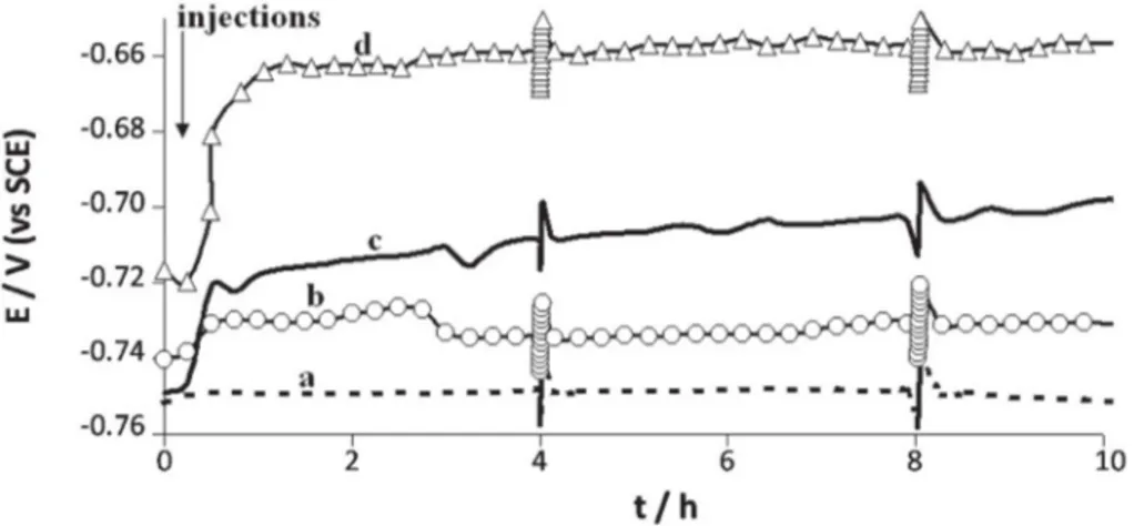
Fig. 1. Open circuit potential versus time for 1145 carbon steel electrode immersed in anaerobic 10 mN phosphate solution pH 7.2, with or without addition of hydrogenase. Fluctuations of +/− 10 mV that appeared on the graph every four hours were due to polarization resistance measurements.
3. Results and discussion
3.1. Influence of active hydrogenase on open circuit potential and deposit composition
Mild steel 1145 coupons were immersed in phosphate solution 10 mM, pH 7.2 for 24 h. The electrochemical cell was hermetically closed and great care was taken to bring the steel coupon into contact with the solution only after it had been strictly deoxygenated (see the
Materials and methodssection). The potential was stabilised for 15 min and hydrogenase was then injected in strictly anaerobic
conditions, because the hydrogenase from C. acetobutylicum is highly sensitive to oxygen. The variation of the open-circuit potential Eocwas
recorded as a function of time for 24 h (Fig. 1).
Seven control experiments were performed without any injection, or with injection of only deoxygenated phosphate solution at t = 15 min, to check that the injection process did not introduce traces of oxygen into the cell. No significant potential evolution was observed. After 24 h immersion, the electrode was covered by a uniform, greyish film (Fig. 2A) that tended to become bluish on exposure to air, a behav-iour that is characteristic of vivianite (Fe3(PO4)2, 8H2O)[42]. SEM
micrography of these coupons (immersed in the absence of hydroge-nase) showed a grey surface with the presence of crystals (Fig. 3). EDX analyses averaged over different spots of the surface did not reveal the presence of carbon although it was clearly detected on clean cou-pons before they were immersed in the phosphate solution, confirming that the deposit coated uniformly the surface of the coupon. In terms of atomic mass percentages, the deposit was mainly composed of iron (51–64%) and oxygen (32–38%) (Table 1). The percentage of phospho-rous, around 3%, was smaller than expected for pure vivianite, which usually contains around 9% phosphorous. As the amounts of iron were rather high, the deposit was probably a mixture of vivianite and iron oxide. Moreover, other products containing simultaneous iron and phosphorous were also suggested in literature: for instance, the forma-tion of an amorphous type of iron phosphide Fe2P is possible, this
deposit was observed under biotic and abiotic conditions, especially when culture media for testing microbial corrosion are supplemented with phosphates and sulphates[43,44]. A compound with average stoi-chiometric formula Zn0·5K1.1PO3·35Fe0.4was also detected during the
protection process of 1138 carbon steel by zinc phosphatation followed by a post-treatment with potassium monofluorophosphate[45].
As shown inFig. 1, injecting hydrogenase caused a fast increase in potential. Most of the potential increase occurred during the first hour Fig. 3. SEM micrograph for 1145 carbon steel surface after 24 hour immersion in anaerobic
10 mM phosphate solution pH 7.2, in the absence of hydrogenase. 10000 × magnification, 10 kV acceleration voltage.
Fig. 2. Photographs of 1145 carbon steel coupons after 24 hour immersion in anaerobic 10 mM phosphate solution pH 7.2 in the absence of hydrogenase (A) and in the presence of 30 µL hydrogenase (B), 50 µL hydrogenase (C) and 80 µL hydrogenase (D).
after injection of the enzyme. Full potential increase values (ΔE) were evaluated by subtracting the value of the potential just before hydroge-nase injection (t = 15 min) from the value at t = 7.50 h (before the sec-ond polarisation resistance measurement). ΔE depended on the amount of hydrogenase injected and increased from 8 mV for 30 μL hydrogenase to 63 mV for 80 μL (Table 2). The visual aspects of the deposits obtained after 24 h were also clearly dependent on the amount of hydrogenase (Table 2). With 30 μL hydrogenase, the coupon was covered with a blu-ish mineral that indicated a marked presence of vivianite (Fe3(PO4)2,
8H2O). A few pits that turned red when exposed to air also indicated
the presence of slight local corrosion (Fig. 2B). Addition of 50 μL hydrog-enase increased the free potential up to 43 mV and the electrode was covered by a grey deposit that seemed more thick (Fig. 2C). 80 μL hy-drogenase led to a green deposit that was unstable and turned red in contact with air, corresponding to a large production of iron hydroxides Fe(OH)2and Fe(OH)3(Fig. 2D)[46]. SEM surface analysis of coupons
ex-posed to 80 μL hydrogenase (coupon D) showed a highly heterogeneous corrosion layer: some surface zones were covered by small crystals (Fig. 4A) and an heterogeneous deposit appeared on others (Fig. 4B). The chemical analysis of the corrosion products on the surface gave
around 61% iron, 22% carbon and 16% oxygen (Table 1). The high percentages of iron and carbon indicated that the steel surface was certainly reached by the EDX probe in the zones where the deposit was not present. The standard deviations of the measurements made on 5 different spots, which were significantly higher than for the previ-ous measurements (Table 1), confirmed that the deposit had an hetero-geneous chemical composition. In contrast with all the other cases, no phosphorous was detected in the presence of 80 μL hydrogenase. This is in agreement with the visual observation of the electrode (Fig. 2D), where the surface of the steel was covered by a reddish iron oxide layer and no vivianite was detected. It can be concluded that a large amount of hydrogenase accelerated the formation of the corrosion products with a FeII/FeIII ratio unfavorable to vivianite deposition. 3.2. Effect of deactivated and denatured hydrogenase
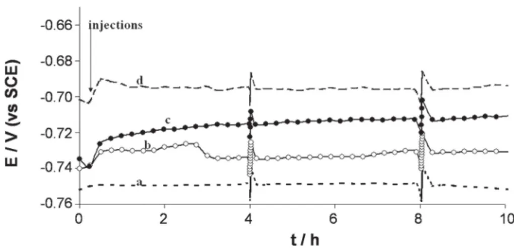
Similar experiments were performed with hydrogenase that was previously deactivated by exposure to air for 2 h and 30 min or dena-tured by heating the aliquot at 100 °C for 30 min until the solution boiled (Fig. 5).
Injection of 30 μL hydrogenase aliquot deactivated by exposure to air increased the free potential by 7 mV. At the end of the experiment, no deposit was visible on the surface of the electrode. On the contrary, the electrode was still electrically conductive and reflected the light as shown inFig. 6where the image of the camera lens can be seen on the coupon surface.
Addition of 30 μL hydrogenase denatured by heating increased the free potential by 26 mV. At the end of the experiment, the mild steel electrode surface was covered by an important non-conductive deposit with cracks spreading all over the layer (Fig. 7).
The surface was analysed carefully discriminating two different zones: on the upper side of the deposit (Fig. 7C spot 1) and in the crack (Fig. 7C spot 2). EDX analysis (Table 1) indicated that the amount of iron on the top of the layer (45%) was around half that in the crack (83%). Phospho-rous was present in the deposit (6%) whereas it was not detected in the
Fig. 4. SEM micrographs of 1145 carbon steel surface after 24 hour immersion in anaerobic 10 mM phosphate solution pH 7.2, containing 80 µL hydrogenase: a zone covered by small crystals (A) and a zone with a heterogeneous deposit (B). 10000 × magnification, 10 kV acceleration voltage.
Table 2
Potential ennoblement ΔE (Et = 7.5 h− Et = 15 min) and visual aspect of the surface at the
end of the experiments (t = 24 h) for 1145 carbon steel coupons immersed in anaerobic 10 mM phosphate solution pH 7.2, with or without hydrogenase.
Hydrogenase amount/corresponding activity
ΔΕ (mV)
Visual aspect of the surface 0 μL/0 U·mL− 1 1 Uniform deposit containing vivianite
30 μL/2.5 U·mL− 1 8 Deposit containing vivianite and a few pits
50 μL/4.25 U·mL− 1 48 Thick greyish deposit
80 μL/6.8 U·mL− 1 63 Marked heterogeneous red deposit
Fe(OH)2/3
30 μL-oxygenated/0 U·mL− 1 7 No visible deposit
30 μL-heated/0 U·mL− 1 26 Thick deposit containing vivianite with
deep cracks Table 1
EDX analysis (atomic mass %) of 1145 carbon steel surface after 24 h immersion in anaerobic 10 mM phosphate solution pH 7.2, with or without hydrogenase. Hydrogenase amount/ element Fe O P C K Cl Mn Na Control, no hydrogenase 0 μLa 64/51 32/38 3/4 − 0/1 1/2 − − 80 μL hydrogenaseb 61 ± 16 16 ± 10 − 22 ± 21 − 1 ± 0.9 − − 30 μL heated hydrogenase
On deposit (spot 1 inFig. 7C)
45 40 6 − 0.5 − 0.5 8
30 μL heated hydrogenase In crack (spot 2 inFig. 7C)
83 14 − − − − − 2
aMaximum/minimum values.
crack. These data indicate that the deposit was made up of corrosion products mixed with vivianite, while only iron and iron hydroxides/ox-ides were present inside the cracks. The cracks were anodic areas where corrosion was occurring, while the phosphate layer was protective.
3.3. Measures of Rp and estimation of corrosion rate
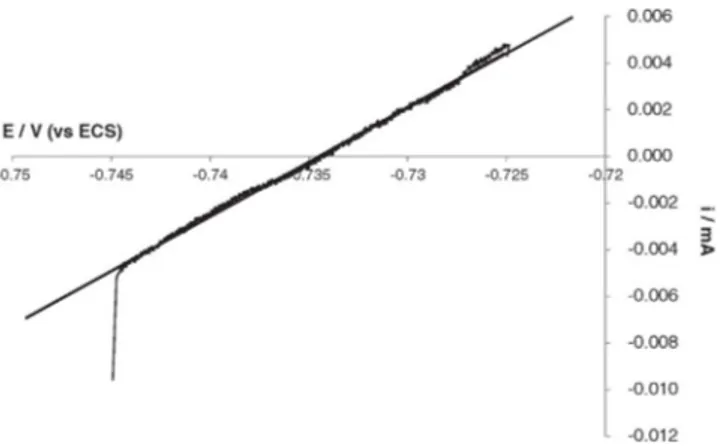
During the immersion, a potential scan was performed every 4 h, at 0.2 mV s− 1around the open circuit potential (E
oc) in the range
[Eoc− 10 mV, Eoc+ 10 mV]. The Stern-Geary model was used to
deter-mine the polarisation resistances Rpfrom the experimental
current-potential measurements (Fig. 8). The values of 1/Rpthat give an
evalu-ation of the corrosion rates (Eq.(10)) are gathered inTable 3. Seven control experiments performed without hydrogenase showed identical 1/Rpthat means identical icorrvalues, which remained stable
during the 24 h experiments. In particular, only a slight modification of 1/Rpvalues was observed during the first 2 h of immersion from 8
to 9.10− 4to 6.10− 4Ω− 1·cm− 2, then it did no longer significantly
vary. A1/Rp value around 6.10− 4Ω− 1·cm− 2(average value from
seven experiments) can be considered as the stable estimated corrosion current that corresponds to the “mild steel/10 mM phosphate solution” interface used here. It can be noticed that these values were close to those reported in the literature for mild steel at open circuit condi-tions[47]. Indeed, in NaH2PO40.1 M, pH 6.0, Rp has been noted to
increase from 188 Ω·cm2(i.e. 1/R
p= 5.3.10− 3Ω− 1·cm− 2) at 1 min
to 1516 Ω cm2(i.e. 1/R
p= 6.7.10− 4Ω− 1·cm− 2) at 60 min.
For the experiments performed with hydrogenase, Tafel plots were recorded every 4 h. No measurement was made in the period t = 0 to t = 4 h to avoid any possible disturbance for the surface state of the cou-pons. In the presence of the hydrogenase, 1/Rpvalues remained almost
constant in all cases and lower than the 6.10− 4Ω− 1·cm− 2obtained in
control experiments, except for the heated hydrogenase. In agreement with the variation of the free potential, 1/Rpvalues indicated that the
main action of hydrogenase occurred before 4 h. After 4 h, 1/Rpvalues
indicate a “passive” behaviour of the mild steel due to the phosphate treatment and the vivianite formation, which was favoured by the presence of hydrogenase. This favourable effect of hydrogenase on the formation of a protective deposit of vivianite has already been shown
[32]. The heated hydrogenase induced more complex behaviour, with a first increase of icorr(high values of 1/Rp) followed by a slow
con-tinuous decrease. Heated hydrogenase increased corrosion rate and then the formation of a thick deposit slowed down corrosion. The pres-ence of cracks where corrosion could continue explained why 1/Rp
(thus icorr) remained higher for some hours with respect to the other
cases that did not show deep cracks. 3.4. Discussion of the mechanisms
In the control experiments no local corrosion was observed after 24 h, icorrwas almost constant (1/Rp around 6.10− 4Ω− 1·cm− 2) and
coupons were covered with a layer containing vivianite, which is known to have a protective effect. Vivianite is an iron (II) phosphate, which may be used as a corrosion inhibiting layer on steel surfaces Fig. 6. Photograph (A) and SEM micrograph (B) of 1145 carbon steel surface after
24 hour immersion in anaerobic 10 mM phosphate solution pH 7.2 in the presence of 30 µL hydrogenase deactivated by air. SEM characteristics: 10000 × magnification, 10 kV acceleration voltage.
Fig. 5. Variation of the open circuit potential versus time for 1145 carbon steel coupons immersed in anaerobic 10 mM phosphate solution pH 7.2, with hydrogenase (in different states) or without hydrogenase. Fluctuations of +/−10 mV that appear every four hours were due to polarisation resistance measurements.
especially because of its low solubility. It is indeed used by some industries as a corrosion protection method; the procedure is acid phosphating carried out at temperatures of up to 95 °C and at pH values between 2 and 3.5 with phosphates of zinc, iron or manga-nese, which leads to vivianite production[48]. Although the detailed mechanisms by which phosphate species lead to the formation of protective layers and the composition of the deposit obtained in phosphate solutions are still research topics, the main point is to have the right amount of Fe II (compared to Fe III) on the material surface that, in contact with the phosphate in the medium, leads to the formation of a vivianite deposit. This is the case in abiotic condi-tions when using chelating agent for instance[49]; the vivianite
layer deposit also depends on phosphate concentration[50,8], on how the preceding oxide layer forms, which is linked to the experi-mental conditions (electrode potential[51]and the presence or not of oxygen[47,52]). In biotic conditions, other mechanisms are sug-gested: oxygen consumption by the biofilm as the driving force to form vivianite[53], acceleration of Fe (III) reduction to Fe (II) in pres-ence of microorganisms (such as Geobacter sulfurreducens[54]).
When hydrogenase was added into the solution, the fast variation of the open circuit potential with time, the visual and microscopic as-pects of the coupon surfaces after 24 h, and the 1/Rp values
(reflecting icorr) confirmed a strong effect of hydrogenase on
corro-sion of carbon steel. It should be noted that the Rpmeasurements
were done 4 h after hydrogenase injection, meaning when the open circuit potential had almost recovered a constant value (Figs. 1 and 5). Rpmeasurements were not performed before to
avoid any disturbance during the first hours, period in which hy-drogenase had the most obvious effect according to Eocrecords.
The different behaviours observed can consequently be attributed to the presence of hydrogenase only, without any parasite effect due to the measurements. 1/Rp values recorded every 4 h were
then almost constant (seeTable 3), except when 80 μL enzyme was added. Except in this latter case, the stability of 1/Rp(thus icorr) and
Eocconsistently indicated that the corrosion state reached 4 h after
hydrogenase injection was roughly stable. Thus, the 1/Rpvalues (as
icorr) did not correspond to the period during which hydrogenase
drastically affected the material, but they corresponded to the new surface steady state that was reached after hydrogenase injection. 1/Rpvalues must consequently be commented not as direct
mea-surements of the hydrogenase action, but as characteristics of the new state resulting from hydrogenase action.
The presence of hydrogenase always led to a fast increase in Eoc, the
amplitude of which increased with the quantity of enzyme. Adding 30 μL hydrogenase resulted in a visually more important deposit than in control experiments, with slight local pitting. It has already been claimed that hydrogenase can induce local cathodic sites that enhance iron dissolution in neighbouring anodic sites; the following precipita-tion of iron ions with phosphate forms a crystalline film partially composed of vivianite. Following this model, hydrogenase enhanced vivianite formation[32]. The observations made here are consistent with this mechanism. Adding 30 μL hydrogenase favoured the forma-tion of a better protective layer, as confirmed by the smaller 1/Rpvalues
(1.5 10− 4Ω− 1cm−2instead of 6.7 10− 4Ω− 1cm−2in control
experi-ments), representing smaller icorr, that were recorded after 4 h. Larger
amounts of hydrogenase (50 or 80 μL) resulted in visually more impor-tant deposits that contained more and more iron oxides/hydroxides. In these cases, iron dissolution and/or ion precipitation was enhanced to an extent that could no longer be balanced by the reaction with Fig. 8. Example of evaluation of Rp by plotting i = f (E) (potential scan rate: 0.2 mV s−1)
for 1145 carbon steel electrode during immersion in anaerobic 10 mM phosphate solution pH 7.2 in the presence of 30 µL of hydrogenase. Regression equation: i (mA) = 0.47(1/kW)xE(V) + 0.35 with R² = 0.9977.
Fig. 7. Photograph (A) and SEM micrographs (B and C) of 1145 carbon steel surface after 24 hour immersion in anaerobic 10 mM phosphate solution pH 7.2 in the presence of 30 µL heated hydrogenase. The markers in (C) indicate the positions where the EDX analyses were performed: on deposit (spot 1) and in a crack (spot 2). SEM characteristics: 10000 × magnification, 10 kV acceleration voltage.
phosphate species, and corrosion products (oxides and hydroxides) accumulated in the layer.
In the presence of 30 μL hydrogenase, the lower icorrcompared
to control experiments can explain the higher value of Eocobtained.
Indeed it means that the anodic reaction decreased, and no supplemen-tary hypothesis is required. With 50 μL and 80 μL hydrogenase, Eoc
enno-blement was roughly proportional to the amount of hydrogenase, but icorrkept similar values. It must be concluded that the catalysis of the
ca-thodic process was also involved in Eocincrease. In the previous work
that dealt with the effect of hydrogenase on vivianite formation[32], the cathodic reaction created by the presence of hydrogenase was due to the presence of phosphate and of a final electron acceptor (the natu-ral redox partner of the enzyme). The cathodic reaction was conse-quently related to the Mechanism 1 described in theIntroduction
section. In contrast, the cathodic reaction detected here can only be the reduction of proton (or water) into hydrogen (Reaction (1)) catalysed by the hydrogenase because no other final reductant (electron acceptor) was present in solution.
Hydrogenase from C. acetobutylicum is highly sensitive to oxygen traces[55]. Keeping it at air, for more than 2 h, ensured complete loss of its catalytic properties for hydrogen oxidation. Adding hydrogenase after deactivating it in air led to an Eocennoblement similar to that
with the same amount of active hydrogenase but, in contrast, the pres-ence of the protein avoided the formation of any deposit and even protected the material surface against corrosion. Such behaviour was not linked to the phosphate medium. Similar observations were made in Tris-HCl pH 6.3: after 24 h, much of the surface remained mirror polished when 30 μL deactivated hydrogenase was added, while control experiments in the absence of enzyme showed a homogeneous grey film (data not shown). In this case, Eocennoblement was due to the
de-crease of icorrinduced by the deactivated protein (1/Rpfrom 6.7 10− 4to
2.4 10− 4
Ω·cm− 2). E
ocennoblement was not linked to a corrosion
pro-cess but to some kind of protection of the material by the protein. Some complex links between “inert” proteins and the corrosion behaviour of metallic surfaces have already been reported in the literature. For in-stance, bovine serum albumin (BSA) adsorbed on iron-chromium alloy showed a protective effect against corrosion at pH 1.3, whereas it accel-erated local corrosion at pH 5.5. In both cases, the protein has been as-sumed to affect the metal behaviour directly, as neither the thickness nor the composition of the protective layer was affected[56]. Using deactivated hydrogenase revealed that the simple protein shell, without enzymatic activity, have a remarkable protective effect. This effect due to the protein nature of hydrogenase was certainly also one of the causes of the icorrdecreases that were recorded with active
hydroge-nase. It must be concluded that hydrogenase affects the electrochemical behaviour of mild steel via different simultaneous effects. As already shown, adsorbed hydrogenase can catalyse the reduction of proton/ water and induce local cathodic/anodic sites that enhance iron dissolu-tion. In the presence of phosphate species this effect favours the forma-tion of a protective layer containing vivianite[32]. This model remains consistent with the data obtained here. The protective effect of the
protein shell must now be added. Moreover large amounts of hydroge-nase lead to the accumulation of iron oxides/hydroxides in the deposit-ed layer, which gets a crackdeposit-ed structure.
Hydrogenase denatured by heating had the greatest effect on the electrochemical parameters and the deposit structure. An important de-posit was observed with a heterogeneous, cracked structure. Obviously the corrosive effect of hydrogenase was not linked to its traditional ac-tivity for hydrogen oxido-reduction. Heating the enzyme completely denatured it, by unwinding and cutting the shell of amino acids that make up its structure. [Fe]-hydrogenases contain numerous Fe\\S clus-ters that provide an electron transfer pathway between the buried ac-tive site and the molecular surface. [Fe]-hydrogenases have a domain with two [4Fe4S]-ferredoxin-like clusters not far from the active site, which are called mesial (FS4A) and distal (FS4B). In addition, the en-zyme has a small domain containing a [4Fe\\4S] cluster (called FS4C) and a plant–like ferredoxin domain with a [2Fe\\2S] cluster (called FS2)[57,58]. Heating the enzyme resulted in its architecture exploding, exposing the metallic clusters to the external surroundings or even releasing the Fe\\S clusters.
As an important conclusion to this work, it can be assumed that the catalysis of proton reduction was caused by the adsorption on the coupon surface of the iron-sulphur clusters contained in the hy-drogenase. This hypothesis perfectly explains that corrosion en-hancement was controlled by the amount of hydrogenase and that the effect was stronger after the enzyme had been denatured by heating. The catalysis of corrosion by hydrogenase may now be thought as another case of catalysis by iron-sulphur compounds. To some extent, the mechanisms suggested here may be compared with the mechanisms generally accepted for the microbial corrosion induced by sulphate reducing bacteria (SRB). SRB reduce sulphate to sulphide ions, which react with the iron ion forming iron sulphide FeS. FeS deposits catalyse proton reduction[16,17]. Cathodic zones (FeS) and anodic zones (Fe) are created on the same electrode, im-plying accelerated deterioration of the material by galvanic corro-sion[59,60].
Hydrogenase from C. acetobutylicum contains 20 atoms of Fe (six in the active site, twelve in the [4Fe\\4S]-type clusters (FS4A, FS4B, FS4C) and two in the [2Fe\\2S]-type clusters (FS2)[39]). The concen-tration of hydrogenase in the initial aliquots was 0.33 × 10− 6 M.
When 30 μL of the aliquot was injected into the 0.05 L electrochemical cell, the final concentration in the cell was 2 × 10− 10 M. The
overall amount of Fe contained in the electrochemical cell was then 4 × 10− 9M. In parallel, the hydrogenase has 18 atoms of sulphur
(four in the H cluster, twelve in the [4Fe\\4S] clusters: FS4A, FS4B, and FS4C and two in 2Fe2S: FS2). The total concentration of sulphur in the electrochemical cell was 3.6 × 10− 9M. These concentrations are
very low, and it must be concluded that the specific iron clusters contained in the enzyme are highly efficient in catalysing proton reduc-tion, certainly much more than the bulk iron sulphide deposits pro-duced by SRB. Actually this conclusion is consistent with the function of these clusters inside the protein that contributes to the efficiency of Table 3
Evolution of 1/Rp versus time during the immersion of 1145 carbon steel coupons in anaerobic 10 mM phosphate solution pH 7.2, with or without hydrogenase; Rp is the polarisation resistance calculated through Stearn-Geary model.
Hydrogenase amount 1/Rp (1/(Ω·cm2)) for t after injection
0 h 2 h 4 h 8 h 12 h 16 h 20 h Control, no hydrogenase 0 μL a7.6 ± 2 10− 4 a9.6 ± 8 10− 4 b6.7 ± 1 10− 4 b6.2 ± 2 10− 4 b5.9 ± 1 10− 4 b6.2 ± 1 10− 4 b6.6 ± 1 10− 4 30 μL – – 1.5 10− 4 1.6 10− 4 1.5 10− 4 1.7 10− 4 1.9 10− 4 50 μL – – 4.0 10− 4 3.4 10− 4 2.9 10− 4 3.1 10− 4 3.3 10− 4 80 μL – – 2.7 10− 4 1.7 10− 4 1.5 10− 4 1.3 10− 4 1.4 10− 4 30 μL-oxygenated – – 2.4 10− 4 2.2 10− 4 2.0 10− 4 1.9 10− 4 1.8 10− 4 30 μL-heated – – 1.3 10− 3 8.7 10− 4 4.0 10− 4 3.1 10− 4 2.6 10− 4 aMean and standard deviation for 3 independent experiments.
the reversible “proton reduction/hydrogen oxidation” reaction. Thanks to this redox chain, hydrogenase has an extremely high activity, of the order of 0.2 mol of hydrogen oxidised per minute per milligram of pro-tein. Suitable adsorption of these clusters on the coupon surface should also be an important factor in efficiency. The presence of amino acids coming from the protein shell, even after unwinding or denaturing by heating, certainly promotes effective adsorption.
4. Conclusion
Hydrogenase from C. acetobutylicum confirmed a high reactivity with surfaces of mild steel. Using less concentrated phosphate solution than in the previous work allowed a gradual effect of hydrogenase to be pointed out, which increased with its concentration in solution. These operating conditions also led to detect different effects of hydrogenase. The action of hydrogenase on mild steel surfaces must now be consid-ered as the result of the complex combination of different elements: local catalysis of proton/water reduction that induces local iron dissolu-tion, protective effect due to the protein shell, formation of a protective layer containing vivianite when phosphate species are present, cracked structure of the deposit that favours local corrosion. Moreover, the elec-trochemical action of hydrogenase is not only linked to its regular en-zyme activity but also to the presence of the ion-sulphur compounds. The denatured enzyme revealed thus to be more active than the active hydrogenase.
Such a versatility of the phenomenon with respect to the experi-mental conditions, in particular the sensitivity of hydrogenase to oxygen, which makes it shift from an active enzyme to a protective protein, is certainly a main cause of the variety of results that have been reported in the literature so far on the possible role of hydroge-nases in microbial corrosion. From this study, a pre-treatment based on the adsorption of inert proteins on steel surface could be proposed as an eco-friend solution in the view to reduce the corrosion in field conditions. Moreover this work may also be a track to develop a new procedure for the deposit of vivianite protective layer on mild steel. Acknowledgements
This work was supported by a grant from CNRS-DRI (BDI PED 2006). It was a part of CNRS European network “Surfaces of materials in living environments (SMILE)”.
The authors gratefully thank Luc Etcheverry (LGC) for his technical support, Marie-Line De Solan (LGC) for EDX facilities and Laurence Girbal and Marie Demuez (Laboratoire d'Ingénierie des Systèmes Biologiques et des procédés LISBP, INSA-Toulouse) for helpful discus-sions and for providing the hydrogenase aliquots.
References
[1] I.B. Beech, Corrosion of technical materials in the presence of biofilms-current understanding and state-of-the art methods of study, Int. Biodeterior. Biodegrad. 53 (2004) 177–183.
[2] W. Lee, Z. Lewandowski, P.H. Nielsen, W.A. Hamilton, Role of sulfate-reducing bacteria in corrosion of mild steel: a review, Biofouling 8 (1995) 165–194.
[3] R. Javaherdashti, R.K. Singh Raman, C. Panter, E.V. Pereloma, Microbiologically assisted stress corrosion cracking of carbon steel in mixed and pure cultures of sulphate reducing bacteria, Int. Biodeterior. Biodegrad. 58 (2006) 27–35.
[4] C. Xu, Y. Zhang, G. Cheng, W. Zhu, Pitting corrosion behavior of 316L stainless steel in the media of sulphate-reducing and iron-oxidizing bacteria, Mater. Charact. 59 (2008) 245–255.
[5] C. Xu, Y. Zhang, G. Cheng, W. Zhu, Localized corrosion behavior of 316L stainless steel in the presence of sulfate-reducing and iron-oxidizing bacteria, Mater. Sci. Eng. 443 (2007) 235–241.
[6] J. Duan, S. Wu, X. Zhang, G. Huang, M. Du, B. Hou, Corrosion of carbon steel influ-enced by anaerobic biofilm in natural seawater, Electrochim. Acta 54 (2008) 22–28.
[7] R. Avci, B.H. Davis, M.L. Wolfenden, I.B. Beech, K. Lucas, D. Paul, Mechanism of MnS-mediated pit initiation and propagation in carbon steel in an anaerobic sulfidogenic media, Corros. Sci. 76 (2013) 267–274.
[8] L. De Silva Muñoz, A. Bergel, R. Basséguy, Role of the reversible electrochemical deprotonation of phosphate species in anaerobic biocorrosion of steels, Corros. Sci. 49 (2007) 3988–4004.
[9] W.P. Iverson, Mechanism of anaerobic corrosion of steel by sulfate reducing bacteria, Mater. Perform. 23 (1984) 28–30.
[10] I.B. Beech, C.W.S. Cheung, Interactions of exopolymers produced by sulphate-reducing bacteria with metal ions, Int. Biodeterior. Biodegrad. 35 (1995) 59–72.
[11] E. Miranda, M. Bethencourt, F.J. Botana, M.J. Cano, J.M. Sanchez-Amaya, A. Corzo, J.G. de Lomas, M.L. Fardeau, B. Ollivier, Biocorrosion of carbon steel alloys by an hydrogenotrophic sulfate-reducing bacterium Desulfovibrio capillatus isolated from a Mexican oil field separator, Corros. Sci. 48 (2006) (2417–243).
[12] R. Javaherdashti, Impact of sulphate-reducing bacteria on the performance of engineering materials, Appl. Microbiol. Biotechnol. 91 (2011) 1507–1517.
[13] Z.H. Dong, T. Liu, H.F. Liu, Influence of EPS isolated from thermophilic sulphate-reducing bacteria on carbon steel corrosion, Biofouling 27 (2011) 487–495.
[14] D. Enning, J. Garrelfs, Corrosion of iron by sulfate-reducing bacteria: new views of an old problem, Appl. Environ. Microbiol. 80 (2014) 1226–1236.
[15] H. Venzlaff, D. Enning, J. Srinivasan, K.J.J. Mayrhofer, A.W. Hassel, F. Widdel, et al., Accelerated cathodic reaction in microbial corrosion of iron due to direct electron uptake by sulfate-reducing bacteria, Corros. Sci. 66 (2013) 88–96.
[16] R. Marchal, Involvment of sulfidogenic bacteria in iron corrosion, Oil Gas Sci. Technol. 54 (1999) 649–659.
[17] W.A. Hamilton, Microbially influenced corrosion as a model system for the study of metal microbe interactions: a unifying electron transfer hypothesis, Biofouling 19 (2003) 65–76.
[18] K. Mori, H. Tsurumaru, S. Harayama, Iron corrosion activity of anaerobic hydrogen-consuming microorganisms isolated from oil facilities, J. Biosci. Bioeng. 110 (2010) 426–430.
[19]R.D. Bryant, W.J. Jansen, J. Boivin, E.J. Laishley, W. Costerton, Effect of hydrogenase and mixed sulphate-reducing bacterial populations on the corrosion of steel, Appl. Environ. Microbiol. 57 (1991) 2804–2809.
[20] C. Chatelus, P. Carrier, P. Saignes, M.F. Libert, Y. Berlier, P.A. Lespinat, et al., Hydrog-enase activity in aged, nonviable Desulfovibrio vulgaris cultures and its significance in anaerobic biocorrosion, Appl. Environ. Microbiol. 53 (1987) 1708–1710.
[21] A.V. Ramesh Kumar, R. Singh, R.K. Nigam, A.V.R. Kumar, R. Singh, R.K. Nigam, Mossbauer spectroscopy of corrosion products of mild steel due to microbiologically influenced corrosion, J. Radioanal. Nucl. Chem. 242 (1999) 131–137.
[22] D.J. Evans, C.J. Pickett, Chemistry and the hydrogenases, Chem. Soc. Rev. 32 (2003) 268–275.
[23]F.A. Armstrong, Hydrogenases: active site puzzles and progress, Curr. Opin. Chem. Biol. 8 (2004) 133–140.
[24] R. Mertens, A. Liese, Biotechnological applications of hydrogenases, Curr. Opin. Biotechnol. 15 (2004) 343–348.
[25] A. Pardo, A.L. De Lacey, V.M. Fernadez, H.J. Fan, Y. Fan, M.B. Hall, Density functional study of the catalytic cycle of nickel-ion [NiFe] hydrogenased and the involvement of high-spin nickel(II), J. Biol. Inorg. Chem. 11 (2006) 286–306.
[26]E.J. Lyon, S. Shima, G. Buurman, S. Chowdhuri, A. Batschauer, K. Steinbach, R.K. Thauer, UV-A/blue-light inactivation of “the metal-free” hydrogenase (Hmd) from methanogenic archaea, Eur. J. Biochem. 271 (2004) 195–204.
[27] M. Frey, Hydrogenases: hydrogen-activating enzymes, Chem. Biochem. 3 (2002) 153–160.
[28] M.D. Yates, M. Siegert, B.E. Logan, Hydrogen evolution catalyzed by viable and non-viable cells on biocathodes, Int. J. Hydrog. Energy 39 (2014) 16841–16851.
[29] R.D. Bryant, E.J. Laishley, The role of hydrogenase in anaerobic corrosion, Can. J. Microbiol. 36 (1990) 259–264.
[30] R.D. Bryant, E.J. Laishley, The effect of inorganic phosphate and hydrogenase on the corrosion of mild steel, Environ. Biotechnol. 38 (1993) 824–827.
[31]S. DaSilva, R. Basseguy, A. Bergel, Electrochemical deprotonation of phosphate on stainless steel, Electrochim. Acta 49 (2004) 4553–4561.
[32] S. DaSilva, A. Bergel, R. Basseguy, Hydrogenase-catalysed deposition of vivianite on mild steel, Electrochim. Acta 49 (2004) 2097–2103.
[33] O.A. Zadvorny, N.A. Zorin, I.N. Gogotov, Transformation of metals and metal ions by hydrogenases from phototrophic bacteria, Arch. Microbiol. 84 (2006) 279–285.
[34] A. Nedoluzhko, I.A. Shumilin, L.E. Mazhorova, V.O. Popov, V.V. Nikandrov, Enzymatic oxidation of cadmium and lead metals photodeposited on cadmium sulphide, Bioelectrochem 53 (2001) 61–71.
[35] J.S. Deutzmann, M. Sahin, A.M. Spormann, Extracellular enzymes facilitate electron uptake in biocorrosion and bioelectrosynthesis, MBio. 6 (2015), e00496 (–15–).
[36] M.J. Lukey, A. Parkin, M.M. Roessler, B.J. Murphy, J. Harmer, T. Palmer, et al., How Escherichia coli is equipped to oxidize hydrogen under different redox condi-tions, J. Biol. Chem. 285 (2010) 3928–3938,http://dx.doi.org/10.1074/jbc. M109.067751.
[37] S. Da Silva, R. Basséguy, A. Bergel, Electron transfer between hydrogenase and 316L stainless steel: identification of a hydrogenase-catalyzed cathodic reaction in anaerobic mic, J. Electroanal. Chem. 561 (2004) 93–102.
[38] M. Mehanna, R. Basséguy, M.L. Délia, L. Girbal, M. Demuez, A. Bergel, New hypothe-ses for hydrogenase implication in the corrosion of mild steel, Electrochim. Acta 54 (2008) 140–147.
[39] L. Girbal, G. Von Abendroth, M. Winkler, P.M.C. Benton, I. Meynial-Salles, C. Croux, J.W. Peters, T. Happe, P. Soucaille, Homologous and heterologous overexpression in Clostridium acetobutylicum and characterization of purified Clostridial and algal Fe-only hydrogenases with high specific activities, Appl. Environ. Microbiol. 71 (2005) 2777–2781.
[40] B. Elsener, Corrosion rate of steel in concrete-measurements beyond the Tafel law, Corros. Sci. 47 (2005) 3019–3033.
[41] M. Stern, A.L. Geary, Electrochemical polarization, J. Electrochem. Soc. 104 (1) (Jan. 1957) 56.
[42] G. McGowan, J. Prangnell, The significance of vivianite in archaeological settings, Geoarchaeology 21 (2006) 93–111.
[43] D. Glindemann, F. Eismann, A. Bergmann, P. Kuschk, U. Stottmeister, Phosphine by bio-corrosion of phosphide-rich iron, Environ. Sci. Pollut. Res. 5 (1998) 71–74.
[44] W.P. Iverson, G.J. Olson, Technical Summary Report No. 1, National Bureau of Standards, National Measurement Lab., Washington DC, 1982.
[45] J.J. Robin, J. Duran, L. Cot, A. Bonnel, M. Duprat, F. Dabosi, Physicochemical and electrochemical study of the protection of a carbon-steel by monofluorophosphates. 1. Influence of a chemical conversion treatment, Appl. Electrochem. 12 (1982) 701–710.
[46] Techniques de l'ingénieur, Corrosion des aciers au carbone section 3.2.1. [47] E.M.A. Martini, S.T. Amaral, I.L. Müller, Electrochemical behaviour of invar in
phosphate solutions at pH = 6, Corros. Sci. 46 (2004) (2907–2115).
[48] W. Rausch, Die Phosphatierung Von Metallen, Leuze Verlag, Saulgau, Germany. The Phosphating of Metals English Electronic Version 1990, 1988.
[49] H. Harms, H.-P. Volkland, G. Repphun, A. Hiltpolt, O. Wanner, A.J.B. Zehnder, Action of chelators on solid iron in phosphate-containing aqueous solutions, Corros. Sci. 45 (2003) 1717–1732.
[50] Y. Gourbeyre, E. Guilminot, F. Dalard, Study of the corrosion layer on iron obtained in solutions of water-polyethilene glycol (PEG400)-sodium phosphate, J. Mater. Sci. 38 (2003) 1307–1313.
[51]C.A. Borrás, R. Romagnoli, R.O. Lezna, In-situ spectroelectrochemistry (UV–visible and infrared) of anodic films on iron in neutral phosphate solutions, Electrochim. Acta 45 (2000) 1717–1725.
[52] A. Paszternák, I. Felhősi, Z. Pászti, E. Kuzmann, A. Vértes, E. Kálmán, L. Nyikos, Surface analytical characterization of passive iron surface modified by alkyl-phosphonic acid layers, Electrochim. Acta 55 (2010) 804–812.
[53] H.-P. Volkland, H. Harms, B. Müller, G. Repphun, O. Wanner, A.J.B. Zehnder, Bacterial phosphating of mild (unalloyed) steel, Appl. Environ. Microbiol. 66 (2000) 4389–4395.
[54] C. Cote, O. Rosas, R. Basséguy, Geobacter sulfurreducens: an iron reducing bacterium that can protect carbon steel against corrosion? Corros. Sci. 94 (2015) 104–113.
[55] M. Demuez, L. Cournac, O. Guerrini, P. Soucaille, L. Girbal, Complete activity of Clos-tridium acetobutylicum [FeFe]-hydrogenase and kinetic parameters for endogeneous redox partners, FEMS Microbiol. Lett. 275 (2007) 113–121.
[56] I. Frateur, L. Lartundo-Rojas, C. Méthivier, A. Galtayries, P. Marcus, Influence of bo-vine serum albumin in sulphuric acid aqueous solution on the corrosion and the passivation of an iron-chromium alloy, Electrochim. Acta 51 (2006) 1550–1557.
[57] J.C. Fontecilla-Camps, A. Volbeda, C. Cavazza, Y. Nicolet, Structure/function relation-ships of [NiFe] and [FeFe]-hydrogenases, Chem. Rev. 107 (2007) 4273–4303.
[58] J.W. Peters, W.N. Lanzilotta, B.J. Lenon, L.C. Seefeldt, X-ray crystal structure of the Fe-only hydrogenase (Cpl) from Clostridium pasteurianum to 1.8 angstrom resolution, Science 282 (1998) 1853–1858.
[59] I. Dupont-Morral, Les bactéries sulfato-réductrices et la corrosion bactérienne, Bull. Soc. Fr. Microbiol. 19 (2004) 108–115.
[60] R.A. King, J.D.A. Miller, Corrosion of mild steels by iron sulphides, Br. Corros. 8 (1973) 137–141.