HAL Id: hal-02267542
https://hal.archives-ouvertes.fr/hal-02267542
Submitted on 15 May 2020
HAL is a multi-disciplinary open access
archive for the deposit and dissemination of
sci-entific research documents, whether they are
pub-lished or not. The documents may come from
teaching and research institutions in France or
abroad, or from public or private research centers.
L’archive ouverte pluridisciplinaire HAL, est
destinée au dépôt et à la diffusion de documents
scientifiques de niveau recherche, publiés ou non,
émanant des établissements d’enseignement et de
recherche français ou étrangers, des laboratoires
publics ou privés.
In-vitro validation of 4D flow MRI measurements with
an experimental pulsatile flow model
A. David, D. Le Touzé, K. Warin-Fresse, P. Paul-Gilloteaux, F. Bonnefoy,
Jérôme Idier, Saïd Moussaoui, P. Guérin, Jean-Michel Serfaty
To cite this version:
A. David, D. Le Touzé, K. Warin-Fresse, P. Paul-Gilloteaux, F. Bonnefoy, et al.. In-vitro validation of
4D flow MRI measurements with an experimental pulsatile flow model. Diagnostic and Interventional
Imaging, Elsevier, 2019, 100 (1), pp.17-23. �10.1016/j.diii.2018.08.012�. �hal-02267542�
ORIGINAL
ARTICLE
/Research
and
new
developments
In-vitro
validation
of
4D
flow
MRI
measurements
with
an
experimental
pulsatile
flow
model
A.
David
a,∗,
D.
Le
Touze
b,
K.
Warin-Fresse
a,
P.
Paul-Gilloteaux
c,
F.
Bonnefoy
b,
J.
Idier
d,
S.
Moussaoui
d,
P.
Guerin
e,
J.-M.
Serfaty
a,faDepartmentofcardiovascularimaging,centrehospitalieruniversitairedeNantes,44800
Saint-Herblain,France
bLaboratoirederechercheenhydrodynamique,énergétiqueetenvironnement(CNRS,UMR
6598),ÉcolecentraleNantes,44321Nantescedex3,France
cStructurefédérativederecherchesantéFranc¸ois-Bonamy,institutderechercheensantéde
l’universitédeNantes,44007Nantescedex1,France
dLaboratoiredessciencesdunumériquedeNantes(LS2N,UMRCNRS6004),Écolecentralede
Nantes,44321Nantescedex3,France
eDepartmentofcardiology,centrehospitalieruniversitairedeNantes,44800Saint-Herblain,
France
fUnitéderechercheInsermUMR1087,institutderechercheensantédel’universitéde
Nantes,44007Nantescedex1,France
KEYWORDS 4Dflowmagnetic resonanceimaging (MRI); Phase-contrast magneticresonance imaging; Experimentalstudies; Pulsatileflowmodel
Abstract
Purpose:Thepurposeofthisstudywastoassesstheprecisionoffour-dimensional(4D) phase-contrastmagneticresonanceimaging(PCMRI)tomeasuremeanflowandpeakvelocity(Vmax) inapulsatileflowphantomandtotestitssensitivitytospatialresolutionandVenc.
Material andmethods:The pulsatile flow phantom consisted ofa straight tube connected to thesystemic circulation ofan experimentalmock circulatory system. Four-dimensional-PCMR images were acquired using different spatial resolutions (minimum pixel size: 1.5×1.5×1.5mm3)andvelocityencoding sensitivities(uptothreetimes V
max).Meanflow andVmaxcalculatedfrom4D-PCMRIwerecomparedrespectivelytothereferencephantomflow parametersandtoVmaxobtainedfromtwo-dimensional(2D)-PCMRI.
Results:4D-PCImeasuredmeanflowwithaprecisionof−0.04%to+5.46%,butslightly under-estimated Vmax when comparedto 2D-PCMRI(differences rangingfrom−1.71% to −3.85%). 4DPCMRImeanflowmeasurementwasinfluencedbyspatialresolution(P<0.001)withbetter
∗Correspondingauthor.
E-mailaddress:art.dav44@gmail.com(A.David). https://doi.org/10.1016/j.diii.2018.08.012
18 A.Davidetal.
resultsobtainedwithsmallervoxelsize.TherewasnoeffectofVenconmeanflow measure-ment.RegardingVmax,neither spatialresolutionnorVencdidinfluence theprecisionofthe measurement.
Conclusion:Usinganexperimentalpulsatileflowmodel4D-PCMRIisaccuratetomeasuremean flowandVmax withbetterresultsobtainedwithhigherspatialresolution.Wealsoshowthat Vencupto3timeshigherthanVmaxmaybeusedwithnoeffectonthesemeasurements. ©2018Soci´et´efranc¸aisederadiologie.PublishedbyElsevierMassonSAS.Allrightsreserved.
Four-dimensional (4D) phase contrast imaging (PCI) using magneticresonanceimaging(MRI)hasbeenrecently devel-opedtoallowabetterunderstandingofbloodflowthrough the heart and large vessels [1—3]. With 4D-PCI, veloc-ity is encoded in all three spatial directions (i.e., three spatialdimensions) along thecardiac cycle(onetemporal direction),resultingina4Dmodel.Unliketwo-dimensional (2D)-PCI or Doppler echocardiography,4D-PCIcan provide information on the spatial and temporal evolution of 3D blood flow within the boundaries of the volume covered [4].Inthepasttwodecades,4D-PCIacquisitionshavebeen reportedinawidespectrumofanatomiclocationssuchas heart[5],especiallyforcongenitalheartdiseases[6], tho-racicaorta[7],visceralvessels[8],orintracranialarteries [9].Currentdisadvantagesof4D-PCIincludelimitedspatial andtemporalresolutionfor flow analysisadjacentto ves-selwalls [10]and long scantimesranging between 5and 20minutesdependingonanatomiccoverage,heartrate,and spatio-temporal resolution [4]. Several studies have com-pared4D-PCItocurrent goldstandard methodsfor in-vivo validation,such asDoppler examination or 2D-PCI, show-inggoodcorrelationandreproducibilityinvariousanatomic territories[11—17].
Inclinicalpractice,incoherentmeasurementsofflowand velocityin the heart and its great vessels raise concerns regarding the accuracy of the technique. Controlled pul-satileorsteadyflow phantomexperimentscanbeusedto assess accuracy for flow and velocity [18]. Hitherto, few studies have compared 4D-PCI to reference methods for invitromeasurementofflowparameters[19—24].
Thepurposeofthisstudywastoassesstheprecisionof 4D-PCItomeasuremeanflowandpeakvelocity(Vmax)ina pulsatileflowphantomandtotestitssensitivitytospatial resolutionandVenc.
Materials
and
methods
Thisworkwasaphantomstudywithoutanyexperimentation onhumans.Therefore,Institutionalreviewboardapproval andinformedconsentwerenotrequired.
Flow
phantom
Toevaluatethequantitativeaccuracyofthe4D-PCI,invitro velocitymeasurements were acquired on a pulsatile flow
phantom previously designed in our center. The phantom consistedofastiffstraighttubewithaninnerdiameterof 20mm,surroundedbyaboxfilledwithagarosegelto min-imizesusceptibilityartifactsandtomimicstatictissuesfor backgroundcorrection.Thetubewasfilledwithwaterand connected topolyvinyl chloride tubingleading out of the magnet room andwas derived from thesystemic circula-tionofanexperimentalmockcirculatorysystem(Syncardia Systems, Inc.) [25]. This system simulated the systemic and pulmonary circulation and consisted of pulsatile left and right cardiac pumps (CardioWestTM) connected with fourtanks.EachartificialventricleCardioWestTMconsisted of a segmented cavity with two chambers, one with air content and the other withwater content, separated by a polyurethane membrane. The four tanks corresponded tothe aorta,thepulmonary arteryand thetwoatriaand containeddifferentvolumesofwater.Aflowmeter,placed betweenthetankssimulatingthesystemiccirculationand therightatrium,providedtheflowrateasareference.The ventricleswerepoweredbycompressedairdeliveredfrom anexternalpneumaticcontroller,mobilizingthemembrane andemptyingthe ejectionchamber. Insufflationpressures intheventricles,pulserateandpressurelevelsinthetanks werecalibratedtodriveapulsatileflowthroughthe phan-tomatarateof100beatsperminute(bpm)withamean flowof4.8L/min(Fig.1).
MRI
acquisitions
Imageacquisitionoftheflowphantomwasperformedona clinical1.5TMRIunit(AERA®,SiemensHealthineers),using an18-channelbodymatrixcoil.Asimulated electrocardio-graphicsignalwithaRRintervalof600ms,correspondingto apulserateof100bpm,wasusedforgating.
Forthe4D flowimaging,several4D-PCIsequenceswith a3Dvolumecoveringtheflowphantomwereacquiredwith differentspatialresolutionsandVenctoassesstheaccuracy ofthesequence(Table1).For2D-PCI,imagesperpendicular tothestraighttubewereacquiredwith60framespercycle, afieldofviewof340mm,amatrixof192,aslabthickness of6mm,andthesameVencasfor4D-PCIacquisitions.
Mean flow (inmL/min), vessel area (inmm2) and V max (in cm/sec) were calculated using a prototype investiga-tional software — 4D Flow (Siemens Healthineers) for 4D flowacquisitionsandusingArgus®software(Siemens Health-ineers)for2D-PCI.
Figure1. Schematicdiagramoftheexperimentalpulsatileflowmodel.
Statistical
analysis
The dataweretabulated usingan Excel2013 spreadsheet (Microsoft) and statistical analysis were performed using XLSTAT and R. Quantitative variables were expressed as means±standard deviation (SD) and ranges. Mean flow and vessel area calculated from4D-PCI acquisitions were comparedwith thereference flow parameters (RFP) by a Wilcoxontest.Kruskal—WallisandWilcoxontestswere con-ductedtoexaminethe differencesbetweentheresultsof 4D-PCIperformedusingdifferentspatialresolutionsand dif-ferentVenc.InWilcoxontests,wecomparedpeakvelocities between2DPCand4D-PCI.Pearson’srwasusedtodescribe thecorrelationbetween2D-PCIand4D-PCIforpeakvelocity measurement.Bland-Altmanplotsweregenerated compar-ingto2DPCIand4D-PCIforpeakvelocitymeasurementand thebias andlimitsof agreementwerecalculated. Signifi-cancewassetatP<0.05(two-sided).
Results
Analysis
of
4D-PCI
mean
flow
and
vessel
area—comparison
with
experimental
values
Theresultsofthecomparisonof4D-PCIderivedmeanflow andvesselareawithRFPandtheeffectsofspatialresolution andVenc arereportedinTables2and3.4D-PCImeasured meanflow with aprecision of −0.04%to+5.46%, Regard-ingspatialresolution,significantdifferenceswerefoundin meanflow and vessel area(P<0.001) between subgroups withidenticalVencbutdifferentspatialresolutions.Better accuracywasobtainedwhensmallervoxelsizeswereused (1.9×1.9×1.9and1.5×1.5×1.5mm3)withnosignificant differencebetween these twohighly resolvedsubgroups. RegardingVenc,Kruskal—Wallistestsshowednosignificant differenceinmeanflowandvesselareainsubgroupswith identicalspatialresolutionbutdifferentVenc(Fig.2).
20 A.Davidetal.
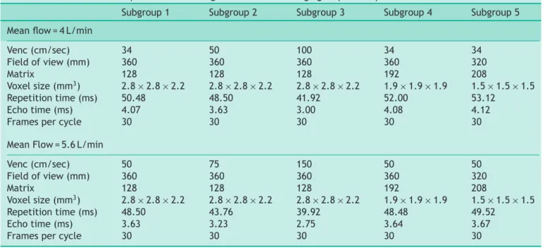
Table1 Four-dimensionalphasecontrastmagneticresonanceimagingacquisitionparameters.
Subgroup1 Subgroup2 Subgroup3 Subgroup4 Subgroup5 Meanflow=4L/min
Venc(cm/sec) 34 50 100 34 34 Fieldofview(mm) 360 360 360 360 320 Matrix 128 128 128 192 208 Voxelsize(mm3) 2.8×2.8×2.2 2.8×2.8×2.2 2.8×2.8×2.2 1.9×1.9×1.9 1.5×1.5×1.5 Repetitiontime(ms) 50.48 48.50 41.92 52.00 53.12 Echotime(ms) 4.07 3.63 3.00 4.08 4.12
Framespercycle 30 30 30 30 30
MeanFlow=5.6L/min
Venc(cm/sec) 50 75 150 50 50 Fieldofview(mm) 360 360 360 360 320 Matrix 128 128 128 192 208 Voxelsize(mm3) 2.8×2.8×2.2 2.8×2.8×2.2 2.8×2.8×2.2 1.9×1.9×1.9 1.5×1.5×1.5 Repetitiontime(ms) 48.50 43.76 39.92 48.48 49.52 Echotime(ms) 3.63 3.23 2.75 3.64 3.67
Framespercycle 30 30 30 30 30
Table2 Meanflowmeasuredinfour-dimensionalphasecontrastmagneticresonanceimaging(4D-PCI)andcomparedto referenceexperimentalvalue.
n Spatialresolution(mm3) Venc(cm/sec) MeanFlow(ml/min) Difference
Referencevalue 4800 — 4D-PCITotal 100 4942.65±873.91[3652.8—6339.6] +142.65(+2.97%) 4D-PCISubgroup1 20 2.8×2.8×2.2 42 5048.82±903.80[3798—6224.4] +248.82(+5.18%) 4D-PCISubgroup2 20 2.8×2.8×2.2 62.5 5010.57±882.56[3903—6189] +210.57(+4.39%) 4D-PCISubgroup3 20 2.8×2.8×2.2 125 5062.11±922.57[3797.4—6339.6] +262.11(+5.46%) 4D-PCISubgroup4 20 1.9×1.9×1.9 42 4798.31±798.32[3809.4—5848.2] −1.69(−0.04%) 4D-PCISubgroup5 20 1.5×1.5×1.5 42 4793.43±904.44[3652.8—5808.6] −6.57(−0.14%)
Resultsareexpressedasmean±standarddeviation(SD).Numbersinbracketareranges.
Table3 Vesselareameasuredinfour-dimensionalphasecontrastmagneticresonanceimaging(4D-PCI)andcompared toreferenceexperimentalvalue.
n Spatialresolution(mm3) Venc(cm/sec) Vesselarea(mm2) Difference
Referencevalue 314.16 — 4D-PCITotal 100 370.5±26.26[303.8—424.6] +56.34(+17.93%) 4D-PCISubgroup1 20 2.8×2.8×2.2 42 384.53±26.33[325.5—424.6] +70.37(+22.40%) 4D-PCISubgroup2 20 2.8×2.8×2.2 62.5 382.92±22.73[337.4—424.5] +68.76(+21.89%) 4D-PCISubgroup3 20 2.8×2.8×2.2 125 378.66±23.27[331.8—422.5] +64.50(+20.53%) 4D-PCISubgroup4 20 1.9×1.9×1.9 42 353.62±16.53[309.7—378.2] +39.46(+12.56%) 4D-PCISubgroup5 20 1.5×1.5×1.5 42 370.5±26.26[303.8—424.6] +56.34(+17.93%)
Resultsareexpressedasmean±standarddeviation(SD).Numbersinbracketareranges.
Analysis
of
4D-PCI
V
max—comparison
with
2D-PCI
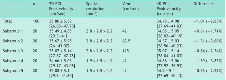
The results of the comparison of 4D-PCI derived Vmax with2D-PCIandtheeffectsofspatial resolutionandVenc are reported in Table 4. 4D-PCI slightly underestimated
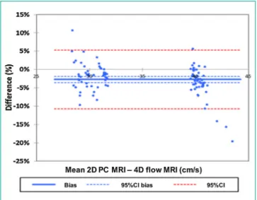
Vmax when compared to 2D-PCI(differences ranging from −1.71% to −3.85%; P<0.001). Correlation was excellent betweenthetwomodalities(r=0.962;95%CI:0.944—0.974; P<0.001). Bland—Altman tests showed a mean of differ-ences (bias) of −2.83% of the overall mean value for Vmax between2D PCand4D-PCI, withlimitsofagreement
Figure2. Columnbarsshowflowandvesselareameasuredwith4D-PCIandcomparedtoreferenceexperimentalvalues.A.Comparison
ofsubgroupswithdifferentspatialresolutionsbutidenticalVencformeanflowmeasurements.B.Comparisonofsubgroupswithdifferent
Vencbut identicalspatialresolutionsfor mean flowmeasurements. C.Comparisonofsubgroups withdifferent spatialresolutionsbut
identicalVencforvesselareameasurements.D.ComparisonofsubgroupswithdifferentVencbutidenticalspatialresolutionsforvessel
areameasurements.
Table4 Peakvelocitymeasuredinfour-dimensionalphasecontrastmagneticresonanceimaging(4D-PCI)andcompared toreferencetwo-dimensionalphasecontrastmagneticresonanceimaging(2D-PCI)values.
n 2D-PCI Peakvelocity (cm/sec) Spatial resolution (mm3) Venc (cm/sec) 4D-PCI Peakvelocity (cm/sec) Difference Total 100 35.80±5.59 [26.88—47.78] 34.78±4.98 [27.69—41.03] −1.01(−2.83%) Subgroup1 20 35.49±4.88 [29.3—41] 2.8×2.8×2.2 42 34.88±5.05 [28.02—40.19] −0.61(−1.71%) Subgroup2 20 35.67±5.98 [26—43.07] 2.8×2.8×2.2 62.5 34.37±5.03 [28.56—40.23] −1.31(−3.66%) Subgroup3 20 35.07±5.14 [27.67—47.78] 2.8×2.8×2.2 125 35.07±5.14 [28.84—41.03] −0.84(−2.34%) Subgroup4 20 34.66±5.06 [29.17—43.08] 1.9×1.9×1.9 42 34.66±5.06 [27.92—39.93] −1.39(−3.85%) Subgroup5 20 35.88±5.1 [29.8—41.65] 1.5×1.5×1.5 42 34.9±5.1 [27.69—40.13] −0.93(−2.59%)
22 A.Davidetal.
Figure3. DiagramshowsBland—Altmanplotsof4D-PCIcompared
with2D-PCIforVmaxmeasurement.
between−10.71%and+5.29%(Fig.3).Therewereno signif-icantdifferencesbetweengroupswithvariedspatial reso-lution(P=0.74),noringroupswithvariedVenc(P=0.39).
Discussion
We developed an experimental methodology to evaluate the accuracy of 4D-PCI flow measurements in a 20-mm straighttube. Weshowedthat 4D-PCIisaccurateat mea-suringmeanflow withbetter resultsobtained withhigher spatial resolution and conversely no effectof Venc when varied up to 3 times Vmax. A good agreement between
4D-PCI and 2D-PCI was found for the measurement of Vmax.
Longtimescansareneededtoacquire4D-PCIespecially whenusingsmallpixelsize.Oneobjectiveofthisworkwas thereforetoevaluatetheimpactof increasingthespatial resolutionontheaccuracyof4D-PCImeasurements.Indeed, inourpresentinvitrostudy,wewereabletodemonstrate thatanincreaseinthespatialresolutionimprovedthe mea-surementofvesselareaandmeanflow,whicharedirectly relatedtothequalityofthesegmentation.Withhigher spa-tial resolution images, the accuracy of the segmentation minimizespartialvolumeeffectsneartotheboundariesof thetube,providingbetterflowmeasurements.Theseresults areconsistentwiththoseof Kweon etal.whoreporteda higheraccuracyofmeanflowmeasurementsonaphantom whenincreasing4Dflowspatialresolutions[21].Oneshould notehoweverthatinourstudy,althoughresultswere bet-terwith1.9mmandwith1.5mmisotropicspatialresolution than with 2.8×2.8×2.2mm resolution, 1.5mm isotropic resolutiondidnot providesignificantlybetter resultsthan 1.9-mmisotropic resolution.This resultmaybe explained bythereduction ofthesignal-to-noise ratiocausedby an important increase of spatial resolution, assuggested by Kweonet al.[21]. Althoughin theory, a higherresolution maygivea betterestimation offlow measurements, con-sideringthesignal-to-noise ratioreductionandthelonger scantime,refiningthe voxelsize below1.9-mm isotropic
mightnotbeusefulunderthepresentconditions.Thisalso supportsthestrategy ofsuper-resolutionat stageof post-processingsothatSNRremainshigh.
Toevaluatethevalvularorarterialstenosisseverity,one ofthemostimportantparameterisVmax[26].Inthepresent study,nodifferenceswerefoundbetweenlowandhigh spa-tialresolutionsubgroups.Thismaybeduetothefactthat thepeakintensityvoxeliscommonlylocatedinthecentral regionofthetube,andtherebylessdependentofthe qual-ityofthe segmentation.Someauthors havereportedthat improving spatial resolution may leadtoa better estima-tionofVmaxby4D-PCI[21].However,thespatialresolution oftheirreferencestandard,(computationalfluiddynamics) was markedlyhigher than 2D-PCI. Our data suggests that currentspatialresolution(2.8×2.8×2.2inourstudy)may besufficienttoprovidereliableresultsregardingthis hemo-dynamicparameter.
Asecondobjectiveofthisstudywastoassesstheimpact of Venconthe measurementof hemodynamicparameters with4D-PCI.Wefoundthattherewasnosignificant differ-enceforallthemeasuredflowparameterswhenusingVenc betweenavelocityequaltoVmaxandavelocitythreetimes greater.ThislowvariationusingloworhighVencmaybe use-fulinclinicalpractice,asonlyoneVencisallowedforthe acquisition ofthe4D-PCIsequence andasitis imperative thatVenc islargelyhigherthan Vmaxtoavoid saturation. This isparticularlytrue whenstudying patientswith arte-rialofvalvularstenosis,whichgenerateshighvelocitiesand velocityaliasing.
Thisstudyhassomelimitations,especiallyregardingthe flow conditionsin our experimental model. For example, the use of a non-MRI compatible pump to generate the flowmadeitnecessarytolengthenthewatercircuit,thus impedingresidualpulsatilitywithinthemeasurementtube. Indeed, the ratio between peak and mean velocitieswas only 1.54. Additionally, analysis of axial velocity profiles acrossthe tube showedthataxialvelocityremained posi-tivethroughoutthecycle,withoutreturntozero.Thetube wasrigid,limitingtheanalysisofvascularcompliance.Our experimentalmodel wasfilledwithwater,whose viscosity is about 10−6m2.s−1 (versus 4.10−6m2 for blood). Finally, regardingVmax,oneshouldnotethatourphantomallowed usto reachapeak ofnearly40cm/s only, whichis lower that peakvelocitiesfound inclinical practicefor valvular andarterialdiseases.WethereforechoselowVenc,farfrom thoseusedinclinicalpractice,sothattheywerehigherthan Vmax,butstillclosetoitinordertomaintainasufficient signal-to-noiseratio.Futurechangesinourmodel, includ-ingsmallertubes(allowinghighervelocities)andtheuseof blood-equivalent fluid,shouldbemade in ordertoobtain more physiologic hemodynamic conditions. Moreover, one shouldnotethattheselowvelocitiesmakeourresultsonly validinturbulentfreeconditions,whicharefoundinvivoin areaslocatedawayfromstenosisandvalves.Assessmentof theaccuracy of4D-PCIinaturbulentenvironmentwillbe thescopeoffuturestudies.
Inconclusion,usinganexperimentalpulsatileflowmodel we showedthat4D-PCIis accuratetomeasuremeanflow and Vmax withbetter results obtained with higher spatial resolution.WealsoshowthatVencupto3timeshigherthan Vmaxmaybeusedwithnoeffectonthesemeasurements.
Disclosure
of
interest
Theauthorsdeclarethattheyhavenocompetinginterest.
References
[1]Firmin DN, Gatehouse PD, Konrad JP, Yang GZ, Kilner PJ,
LongmoreDB.Rapid7-dimensionalimagingofpulsatileflow.
ComputCardiolIEEEComputSocLond1993;14:353—6.
[2]Wigström L, Sjoqvist L, Wranne B. Temporally resolved 4D
phase-contrastimaging.MagnResonMed1996;36:800—3.
[3]MarklM,ChanFP,AlleyMT,WeddingKL,DraneyMT,ElkinsCJ,
etal.Time-resolvedthree-dimensionalphase-contrastMRI.J
MagnResonImaging2003;17:499—506.
[4]StankovicZ,MarklM.4Dflow imagingwithMRI.Cardiovasc
DiagnTher2014;4:173—92.
[5]Bolger AF, Heiberg E, Karlsson M, Wigström L, Engvall J,
SigfridssonA,etal.Transitofbloodflowthroughthehuman
leftventriclemappedbycardiovascularmagneticresonance.
JCardiovascMagnReson2007;9:741—7.
[6]MarklM,GeigerJ, KilnerPJ,FöllD,StillerB, BeyersdorfF,
et al. Time-resolved three-dimensional magnetic resonance
velocitymapping ofcardiovascular flow paths involunteers
andpatientswithFontancirculation.EurJCardiothoracSurg
2011;39:206—12.
[7]BürkJ,BlankeP,StankovicZ,BarkerA,RusseM,GeigerJ,etal.
Evaluationof4Dbloodflowpatternsandwallshearstressin
thenormalanddilatedthoracicaortausingflow-sensitive4D
CMR.JCardiovascMagnReson2012;14:84.
[8]Bächler,PinochetN,SoteloJ, CrelierG, IrarrazavalP,Tejos
C,etal.Assessmentofnormalflowpatternsinthepulmonary
circulationbyusing4Dmagneticresonancevelocitymapping.
MagnResonImaging2013;31:178—88.
[9]Bammer R, Hope TA, Aksoy M, Alley MT. Time-resolved 4D
quantitativeflow MRIof themajorintracranial vessels:
ini-tial experience and comparative evaluation at 1.5T and
3.0T incombinationwithparallelimaging.MagnResonMed
2007;57:127—40.
[10] Boussel L, Rayz V, Martin A, Acevedo-Bolton G, Lawton
MT, Higashida R, et al. Phase-contrast magnetic resonance
imagingmeasurements in intracranial aneurysms in vivo of
flow patterns, velocity fields, and wall shear stress:
com-parisonwithcomputational fluiddynamics.MagnResonMed
2009;61:409—17.
[11] MeckelS,StalderAF,SantiniF,RadüEW,RüfenachtDA,Markl
M,etal.Invivovisualizationandanalysisof3-Dhemodynamics
incerebralaneurysmswithflow-sensitized4-DMRimagingat
3T.Neuroradiology2008;50:473—84.
[12] Frydrychowicz A, Markl M, Hirtler D, Harloff A, Schlensak
C, Geiger J, et al. Aortic hemodynamics in patients with
andwithout repairofaortic coarctation:in vivoanalysisby
4Dflow-sensitivemagnetic resonanceimaging.InvestRadiol
2011;46:317—25.
[13] Stankovic Z, Csatari Z, Deibert P, Euringer W, Blanke P,
KreiselW,etal.Normalandalteredthree-dimensionalportal
venoushemodynamicsinpatientswithlivercirrhosis.
Radiol-ogy2012;262:682—773.
[14]WentlandAL,GristTM,WiebenO.Repeatabilityandinternal
consistencyofabdominal2Dand4DphasecontrastMR flow
measurements.AcadRadiol2013;20:699—704.
[15]MeckelS,LeitnerL,BonatiLH,SantiniF,SchubertT,Stalder
AF,etal.Intracranialarteryvelocitymeasurementusing4DPC
MRIat3T:comparisonwithtranscranialultrasoundtechniques
and2DPCMRI.Neuroradiology2013;55:389—98.
[16]MarklM,WallisW,HarloffA.Reproducibilityofflowandwall
shearstressanalysisusingflow-sensitivefour-dimensionalMRI.
JMagnResonImaging2011;33:988—94.
[17]NordmeyerS,RiesenkampffE,CrelierG,KhasheeiA,
Schnack-enburgB,BergerF,etal.Flow-sensitivefour-dimensionalcine
magnetic resonance imagingfor offline bloodflow
quantifi-cation inmultiplevessels: a validationstudy. JMagnReson
Imaging2010;32:677—83.
[18]DyverfeldtP,BisselM,BarkerAJ,BolgerAF,CarlhällCJ,Ebbers
T,etal.4Dflowcardiovascularmagneticresonanceconsensus
statement.JCardiovascMagnReson2015;17:72.
[19]GuT,KorosecFR,BlockWF,FainSB,TurkQ,LumD,etal.PC
VIPR:ahigh-speed3Dphase-contrastmethodforflow
quantifi-cationandhigh-resolutionangiography.AJNRAmJNeuroradiol
2005;26:743—9.
[20]GhosnMG,JacksonM,MaragiannisD,ChinK,AutryK,IgoS,
etal.Aninvitrovalidationofcardiacmagneticresonance4D
flow measurementswithbioprostheticmitralvalveflow
vol-umesquantification.JCardiovascMagnReson2014;16:67.
[21]KweonJ,YangDH,KimCB,KimN,PaekM,StalderAF,etal.
Four-dimensionalflowMRIforevaluationofpost-stenotic
tur-bulent flow in a phantom: comparison with flowmeter and
computationalfluiddynamics.EurRadiol2016;26:3588—97.
[22]ZhaoSZ,PapathanasopoulouP,LongQ,MarshallI,XuXY.
Com-parativestudyofmagneticresonanceimagingandimage-based
computational fluid dynamics for quantification of pulsatile
flow in a carotid bifurcation phantom. Ann Biomed Eng
2003;31:962—71.
[23]Hollnagel DI, Summers PE, Poulikakos D, Kollias SS.
Com-parative velocity investigations in cerebral arteries and
aneurysms:3Dphase-contrastMRangiography,laserDoppler
velocimetry and computational fluiddynamics.NMRBiomed
2009;22:795—808.
[24]Hollnagel DI, Summers PE, Kollias SS, Poulikakos D. Laser
Doppler velocimetry (LDV) and 3D phase-contrast
mag-neticresonanceangiography(PC-MRA)velocitymeasurement:
validation in an anatomically accurate cerebral artery
aneurysm model with steady flow. J Magn Reson Imaging
2007;26:1493—505.
[25]Senage T, FevrierD, Michel M, Pichot E,Duveau D, Tsui S,
et al.A mockcirculatorysystem toassess theperformance
of continuous-flow left ventricular assist devices (LVADs):
doesaxialflow unloadbetterthancentrifugalLVAD?ASAIOJ
2014;60:140—7.
[26]GachP,DabadieA,SorensenC,QuarelloE,BonelloB,PicoH,
et al.Multimodalityimagingofaortic coarctation:fromthe