Aspects écologiques et physiologiques de la restauration des
récifs coralliens:
Transplantation de coraux de culture sur un récif dégradé
MÉMOIRE PRÉSENTÉ À
L'UNIVERSITÉ DU QUÉBEC À RIMOUSKI Comme exigence partielle de la
Maîtrise ès Sciences (M. Sc.) en Océanographie
par
y AEL HOROSZOWSKI
Avertissement
La diffusion de ce mémoire ou de cette thèse se fait dans le respect des droits de son auteur, qui a signé le formulaire « Autorisation de reproduire et de diffuser un rapport, un mémoire ou une thèse ». En signant ce formulaire, l’auteur concède à l’Université du Québec à Rimouski une licence non exclusive d’utilisation et de publication de la totalité ou d’une partie importante de son travail de recherche pour des fins pédagogiques et non commerciales. Plus précisément, l’auteur autorise l’Université du Québec à Rimouski à reproduire, diffuser, prêter, distribuer ou vendre des copies de son travail de recherche à des fins non commerciales sur quelque support que ce soit, y compris l’Internet. Cette licence et cette autorisation n’entraînent pas une renonciation de la part de l’auteur à ses droits moraux ni à ses droits de propriété intellectuelle. Sauf entente contraire, l’auteur conserve la liberté de diffuser et de commercialiser ou non ce travail dont il possède un exemplaire.
REMERCIEMENTS
Je tiens à remercier le Professeur Buki Rinkevich, chercheur à l'Institut de Recherche Océanographique et Lirnnologique d'Israël (IOLR), pour m'avoir supervisée et guidée tout au long de ce projet. Effectuer ces recherches et partager ces discussions avec vous a formé mon esprit critique et ma démarche scientifique. Votre enthousiasme et votre engagement envers la science sont une source d'inspiration. Je remercie également le Professeur Gaston Desrosiers, chercheur et enseignant à l'Institut des Sciences de la Mer de Rimouski. Merci d'avoir cru en moi et de m'avoir accordé l'opportunité de participer à ce programme; vous demeurerez à jamais en mon cœur. Je suis reconnaissante au Professeur Jean-Claude Brêthes, chercheur et professeur d'océanographie biologique à l'ISMER, qui a bien voulu reprendre la direction de mon projet et en être responsable, Merci pour toute votre aide et votre disponibilité sans faille. Je souhaite remercier le Professeur Réjean Tremblay, chercheur et professeur d'aquaculture à l'ISMER, pour avoir accepté la présidence du jury d'évaluation de ce mémoire. Je remercie également le docteur Menachem Goren, associé de recherche principal à l'Université de Tel-Aviv, pour avoir accepté de faire partie du jury. Je souhaite aussi remercier le Professeur Ido Izhaki, de l'Université de Haifa, pour m'avoir accompagnée pendant toute la durée de l'analyse statistique. Merci pour votre temps, votre patience, vos conseils et votre assistance. Je remercie en outre tous mes collègues du Laboratoire des Invertébrés de l'IOLR, un groupe de gens hors du commun. Un remerciement particulier à Guy Paz pour son aide inappréciable ainsi qu'à Shai Shafir qui m'a beaucoup appris. Un grand merci va aussi à tous ceux qui m'ont aidée pour la transplantation: Guy, Matan, Dror, Baraka, Sharon, Nathaële, Shai, Lotem, Dani, Keren-Or, Oded Ben Shafrut et Nadav Shashar. Merci également à l'équipe de la Pisciculture Ardag, en particulier Dudi Gada et Moshiko Zoaretz, ainsi qu'à l'équipe du Centre de Plongée Meduza. Je suis particulièrement reconnaissante à Nathaële Rahmani pour son aide sur le terrain, pour son amitié et pour les longues heures que nous avons passées sous l'eau. Je remercie aussi mon neveu Paz Eshel, ainsi que Janna Hruby et Lilou et Yuri Vital pour leur lecture critique et leur avis grammatical. Enfin, j'aimerais remercier ma famille et mon mari, Dror, pour leurs encouragements et leur soutien.
ACKNOWLEDGEMENTS
1 would like to thank Prof. Buki Rinkevich, researcher at the Israel Oceanographie and Lirnnological Research Institute, for supervising and gui ding me through this project. Research and discussions with you have shaped my scientific criticism and demarche. Your enthusiasm and commitment to science are inspiring. 1 would like to thank Prof. Gaston Desrosiers, researcher and teacher at the Institut des Sciences de la Mer de Rimouski. Thank you for believing in me and for giving me the opportunity to enroll in this program; you will always stay in my heart. 1 am grateful to Prof. Jean-Claude Brêthes, researcher and professor of biological oceanography at ISMER, who kindly took over the supervision of my project. Thank you for all of your assistance and for always being available for me. 1 would like to thank Prof. Réjean Tremblay, researcher and professor of aquaculture at ISMER, for presiding over my evaluation committee. 1 also wish to thank Dr. Menachem Goren, Principal research associate at Tel-Aviv University, for accepting to be a referee. 1 would like to thank Prof. Ido Izhaki from the Haifa University for coaching me through all of the statistical analysis. Thank you for your time, patience, advice and assistance. 1 would like to thank aU my lab colleagues from the Invertebrates IOLR Laboratory, a unique group of great people. A special thanks to Guy Paz for his invaluable assistance and to Shai Shafir from whom 1 leamed a lot. A big thanks to all the people who helped me set the transplantation: Guy, Matan, Dror, Baraka, Sharon, Nathaële, Shai, Lotem, Dani, Keren-Or, Oded Ben Shafrut and Nadav Shashar. Thanks also to the Ardag Fish Farm 'staff, especially Dudi Gada and Moshiko Zoaretz, and the Meduza Dive Center team. A special thanks to Nathaële Rahmani for her help in the field, for her friendship, and for all of the long hours we spent under water. Thanks aiso to my nephew Paz Eshel, to Janna Hruby and to Lilou and Yuri Vital for their critical reading and grammatical advice. FinaUy, 1 would like to thank my family and my husband, Dror, for their encouragement and support.
RÉSUMÉ
Les récifs coralliens, classés parmi les écosystèmes les plus productifs et bio-diversifiés au monde, protègent les zones côtières limitrophes contre l'érosion, jouent un rôle économique de premier plan pour les populations humaines et fournissent une source importante de protéines à des centaines de millions d'individus. Les activités anthropogènes ont réduit considérablement la capacité des récifs à faire face aux perturbations naturelles et ont mené à une dégradation substantielle de cet écosystème au cours des dernières décennies. L'échec des actions traditionnelles a montré qu~ la restauration active est devenue maintenant inévitable afin d'entraver le déclin des récifs et d'assurer la persistance de cet habitat. Dans le but d'améliorer les pratiques de restauration active et de surmonter certains inconvénients des méthodes traditionnelles, un nouveau concept, le "Jardinage du Récif Corallien", a été proposé. Inspiré de la sylviculture, cette méthode se présente en deux étapes: 1) la génération et la culture de grandes quantités de minuscules fragments de coraux ou de larves dans une pouponnière à corail, 2) la transplantation de ces colonies, une fois adultes, sur des zones de récif dégradé. La réalisation de la première étape de cette méthode à Eilat (sur le bord de la Mer Rouge, en Israël), dans le but d'examiner si ce concept de Jardinage pouvait être mis en application, a été effectuée avec succès et a eu pour résultat la génération d'un nouveau stock de coraux disponibles pour la restauration. Ceci a permis de démarrer la seconde étape de cette méthode, à Eilat également. 554 colonies de Stylophora pistillata et de Pocillopora damicornis issues de la pouponnière ont été transplantées sur cinq massifs coralliens dénudés du récif d'Eilat, afin d'évaluer la faisabilité de l'utilisation de colonies coralliennes issues de pouponnière pour la
transplantation. La transplantation a été divisée en deux activités principales, la préparation des transplants en pouponnière, d'une part, et le transfert et la fixation des colonies sur le site étudié, d'autre part. La phase de préparation a été mise en œuvre avec l'aide de treize bénévoles et a duré une semaine. Le transfert des coraux de culture vers la zone à restaurer et leur fixation sur les massifs coralliens par cinq plongeurs a été terminée en deux semaines. Un suivi de 17 mois a révélé que les deux espèces ont la capacité de s'intégrer dans le nouveau milieu que constitue un récif dégradé. L'étape de pouponnière précédant la transplantation sur récif dégradé a permis de réduire le stress initial du à leur transfert ou à la transplantation elle-même. Les transplants de P. damicornis ont montré une forte capacité d'adaptation aux conditions rudes de l'habitat naturel. Leur taux de survie, de 77,8% ±2,9% après 17 mois, ne différait pas de façon déterminante de celui des colonies naturelles; la proportion des colonies transplantées souffrant de mort tissulaire partielle, ainsi que l'ampleur de la perte de tissu par colonie, étaient comparables à celles des colonies locales. De plus, la prédation des poissons corallivores sur P. damicornis n'excédait pas celle sur les colonies naturelles témoins. Les transplants de S. pistillata se sont avérés moins performants que ceux de P. damicornis face à cet environnement difficile. Leur taux de survie, de 52,2% ±5,7% après 17 mois, était significativement plus faible que celui des colonies naturelles. La mort tissulaire partielle était courante chez les colonies de S. pistil/ata sur le site restauré. Néanmoins, parmi les colonies souffrant de ce syndrome, la proportion de transplantées surpassait celle de colonies naturelles. Il en allait de même de l'importance de la perte de tissu par colonie. Durant les premiers mois qui ont suivi la transplantation, les colonies de S. pistillata issues de pouponnière ont été
sévèrement attaquées par les poissons, attaques dont le nombre a diminué avec le temps . pour atteindre une valeur comparable aux niveaux des colonies témoins au bout de 4 mois. Après avoir passé 16 mois sur le récif naturel, les colonies de S. pistillata transplantées montraient un nombre de zooxanthelles par unité d'aire plus faible que les colonies témoins en pouponnière. La concentration totale de chlorophylle par cellule de zooxanthelle ne présentait cependant aucune variation. Par contraste avec les colonies à croissance naturelle sur le site restauré, les transplants de S. pistillata ont contribué à la reproduction corallienne locale en libérant un nombre important de larves planula. Durant cette étude, nous avons enregistré un taux de détachement de colonies 3 et 10 fois plus important respectivement pour les transplants de S. pistillata et de P. damicornis, en comparaison avec les colonies témoins naturelles. Le taux de croissance des deux espèces transplantées n'a pas été influencé par la transplantation car il est resté identique au taux de croissance élevé des colonies conservées dans la pouponnière à corail. Les deux espèces ont créé de nouveaux espaces de vie sur le récif, de nouvelles niches écologiques, qui ont été utilisées par des invertébrés associés aux coraux. Le nombre de décapodes Trapezia et d'annélides
Spirobranchus comptés dans les transplants, ainsi que le pourcentage de colonies transplantées où ces invertébrés élisaient domicile ont augmenté avec le temps. Néanmoins,
davantage de colonies de transplants de P. damicornis que de colonies de S. pis tilla ta ont été colonisées par les invertébrés associés aux coraux et les premières ont abrité un plus grand nombre de ces invertébrés. Des décapodes Alpheus ont également colonisés les transplants de P. damicornis. 5 mois après la transplantation, de nouveaux bivalves
stimulé la faune récifale par leurs capacités d'ingénieurs écologiques. Nous en concluons que cette nouvelle méthode peut offrir une alternative aux pratiques traditionnelles. Une pouponnière de corail présente l'avantage certain de produire, en peu de temps, un grand nombre de colonies en bonne santé capable de prospérer, de croître et de se reproduire dans des zones dégradées. Toutes les colonies transplantées survivantes ont constitué un accroissement net de la population du récif dégradé car, issues de pouponnière, aucune d'entre elles n'a été prélevée sur la nature. Nous proposons quelques directives pouvant permettre aux praticiens d'obtenir une restauration réussie. Nos résultats suggèrent que l'utilisation des espèces de coraux branchus a des avantages supplémentaires à une simple restauration de la communauté corallienne en zones dégradées. Les capacités d'ingénieurs écologiques de ces espèces sont un avantage important pour la restauration de l'ensemble de l'écosystème du récjf corallien.
ABSTRACT
Coral reefs, one of the most productive and diverse ecosystems on earth, not only protect adjacent costal areas from erosion, but also serve as an economical assess for human populations, providing as ,well a major source of protein to hundreds of million of people. Anthropogenic activities have greatly reduced the reefs' ability to cope with natural disturbances and have led to a severe degradation of this ecosystem during the past few decades. The failure of traditional acts have clarified that active restoration measures are now crucial to impede the reefs' further decline and to ensure the persistence of this habitat. With the aim of improving active restoration practices and overcoming disadvantages of the traditional methods, a new concept, "Gardening Coral Reefs", has been proposed. Inspired from silviculture, this concept consists of two steps: 1) generating and culturing of large pool of minute coral fragments or coral larvae in a coral nursery, 2) transplanting these colonies, when grown up, in degraded reef sites. In order to test the applicability of the
Gardening concept the first step of the method was applied successfully in Eilat (Red Sea, Israel) and has resulted in the generation of a new coral stock for the purposes of restoration. This has permitted to initiate the second step of the method in Eilat. By transplanting 554 nursery-grown Stylophora pistil/ata and Pocillopora damicornis colonies onto five denuded knolls in Eilat's reef, we evaluate the feasibility of using nursery-grown coral colonies for coral transplantation. The transplantation act was divided into two major activities, in-nursery preparation of the transplants and transfer and attachment of the colonies at the study site. The preparation phase was carried out with the help of 13 volunteers and lasted one week. The transfer of the farmed corals to the restoration site and
their attachment on the knolls by 5 SCUBA divers were completed within two weeks. Seventeen months of monitoring revealed that both species have the capacity to acclimate to the new environrnent in a degraded reef. The nursery phase priOf to transplantation was successful in diminishing any initial stress to the transplants due to their transfer or to the transplantation act. P. damicornis transplants showed high adaptability to the harsh conditions at the natural habitat. Their survival, 77.8±2.9% after 17 months, did not differ significantly from naturally growing colonies. The proportion of colonies suffering from partial tissue death and the average magnitude of the tissue loss per colony. were comparable with local colonies. The fish. predation on P. damicornis transplants did not
exceed that ·of the natural colonies. S. pistillata transplants showed lower performance than
P. damicornis transplants once faced with the harsh conditions of the natural habitat. Their
survival, 52.2±5.7% after 17 months, was significantly lower than that of the naturaIly-growing colonies. Partial tissue death was cornrnon for S. pistillata colonies at the restored site, though the average proportion of transplants suffering from this syndrome was higher than natural colonies as weIl as the magnitude of tissue loss per colony. During the first months after transplantation, the nursed S. pis til/a ta colonies were heavily attacked by fish, attacks that decreased with time and became comparable to the control levels after 4 months. After 16 months at the natural reef, transplanted S. pistillata colonies had lower numbers of zooxanthellae per area unit than the nursery-control colonies. Total chlorophyll concentrations per zooxanthella cell, however, showed no change. In contrast to the naturally-growing colonies at the restored site, the S. pis tilla ta transplants contributed to the local coral reproduction by liberating significant numbers of planula larvae. A 3 and 10 fold
higher detachment was recorded during this study for S. pistil/ata and P. damicornis
transplants respectively, in comparison to the natural controls. The growth rates of both transplanted species were not impacted by the transplantation act as they remained identical to the high growth rates of colonies kept. at the coral nursery. Both specie~ created new
living space at the reef, ecological niches that were used by coral associated invertebrates.
The number of Trapezia decapods and Spirobranchus annelids counted in the transplants as well as the percentage of transplanted colonies recruited by those invertebrates increased with time. Nevertheless, more colonies of P. damicornis transplants were colonized by the coral-associated invertebrates than S. pistil/ata and they housed higher numbers of these
invertebrates. Alpheus decapods were also observed settling in P. damicornis transplants.
Five months after transplantation new recruits of Lithophaga bivalves were observed on both species. Thus, both S. pis tilla ta and P. damicornis stimulated the reef-associated fauna
by their ecological engineering capacity. It is conc1uded that this new methodology can
offer an efficient alternative to traditional measures; a coral nursery has c1ear benefits of providing, in a short time, a large number of physiologically fit colonies capable of . thriving, growing and reproducing in degraded areas. AlI of the surviving nursery-grown
transplants at a degraded reef area are a net addition to the coral population since none of the new colonies is collected from the wild. We propose sorne guidelines that could help achieving successful restoration by practitioners. Our results suggest that the use of branching species has additional benefits to simply restoring the coral community in degraded areas. The engineering capacity of branching corals is an important advantage for the restoration of the entire coral reef ecosystem.
CONTENTS
REMERCIEMENTS ... i ACKNOWLEDGEMENTS ... .ii RÉSUMÉ ... iii ABSTRACT ... vii CONTENTS ... xLIST OF FIGURES ... xiii
1. INTRODUCTION ... 1
2. Materials and methods ... 12
2.1 Study sites ... 12
2.2 Coral rearing at the nursery ....... 14
2.3 Preparation of nursery-grown coral colonies ... ........... 14
2.4 Transplantation methodology ... 15
2.5 Monitoring ... 18
2.6 Zooxanthellae abundance and chlorophyll concentrations ... 19
2.7 Growth analysis ... 21
2.8 Larvae collection ... 22
2.9 Statistical analysis ... 23
3. RESULTS ... 25
3.1 Acc1imation of the nursery-grown corals at the restoration site ... 25
3.1.1 Coloration ... 25
3.1.2 Survival ... 25
3.1.3 Detachment ... 29
3 .1.4 Orientation on the knolls ... 34
3.1. 5 Partial tissue death ... 34
3.1.6 Fish attacks ... ; ... 39
.3.2 Zooxanthellae densities and chlorophyll concentrations ... .46
3.3 Growth , ... 47
3.4 Transplanted corals and coral dwelling invertebrates ... .49
3.5 Larval collection ... 56
4. DISCUSSION ... 58
4.1 The acclimation of the nursery-grown coral at the degraded area ... 58
4.2 Growth and reproduction: ... 64
4.3 Impact on the local invertebrates: ecosystem engineering by branching forms ... 66
4.4 The transplantation methodology ... 70
5. CONCLUSION ... 73
6. BIBLIOGAPHIC REFERENCES ... 76
APPENDICES ... 90
7.1 Statistical analysis ofsurvival.. ... 91
Table 7.1.1: Results ofrepeated measures ANOVA ... 91
Tables 7.1.2: Results ofmonthly one way ANOVA ... 91
Table 7.2.1: Results of repeated measures ANOV A ... 99
Tables 7.2.2: Results ofmonthly one way ANOVA ... 99
7.3 S tatistical anal ysis of spatial positioning ... 110
Table 7.3.1: Results of repeated measures ANOV A ... 110
7.4 Stàtistical analysis of partial tissue death ... 111
Table 7.4.1: Results of repeated measures ANOV A ... 111
Table 7.4.2: Results ofrepeated measures ANOVA ... 111
Tables 7.4.3: Results ofmonthly one way ANOVA ... 112 Tables 7.4.4: Results ofmonthly one way ANOVA ... 120
7.5 Statistical analysis offish attacks ... 130
Table 7.5.1: Results ofrepeated measures ANOVA ... 130
Table 7.5.2: Results ofrepeated measures ANOVA ... 130
Table 7.5.3: Results ofrepeated measures ANOVA ... 131
Tables 7.5.4: Results ofmonthly one way ANOVA ... 131
LIST OF TABLES
Table 1: Growth measurements of nursery-grown colonies at transfer (day 0) and after 18 months (543 days) ... 48 Table 2: Results. of correlations and linear regression analyses between time and average percentage of S. pis tilla ta and P. damicornis transplants inhabiting coral-dwelling
invertebrates ... 53
Table 3: Results of the correlation and the linear regression analysis between time and the average number of coral-associated invertebrates counted in S. pistillata and P. damicornis transplants ... 56 Table 4: Time prediction required for aIl transplants of S. pistillata and P. damicornis to inhabit different species of coral-associated invertebrates, and time required for a pair of Trapezia and Alpheus to settle in aIl transplants ... : ... 56 Table 5: Results oflarvae collection on June 19 and June 26,2007. Planulae were collected from transplanted and naturally-growing S. pistillata colonies of the same size, at the Dekel Beach ... : ... 57
LIST OF FIGURES
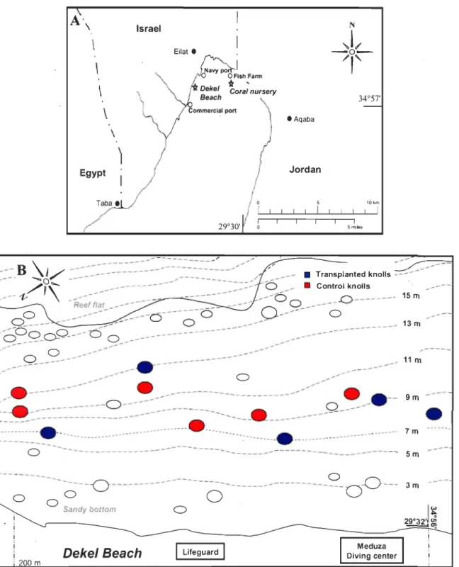
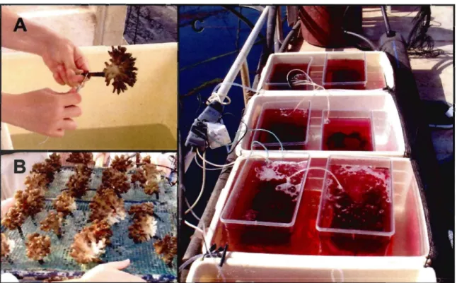
Figure 1: Maps of the study sites. (A) A map of the northern part ofthe Gulf of Eilat (Red Sea), showing the coral nursery and the restoration site (Oekel Beach). (B) Location of the five transplanted knolls and six control knolls at the restoration site (Dekel Beach) ... 13 Figure 2: Preparation of nursery-grown coral colonies. (A) A plastic peg c1eaned of settling algae and fouling organisms with the aide of a scratching dental tool; (B) Corals on trays at the nursery ready to be transferred; (C) Colonies selected for growth analysis incubated with Alizarin Red
s ...
...
...
...
.
....
..
.
...
.
..
...
.
....
16 Figure 3: Transplantation procedures. (A, B) Orilling the substrate of a denuded knoll at the restoration site; (C) Transplantation of a P. damicornis colony; (D) A denuded knollcovered with nursery-grown colonies subsequent to transplantation ... 17 Figure 4: Recruitment of coral-associated invertebrates to the transplanted corals. (A) A
Spirobranchus annelid settled on P. damicornis; (B) Trapezia decapod crab in a P.
damicornis colony; (C) A colony of P. damicornis infested by new recruits of Lithophaga bivalves; (0) Shell of a newly settled Lithophaga in S. pis tilla ta colony (x40); (E) Siphon of a newly settled Lithophaga in S. pistillata colony (x40) ... 20 Figure 5: Qualitative changes observed after the transfer of the transplants to the natural reef. (A) S. pistillata colony subsequent to transplantation (November 2005) showing a dark tissue color pigmentation; (B) The same colony 3 months later, after regaining the typical tissue coloration; (C) White (bleached) zone on P. damicornis colony (November 2005, circ1ed) soon after transplantation; (D) Same tissue are a 3 months later, with normal appearing pigmentation ... 26 Figure 6: Survivorship (Nov. 05-Apr. 07) ofnursery-grown colonies transplanted onto degraded knolls, naturally-growing control colonies on studied knolls and control colonies
at the nursery. (A) S. pistillata; (B) P. damicornis. Data reported as mean ± SE. Letters
denote statistically significant monthly-groups ... 27 Figure 7: Detachment of nursery-grown colonies transplanted onto the 5 degraded knolls,
naturally-growing control colonies on site and control colonies at the nursery. (A) S. pis tilla ta; (B) P. damicornis. Data reported as mean ± SE. Letters denote statistically
significant monthly-groups ... 30
Figure 8: Partial tissue death recorded in transplants, naturally-growing control colonies and control colonies at the nursery, over time. (A) S. pistillata; (B) P. damicornis. Data
reported as mean ± SE. Letters denote statistically significant monthly-groups ... 35
Figure 9: Average magnitude of tissue loss due to partial mortality recorded per
transplanted colony, naturally-growing colony and control colony at the nursery, over time. (A) S. pistillata; (B) P. damicornis. Data reported as mean ± SE. Letters denote statistically significant monthly-groups ... 38
Figure 10: Percentage oftransplanted colonies and naturally-grown colonies damaged by fish. (A) S. pistillata; (B) P. damicornis. Data reported as mean ± SE. Asterisks denote statistically significant monthly-groups ... ~ ... 40
Figure Il: Fish attacks on nursery-grown transplanted colonies. (A) A parrotfish bits S. pis tilla ta transplant; (B) Several broken branches of a S. pistillata transplant are scattered
around the damaged colony; (C) S. pistil/ata transplanted colony that has lost most of its peripheral branches; (D) An exposed peg following the detachment of a colony due to a fish attack; (E) Regeneration by the growth of tissue over damaged skeleton due to fish bites. Arrow points the new tissue growth over the exposed skeleton; (F) The complete regeneration of a damaged colony ... .42
Figure 12: The total numbers offish-damaged transplants and naturally-grown control colonies accumulated over time. (A) S. pistillata; (B) P. damicornis. Data reportedas mean
Figure 13: Fish bites per colony documented on transplanted colonies and naturaUy-grown control colonies. (A) S. pistillata; (B) P. damicornis. Data reported as me an ± SE. Asterisks denote statisticaUy significant monthly-groups ... .45 Figure 14: S. pistillata average chlorophyU a+c concentration and zooxantheUae numbers (± SE) per nursery-control colony (Fish farm) or transplanted colony, after 16 months ... 47 Figure 15: Nursery-grown P. damicornis colony analyzed for growth 18 months after transplantation. The pink Alizarin incorporation in the coral's skeleton represents colony dimension prior to transplantation (day 0). AU white skeletal additions are products of growth at the restoration site ... · ... 50 Figure 16 : Average percentage of nursery-grown transplants inhabiting Trapezia,
Spirobranchus, Alpheus and Lithophaga. (A) S. pistillata; (B) P. damicornis. Data reported as mean ± SE. Pearson Correlation and R-square values are shown ... 51 Figure 17: Average number of Trapezia, Spirobranchus and Alpheus counted in nurSery-grown transplanted colonies. (A) S. pistillata; (B) P. damicornis. Data reported as mean ± SE. Pearson Correlation and R-square values are shown ... 55
1.
INTRODUCTION
The shallow waters of coastal tropical waters are dominated by colorful formations impressive for their variety of motifs and forms--coral reefs. Stretching over a vast region of the tropics, coral reefs form the most biodiversed marine habitats, which represent 5% of aIl marine species (approximately 91 000 species) (Karlson 1999). They are also c1assified among the most productive ecosystems of the world, fixing approximately 700 billion kilograms of carbon annually. In addition, reefs protect adjacent coastal areas from erosion, have a significant economical importance for human populations living in proximity and provide a vital and important source of protein to hundreds of millions of people.
While other reef organisms may contribute to the reefs consolidation, hermatypic corals (phylum Cnidaria, c1ass Anthozoa) are responsible for building the massive biogenic structures that span entire reefs, islands and barrier reefs over the past 200 millions years. Hermatypic corals are colonial animaIs that live in symbiosis relationships with dinoflagellate unicellular algae, thezooxanthellae, situated in their gastrodermic tissue. The coral structures and architectures form three dimensional niches-home to many species of marine invertebrates and fish.
Despite the long history and geological persistence of coral reefs, human activities and anthropogenic pressures have significantly altered their ability to cope with natural disturbances and to maintain themselves (Nystr6m et al. 2000, Pandolfi 2002). Natural
stresses, such as rising sea temperatures resulting in bleaching events (Douglas 2003, Obura 2005, Graham et al. 2007), outbreak of coral diseases (Richardson 1998), along with human stresses such as increased load of sediment and pollution (Aleem 1990, Guest et al.
2007), recreational activities, destructive fishing methods and over-fishing and collection of animaIs for the omamental trade (Lovell and McLardy 2008), have pushed reefs beyond their adaptive capacity (Bell et al. 2006). Worldwide coral reefs are declining at an unprecedented rate (Lesser 2004) and the massive degradation over the past three decades has led in many cases to permanent shifts in reef communities, modifications of the abiotic environrnental conditions and substantial loss of reef areas (Rinkevich 2005b, Aronson and Precht 2006, Hoegh-Guldberg 2006). Alarmingly, 20% of the world's coral reefs have been destroyed and show no immediate potentialities of recovery, 24% are under a severe risk of collapse due to human pressures and 26% face the same threat of collapse in the long run (Edwards and Gomez 2007). Wilkinson (2000) predicts a decline between 40 to 60% of the world's reefs during the next 50 years, unless proper steps are taken. Not only are the biological communities of coral reefs threatened, but also millions of people in over 100 countries who depend on this ecosystem for food and income.
In order to impede the reefs' further decline and to conserve this habitat, restoration measures must be taken. The objective of restoring an ecosystem is to preserve the original ecosystem, in addition to the replacement of lost habitat or destroyed populations (Rinkevich 2005b). Restoration of a coral reef can be of a passive form. Passive restoration is characterized by acts that do not directly interfere with reef organisms, but concentrates on imposing of traditional management efforts, such as no-use zone, imposing of
legislation, etc. By doing that, passive restoration creates the appropriate conditions for reef self-healing through natural processes. On the other hand, active reef restoration requires human intervention (i.e., coral transplantation, coral farming, etc.) and is appropriate anywhere wh en recovery needs to be accelerated in order to protect threatened biodiversity or when natural recovery needs assistance due to a profound change in ecological conditions and reef resilience (Kauffman et al. 1997, McIver and Starr 200 1, DellaSala et al. 2003, Mansourian et al. 2005).
It is becoming more and more evident that degraded reefs rarely recover naturally
from human induced changes without any intervention (Bowden-Kerby 2004, Rinkevich 2005b). In many cases the physical integrity of the reef is damaged, rendering the substrate
inappropriate for new recruitment (Fox and Pet 2001). Marine Protected Areas and "no use zones" are successful in reducing recreational and fishing pressures but are insufficient in countering current-carried pollution and poor to no natural coral recruitment (example of Eilat's "no-use zone", Epstein et al. 1999, Epstein et al. 2005). The rate of coral recruitment is variable, can take up to several years and can be of limited dispersal range for sorne species, impacting damaged areas' diversity (Soong and Chen 2003, Rinkevich 2005a). The anthropogenic (usually chronic) damage acts in the short term, whereas the regeneration of the reef, characterized by a large post-settlement mortality and slow coral growth, is a long term procedure; these two scalesneed to be bridged (Sato 1985, Soong and Chen 2003). In
addition, as was pointed out by Baums (2008), while corals have certain abilities to adapt to changing environmental conditions, their adaptation responses towards human disturbances (such as dynamite fishing) seem improbable, regardless of the time frame. In light of these
facts, it is becoming c1ear that, in addition to conservation and protection, active restoration is now crucial to preserving tl:tis highly diversified and productive ecosystem.
Although active reef restoration is still in its infancy (especially in comparison with forest restoration), various restoration methodologies were employed to address different causes of damage. In cases where the quality of hard substrate was damaged due to ship grounding or blast fishing, primary efforts usually concentrated on consolidating the bottom or adding new hard substrate for colonization (Clark and Edwards 1994, Fox et al. 2003,
Schrimm et al. 2006). While this addresses the physical characteristics of the system,
rehabilitating the substrate alone is not sufficient to ensure the reestablishment of the
habitat's ecological functioning.
Artificial reefs have been widely used as a restoration tool, especially when fish populations were targeted and are usually involved in projects that help promote public awareness (Thailand: Yeemin et al. 2006, Japan: Akakura et al. 2006, French Polynesia: Schrimm et al. 2006. Atlantic Ocean: Koenig 2001, Seaman 2007, Florida: Fahy et al. 2006, and examples reviewed in Spieler et al. 2001). Although artificial reefs have the ability to shift sorne pressure away from the natural reef by creating new dive sites (Leeworthy et al. 2006) and can offer a punctual additional substrate for settlement, they are rarely considered as a promising restoration approach by coral reef restoration ecologists (Abel son 2006, Rinkevich 2005b). An artificial reef can mimic sorne of the characteristics of a natural reef, but nonetheless, it remains artificial and the community development on the artificial reefs can hardly be predicted or controlled. Even after a long
time, their communities rarely resemble the natural reef species' composition (Perkol-Finkel and Benayahu 2005, Perkol-(Perkol-Finkel et al. 2006). Artificial reefs can also reduce the larval supply to natural reefs (Abelson 2006) and cannot counter the problem of lack of seeders stock. Therefore, it could be more appropriate, in the context of coral reefs, to refer to them as "enhancers" rather than "restorers" (Svane and Petersen 2001).
Another common approach, one that is often used in cases of coastal development projects or at locations that have been damaged by' ship grounding, is reattachment and translocation. Threatened or broken colonies are translocated to adjacent un-impacted reefs. When possible, at ship grounding localities or at sites damaged by storms and hurricanes, remaining corals are secured and fixed to hard substrate to prevent their dispersion by currents and water movement that will lead to tissue abrasion (reviewed in Rinkevich 2005b, Bruckner and Bruckner 2006).
By far, the most commonly used approach for active reef restoration has been the transplantation of corals on artificial or natural remaining hard substrates at denuded areas. The addition of live coral colonies aims to reinforce or to re-establish the poor local coral community and thus accelerates or enables recovery. Two main practices were used: the transplantation of whole coral colonies and the transplantation of coral fragments.
In the first approach, whole coral colonies are taken from healthy localities and transferred to degraded sites. Variable degrees of success were reported from such efforts. Bouchon et al. (1981) transplanted coral heads on an artificial reef in the northern Gulf of
colonies were decaying. Schrirnrn et al. (2006) transplanted colonies from nearby donor
sites in French Polynesia and secured them onto concrete blocks or simply placed them in between the blocks. The survivorship after 2.3 years varied between genera from 100% to 73%. A month later, due to phytoplankton bloom, survivorship dropped dramatically to an average of 38%. In contrast, Clark and Edwards (1995) reported 51 % survivorship of colonies transplanted on Armorflex mats in the Maldives after 2.3 years, with most of the mortality occurring during the first seven months of the experiment. They also suggested a
trade-off between growth rates and survivorship of the transplants and conc1uded in a
follow up publication that even when transplants are carefully handled they tend to have higher mortality rates than undisturbed colonies (Edwards and Clark 1998). In Japan,
Akakura et al. (2006) transplanted colonies from a nearby harbor on concrete blocks next to a breakwater. They found varying survival rates between coral species, 80% to less than 20% after 1.5 years.
Beside the lack of uniformity in the method's success, all colonies used by this approach are scarified from the natural reef. Removing corals from healthy reef localities damages those areas and consequently, contributes to the overall damage. In addition, the number of colonies that can be sampled from the reef is very limited, restricting this m~thodology to localized small scale interventions. Facing today's wide reef dec1ine, too many localities are threatened, leaving too few undamaged reefs capable of supplying whole colonies for transplantation.
The second approach, using fragments instead of whole colonies as source material for transplantation, attempts to overcome the disadvantages associated with the fonner approach. Excised branches, fragments and portions of corals have the ability to grow and regain the initial spatial complexity, allowing for a new colony to be established (Epstein and Rinkevich 2001, Shaish et al. 2006). Fragmentation indeed results in a much higher number of "units" to begin with but also has obvious downfalls.
First, the survival capacity of fragments directly transplanted onto a degraded reef is reduced. Yap et al. (1998) observed a survival rate after 1.3 years varying from zero to 40% in fragments of two Po rites species transplanted onto different sites in the Philippines. Dizon et al. (2008) encountered after 5 months 43% mortality of fragments transplanted onto giant clam shells. Van Treeck and Schuhmacher (1997) combined the direct fragment transplantation of four coral species with substrate electrolysis inducing calcium carbonate accretion in the Red Sea (Jordan). While no exceptional mortality was documented soon after transplantation, a 1 year followed up observation revealed 10w survivorship (36% for
Pocillopora damicornis (Linnaeus, 1758), 52% for Stylophora pis tilla ta (Esper, 1797), 72% for AcropiJra variabilis (Klunzinger, 1879) and 68% for Pavona varians (Verrill, 1864)). Only a few studies, such as Guzman (1991) have shown high survival of directly transplanted ramets, indicating that only a few sites are adequate for this methodology.
Most of the studies investigating the relationship between fragment Slze and survivorship have come to the conclusion that survival is size dependent: the larger the fragment is the better chances it has to survive (Smith and Hughes 1999, Lindahl 2003,
Soong and Chen 2003, Bruckner and Bruckner 2006, Latypov 2006, Garrison and Ward 2008). On the other hand, the bigger the fragment, the more stress is inflicted to the donor colony, compromising the mother colony's survival and reproduction (Ward 1995, Zakai et al. 2000, Epstein et al. 2001). Other studies have also shown that direct transfer of coral material resulted in stress associated with transplantation leading to high mortality (Yap et al. 1992). In addition, small fragments are more susceptible to threats encountered in natural reefs, such as predation. When a coralivorous fish or gastropod attacks a small fragment, the resulting damage in comparison to its surface area is much higher than in the case of a colony.
Fragmentation may also affect reproductive activities. Guest et al. (2007) have
shown that, in the case of Goniopora columna (Dana 1846), when fragments are transplanted to more disturbed sites, their oocytes number, oocytes size and polyp size are significantly reduced, suggesting a diversion of energy from reproduction to other needs in response to stressors in the new environment. Zakai et al. (2000) has found that fragmentation of P. damicornis reduces the number of larvae produced by broken colonies and delays their onset of larval release. Small fragments (1-7 cm) studied in this experiment released very few planulae and died within a month. Rinkevich and Loya (1989) have documented a significant reduction in reproductive activity of fragmented S. pistillata colonies, noticing that the effect of breakage on reproduction could take place at least over two reproductive seasons.
In order to overcome the drawbacks of direct transplantation, a new approach,
inspired from silviculture (forest restoration), has been devised by Rinkevich (1995). Reefs are often compared to forests since they share in cornrnon essential ecological and structural traits (Epstein et al. 2003). Forest restoration has been undertaken for over a century in many countries worldwide, allowing the development and refinement of wide array of protocols. Rinkevich proposed to benefit from the knowledge gained in this parallel ecosystem and suggested the "Gardening Coral Reefs" concept. This approach is based on a two steps methodology: 1) generating a huge number of minute coral fragments and their in situ nursery culturing until they form large colonies amenable for transplantation and 2) transplanting these colonies in degraded reef sites (Rinkevich 2006).
The use of a nursery phase allows for coral culture to be initiated from extremely small fragments as small as 1-10 polyps, each (few millimeters, cornrnonly named "nubbins") or from sexual recruits, without compromising their survivorship rate due to the protected idyllic environment (Forsman et al. 2006, Shafir et al. 2006, Shaish et al. 2008). The employment of nubbins substantially reduces the stress inflicted to donor colonies (Shafir et al. 2001, Shafir et al. 2003). This permits a large scale random sampling of corals at a targeted locality that is capable of representing local species abundance and genetic vari abi lit y without damaging a whole reef or compromising its health. Creating new colonies from nubbins and rearing them in a coral nursery allows for the rapid generation of an extremely large stock of corals that can then be used for restoration (Shafir et al. 2006).
The transplantation of whole colonies, rather than coral fragments, could potentially increase their ability to acclimate to the new environment. Adult colonies can also contribute to coral reproduction and enhance the locallarval pool. Moreover, bypassing one of the reef restoration biggest bottlenecks, the availability of source colonies for rehabilitation, allows rapid, larger-scaled restoration acts.
The first step of the gardening concept has been tested in several reef localities in the world, induding the Red Sea, Thailand, Singapore, Philippines, Tanzania and Jamaica, (Rinkevich 2008). Several nursery prototypes have been established: in situ nurseries such as mid-water floating nursery (Shafir et al. 2006) and leg-fixed nursery (Shaish et al. 2008,
Soong and Chen 2003); and ex situ nurseries on land (Shafir et al. 2001, Forsman et al. 2006). Various aspects related to nursery rearing, such as divers nursery structures adapted to different environmental conditions, optimization of coral maintenance at the nursery and elimination of fouling organisms, the use of the nursery as planulae hub, are still being explored (Amar and Rinkevich 2007, Rinkevich 2008, Shafir et al. 2008). The first step of the Gardening methodology has shown promising results and has proven to be successful (Forsman et al. 2006, Shafir et al. 2006, Shaish et al. 2008). Whether nursery-grown coral colonies are suitable for transplantation in damaged reef areas is yet to be proven.
The aim of this study is to evaluate the applicability of the second step of the "Gardening Concept" and to develop guidelines for nursery-grown coral transplantation. We used new branching coral colonies of two species, Stylophora pistillata (Esper, 1797) and Pocillopora damicornis (Linnaeus, 1758), generated in a floating coral nursery in Eilat
for transplantation in a degraded zone of Eilat's reef (Red Sea, Israel). We followed the transplant's acc1imation in their new environment and monitored their survival; growth and contribution to the locallarval stock during 17 months after transplantation. We tested the hypothesis that the transfer of the farmed coral colonies to the natural reef will not influence their survival and, once transferred back to the oligotrophic waters of the reef,
their growth will be reduced. We were also interested whether the spatial positioning on the knolls would impact their survival and detachment. We characterized tissue damage occurring due to partial tissue death or fish action. In addition, we examined the ecosystem
engineering effects the branching nursed colonies might have in the restored site and followed their impact on model species inhabiting living hermatypic corals.
2.
Materials and methods
2. 1 Study sites
In order to examine the applicability of the Gardening concept and develop the methodology for farmed-colonies transplantation, a degraded zone of Eilat's reef (Gulf of Eilat, Red Sea: 29°30'N; 34°S7'E) has been targeted. The reef of Eilat has been in decline for the past four decades as a result of anthropogenic activities, amongst the rapid development of Eilat city, recreational and tourist activities, urban affluences and pollution (Epstein et al. 1999, Rinkevich 200Sa).
A floating in situ nursery was established in 2003 in the northem part of the Bay (Fig. lA), away from the reef, coral predators and recreational activities. The nursery is situated at a depth of 8 m (12 m above the sea bottom) and resides in an enriched nutrient area due to its proximity to Ardag and Dag-Suf fish farms. The intensive mari culture of Gilthead seabream (Sparus aurata) resulted in elevated nutrient concentrations and particulate organic matter that accelerated coral growth (Bongiomi et al. 2003a, b).
The restoration site at the natural reef, the Dekel Beach, is located 2.7 Km south to the nursery between a navy base and the commercial port of Eilat and in front of a busy dive center (Fig. lA). The first 18 meters depth ofthis reef are characterized by a moderate sandy slope with scattered knolls that contain varying amounts of hermatypic-dominance coral covers (from completely bare to weIl covered knoIls, though the latter are very rare).
A
'
\\
,
\
\
Egypt \)
Taba. Israel • Aqaba Jordan 10km 1 1 1 1 5 miles-::_
B
~~/
~~~~~~~~~~~~~:::~~~~~~~~~~~~~--/~/
/
"
~
_---
•
Transplanted knolls-
~~
---/1\...
---
---::::
b
•
Control knolls~--
'V-Re:;;:t--~---
----~~-- ---
6~
_----
-0
---
15 mo
---
_----0 ---0
-
0
---
0 ~--- --- _---- 0 _---/o
0 Q_CY:0 ----o - 0 _---....-
...-
--13 m---
----~---
~
--
~
---
...---
,---
---
_ 11 m 9m---
---
---
~
---
~
-
O~
---o
o
00
Sandy bottom
0Dekel Beach 1 Lifeguard 1 DivMeduza ing center
•
7m
----5m
3m
----Figure 1: Maps of the study sites. (A) A map of the northern part of the Gulf of Eilat (Red Sea), showing the coral nursery and the restoration site (Dekel Beach). (B) Location of the five transplanted knolls and six control knolls at the restoration site (Dekel Beach).
The Israeli Nature and National Park Protection Authority pennit restricted our experiment to 5 denuded knoUs situated in shaUow water (6-13m depth) and aligned from north to
south on a 200 m stretch (Fig. lB).
2.2 Coral rearing at the nursery
New coral colonies were generated at the nursery by re-pruning the initial nursery-grown coral stock of Stylophora pis tilla ta and Pocillopora damicornis established in the nursery at 2003 (Shafir et al. 2006). The donor colonies, clones of three S. pistillata and four P. damicornis mother colonies collected at Eilat's navy port, were pruned by electrician's wire cutters pro vi ding fragments between 1 to 2 cm size. The fragments were glued to the fiat surface of a plastic peg (Red Sea Corals LTD., Israel; 9 cm long, 0.3-0.6 cm wide leg with a 2 cm diameter "head", Shafir et al. 2006) and reared in the nursery on trays constructed from 50X30 cm PVC frames with stretched plastic nets (0.25 cm2 mesh
size) according to the protocol of Shafir et al. (2006). The number of colonies per tray was adjusted every three to four months to space between the new colonies to optimize growth conditions. The new fanned colonies were maintained for a period of eight to 14 months prior to transplantation and reached a diameter of 6 to 9 cm.
2.3 Preparation of nursery-grown coral colonies
During November 2005, 554 nursery-grown colonies of two abundant branching
transplantation at the coral nursery. AIl colonies were prepared during a period of seven days with the help of a team of 13 untrained volunteers. The plastic pegs on which the corals grew were cleaned of settling algae and other sessile fouling organisms using forceps, dish-pads and various scratching dental tools (Fig. 2A). The colonies were examined for existence of coral predators, such Drupella snail, that were removed when found. An average of lOto 15 farmed colonies per hour was prepared by one worker. The prepared colonies were then arranged on trays for transportation (Fig. 2B). Sorne of the colonies (n=100) were placed'in aerated tanks and incubated for 12 ho urs (from sunrise to sunset) with 15 mglL Alizarin Red S (Bames 1970) in order to foIlow future coral's growth. The tanks were placed in plastic containers with a constant water flow generated by a water pump in order to maintain constant water temperature during Alizarin incubation (Fig. 2C). They were shed by plastic net of 0.25 cm2 mesh size to avoid excessive radiation
between 10 am to 14 pm.
2.4 Transplantation methodology
Once cleaned, the coral trays were placed in plastic containers fiIled with seawater and transferred by boat from the coral nursery to the restoration site. The colonies were transplanted onto the five denuded knolIs by five SCUBA divers. Holes were drilled in the
knoIls' hard substrates in regular distances of 20 cm using an underwater pneumatic drilling powered by a SCUBA tank (Fig. 3A, B). A very sm aIl amount of epoxy glue (AquaMend) was placed at the bottom of each hole and the pegs were inserted in the holes, permitting a
Figure 2: Preparation of nursery-grown coral colonies. (A) A plastic peg c1eaned of settling
algae and fouling organisms with the aide of a scratching dental tool; (B) Corals on trays at the nursery ready to be transferred; (C) Colonies selected for growth analysis incubated with
Figure 3: Transplantation procedures. (A, B) Drilling the substrate of a denuded knoU at the restoration site; (C) Transplantation of a P. damicornis colony; (D) A denuded knoll covered with nursery-grown colonies subsequent to transplantation.
good attachment of the corals to the substrata (Fig. 3C, D). Each colony was tagged with a numbered plastic stripe in order to follow each colony's acclimation and survival during the
next months. The whole transplantation operation took approximately 2 minutes per colony transplanted. Two pneumatic drillers powered by SCUBA tanks were used. A 12 L aluminum SCUBA tank compressed to a pressure of 200 bars permitted the drilling of 25 to
30 holes in the hard calcareous substrate of the bare knolls. The most efficient way of transplanting the colonies was to drill and transplant simultaneously. When large substrate
holes occurred. The five untrained volunteer divers perforrned three dives per day (net 3 hours of underwater work per day). One untrained volunteer was capable of transplanting 30 colonies within one hour, inciuding the activities of drilling, epoxy glue mixing, colony tagging and colony anchoring. Two weeks were required in order to complete the transplantation. One square meter of substrate was covered by an average of 15 colonies.
2.5 Monitoring
The transplantation was evaluated on a monthly-basis using SCUBA diving and underwater digital photos. Two control groups were established: a group of 76 naturally growing colonies at the experimental site, of the same species and approximate sizes and a group of 217 nursery-grown colonies prepared for transplantation that was left at the nursery. The two control groups were monitored in parallel to the transplants.
Data on detachment, survival, partial tissue mortality and fish bites was collected on a monthly basis. Partial tissue death and the magnitude of tissue loss per colony due to partial tissue mortality were estimated by eye. Partial tissue mortality is defined as a bare patch of skeleton on the surface of a coral colony due to the loss of part of the living tissue. The proportion of tissue mortality per affected colony was estimated at 10% intervals. Fish bites were counted individually. The bites inflicted lesions due to the removal of tissue, exposing the underlying skeleton. The size and shape of such injuries were not constant. The spatial orientation of each transplanted colony on the knoll (north, south, east, west, or up facet) was deterrnined.
Starting one month after the transplantation, the number of Trapezia (Latreille, 1825) crabs (Fig. 4B) and Alpheus (Fabricius, 1798) shrimps appearing in each colony was counted at each monitoring. The count of settling Spirobranchus (Pallas 1766) worms (Fig. 4A) was added a month later. On April 2006, we witnessed numerous black spots on several S. pis tilla ta and P. damicornis transplants, which appeared to be, after an
examination under a stereomicroscope, metamorphosed bivalves, Lithophaga (R6ding 1798) that settled on the corals (Fig. 4C-E). We followed the Lithophaga recruitment to the transplanted colonies thereafter. Adult Lithophaga are hard to spot since they are found in the coral skeleton with only the ends of their siphons appearing on the coral's surface. Therefore, only colonies that had new settlement which are easily recognized were considered in our survey.
2.6 Zooxanthellae abundance and chlorophyll concentrations
The densities of algal cells in coral tissues as well as the chlorophyll content per algae are variable parameters that are responsive to light and nutrient conditions. Witnessing a color change in the transplants a short time after transplantation, we decided to compare those two parameters with those of the control colonies left in the nursery. For
,
this analysis, fragments of four S. pistillata colonies from each locality were sampled in duplicate .after 16 months. The fragments were taken to the National Center for Mariculture
Figure 4: Recruitment of coral-associated invertebrates to the transplanted corals. (A) A
Spirobranchus annelid settled on P. damicornis; (B) Trapezia decapod crab in a P. damicornis
colony; (C) A colony of P. damicornis infested by new recruits of Lithophaga bivalves; (D) Shell of a newly settled Lithophaga in S. pistillata colony (x40); (E) Siphon of a newly settled Lithophaga in S. pistillata colony (x40).
ethylene diamine tetracetic acid (EDT A) in order to dissociate the living tissue (Rinkevich
et al. 2005). Aliquots of 20l-tl from each sample were spread on a Hemocytometer and counted under a light microscope in order to determine the zooxanthellae cell number. For chlorophyll extraction acetone 100% was added to each sample. The samples were placed on ice in dark conditions and transferred to the laboratory at Haifa (National Institute of Oceanography) where they were kept at 4°C. They were analyzed with a spectrophotometer (630nm, 663nm) within 48h following shipment. The chlorophyll concentrations were calculated using Jeffrey and Humphrey's (1975) equations:
Chlorophyll a = Il.43 E663 - 0.64 E630
Chlorophyll C2 = 27.09 E630 - 3.63 E663
[1] [2]
The fragment's surface area was detennined using a Desktop 3D laser scanner (NextEngine). Prior to scanning, all skeletal bare parts, resulting from the fragment's cuts, were colored in black in order to be omitted from the scan. The fragments were glued on plastic sticks using epoxy glue (Devcon 5 minute Epoxy) and colored in red using a mat spray-paint (Duplicolor, Gennany). Two scans were made for each fragment- a 360 degrees scan (6 scans/3600) and a bracket scan-in order to co ver the entire fragment's surface. The fragments were scanned together with a reference object, enabling an accurate alignment of the 2 scans. The surface area of each fragment was computed by the NextEngine ScanStudio Core software.
2.7 Growth analysis
Unfortunately, sorne of the Alizarin-stained colonies were detached during the course of the experiment, restricting and detennining our choice of quantity for this analysis. Seven S. pistillata colonies (3 transplants and 4 nursery controls) along with ten P.
damicornis colonies (5 transplants and 5 nursery controls) were sampled after 18 months
for growth detennination. The colonies were placed in a recipient containing freshwater followed by an ovemight immersion in a 50:50 freshwater. - bleach (sodium hypochlorite) solution in order to eliminate living tissue. The skeletons were washed from any tissue
remains under tap water and left to dry. They were weighted and measured using an electronic digital caliper to obtain the weight, width (w), length (l) and height (h). The new skeletal additions appearing after the Alizarin red marks were cut using electrician's wire cutter and the colonies were then weighted and measured again. Each colony's diameter and ecological volume were calculated for the initial and the grown structure using the equations (Rinkevich and Loya 1983):
Colony diameter d = (l+w)/2
Ecological volume E= nr2h with r = (l+w)/4
[3 ] [4]
The growth rate constants (k) per day for ecological volumes (E) were obtained using the formula:
Et
=
Eoekt [5]k = (ln EtlEo)/t, t = time in days (0 at the beginning of the study). [6]
2.8 Larvae collection
In order to asSess the transplants' contribution to local coral population's reproduction, 10 transplanted and 10 naturally growing S. pis tilla ta colonies were selected. These colonies were chosen for their size (approximately 10cm in diameter) and for their state ofhealth (without damaged tissue parts). S. pistillata is a brooding species (Shlesinger et al. 1998) that releases planulae larvae during a long reproduction period that stretches
from December to July in Eilat (Rinkevich and Loya 1979, 1987). Recently, a shi ft in the reproductive seasonality was documented by Amar et al. (2007), who reported an extension in the seasonality of planulae shedding, now occurring between January and August, with peak planulation between April and June. Two samplings have been carried out on June 19 and June 26, 2006. Planulae collection devices, consisting of a plankton net sleeve glued to a plastic cup (Amar et al. 2007), were placed over the selected colonies from sunset to sunrise. The plastic cups were drained from water and the planula assemblages over the lid were washed out to a wide petri dish. They were then counted under a stereomicroscope.
2.9 Statistical analysis
Data analyses were performed using SPSS software for Windows version 16.0. The results were examined for each species separately, using the knoll as the sampling unit of repetition. Normality was tested using Kolmogorov-Smirnov or Shapiro-Wilk statistical tests. When needed, the proportions observed were transformed using the arcsine square root transformation in order to approximate normality. Survival, detachment, partial tissue death and fish bites of nursery-grown transplants and control colonies were assessed using a repeated measures analysis of variance (ANOVA). When interactions between the parameters studied appeared, a monthly one way ANOV A was performed using a multiple-comparison Bonferroni correction to account for multiple testing, in order to maintain the 5% error rate (significant differences admitted when the probability is inferior to 0.003). When a significant effect was found, means were compared with a Bonferroni post hoc test (significant differences admitted when the probability is inferior to 0.05).
Zooxanthellae densities and chlorophyll content of transplants and nursery control
colonies, as well as their growth were compared using a Student's t test for independent
samples. The growth rate constant (k) was compared with a test for equality between two percentages.
Both the increase in the average percentage of transplants recruited by invertebrates over time and the increase in the average number of invertebrate specimens residing in each
transplant over time were studied using correlation analysis. When a linear correlation was
found, linear regression was carried out' and statistically significant (ANOVA, p<O.05)
equations of best fit were computed. Data are reported as me an ± standard error of the
3. RESULTS
3.1 Acclimation of the nursery-grown carals at the restoration site
3.1.1 Coloration
Two notable phenomena occurred one to six weeks after transplantation:
1. A change in the colonies' tissue color (Fig. 5 A, B). The colonies at the nursery were characterized by a darker tissue-pigmentation than reef grown colonies. Few weeks
after their relocation tissue colors became pale, and colonies regained the natural appearing color.
2. Transparent tissue areas (appearing white) lacking zooxanthellae due to shading
caused by their proximity to sibling colonies on the nursery trays regained their pigmentation (Fig. 5 C, D)
Those changes occurred in both species.
3.1.2 Survival
Survival rates of nursery-grown transplants, naturally-growing controls and nursery
control colonies of S. pis tilla ta and P. damicornis are shown in Fig. 6. One of the first concerns when carrying out a transplantation is whether the transfer of corals to a new location and the transplantation act are stress fui to the transplants, which could result in an increased mortality during the first months after transplantation. The results of the first four months of monitoring revealed high survivorship (exceeding 95%) for both species'
Figure 5: Qualitative changes observed after the transfer of the transplants to the natural reef. (A) S. pistillata colony subsequent to transplantation (November 2005) showing a dark tissue color pigmentation; (B) The same colony 3 months later, after regaining the typical tissue coloration; (C) White (bleached) zone on P. damicornis colony (November 2005, circled) soon after transplantation; (D) Same tissue area 3 months later, with normal appearing pigmentation.
A
B
80 70
60 a a
• Transplanted knolls (5 knolls; 265 colonies)
50 • Control knolls on site (6 knolls; 41 colonies) a
a
• Control trays at nursery (6 trays; 108 colonies)
40+---r-~--r---r--r-'--r--r~~----~-'r---~r---r
80 70
60 • Transplanted knolls (5 knolls; 289 colonies)
•
Control knolls on site (6 knolls; 35 colonies)50
• Control trays at nursery (6 trays; 109 colonies)
40+---~-r--~--~-'--~~--r-~~--~---r----~----~
o
403 53340 102 365 468
Days after transplantation
Figure 6: Survivorship (Nov. 05-Apr. 07) of nursery-grown colonies transplanted onto degraded knolls, naturally-growing control colonies on studied knolls and control colonies at the nursery. (A) S. pistil/ata; (B) P. damicornis. Data reported as mean ± SE. Letters denote
transplants, a survivorship very similar to that of the two control groups (Fig. 6). In fact, no statistically significant difference was observed between these three groups until 291 days after transplantation (9 months) for S. pistillata (one way ANOVA with Bonferroni correction', p>0.003 for days 40-266) and throughout the entire monitored period in the case of P. damicornis (one way ANOVA with Bonferroni correction, p>0.003 for aIl dates)
(Fig. 6, Appendices Table 7.1.2).
Mortality over time was dependent on the experimental group (transplants, naturally-growing colonies and nursery-kept colonies) as indicated by a repeated measures ANOVA (s. pistillata: FS28,36.97=7.033; p<O.OOl; P. damicornis: FS.37,37.s9= 4.213; p=0.003; Appxs. Table 7.1.1) and differed significantly between the two species (Fig. 6). For S. pistillata, mortality was low and rather constant in the nursery as 98.3± 1.1 % of the colonies were still alive after 17 months. Survivorship was also quite constant for the naturally-growing S. pis tilla ta at the restoration site until September 2006 (313 days after transplantation; 97.2±2.8%). Then, mortality increased revealing, after 533 days, significant higher values than the one recorded at the nursery (one way ANOV A with Bonferroni correction, p<0.003; post hoc Bonferroni multiple comparison, p>0.05 for days 291-486; P<O.05 for day 533; Appxs. Table 7.1.2). After 17 months, 81.3±6.1 % ofnaturally-growing S. pis tilla ta controls at the restoration site remained alive. The survivorship of transplanted
1 The multiple-comparison Bonferroni correction was performed to account for multiple testing, as the same
hypothesis was repeated 14 times (for each month). In arder to maintain the 5% error rate, we divided 0.05 by
the number of repetitions (14). Therefore, significant differences between groups are only adrnitted when
p<0.003. When significant values were found (p<0.003), means were compared with post hoc tests, admitting statistically significant differences for p<0.05. .