HAL Id: dumas-02076049
https://dumas.ccsd.cnrs.fr/dumas-02076049
Submitted on 21 Mar 2019HAL is a multi-disciplinary open access archive for the deposit and dissemination of sci-entific research documents, whether they are pub-lished or not. The documents may come from teaching and research institutions in France or abroad, or from public or private research centers.
L’archive ouverte pluridisciplinaire HAL, est destinée au dépôt et à la diffusion de documents scientifiques de niveau recherche, publiés ou non, émanant des établissements d’enseignement et de recherche français ou étrangers, des laboratoires publics ou privés.
Ludivine Oliveira da Silva
To cite this version:
Ludivine Oliveira da Silva. Évaluation des facteurs de risque de mortalité et de récurrence de troubles du rythme ventriculaires chez les patients avec une cardiopathie structurelle dans les suites d’une ablation d’orage rythmique. Human health and pathology. 2017. �dumas-02076049�
N°2017ANTI0178
Évaluation des facteurs de risque de mortalité et de récurrence de troubles
du rythme ventriculaires chez les patients avec une cardiopathie structurelle
dans les suites d’une ablation d’orage rythmique
THESE
Présentée et soutenue publiquement à la Faculté de Médecine Hyacinthe BASTARAUD des Antilles
Et examinée par les Enseignants de la dite Faculté Le 9 Mai 2017
Pour obtenir le grade de DOCTEUR EN MEDECINE Par OLIVEIRA DA SILVA Ludivine
Jury
Président : Professeur NEVIERE Rémi Membres : Professeur ROQUES François
Professeur FARID Karim Docteur INAMO Jocelyn
Docteur GRANDJBAKHCH Estelle
Faculté de Médecine Hyacinthe Bastaraud UFR DES SCIENCES MÉDICALES
NOM ET PRENOM : OLIVEIRA DA SILVA Ludivine
SUJET DE LA TEHSE : Évaluation des facteurs de risque de mortalité et de récidive de troubles du rythme ventriculaires chez les patients avec une cardiopathie structurelle dans les suites d’une ablation d’orage rythmique
THESE : MEDECINE
Qualification : Médecine Spécialisée ANNEE : 2017
NUMERO D’IDENTIFICATION : 2017ANTI0178
MOTS CLEFS : Electrical storm ; Radiofrequency ; Ventricular tachycardia ; Structural cardiomyopathy
RESUME
Background- Electrical storm (ES) occurring in patients with a cardiomyopathy can lead to bad evolution. Caheter ablation (CA) has evolved into a prior treatment and this study aims to identify predictive factors of mortality and recurrence of ventricular tachycardia (VT) after a radiofrequency.
Methods and results- 87 patients that had CA for a drug-resistant ES were included : 36 patients with chronic artery disease, 20 with recent myocardial infarction, 14 with arrhythmogenic right ventricular dysplasia, 11 with idiopathic dilated cardiomyopathy, 3 with valvular cardiomyopathy, 2 with hypertrophic cardiomyopathy and 1 with a previous myocarditis. 37 patients (43%) presented a recurrence of tachyarrhythmia within 1 month after the first radiofrequency, 18 patients underwent multiple CA. Overall, 48 patients (55%) did not have a recurrence of VT after the last CA. Independent predictive factor of early-VT recurrence were recent myocardial infarction (OR=2,343 IC95%[1,149 ; 4,778] p=0,019) and positive or undone ventricular stimulation (OR=0,425 IC95%[0,218 ; 0,828] p=0,012). 20% of patients died within one month (n=17). Male gender (OR=0,208 IC95%[0,063 ; 0,693] p=0,011), low tolerance of VT (OR=8,486 IC95%[1,084 ; 66,40] p=0,042), general anaesthesia (OR=3,163 IC95%[1,012 ; 9,885] p=0,048] and VT recurrence after first CA (OR=4,393 IC95%[1,209 ; 15,97] p=0,025) were associated with early mortality.
Conclusion- Catheter ablation is an effective treatment of electrical storm when procedure is a complete success. A population at high risk of death may be identified and would benefit of a strategy considering heart transplantation and ventricular assistance device in case of CA failure.
JURY : Président : Professeur Rémi Nevière Juges : Professeur François Roques : Professeur Karim Farid
: Docteur Jocelyn Inamo
Le Président de l'Université des Antilles : Jacky NARAYANINSAMY Doyen de la Faculté de Médecine : Raymond CESAIRE
Vice-Doyen de la Faculté de Médecine: Suzy DUFLO
Professeurs des Universités - Praticiens Hospitaliers
Bruno HOEN Maladies Infectieuses
CHU de POINTE- À -PITRE/ABYMES Tel : 05 90 89 15 45
Pascal BLANCHET Chirurgie Urologique
CHU de POINTE- À -PITRE/ABYMES Tel : 05 90 89 13 95
André-Pierre UZEL Chirurgie Orthopédique et Traumatologie
CHU de POINTE-A-PITRE/ABYMES Tel : 05 90 89 14 66
Pierre COUPPIE Dermatologie
CH de CAYENNE Tel : 05 94 39 53 39
Thierry DAVID Ophtalmologie
CHU de POINTE-A-PITRE/ABYMES Tel : 05 90 89 14 55
Suzy DUFLO ORL – Chirurgie Cervico-Faciale
CHU de POINTE-A-PITRE/ABYMES Tel : 05 90 93 46 16
Eustase JANKY Gynécologie-Obstétrique
CHU de POINTE-A-PITRE/ABYMES Tel 05 90 89 13 89
François ROQUES Chirurgie Thoracique et Cardiovasculaire
CHU de FORT- DE - FRANCE Tel : 05 96 55 22 71
Jean ROUDIE Chirurgie Digestive
CHU de FORT- DE - FRANCE Tel : 05 96 55 21 01 - Tel : 05 96 55 22 71
Jean-Louis ROUVILLAIN Chirurgie Orthopédique
CHU de FORT- DE - FRANCE Tel : 05 96 55 22 28
André CABIE Maladies Infectieuses
CHU de FORT- DE - FRANCE Tel : 05 96 55 23 01
CHU de FORT- DE - FRANCE Tel : 05 96 55 22 61
Vincent MOLINIE Anatomopathologie
CHU de FORT- DE - FRANCE Tel : 05 96 55 23 50
Raymond CESAIRE
Bactériologie-Virologie-Hygiène option virologie
CHU de FORT- DE - FRANCE Tel : 05 96 55 24 11
Professeurs des Universités - Praticiens Hospitaliers
(Suite)
Philippe DABADIE Anesthésiologie/Réanimation
CHU de POINTE- À -PITRE/ABYMES Tel : 05 96 89 11 82
Maryvonne DUEYMES-BODENES Immunologie
CHU de FORT- DE - FRANCE Tel : 05 96 55 24 24
Régis DUVAUFERRIER Radiologie et imagerie Médicale
CHU de FORT- DE - FRANCE Tel : 05 96 55 21 84
Annie LANNUZEL Neurologie
CHU de POINTE- À -PITRE/ABYMES Tel : 05 90 89 14 13
Louis JEHEL Psychiatrie Adulte
CHU de FORT- DE - FRANCE Tel : 05 96 55 20 44
Mathieu NACHER
Epidémiologie, Economie de la Santé et Prévention
CH de CAYENNE Tel : 05 94 93 50 24
Guillaume THIERY Réanimation
CHU de POINTE-A-PITRE/BYMES Tel : 05 90 89 17 74
Magalie DEMAR - PIERRE Parasitologie et Infectiologue
CH de CAYENNE Tel : 05 94 39 53 09
Vincent MOLINIE Anatomie Cytologie Pathologique
CHU de FORT DE FRANCE Tel : 05 96 55 20 85/55 23 50
Michel DEBANDT Rhumatologie
CHU de FORT- DE - FRANCE Tel : 05 96 55 23 52
Jeannie HELENE-PELAGE Médecine Générale
CHU de Pointe-à-Pitre / Cabinet libéral Tel : 05 90 84 44 40
Karim FARID Médecine Nucléaire
CHU de FORT- DE - FRANCE Tel : 05 96 55 21 67
Mehdi MEJDOUBI Radiodiagnostic et imagerie Médicale
CHU de FORT- DE - FRANCE Tel : 05 96 55 21 84
Rémi NEVIERE Physiologie
CHU de FORT- DE - FRANCE Tel : 05 96 55 .. .. .
Christian SAINTE-ROSE Radiodiagnostic et imagerie Médicale
CHU de FORT- DE - FRANCE Tel : 05 96 55 .. .. .
Professeurs Associés de Médecine Générale
Franciane GANE-TROPLENT Médecine générale
Cabinet libéral les Abymes Tel : 05 90 20 39 37
Maître de Conférences des Universités - Praticiens Hospitaliers
Christophe DELIGNY Médecine Interne
CHU de FORT- DE - FRANCE Tel : 05 96 55 22 55
Jocelyn INAMO Cardiologie
CHU de FORT- DE - FRANCE
Tel : 05 96 55 23 72 - Fax : 05 96 75 84 38
Fritz-Line VELAYOUDOM épse CEPHISE Endocrinologie
CHU de POINTE- À -PITRE/ABYMES Tel : 05 90 89 13 03
Marie-Laure LALANNE-MISTRIH Nutrition
CHU de POINTE- À -PITRE/ABYMES Tel : 05 90 89 13 00
Sébastien BREUREC Bactériologie &Vénérologie
CHU de POINTE- À -PITRE/ABYMES Tel : 05 90 89 12 80
Narcisse ELENGA Pédiatrie
CH de CAYENNE Tel : 05 94 39 77 37
Moana GELU-SIMEON Gastroentérologie hépatologie
CHU de POINTE-A-PITRE/ABYMES Tel : 05 90
Chefs de Clinique des Universités - Assistants des Hôpitaux
BANCEL Paul
ORL/Chirurgie maxillo faciale
CHU de Pointe-à-Pitre Tél. : Tél. : 0590 89 14 60
BORJA DE MOZOTA Daphné Gynécologie-Obstétrique
CHU de POINTE- À -PITRE/ABYMES Tél. : 0590 89 19 89
DARCHE Louis Chirurgie Digestive et Viscérale
CHU de Martinique Tél. : 0596 55
DE RIVOYRE Benoit Ophtalmologie
CHU de Pointe-à-Pitre Tél. : 0590 89 14 50
DEBBAGH Hassan Chirurgie thoracique
CHU de Martinique Tél. : 0596 55 22 71
DOURNON Nathalie Maladies infectieuses
CHU de Pointe-à-Pitre Tel : 05 90 89 10 10
GALLI-DARCHE Paola Neurologie
CHU de Martinique Tél. : 0596 55 .. .. .
Chefs de Clinique des Universités - Assistants des Hôpitaux
(Suite)
GHASSANI Ali Gynécologie-Obstétrique
CHU de Pointe-à-Pitre Tél. : 0590 89 19 89
JACQUES-ROUSSEAU Natacha Anesthésie-Réanimation
CHU de Pointe-à-Pitre Tél. : 0590 89 11 82
MARY Julia Rhumatologie
CHU de Martinique Tél. : 0596 55 23 52
MOINET Florence Rhumatologie-médecine interne
CHU de Martinique Tél. : 0596 55 22 55 MONFORT Astrid Cardiologie CHU de Martinique Tél. : 0596 55 23 72
MOUREAUX Clément Urologie
CHU de Pointe-à-Pitre Tél. : 0590 89 13 95
NABET Cécile CH “Andrée ROSEMON” de Cayenne Parasitologie et Mycologie
Tél. : 0594 39 53 59
PARIS Eric Réanimation
CHU de Pointe-à-Pitre Tél. : 0590 89 10 10
PIERRE-JUSTIN Aurélie Neurologie
CHU de Pointe-à-Pitre Tél. : 0590 89 13 40
SAJIN Ana Maria Psychiatrie
CHU de Martinique Tél. : 0596 55 20 44
SEVERYNS Mathieu Chirurgie orthopédique
CHU de Martinique Tél. : 0596 55 22 28
Chefs de Clinique des Universités – Médecine Générale
CARRERE Philippe
Médecine Générale CHU de Pointe-à-Pitre /Cabinet
Tél. : 0690 99 99 11
PLACIDE Axiane Médecine Générale
CHU de Martinique / Cabinet
NIEMETZKI Florence Médecine Générale
CH « Andrée Rosemon » de Cayenne/Cabinet
MOUNSAMY Josué Médecine Générale
Professeurs EMERITES
CHARLES-NICOLAS Aimé
Psychiatrie Adulte
Georges JEAN-BAPTISTE Rhumatologie
CHU de FORT- DE - FRANCE Tel : 05 96 55 23 52 - Fax : 05 96 75 84 44
Serge ARFI Médecine interne
CHU de FORT- DE – France Tel : 05 96 55 22 55 - Fax : 05 96 75 84 45
Bernard CARME Parasitologie
Dédicaces
A mes maitres
Madame le Docteur Estelle Grandjbakhch, ma directrice de thèse, merci de m’avoir confié ce travail passionnant, pour ton investissement dans ce travail. Tes orientations et remarques m’ont permis de pousser mes réflexions toujours plus loin.
Monsieur le Professeur Remi Nevière, merci de m’avoir fait l’honneur d’être le président de mon jury, vous m’avez permis d’appréhender un peu plus les statistiques, merci de l’aide que vous m’avez apporté avec toujours beaucoup de bonne volonté et de gentillesse.
Monsieur le Professeur François Roques, j’ai découvert la chirurgie cardiaque avec vous au début de mon internat et je ne me lasse pas de toujours apprendre à vos côtés.
Monsieur le Docteur Jocelyn Inamo, vous me connaissez depuis mes débuts de petit cardiologue et m’avez donné l’opportunité de rapidement toucher à tout dans ce domaine. Monsieur le Professeur Karim Farid, merci d’avoir eu la gentillesse d’accepter de faire partie de mon jury, même si je sais bien que la rythmologie n’est pas votre domaine de prédilection.
A ma famille
Vous avez eu quelques difficultés à comprendre mes tribulations mais vous avez toujours été présents pour me soutenir.
A mes amis
Ma bande de bobbys, Chouchou, Lylou, La Moz, Noé, Alex, Elise, Laetitia et Hadrien, on a survécu tous ensemble à ces 6 années et plus encore, toujours autant d’amour et de folie avec vous !
Charlotte et Ben (la vie avec vous fut un délice, j’espère qu’il y aura toujours une petite place pour moi au fond du couloir avec vous) Aydine (j’ai mal aux pieds d’avoir voyagé dans tes fractures temporelles)
Pauline (mon buddy des fonds marins)
A vous que j’ai connu sous le soleil au fil des stages ou de la vie : Anais, Romain (Dr Pasqualottoooo), Anne Lise, Emma (la blonde la plus blonde que je connaisse), Hossam (le monarque de la principauté tartanaise), Leila (que d’aventures ensemble autant dans les murs de l’hôpital qu’en dehors), Tito (l’homme aux m&ms), Rebecca, Laura, David, Pierre, Sophie, Tiana, Nadia et Alison (les Cadettoises), Sask et Julien (les geuneuvois), Marie et Bastien, Elodie (la meilleure des coiffeuses), Xavier.
Mes parents de la Martinique : Olivier (l’homme qui peut être brillant et beauf à fois cf PSG et Maitre Gimms) et Ludivine (La Blonde), vous auriez été fiers de moi !
La Team Pitié, autant les meilleurs co internes : Alex et Nico (trop de pintes avec vous), Sophie (mon rayon de soleil), Stefan, Louis, Amel et Quentin, que les chefs
complètements fous : Riadh, Flo et Mathieu.
Le groupe de meufs de Bichat : Sophie, Raphaëlle, Miléna, Charlène et même toi Morgan (le pire de nous toutes).
Mes rythmologues préférés, ce n’était pas de la folie, c’était au-delà de la folie avec vous : Romain, Alexis et Mickael.
Mention spéciale à Léa, ma poule sans toi Paris et le reste du monde n’auraient pas eu la même saveur.
Chloé, qui essaye de me suivre depuis pratiquement toujours, merci pour ton soutien sans faille et tes mots toujours justes.
Ceux qui étaient là avant et qui ont le mérite de savoir situer Clamecy sur la carte : Ophélie (toi et ta bonne étoile vous nous emmènerez au bout du monde), Mériam (ma biatch préférée), Laurence (ma p1 parallèle avec toi, c’est bien les bancs des bars qu’on a usés ensemble) et Benoit (le marathonien au QT long)
Sophiane, Lorie et René, j’ai fait mes premiers pas dans la cardiologie avec vous, sans vous je n’en serais certainement pas là.
A mes collègues
Ceux qui m’ont permis d’être le médecin que je suis aujourd’hui, surtout Fabrice qui me fait confiance depuis le début et Bruno Sanchez qui est une source d’inspiration quotidienne grâce à ses multiples anecdotes et ses connaissances illimitées.
Table des matières
I-
Introduction……….Page 13
1- Contexte et Définition Page 13 2- Traitement de l’orage rythmique Page 14 3- Différentes stratégies d’ablation d’orage rythmique Page 14 4- Objectif Page 20
II- Methodes………..Page 20
1- Population d’étude Page 20 2- Procédure Page 21 3- Information collectées Page 22 3.1- Processus de recueil Page 22 3.2- Caractéristiques démographiques et cliniques du patient Page 22 3.3- Caractéristiques électrophysiologiques et procédurales Page 23 3.4- Suivi Page 24
III- Analyse statistique………...Page 24
IV- Resultats………...Page 25
1- Population d’étude Page 25 1.1- Caractéristiques de la population au moment de l’orage
rythmique Page 25 1.2- Données de l’ablation Page 26 2- Résultats à court terme Page 29 2.1- Récidive de tachycardie ventriculaire à 30 jours Page 29 2.2- Mortalité à 30 jours Page 32 3- Résultats au long court Page 39 3.1- Mortalité à 1 an Page 39 3.2 Récidive de tachycardie ventriculaire et Transplantation
cardiaque ou assistance ventriculaire gauche à 1 an Page 41
V-
Limites de l’étude………….………...…Page 42
VI- Discussion……….…………Page 43
VII- Conclusion……….………...Page 48
VIII- Bibliographie……….……...Page 49
Liste de des abréviations
ES : Electrical storm
VT : Ventricular tachycardia VF : Ventricular fibrillation
ICD : Implantable cardioverter device CA : Catheter ablation
IDCM : Idiopathic dilated cardiomyopathy
LAVA : Local abnormal ventricular activity
HCM : Hypertrophic cardiomyopathy CAD : Coronary artery disease ARVD : Arrhythmogenic right ventricular cardiomyopathy
PVC : Premature ventricular complex
ECLS : Extracorporeal cardiac life support
LVAD : Left ventricular assist device CT : Cardiac transplantation
LV : Left ventricule RV : Right ventricule
TTE : Trans thoracic echocardiography MRI : Magnetic resonance imaging MI: Myocardial infarction
ICM: Ischemic cardiomyopathy
LVEF : Left ventricle ejection function HD : Hemodynamic
I-
Introduction
1-Context and definition
An electrical storm (ES) is defined as (a) three or more episodes of ventricular tachyarrhythmia (ventricular tachycardia (VT) or ventricular fibrillation (FV)), separated by more than 5min in sinus rhythm, or (b) three or more appropriate implantable cardioverter device (ICD) therapies for VT/FV occurring within 24 hours.(1) It is associated with high mortality and morbidity (2). In AVID trial, occurrence of electrical storm was associated with a higher mortality (RR=2,4 ; p=0,003), compared to patients with VT/VF or without arrhythmia.(3) Treatment of electrical storm also lead to more frequent and prolonged hospitalizations. Credner and al. retrospectively studied a cohort of patients receiving an ICD, electrical storm occurred in 14 patients (10%) and 12 patients needed a hospitalization for intravenous treatment. (4)
Incidence of electrical storm varies in the literature: the incidence ranges between 10-30% in patients with ICD in secondary prevention(5), whereas it is around 3% in primary prevention(3).
Catheter ablation (CA) has evolved into an important treatment option for patients with scare-related heart disease presenting VT or VF(6). The efficiency of CA in reducing or prevent recurrence of sustained VT has been shown and its place in the strategy for treating ventricular arrhythmia gets from a second-hand strategy to a prior treatment in some cases(7).
However, data on mortality and outcomes associated with catheter ablation in patients presenting with electrical storm are poor. Two major studies included between
50 and 95 patients. Absence of recurrence was obtained in 48% to 66% of patients and mortality rate was between 16% and 28%. Predictive factors of cardiac mortality found were: age, idiopathic cardiomyopathy and acute failure of catheter ablation.(8) (9)
2- Treatment of ES
Firstable, a treatable aetiology (myocardal ischaemia, electrolyte disorder, heart failure, hyperthryroidism, infection …) need to be investigated, but is absent in 70% to 80% cases(10). Electrical storm treatment depends on the underlying heart disease. For example, sympathetic blockage is effective in controlling electrical storms in patients with the majority of structural heart diseases, congenital long QT syndrome and catecholaminergic polymorphic VT, whereas isoproterenol is used to control electrical storm in Brugada syndrome, early repolarization syndrome and short QT syndrome(2). Catheter ablation has been recognized as an effective treatment of electrical storm, when other therapeutics failed (4).
3- Different strategies of ES ablation
Reentry is the most common mechanism of monomorphic VT(2). A dense fibrosis creates a conduction block, close by a region associating myocytes intercalated with fibrosis leading to a slow conduction providing the substrate for re-entry(8).
Different VT ablation approaches exist. The first approach targets the VT circuit and critical isthmus in case of incessant monomorphic reentrant VT. Critical isthmus of reentrant ventricular tachycardia can be identified by 3D electroanatomical activation
mapping during ventricular tachycardia, by entrainment mapping if the ventricular tachycardia is stable and hemodynamically tolerated during the procedure(11). The inner loop site is localised, when post pacing interval is equal or within 30ms of the tachycardia cycle length at any site of the re-entrant circuit, during entrainment stimulation. The ideal site for ablation within a central isthmus is a mid-diastolic potential during VT, with a post pacing interval that is equal to the tachycardia cycle length(12). To localize different parts of the circuit, we measure interval from stimulus to QRS over tachycardia cycle length. The exit site is localized by a ratio < 0,3, central isthmus between 0,3 and 0,7 and the entrance at a ratio >0,7(13). Ablation line is done at the entrance and exit sites to eliminate the conducted channel(14).
VT critical isthmus of monomorphic re-entrant VTs can also be localized by pace-mapping maps. Pace-pace-mapping consists in pacing at a particular site in the ventricle, producing a 12-lead ECG morphology that is compared to the clinical ventricular VT. Percentage of matching is expressed according to the number of matching leads using a scale from 0 to 12. It is possible only if the 12-leads ECG of the clinical VT is available during the procedure (spontaneous or inducible VT). Computered-assisted comparisons allow precision of pace-mapping. Exit site of a critical isthmus can be localised by a matched QRS morphology with a short stimulus-to-QRS interval, getting longer when moving to the entrance site. Abrupt transition between a matching paced-QRS to clinical VT and poor correlation identifies the core of a VT isthmus. (15)
The second approach targets the VT substrate by identifying local abnormal ventricular activities (LAVAs) corresponding to slow conduction areas that could promote VT. Low voltage amplitude < 0,5mV electrograms, fractionation and late
potentials are targets of catheter ablation.(16) This strategy is associated with better outcomes in patients who had a complete abolition or dissociation of LAVA. (17) Other substrate ablation strategies exist, as ablation lines getting around the scar border within the low-voltage area and going through the exit region and ablation lines perpendicular form healthy myocardium from one part of the scar to the other part through the isthmus and unexcitable tissue. (18) Complete scar homogenization have also been proposed to eliminate all abnormal electrograms(13). Such strategy could be associated with higher success rate (19). The advantage of substrate mapping is that this strategy can be performed in case of non-inducible VT or in case of hemodynamically unstable VT(5). However, no study has shown the superiority of one strategy over the other yet. Both strategies (VT isthmus mapping or substrate ablation) can also be associated(13).
The other mechanism of VT consists in triggered VT induced by local abnormal automaticity associated with abnormal Purkinje fiber, conducing to polymorphic VT or FV. Abnormal automaticity of Purkinje fibers results from a deficient intracellular Ca2+ regulation. Triggered VT occurs as a result of early or delayed after-depolarization occurring during the action potential. (20) This mechanism is more frequently related to hypertrophic cardiomyopathy (HCM), acute myocardial infarction(21) or in electrical storms without underlying structural heart diseases. In those cases, ablation can target the PVC that triggers the arrhythmia.
Figure 1. Endocardic catheter ablation of a critical isthmus as determined by pace-mapping in patient with an ischemic cardiomyopathy
Epicardial mapping and ablation is more often required in patients with dilated cardiomyopathy or arrhythmogenic right ventricular cardiomyopathy (ARVC) because of a subepicardial substrate (6) (1). Coronary angiography has to be performed at the beginning of the procedure to localize coronary arteries and avoid radiofrequency at less than 5 mm from any vessel.
Figure 2. Catheter ablation of an electrical storm in a patient with a dilated cardiomyopathy, endocardial activation mapping on left side and epicardial voltage mapping on right side
Figure 3. Catheter ablation of an electrical storm in a patient with a dilated cardiomyopathy: endocardial activation-mapping
Figure 4. Epicardial activation-mapping during catheter ablation of an electrical storm in patient with a coronary artery disease
Hemodynamic support using left ventricular assist devices (LVAD) as Heartmate, extracorporeal cardiac life support (ECLS) or Impella may be used in patients in refractory cardiogenic shocks or hemodynamiccaly non tolerated VT. It allows activation mapping of fast ventricular tachycardia or unstable VT in patients with low left function and haemodynamic insufficiency(14) and has been associated with better outcomes(22). Baratto and al. studied a cohort of 64 patients, undergoing an unstable VT catheter ablation procedure under ECLS. After a median follow-up of 21 months, the overall survival was about 88%. ECLS was used as a bridge to LVAD in 6,9% and to heart transplantation in 3,5% of patients(22).
4- Objective of the study
The objective of this study was to evaluate the early and late outcomes of patients with structural heart disease with electrical storm undergoing radiofrequency ablation and to identify predictive factors of mortality and recurrence of ventricular arrhythmia.
II- Methods
1- Patient population
We realized a retrospective multicentre study of all patients with a structural heart disease, who underwent a catheter ablation for an electrical storm between 2006 and 2016. Four different centres from the APHP hospitals in Paris participated to the study: Hôpital Pitié Salpétrière, Hôpital Mondor, Hôpital Bichat and Hôpital Européen Georges-Pompidou.
The heart disease was classified as: chronic ischaemic cardiomyopathy, acute myocardial infarction, dilated cardiomyopathy, hypertrophic cardiomyopathy, valvular cardiomyopathy (with severe organic valvular stenosis or insufficiency) and arrhythmogenic right ventricle dysplasia. Patients with no structural heart disease (idiopathic ventricular fibrillation, short-couplet “torsades-de-pointe”, canalopathies) were excluded from the analysis.
Each patient was admitted with a diagnosis of electrical storm. Catheter ablation was proposed in case of failure of anti-arrhythmia therapy, according to the recommendations.
(6) In case of refractory non tolerated VT with cardiogenic shock, a general anaesthesia was performed and could be associated with a mechanical support such as ECLS.
2- Procedure
Radiofrequency VT ablation was performed with a moderate sedation or under general anesthesia according to operator preference or clinical presentation of the patient, with a direct arterial blood pressure and O2 saturation monitoring. Electroanatomical maps of ventricle were obtained using different 3D electromapping systems as CARTO (Biosense©) or SAINT JUDE MEDICAL© systems (Ensite©, Velocity© and Navix©). Endocardial and/or epicardial bipolar and unipolar voltage maps were performed to identify scar areas. Late abnormal electrograms and late potential were tagged. When clinical VT was spontaneously present or could be induced by ventricular stimulation and was haemodynamically well tolerated, 3D activation maps and entrainment manoeuvres were performed to characterize the arrhythmia circuit. When possible, pace-mapping was performed to confirm the isthmus location. In case of haemodynamic instability, difficulty of induction or unstable morphology, substrate mapping during sinus rhythm, using electroanatomic mapping system were performed. Mapping and ablation were usually performed with an irrigated-tip catheter. Left ventricle was mapped through a retrograde trans-aortic and/or trans-septal approach. In case of epicardial ablation, coronary angiography or computed tomography was performed to localize coronary arteries and radiofrequency was avoided at less than 5 mm from any vessel. Phrenic nerve was also tagged.
At the end of the procedure, if the hemodynamic state of the patient was stable, a programmed ventricular stimulation was performed to search for a non-inducibility of any VT. The primary results were classified as complete success in absence of any sustained VT/FV triggered (classe A), intermediate result when clinical VT was no more inducible but with persistant inducibility of sustained non-clinical VT/VF (classe B) and failure when the clinical VT was still inducible (classe C).
3- Collected informations
3.1 Collect proceeding
All medical informations were collected from medical records and data files. Electrophysiologycal informations of radiofrequency procedures were reported from operating reports. Outcome was obtained from medical records and phone calls to the patient or patient family when the patient was lost of view.
3.2 Demographic and clinical characteristics of the patient
We collected clinical characteristics of the patient: sex and age, presence of comorbidities (renal failure, respiratory disease, and diabetes), first episode of VT, previous catheter ablation and previous catheter ablation for this episode, medication used at the time of ES occurrence and medication used to control the electrical storm, and presence of an ICD and cardiac resynchronization therapy. The diagnosis of the cardiomyopathy was made by performing a trans-thoracic echocardiography (TTE), a coronary angiogram, a right ventriculography or a magnetic resonance imaging (MRI). Right and left ventricular functions were evaluated during the hospitalization by TTE.
Type of cardiomyopathy (chronic ischaemic cardiomyopathy, acute coronary artery disease, dilated cardiomyopathy, hypertrophic cardiomyopathy, valvular cardiomyopathy and arythmogenic right ventricle dysplasia) according to usual definitions and date when the cardiomyopathy was first diagnosed was reported. When electrical storm occurred during the same hospitalization of the myocardial infarction, the underlying cardiomyopathy was defined as acute coronary artery disease.
3.3 Electrophysiological and procedural characteristics
We collected the date of onset of tachyarrhythmia and duration of ES, hemodynamic tolerance of VT, the need of hemodynamic support (ECLS, Heartmate and Impella) and its timming, the use of inotropic drugs (Adrenaline, Noradrenaline and Dobutamine). Concerning the procedure, if general anaesthesia was necessary, its indication was reported as: presence of a hemodynamic support, a treatment of an incessant VT despite classical treatment or only for the radiofrequency. Informations of tachyarrhytmia included type (monomorphic or polymorphic VT), origin of VT, number of different morphologies, cycle of VT. The number of inducible VT, strategy of ablation (activation mapping, pace-mapping, LAVAs), type of ablation (substrat or ongoing VT), epicardial ablation, complication of the procedure and result of the catheter ablation after a programmed ventricular stimulation were indicated. Also type of 3D elctromapping system and type of radiofrequency catheter.
Data concerning follow-up were retrospectively collected from medical records or by phone call to achieve a 1 year follow-up. The recurrence of VT was diagnosed during the telemetry watching during hospitalization or at ICD consultation. Vital status, heart transplantation or LVAD were evaluated. Events were separated in early outcomes when occurring within 30 days, and late outcomes when occurring between 30 days and one year.
III- Statistical analysis
Categorical variables were reported as n (%) and continuous variables were displayed as means and standard deviations (SD). Correlation studies were performed using the Pearson correlation coefficient and p-values<0.05 were considered as statistically significant. A survival analysis of early and late VT recurrences and death was performed using the Kaplan-Meier and cumulative survival rates were compared with the log-rank test. A backward stepwise univariate and multivariate Cox regression was performed to identify parameters independently associated with the occurrence of early recurrences of VT and early and late mortality in the waiting list. All variables with p<0.10 in univariate analysis were included in the multivariate model. A p<0.05 was considered as statistically significant. Statistical analyses were performed using the SPSS 22.0 software.
1- Study population
1.1 Characteristics of the population at the time of ES
87 patients (77 men with a mean age of 59 ± 13 years) with electrical storm treated by catheter ablation and structural heart disease were identified. The mean left ventricle (LV) ejection function was 31 ± 14 %. 36 patients (43%) had a chronic coronary artery disease (CAD), 20 patients (23%) had a an acute coronary artery disease, 14 patients (16%) presented with an ARVD, 11 patients (13%) had an IDCM, 3 patients (4%) had a valvular cardiomyopathy, 2 patients (2%) had an HCM and one had previous myocarditis (1%). 62 patients (73%) where on Beta-blocker treatment, 29 patients (33%) were treated by Amiodarone before the electrical storm and 59 patients (68%) already had an ICD implantation at the time of admission (Figure 4).
Figure 5. Distribution of structural heart disease
Total population n=87 N(%) or mean ±SD Chronic CAD Acute CAD ARVD IDCM Valvular cardiomyopathy HCM Myocarditis
Age (mean±SD) 59 ± 13
Sexe (male) 77 (89%)
Creatinine (mean±SD) µmol/L 115 ± 56 LV ejection fraction (mean±SD) % 31 ± 14
Underlying heart disease n (%)
Chronic CAD 36 (41) Acute CAD 20 (23) ARVD 14 (16) IDCM 11 (13) Valvular cardiomyopathy 3 (4) HCM 2 (2) Myocarditis 1 (1) Diabetes n (%) 14 (16)
Chronic lung disease n (%) 15 (17)
ICD n (%) 59 (68)
Beta blockers n (%) 62 (71) Amiodarone n (%) 29 (33)
Table 1. Baseline clinical and demographic characteristics of the study population ; LV : left ventricle ; CAD : coronary artery disease ; ARVD : Arrhythmogenic right ventricular cardiomyopathy ; IDCM : Idiopathic dilated cardiomyopathy ; HCM : Hypertrophic cardiomyopathy ; ICD : Implantable cardioverter device
1.2 Ablation datas
Overall, they underwent 108 procedures (median 1.5 [1-11]). The mean delay between beginning of ES and catheter ablation was 7 days (± 6 days). 21 of them (24%) had a previous CA for VT or ES, among them 7 patients (8%) were referred for CA in one of the four study-centers after failure of a first catheter ablation for this electrical storm. 28 patients (32%) needed an inotropic treatment and 13 patients (15%) benefited from a haemodynamic support.
Most of patients had a monomorphic VT (71 patients, 82%) with only one morphology (59 patients, 66%). The origin of VT was the left ventricle in 66 patients (76%) and both ventricles for 1 patient.
9 epicardial ablations were performed (10%) and 18 patients (21%) had repeat catheter ablation because of a ventricular arrhythmia recurrence including 15 patients (17%) who had repeat ablation within 30 days for early VT recurrence. Acute success of
the procedure was obtained in 49 patients (56%) with non inducibility of any VT . Electrophysiological study at the end of the procedure was not tested or triggered only non-clinical VT in 33 patients (38%) clinical VT remained inducible in 3 patients (3%). (Table 2).
Total population n=87 N(%) or mean ±SD Ablation before n (%) 7 (8) Duration of ES (mean±SD) days 7 ± 6 Vasopressor drugs n (%) 28 (32) General anaesthesia n (%) 32 (37) Hemodynamic support n (%) 13 (15) Cycle (mean±SD) ms 430 (134) Polymorphic VT n (%) 16 (18) Monomorphic VT n (%) 71 (82) Multiple morphologies n (%) 28 (34) ≥1 inductible VT n (%) 54 (62) Origin of VT n (%) Left VT 66 (76) Right VT 19 (22) Both 1 (1)
Epicardial catheter ablation n (%) 9 (10)
Type of ablation n(%)
Activation mapping (VT or pacemap) 18 (21)
LAVA 17 (20)
PVC 8 (9)
LAVA + activation mapping 41 (47)
LAVA + PVC 1 (1) Result n (%) Classe A 49 (56) Classe B 33 (38) Classe C 3 (3) Complication of procedure n (%) 19 (23) Multiple ablations n (%) 18(21)
Table 2. Electrophysiological and procedural characterictics of the study population ; ES : Electrical storm ; VT : Ventricular tachycardia ; LAVA : Local abnormal ventricular activity ; PVC : Premature ventricular complex; Classe A: no inducible VT; Classe B: simulation pacing not performed or persistant non-clinical VT; Classe C: Inducible clinical VT
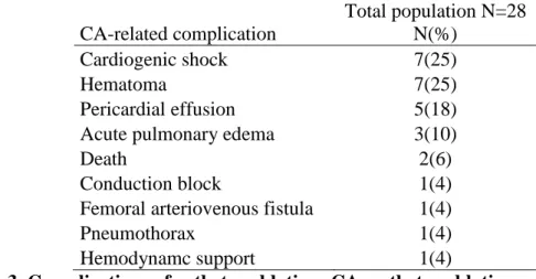
Complication rate was 26% (28 complications on 108 procedures). Most patients (n=19, 23%) suffered from procedural complications during their first procedure in the study center: 7 cardiogenic shocks, 6 hematomas of the punction site, 3 pericardial effusion, 1 acute pulmonary edema, 1 apparition of a block of conduction, 1 femoral arteriovenous fistula. 2 patients died because of the second procedure, 1 due to a tamponade and 1 due to decanulating of ECLS (Table 3).
CA-related complication Total population N=28 N(%) Cardiogenic shock 7(25) Hematoma 7(25) Pericardial effusion 5(18) Acute pulmonary edema 3(10)
Death 2(6)
Conduction block 1(4) Femoral arteriovenous fistula 1(4) Pneumothorax 1(4) Hemodynamc support 1(4)
Table 3. Complications of catheter ablation; CA: catheter ablation
2- Short-Term results
2.1 Recurrence of VT within 30 days
A total of 37 patients (43%) presented a VT recurrence within 1 month after the first CA performed in one of the four study-centers. 45 patients (52%) had a early recurrence after the first ablation when previous CA for the same episode were included.
Overall, 17 patients underwent multiple CA (mean 1.5 [1-11]). Among them, 10 presented no recurrence within 30 days after the last CA, including 2 patients who died within 30 days of cardiogenic shock. Overall, 48 patients (55%) did not have early VT recurrence after the last CA.
Most VT recurrences occurred within five days after the procedure. Among the 37 patients with VT recurrence, 23 patients had a recurrence of the clinical VT, 12 patients presented with another VT. 16 patients had another electrical storm.
Number of patients
87 53 51 47
Firgure 6. Kaplan-meier event-free for recurrence of VT; VT: Ventricular tachycardia
Acute CAD, multiple morphologies of clinical VT and negative ventricular stimulation at the end of CA were predictors of early VT recurrence in Cox regression univariate analysis. Only acute CAD and negative ventricular stimulation at the end of CA remained predictors of early VT recurrence in multivariate cox regression analysis (Table 5). ES occurring in the setting of acute CAD had higher risk of CA failure than other cardiomyopathies (figure 7).
p OR 95,0% CI Lower Higher p OR 95,0% CI Lower Higher Sex .552 .751 .292 1.929 Age .532 1.008 .983 1.034 Recent MI .043 2.049 1.024 4.098 .019 2.343 1.149 4.778 LVEF 0.922 1.001 0.979 1.023 Inotropic drugs .887 1.051 .528 2.094 Haemodinamic support before CA .807 1.115 .465 2.674 Hemodynamic tolerance of VT .828 .931 .485 1.784 Monorphic VT .440 1.383 .607 3.149 ≥1 morphology .053 1.906 .992 3.660 .318 1.425 .711 2.856 LV VT .391 1.393 .653 2.969 ≥1 inducible VT .616 1.192 .599 2.374 CA in VT .562 .846 .482 1.487 Epicardial CA .808 1.138 .403 3.213 No VT inducible at the
end of procedure (class B/C)
.035 .496 .258 .952 .012 .425 .218 .828 General anesthesia .417 1.313 .680 2.533
Table 4. Univariate and multivariate analysis for ventricular tachycardia recurrence at 1month ; MI : Myocardial infarction ; LVEF : Left ventricle ejection function ; CA : Catheter ablation ; VT : Ventricular tachycardia ; LV VT : Left ventricule ventricular tachycardia
Figure 7. Kaplan-meier event-free for early ventricular tachycardia recurrence within 30 days according to the cardiomyopathy; VT: Ventricular tachycardia; ICM: Ischemic cardiomyopathy, DCM: Dilated cardiomyopathy, MI: Myocardial infarction
2.2 Death within 30 days
17 patients (20%) died within 30 days after the first CA. 8 patients died because of the persistant tachy-arrhythmia conducing to cardiac arrest, 6 patients had a severe cardiac dysfunction (refractory cardiogenic chock or electro-mechanic dissociation) and 3 patients because of other complication. 5 patients (6%) get to a ventricular assistance or a heart transplant (Figure 8).
Compared with patients without structural heart disease, patients with underlying cardiomyopathy were at higher risk of early death (figure 9). Patients with electrical storm and acute CAD had a lower survival than other cardiomyopathies. No difference was
observed between stable CAD and IDCM. Patients with other cardiomyopathies (mostly ARVC) presented no early death (figure 10).
Figure 8. Cause of death within the 87 patients
Number at risk
CPT 87 73 69 65 No CPT 13 13 13 12
Figure 9. Early death Kaplan-Meier survival depending on underlying structural heart disease; No CPT: no cardiomyopathy, CPT: cardiomyopathy
Tachy-arrhythmia Cardiac dysfunction Intensive care complication Ventricular assistance Heart transplantation
Figure 10. Early death Kaplan-Meier survival depending on type of cardiomyopathy ; ICM: Ischemic cardiomyopathy, DCM: Dilated cardiomyopathy, post MI: acute myocardial infarction
p HR 95,0% CI Lower Higher p HR 95,0% CI Lower Higher Gender (male) .003 .208 .073 .595 .011 .208 .063 .693 Age .248 1.023 .985 1.062 Acute CAD .023 3.086 1.171 8.131 .533 1.688 .326 8.748 Diabetes .912 1.073 .308 3.734
Lung chronic disease .271 1,311 .427 4.022 Renal failure .239 1.818 .672 4.918 LVEF .039 .953 .911 .998 .257 .967 .913 1.025 Low HD tolerance of VT .012 13.359 1.770 100.81 .042 8.486 1.084 66.40 HD support .001 5.157 1.952 13.623 .760 .777 .154 3.917 Inotropic drugs .151 2.011 .776 5.215 General anaesthesia .003 4.847 1.705 13.780 .048 3.163 1.012 9.885 Polymorphic VT .002 4.773 1.812 12.573 .149 2.238 .749 6.689 ≥1morphology .001 5.119 1.890 13.870 .707 1.325 .305 5.754 LV VT .113 2.764 .785 9.730 ≥1 inducible VT .637 1.286 .453 3.650 Epicardial CA .862 1.140 .261 4.985 Multiple CA .370 1.613 .568 4.582 No VT inducible at the end of procedure .556 1.348 .499 3.647 CA complication .050 2.693 1.002 7.239 .467 1.608 .447 5.783 VT recurrence after last CA .038 2.865 1.059 7.752 .712 .692 .098 4.901 VT recurrence after 1st CA .010 5.158 1.481 17.961 .025 4.393 1.209 15.97
Table 5. Univariate and multivariate analysis for early death at 1 month; CAD: coronary artery disease; LVEF: Left ventricle ejection function; HD: Hemodynamic; VT: ventricular tachycardia; CA: Catheter ablation
In univariate cox analysis, male gender, acute CAD, LVEF, low hemodynamic tolerance of VT, hemodynamic support before ablation, general anaesthesia before or during CA, polymorphic VT, multiple morphologies of clinical VT, CA-related complication and VT recurrence after first or last ablation were predictors of early death. In multivariate analysis, only male gender, low tolerance of VT, general anaesthesia and VT recurrence after first CA were independent predictors of early death within one month
(table 6 and figure 11). General anaesthesia was widely used as an anti-arrhythmia treatment in patients with refractory electrical storm. Thus it was closely correlated with acute CAD, low tolerance of VT, use of a hemodynamic support and inotropic drugs (p<0.001). Patients with electrical storm in context of acute CAD had a more severe clinical presentation with more frequent use of inotrope drugs (p=0.012), general anaesthesia (p<0.001) and hemodynamic support (p<0.001). Within the group of patients with VT recurrence after the first catheter ablation, repeat procedure was not associated with better outcome (figure 12).
Number at risk
VT recurrence 44 34 32 28 No VT recurrence 40 38 38 36
Figure 11. Early death within 30 days according to ventricular tachycardia recurrence after the first VT catheter ablation; VT: Ventricular tachycardia
No VT recurrence VT recurrence
Number at risk
Repeat 14 10 9 8 No repeat 28 24 23 19
Figure 12. Early death within 30 days in the 45 patients with early VT recurrence after the first CA according to repeat procedure or not; VT: Ventricular
Figure 13. Flow chart regarding evolution of patients who had a recurrence after the first catheter ablation; CA: Catheter ablation, VT: Ventricular tachycardia, LVAD: Left ventricular assistance device, HT: Heart transplantation
VT recurrence after 1st CA N=44 Redo n=17 No redo N=29 13 Deaths 3 LVAD/HT 3 Deaths 0 LVAD/HT VT recurrence n=7 No VT recurrence n=10 0Deaths 0 LVAD/HT
Figure 14. Flow chart of evolution of patients depending on presence or absence of recurrence of VT after the first catheter ablation; VT: ventricular tachycardia, CA: catheter ablation, HT: heat transplantation, LVAD: Left ventricular assistance device, CAD: Coronary artery disease, IDCM: Idiopathic dilated cardiomyopathy
3- Long-Term results
3.1 Death within 1 year
After 1 year of follow-up, the death-rate is about 24 patients (29%) with 9 deaths because of tachyarrhythmia, 6 because of heart failure, 8 of non-cardiovascular etiology and 1 with an unknown cause.
There was no difference in mortality at one 1 year between type of cardiomyopathy (Figure 15). Patients with other cardiomyopathy as ARVC or HCM had better long term outcomes.
Survival at one year was not influenced by repeated ablation. (figure 14) VT recurrence after 1st CA N=44 No VT recurrence after 1st N=39 HT/LVAD N=3 No HT/LVAD N=41 14 Deaths 5 acute CAD 3 IDCM 6 chronic CAD 5 Patients had 2 to 3 CA 1 patient had 7 CA
1 Death (J38,chronic CAD,5 previous CA)
HT/LVAD N=2
Figure 15. Death within 1 year depending on cardiomyopathy; ICM: Ischemic cardiomyopathy, DCM: Dilated cardiomyopathy, MI: Myocardial infarction
p OR 95,0% CI Lower Higher Sexe ,596 22,353 ,000 2159662,713 Duration of ES ,103 1,048 ,991 1,110 IDCM ,078 3,478 ,869 13,927 Post MI ,368 ,036 ,000 49,841
Chronic lung disease ,699 1,364 ,283 6,568 Diabetes ,191 2,524 ,631 10,100 Renal impairment ,721 ,787 ,211 2,930 LVEF ,043 ,927 ,862 ,998 Inotropic drugs ,754 1,249 ,312 4,994 General anesthesia ,701 ,665 ,083 5,318 Hemodynamic support ,956 1,060 ,133 8,481 Hemodynamic tolerance of VT ,491 1,653 ,395 6,918
Polymorphic VT ,509 ,042 ,000 512,225 ≥1morphology ,301 ,032 ,000 21,559 ≥1 inducible VT ,136 4,867 ,608 38,926 Epicardial CA ,005 7,434 1,832 30,159 Multiple CA ,247 2,269 ,567 9,078 Success of procedure ,518 ,648 ,174 2,416 CA-related complication ,509 ,496 ,062 3,967 VT recurrence after 1st CA ,871 1,115 ,299 4,153 VT recurrence after last CA ,554 1,487 ,399 5,541 TV recurrence at 1year ,535 1,551 ,388 6,201
Table 6: Univariate Cox analysis of long term survival; ES: Electrical storm; IDCM: Idiopathic dilated cardiomyopathy; MI: Myocardial infarction; LVEF: Left ventricle ejection function; VT : Ventricular tachycardia ; CA : Catheter ablation
In univariate Cox analysis, idiopathic dilated cardiomyopathy, LVEF and epicardial CA were predicting factors of late death within one year. In multivariate analysis, there was no persistent predicting factor (Table7).
Figure 16. Long term death within one year in the 45 patients with ventricular tachycardia recurrence after the first ablation according to repeated ablation or not.
3.2 Recurrence of VT and heart transplantation or ventricular assistance
within 1 year
46 patients (55%) presented a recurrence of VT. There was no difference in long term VT recurrence depending on type of cardiomyopathy.
V-
Limitations
There are inherent limitations linked to the retrospective design of the study. However this study is one of the largest recent study in that setting.
As we included only patients with catheter ablation, we cannot compare catheter ablation efficacy to medical strategy.
VI- Discussion
Carbucicchio and al. evaluated short and long-term effects of catheter ablation in a population of patients with implantable cardioverter-defibrillator, presenting with electrical storm. This population was composed with 95 patients with a cardiomyopathy, mainly coronary artery disease. After catheter ablation, 92% of patients were free of electrical storm, 66% of patients didn’t have any VT recurrence after a median follow-up of 22 months and 15 patients (16%) died. The predictive factors of cardiac mortality were: age, idiopathic dilated cardiomyopathy (IDCM) and an acute failure of catheter ablation (8). Another study of Kozeluhova and al. concerned 50 patients with a structural heart disease who underwent a catheter ablation for electrical storm. After a median follow-up of 18 months, 48% of patients didn’t have any recurrence of VT, 26% of patients needed repeated procedure and 14 patients (28%) died during the follow-up(9). Arya and al. evaluated the efficiency of radiofrequency catheter ablation of electrical storm due to monomorphic VT in patients with idiopathic dilated cardiomyopathy in 13 patients who underwent 17 procedures. VT recurrence was prevented in 61,5% of patients. Patients with a complete success of the radiofrequency had better outcomes in recurrence of VT and survival(23). Two others studies evaluated the place of catheter ablation in structural heart disease, in very limited cohort (5 and 9 patients) (24).
In our study, recurrence of VT occurred in 43% of patients, which is similar to the results of different studies with the same population, that is between 38,5% and 52%(8) (9) (23). The predictive factors of VT recurrence found in our cohort were electrical storm occurring in patient with acute coronary artery disease and inducible VT at the end of the procedure. Success of the procedure is a recurrent predictive factor of absence of VT recurrence found in different studies.
Catheter ablation of ventricular tachycardia for ES evolved into a prior treatment because of data suggesting an increased mortality due to ICD shocks(25) (26). Suppressing ventricular tachycardia could decrease mortality due to ventricular arrhythmias and consequences of ICD shocks(27). But analyzing different studies, the mortality does not decrease despite a significant reduction in ventricular tachycardia recurrence and ICD-therapies. Mallidi J and al. did a meta-analysis on 457 patients from five studies, and catheter ablation was associated with a decreased VT recurrence of 35 % without any effect on mortality.(28) The VTACH tri al enrolled 107 patients with previous myocardial infarction to catheter ablation associated with ICD or ICD alone. Freedom of VT/VF recurrence of higher in the ICD and ablation group (46%) versus the ICD group (29%). Nine patients died during the follow-up, without difference between the two groups. (29)
Regarding patients with structural heart disease, suppression of VTs does not guarantee a good prognosis and does not prevent death due to progression of heart failure. This suggests that cardiac damage is apparently too extensive, and VT recurrence is more likely to reflect severity of the cardiomyopathy. (30) (29)
Mortality was high, around 29% during the first year and mostly occurring during the first month, decreased slowly during the follow-up. The high mortality in patients with ES would reflect the deterioration of heart function and evolution of the cardiomyiopathy(31). Early-mortality rate during the first month was about 20% and predictive factors of death were male gender, low hemodynamic tolerance of the VT, general anesthesia and recurrence of VT after the first catheter ablation. Beside, when we analyze mortality depending on the cardiomyopathy, it seems that a recent myocardial infarction complicated with an electrical storm is associated with significantly higher
mortality than other cardiomyopathies (p=0,027). Long term mortality was mostly drived by hemodynamic factors as LVEF or ICDM and was not associated with catheter ablation success. Patients with recent myocardial infarction had a more severe clinical presentation and a worst outcome with higher risk of VA recurrence and early mortality.
General anesthesia was found as an independent predictor of early death, probably witnessing the more severe clinical presentation of patients necessitating general anesthesia because of low tolerance of VT or hemodynamic support. Thus, the decision to perform catheter ablation in these patients should be carefully stated by a multidisciplinary team and alternative strategies as emergency heart transplantation should be discussed in case of CA failure. In the same way, electrical storm could be the sign of advanced cardiomyopathy that could be candidate for heart transplantation or assistance devices. Most of death occurred within the first month after CA and 47% of deaths were directly linked to VA. Very interestingly, we found that VA recurrence after the first CA was independently associated with higher early mortality and repeat procedure was not associated with better outcome. Similar results were found in previous studies. Nagashima and al that studied VT CA ablation of 370 patients found that early recurrence was an independent predictor of mortality and repeat procedure did not modify the outcome (32). As in the present study, early VA recurrence was associated with acute failure or no final induction test. Kozeluhova et al also showed that recurrence of ES was associated with higher mortality and heart transplantation (9). As groups with repeat and no repeat ablation were not randomized, it is difficult to access the efficacy of repeat ablation procedures but the inner and previous studies suggest no clear benefice. In Nagashima and al study, comparing 44 patients who underwent repeat ablation and 37 patients without repeat ablation, there was no significant difference in survival(32).
In case of catheter ablation failure, alternative strategies to repeat CA should be discussed. However, apart from acute CAD and severity of the initial presentation with low tolerated VT and need for general anesthesia, no clear clinical predictive factors of early mortality was found. This underlies how difficult it is to identify patients at high risk that would better benefit from emergency heart transplantation than first line or repeat CA. In this high risk population, CA was associated with frequent CA-related complications (26% of procedures) and two patients directly died of CA-related complications. This result suggests that catheter ablation in these high-risk patients is not a benign procedure. In our cohort, even if a second procedure was associated with less recurrence of VT (61% of patients without any recurrence of VT within 1 month after CA), it didn’t lead to better survival.
As we only included patients treated with CA, we were not able to compare CA to medical strategy alone. Izquierdo and al compared outcomes of patients who underwent a catheter ablation for ES, versus a medical treatment alone. There was no difference in mortality and in ES recurrence after a median follow-up of 28 months. They showed only a decreased ES recurrence in the population with LVEF > 25% who underwent a catheter ablation (21%) compared to the population with medical treatment (62%). Leading to the idea that catheter ablation is less effective in patients with severe heart failure. None of patients in this study benefited of repeat catheter ablation(11).
It is not clear if catheter ablation failure is caused by the severity of the underlying disease or incomplete procedure. Our results with others suggest that CA ablation of all VT should be performed to obtain negative ventricular stimulation as a primary end-point as this parameter was associated with less VA recurrence. However, this parameter was not associated with early mortality, even in univariate analysis. A more aggressive
strategy, using epicardial mapping if necessary (in particular in ICDM and ARVC) and ablation of all VTs, may improve outcome. In a population of 13 patients with IDCM who had a catheter ablation for ES, 8 patients had a failure of endocardial ablation and four of them had an epicardial CA. In this population who underwent an epicardial CA, 3 patients had a complete success of the procedure. Finally, a successful CA was associated with improved survival(23).
VII- Conclusions
Catheter ablation is an effective treatment of electrical storm in patients with structural heart disease. In this study, nearly 50% of patients were free of VA after the first procedure and 55% after the last procedure. Electrical storm in patients with recent
myocardial infarction and severe clinical presentation with low tolerated VT and general anesthesia was associated with higher early ventricular arrhythmia recurrence and mortality. Early recurrence after the first CA was also independently associated with higher early-mortality and repeat procedure did not influence significantly the outcome. Absence of early VA recurrence was predicted by negative elctrophysiological study at the end of the procedure. Identification of patients that would benefit better from alternative strategies as emergency heart transplantation or ventricular assistance remain difficult. These options should be multidisciplinary discussed in case of catheter ablation failure on higher risk patients.
VIII- Bibliography
1. Gao D, Sapp JL. Electrical storm: definitions, clinical importance, and treatment. Curr Opin Cardiol. janv 2013;28(1):72‑9.
2. Maruyama M. Management of electrical storm: The mechanism matters. J Arrhythmia. 1 août 2014;30(4):242‑9.
3. Exner DV, Pinski SL, Wyse DG, Renfroe EG, Follmann D, Gold M, et al. Electrical storm presages nonsudden death: the antiarrhythmics versus implantable
defibrillators (AVID) trial. Circulation. 24 avr 2001;103(16):2066‑71.
4. Credner SC, Klingenheben T, Mauss O, Sticherling C, Hohnloser SH. Electrical storm in patients with transvenous implantable cardioverter-defibrillators: incidence, management and prognostic implications. J Am Coll Cardiol. déc 1998;32(7):1909‑15.
5. Gatzoulis KA, Andrikopoulos GK, Apostolopoulos T, Sotiropoulos E, Zervopoulos G, Antoniou J, et al. Electrical storm is an independent predictor of adverse long-term outcome in the era of implantable defibrillator therapy. Eur Eur Pacing Arrhythm Card Electrophysiol J Work Groups Card Pacing Arrhythm Card Cell Electrophysiol Eur Soc Cardiol. mars 2005;7(2):184‑92.
6. Priori SG, Blomström-Lundqvist C, Mazzanti A, Blom N, Borggrefe M, Camm J, et al. 2015 ESC Guidelines for the Management of Patients With Ventricular
Arrhythmias and the Prevention of Sudden Cardiac Death. Rev Espanola Cardiol Engl Ed. févr 2016;69(2):176.
7. Bella PD, Baratto F, Tsiachris D, Trevisi N, Vergara P, Bisceglia C, et al.
Management of Ventricular Tachycardia in the Setting of a Dedicated Unit for the Treatment of Complex Ventricular ArrhythmiasClinical Perspective. Circulation. 2 avr 2013;127(13):1359‑68.
8. Carbucicchio C, Santamaria M, Trevisi N, Maccabelli G, Giraldi F, Fassini G, et al. Catheter ablation for the treatment of electrical storm in patients with implantable cardioverter-defibrillators: short- and long-term outcomes in a prospective single-center study. Circulation. 29 janv 2008;117(4):462‑9.
9. Kozeluhova M, Peichl P, Cihak R, Wichterle D, Vancura V, Bytesnik J, et al. Catheter ablation of electrical storm in patients with structural heart disease. Eur Eur Pacing Arrhythm Card Electrophysiol J Work Groups Card Pacing Arrhythm Card Cell Electrophysiol Eur Soc Cardiol. janv 2011;13(1):109‑13.
10. Brigadeau F, Kouakam C, Klug D, Marquié C, Duhamel A, Mizon-Gérard F, et al. Clinical predictors and prognostic significance of electrical storm in patients with implantable cardioverter defibrillators. Eur Heart J. mars 2006;27(6):700‑7. 11. Izquierdo M, Ruiz-Granell R, Ferrero A, Martínez A, Sánchez-Gomez J, Bonanad
C, et al. Ablation or conservative management of electrical storm due to monomorphic ventricular tachycardia: differences in outcome. Europace. 1 déc 2012;14(12):1734‑9.
12. Tung R. Evolution of ventricular tachycardia ablation in structural heart disease. J Arrhythmia. 1 août 2014;30(4):250‑61.
13. Tanawuttiwat T, Nazarian S, Calkins H. The role of catheter ablation in the management of ventricular tachycardia. Eur Heart J. 14 févr 2016;37(7):594‑609.
14. Baldinger SH, Stevenson WG, John RM. Ablation of ischemic ventricular
tachycardia: evidence, techniques, results, and future directions. Curr Opin Cardiol. janv 2016;31(1):29‑36.
15. de Chillou C, Sellal J-M, Magnin-Poull I. Pace Mapping to Localize the Critical Isthmus of Ventricular Tachycardia. Card Electrophysiol Clin. mars
2017;9(1):71‑80.
16. Komatsu Y, Daly M, Sacher F, Derval N, Pascale P, Roten L, et al.
Electrophysiologic characterization of local abnormal ventricular activities in postinfarction ventricular tachycardia with respect to their anatomic location. Heart Rhythm. nov 2013;10(11):1630‑7.
17. Komatsu Y, Maury P, Sacher F, Khairy P, Daly M, Lim HS, et al. Impact of Substrate-Based Ablation of Ventricular Tachycardia on Cardiac Mortality in Patients with Implantable Cardioverter-Defibrillators. J Cardiovasc Electrophysiol. 1 sept 2015;
18. Marchlinski FE, Callans DJ, Gottlieb CD, Zado E. Linear ablation lesions for control of unmappable ventricular tachycardia in patients with ischemic and nonischemic cardiomyopathy. Circulation. 21 mars 2000;101(11):1288‑96.
19. Jaïs P, Maury P, Khairy P, Sacher F, Nault I, Komatsu Y, et al. Elimination of local abnormal ventricular activities: a new end point for substrate modification in patients with scar-related ventricular tachycardia. Circulation. 8 mai
2012;125(18):2184‑96.
20. Haissaguerre M, Vigmond E, Stuyvers B, Hocini M, Bernus O. Ventricular arrhythmias and the His-Purkinje system. Nat Rev Cardiol
21. Wolk R. Arrhythmogenic mechanisms in left ventricular hypertrophy. EP Eur. 1 juill 2000;2(3):216‑23.
22. Baratto F, Pappalardo F, Oloriz T, Bisceglia C, Vergara P, Silberbauer J, et al. Extracorporeal Membrane Oxygenation for Hemodynamic Support of Ventricular Tachycardia Ablation. Circ Arrhythm Electrophysiol. déc 2016;9(12).
23. Arya A, Bode K, Piorkowski C, Bollmann A, Sommer P, Gaspar T, et al. Catheter ablation of electrical storm due to monomorphic ventricular tachycardia in patients with nonischemic cardiomyopathy: acute results and its effect on long-term
survival. Pacing Clin Electrophysiol PACE. déc 2010;33(12):1504‑9.
24. Peichl P, Cihák R, Kozeluhová M, Wichterle D, Vancura V, Kautzner J. Catheter ablation of arrhythmic storm triggered by monomorphic ectopic beats in patients with coronary artery disease. J Interv Card Electrophysiol Int J Arrhythm Pacing. janv 2010;27(1):51‑9.
25. Villacastín J, Almendral J, Arenal A, Albertos J, Ormaetxe J, Peinado R, et al. Incidence and clinical significance of multiple consecutive, appropriate,