Development and optimization of new sclerosing embolizing
agents for vascular diseases treatment
by
Fatemeh
ZEHTABI
MANUSCRIPT-BASED THESIS PRESENTED TO ÉCOLE DE
TECHNOLOGIE SUPÉRIEURE IN PARTIAL FULFILLMENT FOR THE
DEGREE OF DOCTOR OF PHILOSOPHY Ph.D.
MONTREAL, 16 APRIL 2018
ÉCOLE DE TECHNOLOGIE SUPÉRIEURE UNIVERSITÉ DU QUÉBEC
This Creative Commons licence allows readers to download this work and share it with others as long as the author is credited. The content of this work can’t be modified in any way or used commercially.
BOARD OF EXAMINERS
THIS THESIS HAS BEEN EVALUATED BY THE FOLLOWING BOARD OF EXAMINERS
Mrs. Sophie Lerouge, Thesis Supervisor
Mechanical Engineering department at École de technologie supérieure
Mr. Mircea A. Mateescu, Thesis Co-supervisor
Department of Chemistry at Université du Québec à Montréal (UQAM)
Mr. Natalia Nũno, President of the Board of Examiners
Department of Automated Production Engineering at École de technologie supérieure
Mrs. Nicole Demarquette, Member of the jury
Mechanical Engineering department at École de technologie supérieure
Mr. Gilles Soulez, External Examiner
Department of Radiology, Radio-Oncology and Nuclear Medicine at Université de Montréal
Mr. Gregoire Leclair, External Examiner Faculty of Pharmacy at Université de Montréal
THIS THESIS WAS PRENSENTED AND DEFENDED
IN THE PRESENCE OF A BOARD OF EXAMINERS AND PUBLIC 19 FEBRUARY 2018
ACKNOWLEDGMENT
First, I would like to thank the members of the jury for having accepted to evaluate my manuscript and attend my defense.
For the past five years, my research directors, Sophie Lerouge and Mircea A. Mateescu, have guided, counselled and encouraged me. Thank you for sharing with me your scientific expertise and your human qualities. Sophie, thank you for having welcomed me to the laboratory, your determination has often encouraged me to persevere and to surpass myself. Mircea A. Mateescu and Pompilia Ispas, your positive energy is contagious; thank you for your support and the constructive advice you have given me throughout the project. Thanks to the researchers who collaborated with us on the different aspects of the project, in particular Gilles Soulez and his team for their expertise in in vivo testing and advice on clinical aspects. I would especially like to thank Ahmed Fatimi, Marion Maire, Dr. Annabi, and Helen Hoen for their help and valuable advice during this project. Working as a team with you has been a very rewarding experience. A thought to my office colleagues Elias, Pauline, Yasaman, Caroline, Matthew, Maedeh, Sepideh, Nick, Capucine, Atma, Francesco, Eve and Melusine. I owe thanks to a very special person, my husband, Ehsan for his continued and unfailing love, support and understanding during my pursuit of Ph.D. degree that made the completion of thesis possible. You were always around at times I thought that it is impossible to continue, you helped me to keep things in perspective. I greatly value his contribution and deeply appreciate his belief in me. Thank you to my friends and family for the support you have given me during these five busy years.
Finally, I would like to thank the organizations that funded me during this Ph.D., Canada Research Chair, the CRCHUM and the École de Technologie Supérieure.
DÉVELOPPEMENT ET OPTIMISATION DE NOUVEAUX AGENTS EMBOLISANTS ET SCLÉROSANTS POUR LE TRAITEMENT DES
MALADIES VASCULAIRES
Fatemeh ZEHTABI
RESUME
Le traitement endovasculaire des anévrysmes de l'aorte abdominale (AAA), une dilatation localisée de l'aorte, consiste à déployer une endoprothèse pour exclure l'anévrisme du flux sanguin et l'empêcher de se rompre. Le succès clinique de cette procédure est toutefois limité par la persistance fréquente ou la récidive de la circulation sanguine à l'intérieur de l'anévrisme, appelée endofuite, qui peut entraîner une croissance anévrismale et des risques accrus de rupture. Pour traiter ou prévenir les endofuites, l'embolisation du sac est de plus en plus effectuée par injection d'un agent embolique pour occlure le flux sanguin qui persiste à pénétrer dans l'anévrisme. Cependant, les agents d'embolisation actuels sont loin d'être idéaux et les cliniciens recherchent de nouveaux agents injectables. L’agent embolisant idéal doit être injectable avec un bon contrôle, occlure efficacement le flux sanguin, biocompatible et radio-opaque. L'hypothèse de cette thèse est que l’ajout d’un agent sclérosant (induisant une dénudation endothéliale) et un inhibiteur des metalloproteinase (pour réduire la progression de l’anévrisme) permettrait de mieux prévenir les endofuites.
Tout d’abord, nous avons montré qu’un thermogel de chitosane radio-opaque contenant du tétradécylsulfate de sodium (STS) comme agent sclérosant / embolisant, préalablement développé au laboratoire, tend à réduire le nombre d’endofuite par rapport à un agent embolisant sans effet sclérosant (hydrogel sans STS) dans un modèle canin de réparation d'anévrisme endovasculaire. Cependant, la différence entre les groupes n'était pas statistiquement significative, peut-être en raison du petit nombre d'animaux.
Dans un second temps, nous avons évalué la faisabilité de développer un agent embolisant sclérosant à partir d’un agent embolique actuellement commercialisé (Onyx) et d’éthanol, produit qui pourrait être approuvé plus rapidement pour utilisation clinique.
Enfin, un hydrogel thermosensible combinant les propriétés occlusives, sclérosantes et inhibitrices de MMP a été développé à base de chitosane et de doxycycline (DOX). Ce gel présente des propriétés prometteuses d'ablation endothéliale et d'inhibition de la MMP. L'expérience in vivo à court terme des gels CH-DOX dans les artères rénales du porc a validé son injectabilité, une bonne radiopacité et une bonne embolisation initiale des petits vaisseaux sanguins. Cependant, une réouverture du flux sanguin a été observée quelques minutes après l'embolisation. Nous croyons que l'augmentation de la quantité de médicament serait une bonne option pour supprimer cette limitation du produit, mais une analyse plus approfondie est nécessaire.
Mots-clés: Anévrisme de l’aorte abdominale, agent embolisant, sclérosant, doxycycline,
DEVELOPMENT AND OPTIMIZATION OF NEW SCLEROSING EMBOLIZING AGENTS FOR VASCULAR DISEASES TREATMENT
Fatemeh ZEHTABI
ABSTRACT
Endovascular treatment of abdominal aortic aneurysms (AAA), a localized dilatation of the aorta, involves deploying a stent to exclude the aneurysm from the blood stream and prevent it from breaking. The clinical success of this procedure, however, is limited by the frequent persistence or recurrence of blood flow within the aneurysm, called endoleak, which can lead to aneurysmal growth and increased risk of rupture. To treat or prevent endoleaks, embolization of the sac is increasingly performed by injection of an embolic agent to occlude the blood flow that persists in entering the aneurysm. However, current embolization agents are far from ideal and clinicians are looking for new injectables. The ideal embolizing agent should be injectable with good control, effectively occlude blood flow, be biocompatible and radiopaque. The hypothesis of this thesis is that the addition of a sclerosing agent (inducing endothelial denudation) and a metalloproteinase inhibitor (to reduce the progression of the aneurysm) would better prevent endoleaks.
First, we have shown that a radiopaque chitosan thermogel containing sodium tetradecyl sulphate (STS) as a sclerosing / embolizing agent, previously developed in the laboratory, tends to reduce the number of endoleaks compared to an embolizing agent without sclerosing effect (hydrogel without STS) in a canine model of endovascular aneurysm repair. However, the difference between the groups was not statistically significant, perhaps because of the small number of animals.
In a second step, we assessed the feasibility of developing a sclerosing embolizing agent from a currently marketed embolic agent (Onyx) and ethanol, which could be approved more quickly for clinical use.
Finally, a thermosensitive hydrogel combining the occlusive, sclerosing and inhibitory properties of MMP was developed based on chitosan and doxycycline (DOX). This gel has promising endothelial ablation and MMP inhibition properties. Short-term in vivo CH-DOX gels in the renal arteries of pork validated its injectability, good radiopacity and good initial embolization of small blood vessels, however, reopening of blood flow was observed a few minutes later embolization. We believe that increasing the amount of medication would be a good option to remove this limitation of the project, but further investigation is needed.
Keywords: Abdominal aortic aneurysm, embolizing agent, sclerosing agent, doxycycline,
TABLE OF CONTENTS
Page
INTRODUCTION ... 23
CHAPTER 1 LITERATURE REVIEW ... 25
1.1. Clinical problematic ...25
1.1.1. Structure of the arteries ... 25
1.1.2. The abdominal aortic aneurysm (AAA) ... 27
Definition ... 27
Risk factors ... 28
Pathophysiology of AAA ... 29
1.1.3. Treatments of AAA ... 31
Endovascular aneurysm repair (EVAR) ... 32
Endoleak ... 33
Sac embolization procedure... 35
Cons and pros of current embolic agents ... 36
Design criteria of an ideal embolizing agent ... 39
1.1.3.5.1. Occlusive properties ...40
1.1.3.5.2. Endothelial ablation ...40
1.1.3.5.3. Prevent aneurysm progression ...42
1.2. Hydrogels ...43
1.2.1. Definition ... 43
1.2.2. Advantages and limitations of hydrogels ... 43
1.2.3. Types of Hydrogels ... 44
1.2.4. Natural polymers versus Synthetic hydrogels ... 44
1.2.5. Physical versus chemical hydrogel ... 45
1.2.6. Environmentally-sensitive hydrogels ... 46
1.2.7. Injectable hydrogels ... 47
1.2.8. Drug delivery ... 48
1.3. Previous work by the team ...50
1.3.1. Chitosan radiopaque injectable gels ... 50
1.3.2. Chitosan-sclerosing gels ... 54
CHAPTER 2 PROJECT OBJECTIVES ... 56
CHAPTER 3 CHITOSAN-SODIUM TETRADECYL SULFATE HYDROGEL: CHARACTERIZATION AND PRECLINICAL EVALUATION OF A NOVEL SCLEROSING EMBOLIZING AGENT FOR THE TREATMENT OF ENDOLEAKS ... 61
3.1. Context ... 61
3.2. Abstract ... 62
3.3. Introduction ... 63
3.4. Methods ... 64
3.4.2. In Vivo Embolization of Endoleaks ... 65 3.5. Results ... 68 3.5.1. In Vitro Characterization ... 68 3.6. Discussion ... 74 3.7. Limitations ... 76 3.8. Conclusions ... 77 3.9. Acknowledgements ... 77
3.10. Compliance with Ethical Standards ... 77
CHAPTER 4 NEW ALCOHOL AND ONYX MIXTURE FOR EMBOLIZATION: FEASIBILITY AND PROOF OF CONCEPT IN BOTH IN VITRO AND IN VIVO MODELS ... 79
4.1. Context ... 79
4.2. Abstract ... 80
4.3. Introduction and Rationale ... 80
4.4. Materials and Methods ... 83
4.4.1. Part I: In Vitro Study ... 83
4.4.2. Part II: In Vivo Study in a Swine Model ... 84
4.5. Results ... 88
4.5.1. Part I: In Vitro Testing ... 88
4.5.2. Part II: In Vivo Study ... 90
4.6. Discussion ... 92
4.7. Study Limitations ... 93
4.8. Potential Clinical Impact ... 94
4.9. Compliance with Ethical Standards ... 94
CHAPTER 5 CHITOSAN-DOXYCYCLINE HYDROGEL: AN MMP INHIBITOR / SCLEROSING EMBOLIZING AGENT AS A NEW APPROACH TO ENDOLEAK PREVENTION AND TREATMENT AFTER ENDOVASCULAR ANEURYSM REPAIR ... 97
5.1. Context ...97
5.2. Abstract ...97
5.3. Statement of Significance ...98
5.4. Introduction ...99
5.5. Materials and methods ...101
5.5.1. Preparation of hydrogels ... 101
5.5.2. Characterization of hydrogels as an embolic agent ... 102
5.5.2.1. Rheology ... 102
5.5.2.2. Occlusive properties ... 102
5.5.2.3. Gel injectability ... 102
5.5.3. DOX cytotoxic dose-response on endothelial cells ... 103
5.5.4. Ex Vivo de-endothelialization ... 104
5.5.5. Indirect cytotoxicity tests ... 104
5.5.6. In Vitro drug release and swelling studies ... 105
5.5.8. Structural and morphological analyses ... 106
5.5.8.1. Scanning electron microscope (SEM) ... 107
5.5.8.2. Fourier transform infrared spectroscopy (FTIR) ... 107
5.5.9. In Vivo assays ... 107
5.5.10. Statistical analysis ... 108
5.6. Results ...109
5.6.1. Hydrogel rheological and in vitro occlusive properties ... 109
5.6.2. DOX effect on endothelial cell viability ... 112
5.6.3. Indirect cytotoxicity of CH hydrogels ... 112
5.6.4. DOX In Vitro release rate and hydrogels swelling ... 112
5.6.5. MMP inhibition ... 114
5.6.6. FTIR and SEM ... 115
5.6.7. In vivo testing ... 116
5.7. Discussion ...118
5.8. Conclusion ...124
5.9. Funding ...125
5.10. Acknowledgments ...125
5.11. Appendix A. Supplementary data ...125
CHAPTER 6 DISCUSSION ... 127
CONCLUSION ... 143
LIST OF TABLES
Page
Table 3.1 pH and concentration of each compound in CH and CH–STS hydrogels used in this study ... 64 Table 3.2 The presence of residual endoleak immediately after embolization (based on
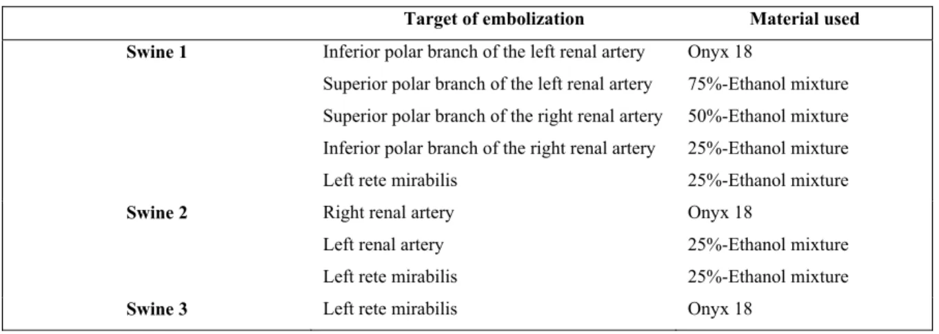
angiography) and before sacrifice at 3 or 6 months (according to CT scan and confirmed by defect on macroscopic tissue slides). Complications are also indicated. No SG thrombosis was observed .. 71 Table 4.1 Various mixtures used for the embolization of target arteries in each
swine ... 86 Table 4.2 Consensus evaluation of the in vivo behavior and characteristics of different
mixtures of Onyx and ethanol ... 86 Table 4.3 Penetration depth score (in mm) for each product tested in swine 1, as
measured on CT MPR reconstructions between the renal cortex and the nearest opaque arterial segment at 5 points ... 86 Table 4.4 Volume decrease during the solidification of different mixtures - most of the
decrease is assumed to be due to the release of ethanol ... 91 Table 5.1 Initial and final concentrations (in the syringe and in the gel respectively) of
each component and pH of the various hydrogels tested. ... 103 Table 5.2 Percentage of immediate and delayed (after 17 min ± 5 min) successful
occlusion of porcine renal caudal artery branches at various division levels and overall after embolization with CH/SHC0.075PB0.08/VIS50/DOX0.1, as measured by digital subtraction angiography ... 121 Table 6.1 Summary of the strength and weaknesses of each embolizing agent
according to the results gathered in this PhD and in previous literature data and design criteria ... 139
LIST OF FIGURES
Page
Figure 1.1 Structure of the arterial wall. Image from (Servier-Medical-Art) ... 26
Figure 1.2 Difference between normal blood vessels and AVM ... 27
Figure 1.3 Normal aorta versus abdominal aortic aneurysm. ... 28
Figure 1.4 Summary of the pathogenesis of AAA. ... 31
Figure 1.5 Treatment of AAA by open surgery (a) and endovascular treatment (b) .. 32
Figure 1.6 Stent-graft ... 33
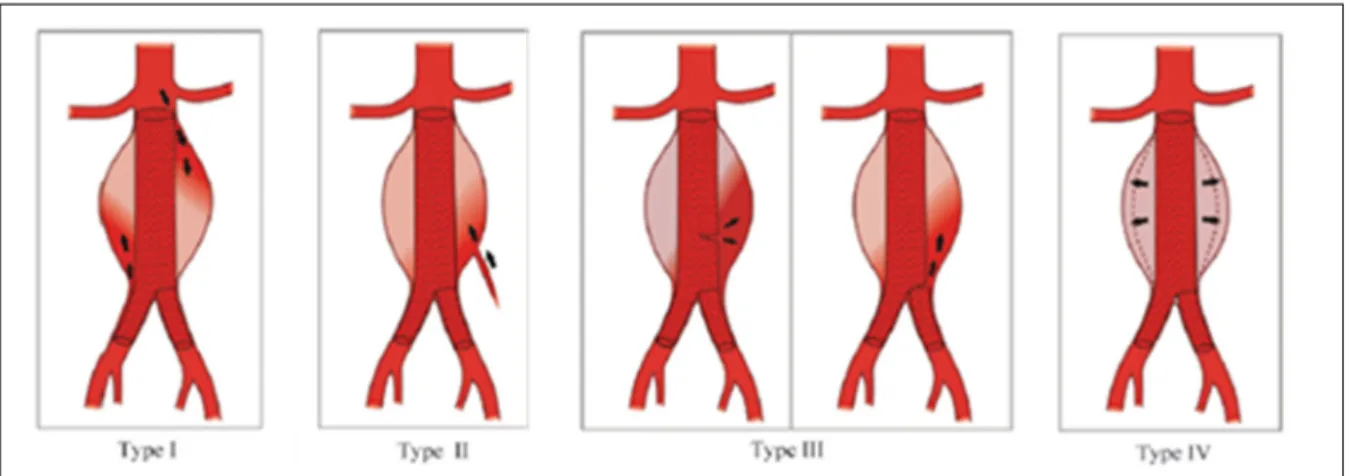
Figure 1.7 Various types of endoleak: type I, leak at the attachment site; type II, leak from a branch artery; type III, graft defect; and type IV, graft porosity ... 35
Figure 1.8 Schematic diagram of (a) a chemical hydrogel with point crosslinks and (b) a physical hydrogel with multiple-junction zones ... 46
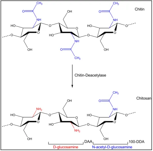
Figure 1.9 Typical structure of chitosan obtained by alkaline deacetylation of chitin. ... 52
Figure 1.10 Chemical structure of (a) chitosan and (b) βglycerophosphate (β-GP) ... 53
Figure 1.11 Chemical structure of sodium tetradecyl sulfate (C14H29NaO4S) ... 54
Figure 3.1 Schematic representation of the bilateral iliac aneurysm model with macroscopic image of the misfit created at the proximal neck using SG deformation ... 67
Figure 3.2 A) Rheological properties and injectability of the embolizing agents ... 69
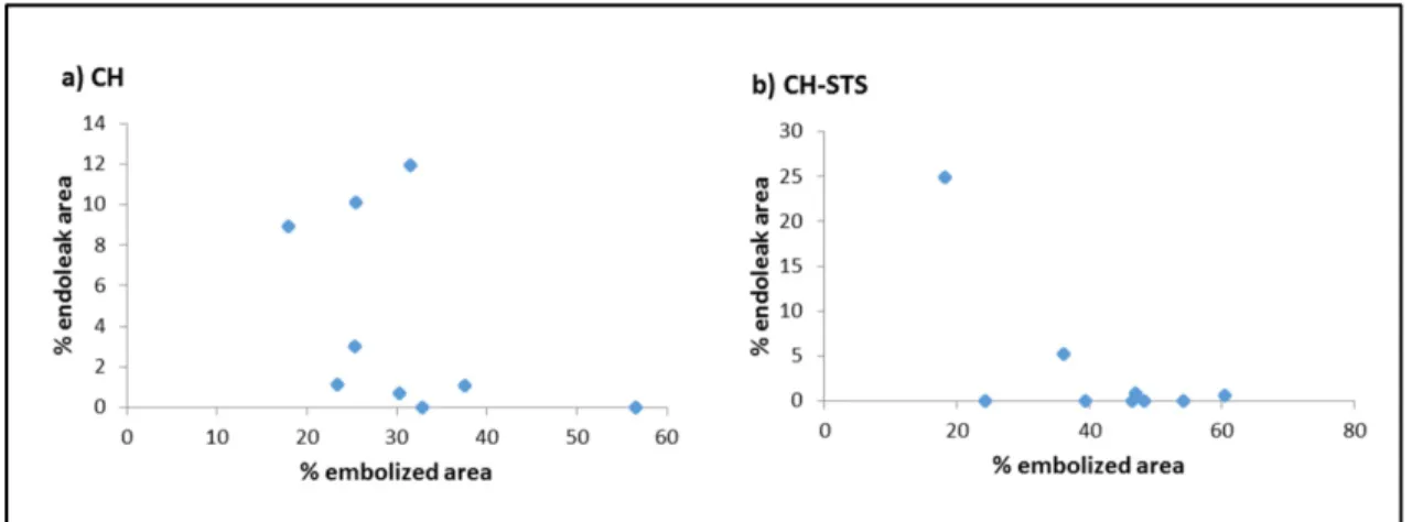
Figure 3.3 Percentage of endoleak area (%) as a function of percentage of aneurysm embolized for a) CH gel; b) CH-STS gels. ... 70
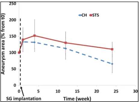
Figure 3.4 Evolution of aneurysm mean area in both groups normalized to the initial area (mean + SD)... 72
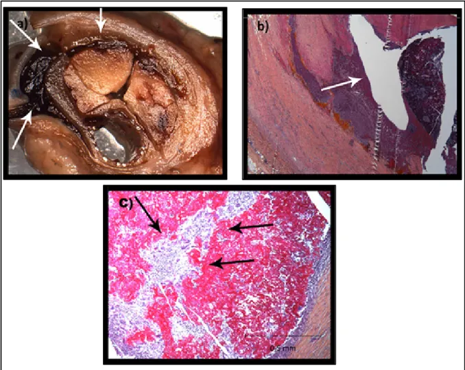
Figure 3.5 Macroscopic pictures of aneurysms treated by A CH and B CH–STS gels in the same animal (#5). ... 72
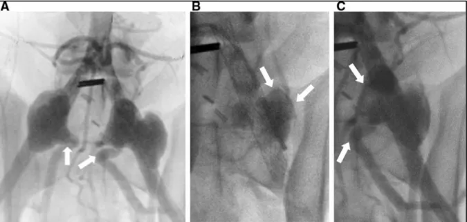
Figure 3.6 A Angiography of the bilateral aneurysm model, showing the bilobal form of the left aneurysm (Dog #4); B fluoroscopic image of the left aneurysm, showing incomplete embolization by CH–STS; C angiography post-embolization with residual type I endoleak ... 73 Figure 3.7 A, B Transverse tissue section and histological slide (HPS staining)
showing the presence of an endoleak throughout the CH matrix; C inflammation and resorption of the embolization agent ... 74 Figure 4.1 Examples of penetration score measurement; coronal oblique CT images of
swine 1 with MPR reconstruction. A Right kidney upper pole (50%-ethanol mixture). B Left kidney upper pole (75%-(50%-ethanol mixture). C Right kidney lower pole (25%-ethanol mixture). D Left kidney lower pole (Onyx 18). ... 87 Figure 4.2 Final shape of A Onyx, B 25%-ethanol mixture and C 50%-ethanol mixture
after injection in a saline solution bath and polymerization. Note the non-cohesive aspect of 50%-ethanol mixture (C) ... 88 Figure 4.3 A Kinetics of the solidification of the test mixtures in contact with saline as
assessed by the evolution of the storage modulus, G0, for Onyx, 25%-ethanol mixture and 50%-25%-ethanol mixture. B In vitro embolization results show good resistance to pressure for Onyx, and 25%-ethanol mixture; 50%-ethanol mixture was resistant only to 10 mmHg ... 89 Figure 4.4 Swine 1 left kidney. A Fluoroscopy image of the left kidney of swine 1
compared to Onyx 18 in the inferior polar artery branches compared to Onyx 18 in the inferior polar artery. B High-resolution X-ray examination of a thick slice of the explanted left kidney. ... 91 Figure 4.5 Swine 1 right kidney. A Fluoroscopic image of the right kidney of swine 1.
B Coronal CT with MPR reconstruction. ... 92 Figure 4.6 Characteristics of the different mixtures compared to those of Onyx 18. A
Fluoroscopic image of both kidneys showing Onyx 18 in the left lower pole, 75%-ethanol mixture in the left upper pole, 50%-ethanol mixture in the right upper pole and 25%-ethanol mixture in the right lower pole. B Swine 2: Fluoroscopic image of both kidneys: Onyx 18 in the right renal artery and 25%-ethanol mixture in the left renal artery. C Oblique axial CT image for swine 1 at the lower pole of the kidneys, showing distal penetration of the 25%-ethanol mixture in the right inferior polar branches compared to Onyx 18 on the left side. D Oblique axial CT image with MIP reconstruction for swine 2……….95
Figure 5.1 Division system method: D1 referred to the immediate and largest branches of the caudal artery; subsequent branches from this artery were labelled D2, and further branches were labelled as D3 ... 109 Figure 5.2 Evolution of the storage modulus (G´) at 37 °C as a function of time for
chitosan hydrogels prepared with different gelling agents ... 110 Figure 5.3 Maximum liquid pressure sustained by chitosan hydrogels prepared with
different gelling agents ... 111 Figure 5.4 a) Dose response of DOX on HUVECs showing LD50% around 0.8 mg/mL
in media solution. b) Factor VIII immunostaining of aortic vessels: (I) untreated or embolized ex vivo with (II) CH gel; (III) CH-DOX0.1 gel; (IV) CH-DOX0.3 gel; (V) CH-DOX1 gel ... 113 Figure 5.5 Viability of L929 fibroblasts exposed to extracts recovered at days 1, 2, 3
and 7 during incubation with various CH hydrogels (n = 18, mean ± SD). (Ctrl- = cells in medium culture, Ctrl+ = cells exposed to 10% DMSO). ... 114 Figure 5.6 Gelatin zymogram showing the MMP inhibition by gels containing different
concentrations of DOX ... 115 Figure 5.7 FT-IR spectra taken a) chitosan powder b) DOX powder c) physical mixture
of CH and DOX d) lyophilized CH gels without DOX e) lyophilized CH-DOX gels ... 116 Figure 5.8 a) DOX release rate from CH-DOX0.1 and CH-DOX1 gels done by release
test. b) Evaluation of swelling behaviour of the hydrogels in PBS at 37 ºC using weight loss measurements at equal time intervals to the release test ... 117 Figure 5.9 a) Effect of DOX on MMP-2 gelatinolytic activity after 24 h contact with
cells. Data are shown as mean ± SD. * p < 0.05 compared to control ... 120 Figure 5.10 SEM images of freeze dried a) CH hydrogel b) CH -DOX hydrogel ... 120 Figure 5.11 DSA before a) and after b) complete embolization of the right lower polar
artery of kidney by CH-DOX0.1 gels c) radiopaque embolizing gel visible without substraction (arrows) ... 124 Figure 5.12 Factor VIII immunostaining of in vivo embolized vessels: (a) untreated;
(b) embolized with CH-DOX0.1. ... 124 Figure 6.1 Final Storage modulus, G’ (Pa) after 24 h of gelation at 37ºC for
Figure 6.2 Maximum force required to inject the gel through the catheter (Øint = 0,61mm) after different times of gelation (mean ±SD; n=3) ... 133 Figure 6.3 the shape of Onyx 18, CH-STS3% and CH-DOX 0.1% after coming out of
0.21” catheter ... 134 Figure 6.4 Evolution of the storage modulus (G’) with time for different formulations
at 37°C (n=3) ... 135 Figure 6.5 Viability of L929 fibroblasts exposed to extracts recovered at days 1, 2, 3
and 7 during incubation with various CH hydrogels (n = 18, mean ± SD). (Ctrl- = cells in medium culture, Ctrl+ = cells exposed to 10% DMSO). ... 136 Figure 6.6 Tentative model of the Chitosan-STS (anionic surfactant) complex
LIST OF ABBREVIATIONS
AAA Abdominal Aortic Aneurysm ANOVA Analysis of variance
AVM ArterioVenous Malformation
BGP β-Glycerol Phosphate CH Chitosan
CHRU Centre Hospitalier Regional Universitaire CIPA CHUM institutional animal care committee
CRCHUM Centre de Recherche du Centre Hospitalier de l'Université de Montréal DDA Degree of Deacetylation
DMSO Dimethyl Sulfoxide
DNA Deoxyribonucleic acid
DOX Doxycycline
DSA Digital Subtraction Angiogram ETS École de Technologie Supérieure
EVAR Endovascular Aneurysm Repair
EVOH Ethylenevinyl-Alcohol
FTIR Fourier-Transform Tnfrared Spectroscop GAG Glycosaminoglycane
GIT Gastrointestinal Tract
LCST Lower Critical Solution Temperature LMEM Linear Mixed Effect Model
LMs Lymphatic Malformations
MMP Matrix Metalloproteinase
MW Molecular Weight
NSERC Natural Sciences and Engineering Research Council of Canada
PB Phosphate Buffer
PEG Poly Ethylene Glycol
RNA Ribonucleic Acid
RPMI Roswell Park Memorial Institute SEM Scanning Electron Microscopy
SG Stent Graft
SHC Sodium Hydrogen Carbonate
SMC Smooth Muscle Cells
SPD Sodium Phosphate Dibasic
SPM Sodium Phosphate Monobasic
STS Sodium Tetradecyl Sulphate
VIS Visipaque
INTRODUCTION
Today, both diagnostic and treatment of vascular diseases remain challenging. These diseases include the well-known stenosis of blood vessels due to atherosclerosis but also localized dilatation of arteries (called aneurysms) (Moxon et al., 2010), abnormal connection between arteries and veins (called arteriovenous malformations, AVM) (Cho et al., 2006) and abnormally formed blood vessels such as venous or lymphatic malformations (Bloom et al., 2004). A possible treatment of the aforementioned diseases is blood vessel embolization, which involves the injection by catheter of an embolic agent to stop the flow of blood.
In this Ph.D. project, the main focus is on abdominal aortic aneurysm (AAA), a permanent dilatation of the abdominal aorta due to the weakened vessel wall, which can cause vessel bursts and lead to death within minutes. This chronic disease can be treated via open surgery or, a minimally-invasive alternative to open repair, endovascular repair (EVAR). The long-term success of EVAR remains limited in part due to the development of endoleaks, i.e. the perfusion of the aneurysm outside of the stent graft, which can be treated by sac embolization (Siracuse et al., 2016a).
However, the efficacy of the presently commercialized embolic agents is limited by several drawbacks such as cost, difficulty of handling and the frequent and repeated occurrence of endoleaks (Stavropoulos, Kim, et al., 2005). Therefore clinicians seek for new embolic agents which could address the present gap in the market. The main hypothesis of this thesis is that endoleak recurrence are due to the fact that the current embolic agents used for AAA have not been developed to address the underlying pathophysiology of the disease, but only as basic occlusive agents thanks to their mechanical properties.
The development of AAAs has been linked to persistent, localized inflammation associated with excessive activity of matrix metallopeptidases (MMPs), resulting in the progressive destruction of the vessel’s extracellular matrix (Grzela et al., 2011). Additionally, as previously shown by our team (Fatimi et al., 2012), the presence of the endothelial layer has been implicated in the development and persistence of endoleaks. Therefore, the general aim of this work is to develop an embolization agent that is occlusive, able to inhibit MMP and promote
endothelial sclerosis, these being numerous potential approaches for AAA/endoleak prevention and treatment.
In chapter 1, the literature review presents the basic knowledge regarding the pathophysiology of aneurysms and their risk factors, as well as the current available treatment options. Moreover the advantages and disadvantages of various commercialized embolic agents have been reviewed and the design criteria of an ideal embolic agent are presented.
The objectives and hypothesis of the project are established in the Chapter 2. Chapters 3, 4 and 5 present the three papers published during this Ph.D, related to these objectives. The first article reports the superiority of an embolizing/sclerosing agent (CH-STS hydrogel), compared with an embolizing but non sclerosing agent (CH hydrogels) to treat endoleaks in a canine model of endovascular aneurysm repair. The second article presents the integration of a well-known sclerosing agent (Ethanol) to a currently commercialized embolic agent (Onyx) and investigates its potential as an embolizing sclerosing agent. The third and final article is about designing and developing a new MMP inhibitor/sclerosing embolizing agent by integration of Doxycycline (DOX) in chitosan hydrogel, a well know MMP inhibitor and sclerosing agent, in order to address all the gaps present in the current embolic agents.
Finally in Chapter 6, a general discussion to compare the entire developed agent based on some unpublished results, literature data and design criteria with some recommendations for future steps will be presented, followed by a general conclusion.
CHAPTER 1 LITERATURE REVIEW 1.1. Clinical problematic
1.1.1. Structure of the arteries
Arteries are large and elastic vessels, which carry blood away from the heart to the organs to provide tissues with oxygen, nutrients and hormones essential for their proper function. The closer the arteries are to the heart, the higher their diameter and the thicker they are, the largest artery being the aorta. The structure of their walls gives them the mechanical strength and elasticity necessary to support blood flow.
On the contrary, the capillaries which infiltrate the tissues have thin walls which allow nutrient exchanges with surrounding cells (Nicpon-Marieb et al., 1998).
The arterial wall is composed of three layers: the tunica intima, tunica media and tunica adventitia (Figure 1.1).
The tunica intima is the thinnest layer and consists of endothelium, a simple epithelium squamous covering the inside of all blood vessels. In a healthy organism, the endothelium has a smooth surface to reduce friction with blood. The endothelium is a monolayer of endothelial cells arranged longitudinally resting on a layer of connective tissue called basement membrane. It forms a semi-permeable and thromboresistant membrane controlling activation, adhesion and aggregation of platelets, leukocyte adhesion and migration and the proliferation of smooth muscle cells (Nicpon-Marieb et al., 1998).
The tunica media is composed of elastin fibres and vascular smooth muscle cells (VSMC) and gives its mechanical strength and elasticity to the vessel.
Tunica adventitia is the thickest and outermost layer of a blood vessel. It is mainly made of a network of collagen and elastin but fibroblast cells are also found. The collagen serves to anchor the blood vessel to nearby organs, giving it stability. It also contains nerve fibres, lymphatic vessels and in the case of large vessels, it has many tiny blood vessels to irrigate the
outer vessel wall, called vasa vasorum. The vessel provides the external elasticity and resistance to deformation (Nicpon-Marieb et al., 1998).
Figure 1.1 Structure of the arterial wall. Image from (Servier-Medical-Art)
Several vascular diseases can affect the vascular system. This thesis will mainly focus on abdominal aortic aneurysms, an abnormal dilation of the aorta which will be presented in more details in the next section. Other diseases which interest us include arteriovenous malformations (AVM) and lymphatic malformations. AVM is an abnormal connection between arteries and veins, bypassing the capillary system (abnormal communications). Normally, arteries supply oxygen-rich blood from the heart to the body, and veins carry oxygen-depleted blood away back to the heart. While in an AVM, a tangle of blood vessels bypasses normal tissue and directly diverts blood from the arteries to the veins (Crotty et al., 1993) (Figure 1.2).
Furthermore a localized abnormality of lymphatic vessels could lead to a mass in the head or neck named lymphatic malformation. Lymphangioma and Cystic hygroma are two main types
of lymphatic malformations. Lymphangioma are malformations which are sometimes found in the mouth, cheek, and tissues surrounding the ear, as well as other parts of the body consisting of a collection of blood vessels and greatly enlarged lymphatic vessels that are overgrown and clumped together. Cystic hygroma are large cyst or pocket of lymphatic fluid that result from blocked lymphatic vessels and usually occur in the neck (Bloom et al., 2004).
One of the treatment options for all these diseases is embolization with an embolic agent although there are some differences in the details of the embolotherapy. AVM and lymphatic malformation and their treatment options will be explained in greater details in the discussion section. In the next section, we explain the definitions, causes and risk factors and treatment options of abdominal aortic aneurysm.
Figure 1.2 Difference between normal blood vessels and AVM (© 2015 Medical Foxx)
1.1.2. The abdominal aortic aneurysm (AAA)
Definition
An aneurysm is a localized dilatation, more than 50% of the normal diameter, permanent and irreversible of a vessel caused by weakening of the wall (Fievez, 1989). The locations of
aneurysms are mainly the abdominal aorta and arteries of the brain. AAA, which is the main interest of this PhD project, is a dilatation of the infrarenal aorta, the main blood vessel that carries blood from the heart to the rest of the body (Figure 1.3).
If the aneurysm is not detected and managed preventively, it may rupture suddenly when the vessel wall can no longer resist pulsatile blood flow. The patient then undergoes massive hemorrhage which can cause death in minutes. Rupture of AAA is the 10th leading cause of death in men over the age of 55 (Guidebook, 2012). When an AAA is diagnosed in a patient, follow-up is done by imaging to allow the physician to assess the risk of rupture and the risk of complications in an intervention based on the patient's state of health. Generally, when the diameter of the AAA exceeds 5 cm or the increase exceeds 0.5 cm / year the doctors recommend surgery or endovascular therapy, as developed in the next section (Thompson et al., 2012). Approximately 36 of every 100,000 surgical operation performed in the U.S. is for treating AAA.
Figure 1.3 Normal aorta versus abdominal aortic aneurysm. Image from (Servier-Medical-Art)
Risk factors
The origin of the AAA remains unclear, although several risk factors for the disease have been identified. Indeed, risk factors commonly associated with AAA are male gender, age, hypertension, environmental factors (smoking) or family history (Vardulaki et al., 2000). Male sex is the greatest risk factor for AAA and the risk of having AAA increases with age, such
that women between 65 and 79 years are six times less likely to develop an AAA than men of the same age (Vardulaki et al., 2000). Also smoking is the largest avoidable risk factor for AAA, such that male smokers are 2.5 times more likely to develop an AAA than non-smokers. Hypertension may be a factor predisposing to AAA by increasing the growth rate or risk of rupture of an existing AAA. Overall, the most effective parameter on AAA is male sex following by smoking and age, also effects of hypertension may be involved (Vardulaki et al., 2000).
Pathophysiology of AAA
Based on the researches, various genetic risk factors and biological processes have been recognized that contribute to AAA pathogenesis. Although there are a number of visible signs of AAA pathogenesis such as inflammation, vascular smooth muscle cell (VSMC) apoptosis, extracellular matrix degradation, and oxidative stress on the histological level, the order of the pathological events and their direct contribution to AAA, are not yet understood (Kuivaniemi, Ryer, Elmore, et al., 2015) (Figure 1.4).
In an AAA, alterations in the aortic wall are believed to be based on three main mechanisms: (i) the presence of inflammatory cells, (ii) cellular apoptosis, and (iii) proteolysis of structural proteins.
The wall of the aneurysms plays the role of infiltration of inflammatory cells, macrophages and T and B lymphocytes, which release cytotoxic mediators such as cytokines, perforin and Fas ligand (FasL), which in turn can trigger apoptosis of VSMCs (Choke et al., 2005; Henderson et al., 1999).
Due to the dual role of VSMCs, protection against inflammation and proteolysis, and maintenance of the vascular wall through the production of elastin and collagen, the reduction of the density of smooth muscle cells will play an important role in the development of AAA (Sakalihasan et al., 2005). The depletion of VSMCs in the media is explained by a high rate of apoptosis observed in aneurysms (Henderson et al., 1999). Apoptosis is a programmed cell death mechanism that allows tissue homeostasis in a healthy context. The death of cells by apoptosis involves caspase activation whose role is to cleave important cellular proteins (for
example cytoskeletal proteins) and to trigger fragmentation of the chromosomal DNA (Durdu et al., 2012; Rowe et al., 2000). It can be triggered extrinsically (such as presence of oxidizing agents or proapoptotic cytokines) or intrinsic (such as hypoxia or serum deprivation). However, when these stimuli are pathologically present, such as in the case of aneurysms, smooth muscle cells begin to produce markers of apoptosis leading to a phenomenon of successive deaths of smooth muscle cells of the tunica media which can lead to cell depletion that affects tissue function (Henderson et al., 1999).
In a healthy vessel, the structural proteins of the arterial wall provide a uniform distribution of the pulsatile mechanical stresses exerted by the blood (Wills et al., 1996) while in the case of an aneurysmal aorta, there is a fragmentation of the elastin and collagen fibres and approximately 60% of Glycosaminoglycane (GAG) deficiency (Ailawadi et al., 2003; Theocharis et al., 1999). The expansion and contraction of vessels caused by blood flow, and also the lack of smooth muscle cells (as a mechanical support of vessels) can lead to growth of small aneurysms.
An important part of the decomposition of the protein structure is related to an amplified activity of proteolytic enzymes, a family of zinc endopeptidases, which are called matrix metalloproteinases (MMP). In a healthy vessel, the action of MMPs is countered by inhibitors in order to maintain a balance between fibre fragmentation and regeneration. In the case of an AAA, because of the imbalance between these enzymes and their inhibitors, the phenomenon
becomes excessive and compromises the mechanical strength of the walls. MMP-2 and MMP-9 are two of identified MMPs in AAA tissue, and expressed principally by
infiltrating macrophages, vascular smooth muscle cells (SMCs) and endothelial cells and they have destructive effect on the basement membrane collagen (collagen IV) (Sho et al., 2004). MMP-2 is able to degrade elastin and fibrillar collagen which are two major macromolecules of aortic extracellular matrix. Increased MMP-2 protein and messenger RNA levels have been reported in AAAs, which suggests a role in pathogenesis or progression of aneurysms (Liu et al., 2003). MMP-9, produced mainly by macrophages, plays an important role in the early stages of aortic aneurysm formation; MMP-9 degrades elastin and other extracellular matrix components to result in progressive expansion of aneurysms.
It was demonstrated that MMP-9 was the dominant MMP in large aneurysms, while elastolytic MMP-2 was predominant MMP type in the small aneurysms. In view of this conclusion, it is suggested that MMP-2 participates in initiation of aneurysm formation, while MMP-9 has a significant role in the dilation of larger aneurysms (Kaito et al., 2003).
Although the mechanism is not well understood, autoimmunity may also contribute in AAA initiation and progression. There are also hypotheses that suggest the possibility of a breakdown of the immunoregulatory mechanisms or some type of a molecular mimicry following a bacterial or viral infection (Hinterseher et al., 2012; Kuivaniemi, Ryer, Elmore, et al., 2015).
Figure 1.4 Summary of the pathogenesis of AAA. Several biological processes and risk factors have been identified that contribute to AAA pathogenesis such as inflammation, vascular smooth muscle cell (VSMC) apoptosis, extracellular matrix degradation, and oxidative stress (from (Kuivaniemi, Ryer, Richard Yoon, et al., 2015).
1.1.3. Treatments of AAA
There are presently two ways to prevent AAA rupture: Open surgery and Endovascular aneurysm repair (EVAR) (Figure 1.5).
Figure 1.5 Treatment of AAA by open surgery (a) and endovascular treatment (b) (adapted from http://www.mayoclinic.org/aortic-aneurysm/)
The standard procedure for the treatment of aneurysms is open surgery. The operation involves making a large incision along the abdomen, locate the neck of the aneurysm and its extent, clamp the aorta and the iliac arteries, opening the aneurismal sac in length, then a woven vascular graft is sutured to the distal and proximal ends. Finally, the aneurismal sac is wrapped around the implant and sutured to protect it and the incision site is closed with sutures and/or staples (Creech, 1966; Schermerhorn, 2009). The most important complication of this treatment is operation risks, which are high in some patients with comorbidities (van Zeeland et al.). Therefore, although excellent results for open surgery have been reported, EVAR has been proposed as an appealing, less invasive, alternative to open surgery, especially in elderly patients due to reduced patient operating risks and time of recovery (Geisbusch et al., 2011).
Endovascular aneurysm repair (EVAR)
EVAR is an endovascular procedure, used to treat pathology of the aorta, most commonly an AAA. In the EVAR method, a tubular implant, called stent-graft (SG) (Figure 1.6) is inserted through an artery and serves as a blood flow conduit through the aneurysm sac. The SG excludes the aneurysm and acts as a barrier occluding blood flow at systemic pressures from coming in contact with the weakened wall of aneurysm, thereby preventing continued aneurysm expansion and rupture (Figure 1.5b) (S. W. Stavropoulos et al., 2007).
The EVAR was introduced in humans for the first time in 1991 by Parodi (Parodi et al., 1991). Endovascular treatment is mainly used today on fragile patients (age, vascular or respiratory diseases, etc.) for whom surgery is high risk because of possible postoperative complications. However, this technique cannot be used in patients whose iliac arteries form does not allow access catheter or for those who do not have a healthy portion of infrarenal aorta to allow attachment of the stent (Sakalihasan et al., 2005; Schermerhorn, 2009).
Moreover, on average, 3.7%/year of patients from the US population who underwent EVAR require secondary intervention (Nordon et al., 2010). Reoperation may range from additional stent placement to surgical conversion (Moulakakis et al., 2010). These secondary interventions are mostly due to complications such as endoleak and stent-graft migration or stenosis or thrombosis inside the implant.
Figure 1.6 Stent-graft (from
http://www.cookmedical.com/zenithflex/) Endoleak
Endoleak, the persistent or recurrence of blood flow inside the aneurismal sac is one of the most significant challenges related to EVAR. There are various classifications for endoleak. Anatomic factors and patient selection are some reasons for certain of the leaks, others are
device related like an equipment failure or poor integration of the implant into the surrounding tissue and some others are intrinsic (Hobo et al., 2006).
Type I endoleak: In this type of endoleak, blood flow into the aneurysm sac because of inadequate or ineffective seal at the proximal (type IA) or distal (type IB) end of the graft. it mostly appears in the early course of treatment, but may also appear later (Figure 1.7 Various types of endoleak: type I, leak at the attachment site; type II, leak from a branch artery; type III, graft defect; and type IV, graft porosity (Bastos et al., 2011); (Golzarian et al., 2005). Type II endoleaks: In this type of endoleak, retrograded blood flow from collateral vessels fills the aneurysm sac. Collateral vessels are the ones which connect portions of the same artery or link two different arteries (Figure 1.7) (Golzarian et al., 2005).
Type III endoleak: Blood flows into the aneurysm sac due to incomplete or ineffective sealing between two component of the stent graft (SG) or rupturing of the grafted fabric, which can lead to direct connection between the aorta and aneurysm sac (Figure 1.7)(Golzarian et al., 2005).
Type IV endoleak: In this type, blood can pass from the graft into the aneurysm sac due to the porosity of the graft fabric (Figure 1.7) (Golzarian et al., 2005).
Type V endoleaks (ie, endotension): This type of endoleaks have been defined as a continuous enlargement of the aneurysmal sac without evidence of endoleak on imaging.
Certain endoleaks have to be treated as soon as possible, since the aneurysm sac is under the continuous pressurization. The majority of these complications can be resolved to a great extent by applying endovascular techniques, without using open surgery (Rosen et al., 2008).
Figure 1.7 Various types of endoleak: type I, leak at the attachment site; type II, leak from a branch artery; type III, graft defect; and type IV, graft
porosity (Bastos et al., 2011)
Sac embolization procedure
To treat or prevent endoleaks (especially type II and some of type I leaks) (Golzarian et al., 2005) sac embolization is increasingly performed by injection of an embolic agent to block the blood flow. In this method, an embolic agent is used to fill the aneurysmal sac (Bosman et al., 2010).
For type II endoleaks, the key to success is to disrupt the communications between the vessels involved in the leak and the perigraft space inside the AAA. Since type II endoleak can resolve itself, sac embolization is now generally used when aneurysm sac continues to grow (Gandhi et al., 2016). For a successful embolization of type II endoleaks, the inflow vessels and the leak area inside the aneurysm sac need to be embolized (Golzarian et al., 2005).
In type IA and IB endoleak, this technique is used when there are no other treatment options (Golzarian et al., 1997; Sheehan et al., 2004).
Treatment of type II endoleaks via embolization might be performed by transarterial (TA) approach: embolization of the dominant feeding artery (either the inferior mesenteric artery (IMA) or the lumbar artery), or translumbar (TL) approach: embolization of entire endoleaks cavity. Previous study showed that the TL thechnique might be more durable than TA because
in this method endoleak cavity in addition to feeding artery would be embolized which would potentially address the weakness of the TA technique and result in a more durable embolization (Stavropoulos et al., 2009).
The embolizing agent (in our study, mixing precursor solutions of the hydrogel outside of the catheter to form a mixed hydrogel) would be injected using catheter in the implantation site. Off-target embolization does remains problematic of all embolic agents, particularly those that are liquid based (Vaidya et al., 2008). As a fail-safe, transcatheter embolization could be performed through a balloon catheter. This will prevent not only off target embolization but reflux as well.
Cons and pros of current embolic agents
Presently there are just few commercialized embolizing materials, namely 1) Coils (made from platinum or stainless Steel) or 2) liquid embolic agents like Onyx (ethylenevinyl-alcohol (EVOH))® (Micro Therapeutics Inc, Irvine, USA), NBCA (n-butyl-cyanoacrylate) and fibrin
glue combined or not with coils (Martin et al., 2001). However each of them have some drawbacks such as high cost, hard to handle, high toxicity, and extensive medical comorbidities and also they do not fully prevent endoleak recurrence (Stavropoulos, Kim, et al., 2005). Coils are commonly used to embolize intracranial aneurysms and are sometimes used to occlude collateral arteries leading to type II endoleaks. However in some cases, embolizing some endoleak sac and arteries by coil is impossible because it is hard to control (Stavropoulos, Kim, et al., 2005). The main drawback of using coils to treat endoleaks is the high cost of coils, increased procedure time, and risk of backing the access catheter out of endoleak when deploying the coils (Stavropoulos, Kim, et al., 2005) and the high radiopacity which impair future CT scan examination. Also effectiveness and durability of coil is limited because of recanalization of blood flow within the interstices of coils and coil compaction. For this reason, it is increasingly replaced or used in combination with a liquid embolic agent.
Unlike coils (Bonvini et al., 2003; Golzarian et al., 2002), liquid embolic agent can fill the endoleak sac completely, including all inflow and outflow vessels, without selective
catheterization of each parent vessel. Besides owning to structure of the solid cast formed by a liquid embolic agent in the aneurysm sac, the possibility of recanalization is reduced and finally more durable repair is achieved (Martin et al., 2001).
One of the available liquid embolic agent is n-butyl-cyanoacrylate (NBCA) (Cordis Neurovascular, Miami Lakes, USA), which is approved by the U.S. Food and Drug Administration for presurgical embolization of cerebral arteriovenous malformations. The most important advantage of this liquid embolic agent over other current embolic agents is the variety of its viscosity and hardening time.
By cons, one of the most significant drawbacks of NBCA is the possibility of gluing at the time of embolization; therefore injection of NBCA needs a remarkable skill (Stavropoulos, Kim, et al., 2005).
- Onyx
Onyx is a nonviscous biocompatible liquid embolic agent of ethylenevinyl-alcohol (EVOH) copolymer dissolved in dimethyl sulfoxide (DMSO) that can be injected through DMSO-compatible microcatheters and due to its content of tantalum powder is very well visualized under fluoroscopy (Martin et al., 2001). Once injected, when Onyx is exposed to blood or saline solution, the solidification process starts, DMSO dissipates and the EVOH precipitates (Ayad et al., 2006). Two formulations of Onyx can be used: Onyx 18 (6% EVOH) and Onyx 34 (8% EVOH). Onyx 18 is less viscous and may flow further in the endoleak cavity. Both formulas solidify within 5 minutes of injection (Abularrage et al., 2012). This is presently the only liquid embolic agent approved by the FDA for the treatment of endoleaks in AAA. Unlike NBCA (n-butyl-cyanoacrylate), catheter occlusion with Onyx is uncommon, provided that the delivery catheter has been flushed with DMSO before Onyx delivery. The risk of ischemic complications, which may result from inadvertent occlusion of downstream vessels, decreases by using Onyx. This is because it can be delivered in a slower, more controlled manner than other available liquid embolic agents (Martin et al., 2001).
However Onyx has its own particular difficulties. First of all, Onyx is highly radiopaque so it is possible, when the endoleak cavity is filled, that the micro catheter tip becomes hidden. Therefore using Onyx as an embolic agent can increase the risk of non-target embolization, unless the accurate location of the microcatheter tip is known. Second, embolisation by Onyx is time consuming because it must be injected slowly. Third disadvantage of Onyx is its cost and this may be the determining factor in using alternative embolic agents. Fourth, the tantalum which is added to Onyx as a contrast agent causes long term radiopacity which would interfere with follow-up imaging (Chun et al., 2013).
Studies by Chaloupka et al. demonstrated that injection of DMSO into a porcine rete mirabile exhibited high toxicity with vascular failure, bleeding, and vascular necrosis (Chaloupka et al., 1999). These effects in an animal test have been reduced by decreasing the dosage and injection speed of DMSO. Generally, those embolic agents that used organic solvents as a vehicle cause some undesirable effects in vivo. Because the organic solvents spread into the blood flow and dissolved the polymers precipitate (Stavropoulos, Kim, et al., 2005).
Both Onyx and NBCA have some difficulties during the injection, are non-biodegradable and non porous and prevent tissue healing in the cast. Also their long term biocompatibility is a concern. Injection of fibrin glues in aneurysm sac, reduce the rate but cannot entirely prevent type II endoleak. In general, while embolic agents have shown the potential to minimize endoleak occurrence, embolization failures (recurrence and recanalization) was reported with all of tested agents (S Lerouge et al., 2011).
Due to the inconvenience of these agents, clinicians seek for new liquid embolic agents that do not require high levels of injection skill or solvents with harmful effects in vivo. Over the last decades, multiple variations of new embolic materials have been evaluated. While each agent has its own benefits and limitations, no single embolic agent is suitable for all indications. Therefore, for each case, the most appropriate agent among the available agents should be chosen based on some factors: the pathovascular nature, site and hemodynamic status of the lesion to be embolized, systematic and local effects of embolization and some potential complications like the availability and cost of embolic material and practicability of injection (Dondelinger et al., 1990).
Design criteria of an ideal embolizing agent
In order to develop better embolizing agents, it is important to understand the main design criteria of an ideal embolic agent, as briefly summarized below:
• Deliverability under control is one of the most important design criteria of an embolic agent. First, the product must be injectable and compatible with microcatheters. Moreover it should present an adequate solidification kinetic. If the solidification speed is too fast, the catheter might be blocked or lead to limited control during injection because of the great injection force required. And if solidification speed is too slow, the embolic agent might not be able to fill the cavity properly and migrate into distal vessels that may occlude.
• Radiopacity of the embolic agent is indispensable to visualize the injection of embolic agent under fluoroscopy.
• Occlusive properties of the embolic agent have a significant impact on its functions. It should have a reliable occlusion of both the primary and collateral vessels with a low failure and recurrence rate. This feature is explained in detail in the next section. • Should address the underlying pathophysiology of aneurysm formation and
progression. As explained in detail in the next section, the embolization agent should ideally not only occlude flow in the short term, but also stop the pathophysiological mechanism leading to aneurysm progression and formation. As discussed above, inducing endothelial denudation can help for preventing recanalisation processes and promoting fibrosis and healing (Fatimi et al., 2012), Besides inhibiting aneurysmal wall degradation may also prevent aneurysm progression.
• Biocompatibility is another important factor that should be considered when designing an embolic agent. An embolic agent should have a low toxicity and no adverse reaction on the adjacent tissues and promote fibrous healing
• Biodegradability: while not obligatory, the biodegradability and porous structure of the embolizing agent is very important to favor replacement by fibrous tissue, resulting in the aneurysm shrinking and healing completely after degradation.
• Universal availability at reasonable cost (Stavropoulos, Kim, et al., 2005). 1.1.3.5.1. Occlusive properties
Embolization consists of occluding the aneurysmal sac (type I endoleak) or the collateral artery feeding the aneurysmal sac (type II endoleak). In both cases, it is sought to prevent the flow of blood to the aneurysmal sac. The main force applied to hydrogel after implantation is due to the blood flow. The mechanical properties can be measured with a rheometer and make it possible, inter alia, to compare the different gels from one to another and to be able to measure the effect of the modification of the composition or components of the hydrogel. Moreover, it is essential to evaluate their ability to block blood flow. This cannot be extrapolated from the rheometry data. For that reason, in this study, an in vitro embolization bench test was designed to evaluate embolization properties by measuring the maximal pressure sustained by the gel before breaking (Fatimi et al., 2016). The mechanical force applied to the gel is a function of the blood pressure and vessel diameter. The in vitro mechanical test was performed using a tube with a diameter equivalent to a segmental renal artery (3 mm) with a liquid having viscosity similar to blood. Despite the limitation of in vitro models such as excluding blood-biomaterials interactions from the study and the absence of circulating fluid, rheometry results along with in vitro embolization results could suggest that if their mechanical properties are sufficient to occlude aneurysm in vivo or not. 800 Pa was previously demonstrated as an in vitro threshold that correlates to in vivo embolization success (Fatimi et al., 2016). However, the real ability to occlude the blood flow should be confirmed by the in vivo experiments. 1.1.3.5.2. Endothelial ablation
Endoleaks are endothelialized neochannels between the aneurysm wall and the stent-graft. They are connecting the residual aneurysm with the parent vessel or a collateral branch. The formation of such neovascular channels is believed to play a significant role in EVAR failures
due to limitations in thrombosis of circulating blood into the aneurysm (Soulez et al., 2008). Moreover endoleaks cause formation of an active self-perpetuating endothelialized space which can provide the essential conditions to maintain abnormal flows, aneurysm persistence, ongoing vessel wall disease, and, sometimes, aneurysm growth (Soulez et al., 2008). Raymond et al., studied the role of the endothelial lining in persistence or recurrence of endoleak after embolization of intracranial aneurysms (Raymond et al., 2004; Raymond et al., 2002) According to their results, endothelial ablation can inhibit recanalization after coil occlusion of arteries (Raymond et al., 2004; Raymond et al., 2002). Our team also previously showed that endothelial lining, which lines the interior surface of blood vessels and exhibits anti-thrombotic properties, plays an important role in endoleak persistence or recurrence in AAA. In vivo studies suggested that the combination of occlusive properties and endothelial denudation of the aneurysm could provide a new strategy for improving the long-term results of stent-graft repair of abdominal aortic aneurysms. Therefore one additional criterion should be the ability to remove the endothelial layer to avoid recanalization process. This can be achieved by Sclerotherapy.
Sclerotherapy is a technique to apply an agent with specific chemical, physical, and biological
properties to disrupt target tissue which can lead to the formation of sclerosed or ‘‘hardened’’ by-products (Albanese et al., 2010). For instance, this procedure aims to restrict recurrence, proliferation, or collateralization by irreversibly destroying the endothelium layer of targeted vein (Albanese et al., 2010). Provoking a permanent endothelial injury in the target vascular structures is the fundamental basis of a successful sclerotherapy. It could be done by irritating, dehydrating, changing the surface tension of the endothelium with inducing a controlled acute inflammatory response without causing any chronic effects (Chu, 2006). This induced localized inflammatory reaction can produce small thrombosis that eventually leads to permanent fibrosis and elimination of the vein. It is used for instance for varicose veins. The procedure involves direct injection of a sclerosing agent into the varicose veins (Wiegand et al., 2011). After that, compression therapy may be applied to assist healing for a few days or as long as 8 weeks (Pollack et al., 1949). The most commonly used sclerosing agents are Ethanol, STS, Ethanolamine, Bleomycin, DOX, etc.
1.1.3.5.3. Prevent aneurysm progression
Aneurysm progression has been shown to occur even after EVAR treatment and has been related to endoleak late occurrence of recurrence (Liu et al., 2003). This is believed to be due to continuous destruction of extracellular matrix components of the artery, leading to progression of the disease to the adjacent vessel segments.
Several drugs have been proposed to stop aneurysm progression. DOX is probably the most investigated drug in preclinical models of AAA (Kroon et al., 2015). Oral administration of DOX, in particular, had been proven effective in multiple animal models of AAA and short term human studies; these studies have consistently reported that doxycycline limit AAA growth by limiting the extracellular matrix remodelling and inflammation (Kroon et al., 2015; Prall et al., 2002; R. W. Thompson et al., 1999). However, in some studies, long-term DOX therapy neither reduce aneurysm growth nor influence the need for AAA repair or time to repair, supposedly due to the suboptimal dose of drug. High, long-term, systemic dosing may be avoided due to the risk of negative side-effects (Meijer et al., 2013).
Doxycycline inhibits MMP-2 and MMP-9 derived from human vascular cell types and from tissue explants from AAAs by binding to the active zinc sites and also by binding to an inactive calcium site, which causes conformational change and loss of enzymatic activity (Liu et al., 2003). Petrinec et al first demonstrated that doxycycline therapy preserved elastic lamellar structure, and reduced enlargement of experimental AAAs by inhibition of MMP-9 activity (Petrinec et al., 1996). In addition to these effects, doxycycline may influence connective tissue degradation within human aneurysms by reducing the monocyte/macrophage expression of MMP-9 mRNA and by suppressing the post-translocational processing (activation) of proMMP-2 (Kaito et al., 2003). Moreover, there might be other non-MMP dependent mechanisms involved in AAA formation that could be stopped by DOX (Kroon et al., 2015). Indeed, AAA can be considered an autoimmune disease given the observation that AAA tissue shows clonal expansion of T cells. Lindeman et al. proposed that doxycycline can also inhibit mitochondrial protein synthesis which will result in a proliferation arrest, especially of clonally expanding T cells (Lindeman et al., 2009).
According to this list of design criteria, hydrogels appear as interesting candidates to act as embolic matrices able to deliver drugs (such as sclerosing agent (STS) or MMP inhibitor such as DOX) locally due to their favourable features such as biocompatibility, biodegradability, injectability and tunable mechanical properties. Hydrogels and their properties are described in the hydrogel section.
1.2. HydrogelsDefinition
Hydrogels are crosslinked networks of 3Dِِِ, hydrophilic polymers composed of copolymers or homopolymers. Chemical crosslinks (tie-points, junctions), or physical crosslinks, such as entanglements or crystallites provide the physical integrity and network structure and also make the hydrogel insoluble (Peppas, Bures, et al., 2000).
1.2.2. Advantages and limitations of hydrogels
Hydrogels can absorb a huge amount of aqueous solution compared to other types of materials. Owning to their large water content, soft consistency and high flexibility, they have a certain similarity to living tissues. Moreover, their low polymer concentration limits their toxicity. Sensitive Hydrogels are called ‘Intelligent’ or smart’ hydrogels and they have the ability to sense external physical and chemical stimuli and respond in different ways (Qiu et al., 2001). Moreover, some types of hydrogels are injectable exhibiting a sol–gel phase transition in respond to environmental stimuli, such as pH and/or temperature changes (Nguyen et al., 2010). Also, the properties of hydrogels are tunable and can be modified. Regarding to these properties, hydrogels find plenty of applications, ranging from hygiene products, to soil conditioner, to biomedical and pharmaceutical devices. Some specific examples of applications are biosensor membranes, contact lenses, artificial heart coating, synthetic vascular grafts, artificial skin, and drug delivery and release devices (Joseph D., 1976). However, these highly hydrated polymer networks have some limitations. Due to high water content in hydrogels, they generally have a low mechanical strength and need further modifications to reach the required mechanical features based on their applications (Mujumdar et al., 2008).
1.2.3. Types of Hydrogels
Hydrogels can be classified based on the polymer origin (natural, synthetic and synthetic/natural hybrid hydrogels) or the type of crosslinks between polymer chains (chemical crosslinks (tiepoints, junctions), or physical crosslinks, such as entanglements or crystallites) (Gulrez et al., 2011).
1.2.4. Natural polymers versus Synthetic hydrogels
Polymers of natural origin are one of the most attractive options for biomedical applications, mainly owing to their similarities with the extracellular matrix and other polymers found in the human body, their biocompatibility, critical biological functions and inherent biodegradability. Such systems are also chemically versatile, may be modified by well-established chemical methods and mostly show a rather good biological performance (Reis et al., 2008).
Natural polymers are present in, or created by, living organisms including polymers from renewable resources. There are three main types of natural polymers: 1) polymers derived from living organisms including carbohydrates (chains of sugar), 2) proteins (chains of amino acids), and 3) polynucleotides (chains of nucleotides) (DNA, RNA). Among them, most commonly used hydrogel of natural origin are: cellulose, chitosan, collagen etc.
Hydrogels can be prepared with synthetic polymer for using in various biomedical disciplines. They are still developing for new promising applications. These polymers have great versatility in structure, molecular weight and composition so they can be designed to meet specific needs. Using this kind of hydrogels has some advantages including precise control and mass produced, that can be tailored to give a wide range of properties, low immunogenicity and minimize risk of biological pathogens or contaminants. In contrast, they have some drawback such as low biodegradability and also they can contain toxic substances (Park et al., 2010). Synthetic polymer hydrogels exhibit different characteristics due to various chemical structures, methods of preparation, water content and cross-linking degree. So by using synthetic hydrogels, it is possible to design a new desired hydrogel with specific functions for a specific application. These changes can be performed in chemical composition and
concentration of material or even in one of the synthesis factors (linking method, cross-linking agent, method and conditions of the synthesis) which can lead to new functional intelligent biomaterials (Gibas et al., 2010). Among them, most commonly used synthetic hydrogel are: Poly (ethylene glycol) (PEG), poly (hydroxyethyl methacrylate) etc. Often, hydrogels synthesized from natural polymers are more biocompatible, more biodegradable and have less toxic byproducts compared to those synthesized from synthetic constituents (Piai et al., 2009). Natural hydrogels synthesised from natural polymers have a broad application range in tissue engineering due to their biocompatibility, intrinsic biodegradability and critical biological properties (Zhu et al., 2011).
1.2.5. Physical versus chemical hydrogel
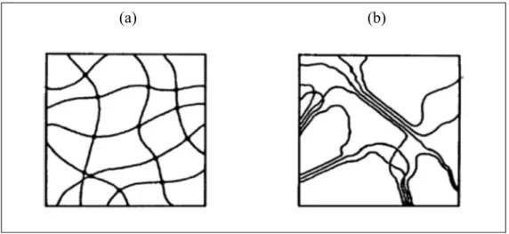
Referring to the forces involved in the building up of networks, two main classes of hydrogels can be determined: (i) chemical hydrogels and (ii) physical hydrogels (Figure 1.8).
In chemical gels, polymer chains are covalently cross-linked and make three dimensional networks (Hennink et al., 2002). In this type of hydrogels the equilibrium swelling levels depends on crosslink density and the polymer-water interaction parameters like hydrophilicity of the polymer chains (Rosiak et al., 1999). In Physical or Reversible gels, the polymer chains bond together by physical crosslinks, such as entanglements or crystallites and/or other weak forces (van der Waals, hydrogen and ionic bonding). Existence of physical cross-links between diverse polymer strings, prevent dissolution of the hydrogels (Hennink et al., 2002). However applying stress or changing physical conditions can break all of these links. So this kind of hydrogels is called reversible hydrogels (Rosiak et al., 1999).
Figure 1.8 Schematic diagram of (a) a chemical hydrogel with point crosslinks and (b) a physical hydrogel with multiple-junction zones
1.2.6. Environmentally-sensitive hydrogels
Environmentally-sensitive hydrogels can sense external environment stimuli like changing in pH, temperature, ionic strength, surrounding biological fluid, etc. and also they can respond to them. These responses can be exhibited in various manners like changing in swelling behavior, network structure, permeability or mechanical strength.
These types of hydrogels are also called ‘smart’ or ‘Intelligent’ hydrogels and categorized based on the type of their stimuli. They have been used in diverse biomedical applications, such as in making artificial valves, inducing vascular angiogenesis and immobilization of enzymes cells (Qiu et al., 2001).
Temperature-sensitive hydrogels are part of the family of environmentally stimulus-sensitive hydrogels, which are able to respond to environmental temperature changes by changing their physical properties. Thermo-responsive hydrogels exhibit a change of the degree of swelling with temperature of the surrounding fluid. They are classified into three groups including negatively thermo-sensitive, positively thermo-sensitive, and thermally reversible gels. Negative temperature-sensitive hydrogels have a lower critical solution temperature (LCST) and swelling at low temperature (<LCST) and shrinking at high temperature (>LCST). Conversely, positively thermosensitive swell at high temperature and shrinking at low
temperature (Fang et al., 2008). Thermally reversible gels are copolymers made of different monomers with different LCST values.
1.2.7. Injectable hydrogels
For the purpose of AAA embolization, hydrogels must be injectable by small catheter but rapidly demonstrate enough mechanical properties to occlude blood flow. This limits the number of available materials. To that purpose, the hydrogel should be gel in response to an external stimulus such as calcium ions, light, chemical crosslinker, pH, temperature, etc. Injectable hydrogels have lots of applications in biomedical and pharmaceutical fields like drug delivery, cell growth, and tissue engineering. Solution of hydrogels can be mixed with drugs, proteins, or cells and then injected and formed in situ by chemical or physical crosslinking methods (Madan et al., 2009). Chemical crosslinked hydrogels are formed by photo polymerization, disulfide bond formation, or reaction between thiols and acrylate or sulfones methods. Physical crosslinked hydrogels are formed by the self-assembly in response to environmental stimuli (Nguyen et al., 2010). Therefore, physical hydrogels are more attractive for biomedical applications, because they do not use any organic solvents, crosslinking agents or photo irradiation. Moreover unlike chemical crosslinked hydrogels, they do not damage incorporated proteins, embedded cells and surrounding tissues by the heat of reaction (Nguyen et al., 2010).
Nowadays, only a few injectable hydrogels are proposed as embolic agents for blood vessels. These are EmboGel and UltraGel. EmboGel is a hydrogel embolic agent consisting of a mixture of Iohexol and alginate, with a calcium chloride solution used to initiate polymerization. UltraGel consists of irgacure, iohexol and polyethylene glycol diacrylate (Barnett et al., 2011). These materials show a rapid polymerization. Also when used in an AAA endoleak model, the agent is delivered in a strand-like form. Due to this form, EmboGel is able to effectively fill the aneurismal cavity and occlude it from the blood flow (Barnett et al., 2009). However EmboGel also has a problem in terms of insufficient inherent strength. Recently, new embolizing agents based on Chitosan thermogels have been proposed by Lerouge’s team. These are detailed in the “Previous work by the team” section.