Controlled Liposome Production using Micromixers Based on
Dean Flow Dynamics
by
Rubén Rodrigo LÓPEZ SALAZAR
THESIS PRESENTED TO ÉCOLE DE TECHNOLOGIE SUPÉRIEURE
IN PARTIAL FULFILLMENT FOR THE DEGREE OF
DOCTOR OF PHILOSOPHY
Ph.D.
MONTREAL, JUNE 25, 2020
ÉCOLE DE TECHNOLOGIE SUPÉRIEURE UNIVERSITÉ DU QUÉBEC
This Creative Commons license allows readers to download this work and share it with others as long as the author is credited. The content of this work can’t be modified in any way or used commercially.
BOARD OF EXAMINERS
THIS THESIS HAS BEEN EVALUATED BY THE FOLLOWING BOARD OF EXAMINERS
Mr. Vahé Nerguizian, Thesis Supervisor
Department of Electrical Engineering, École de Technologie Supérieure
Mr. Ion Stiharu, Thesis Co-supervisor
Department of Mechanical and Industrial Engineering, Concordia University
Mrs. Sophie Lerouge, President of the Board of Examiners
Department of Mechanical Engineering, École de Technologie Supérieure.
Mr. Frédéric Nabki, Member of the jury
Department of Electrical Engineering, École de Technologie Supérieure
Mr. Miguel Burnier, External Evaluator
Department of Ophthalmology, Faculty of Medicine, McGill University
THIS THESIS WAS PRESENTED AND DEFENDED
IN THE PRESENCE OF A BOARD OF EXAMINERS AND PUBLIC JUNE 15, 2020
FOREWORD
The completion of this thesis has been an incredible journey that started several years ago, in 2014, where I met my research supervisor. Six years after, and it feels that it was yesterday when we talked about small vesicles called “liposomes.” We theorized about how we could produce them and how these advanced delivery systems could help patients. Two years later, after this first meeting, I decided to join the ÉTS Ph.D. program. Immediately after starting my Ph.D., my co-supervisor joined us with great enthusiasm in the endeavor of starting from scratch a completely new project for the laboratory.
This journey has taken us to four different laboratories and collaborated with eight academic institutions situated in three different countries, as well as a company. It allowed me to meet incredible people, enthusiastic about teaching me what they knew about chemistry, biology, and engineering, as well as sharing their laboratories to complete the puzzle of this multidisciplinary research.
This work is, in a sense, is a summary of this journey, a personal and a shared one with all the people who put something into it. A journey that I hope one day can contribute to patients and families waiting for next-generation treatments.
ACKNOWLEDGMENT
I would like to thank my research supervisor Vahé Nerguizian, and my co-supervisor, Ion Stiharu, for welcoming me to this wonderful world of biomedical engineering and for all these years of guidance, confidence in this project, and financial support. Their enthusiasm for science is inspiring; it pushed me to do better every day.
Also, I would like to thank the Mexican National Council of Science and Technology (CONACyT 389081) for its financial support through all these years.
Additionally, I would like to thank Anas Alazzam for supporting this project with both intellectual and financial resources; without him, this project would not be possible. Likewise, I would like to thank Karl F. Bergeron, Alexandre Legiot, Sabri Rial, Denis Flipo, and Catherine Mounier from the Université du Québec à Montréal for providing me with resources, ideas, support, and training to complete this research.
Similarly, I would like to thank the group CREER from Polytechnique Montréal, especially to Jean-Sébastien Décarie. Moreover, I would like to thank the NanoQAM team, especially to Ricardo Izquierdo and Alexandre Robichaud, as well as the UQAM chemical department support staff, especially to Galyna Shul, Jacqueline Hue Tieu, and Luc Arsenault.
Correspondingly, I would like to thank Amber Yasmeen and Tahira Baloch from the Lady Davis Institute at the Jewish General Hospital for their support and training in cancer research. Thank you for opening my mind to new possibilities.
Equally, I am grateful to Miguel Burnier, Julia Burnier, Thupten Tsering, Janusz Rak, and Laura Montermini from the Research Institute of the McGill University Health Center for opening me the doors of their laboratories to propel this research even further. Finally, I am grateful to Sophie Lerouge, Frederik Nabki, and Miguel Burnier for their participation in the thesis defense committee.
I would like to express my deepest gratitude to the laboratory members and collaborators. Their support and contributions were crucial to complete this work. These are not in a particular order, Laura Gonzalez, Ixchel Ocampo, Luz-Maria Sanchez, Sergio Camacho, Isaias Cueva, Mohsen Delavari and Farnaz Darooeizadeh.
Similarly, I would like to acknowledge my friends, the new and the old as well as my family, the ones here, and the ones who are not anymore for supporting me through this journey. Especially to my parents Paulina Salazar, Rodrigo Lopez, and my sisters Ana Lopez and Yolanda Salazar. Your love reaches all the planet, and I am sure that the universe too.
Last but not least, I would like to thank my life and adventures partner Prisca Bustamante for her unconditional support, generosity, and love. Her perseverance and dedication to science are inspiring. I always found in her beautiful and sharp mind a colleague and a friend to share a cup of chocolate to talk about life, science, and all.
Production contrôlée de liposomes à l'aide de micromélangeurs basés sur Dean la dynamique des écoulements
Rubén Rodrigo LÓPEZ SALAZAR RÉSUMÉ
Les liposomes constituent des transporteurs polyvalents, car ils ont la capacité de cibler spécifiquement des organes, des tissus et des cellules. Ils tiennent cette spécificité de leurs propriétés physicochimiques, qui régissent les interactions avec les systèmes biologiques. De ce fait, afin que les liposomes soient des transporteurs efficients, il est crucial de contrôler ces propriétés.
Afin d'être déployés de manière efficace comme nanomédicaments, les liposomes doivent être produits à une échelle industrielle, et ce, d'une façon reproductible. Le recours à des méthodes conventionnelles de production de masse ne permet pas d’exercer un contrôle adéquat sur les propriétés des liposomes. En effet, ces méthodes requièrent de multiples étapes d'homogénéisation et d'encapsulation de médicaments, rendant particulièrement difficile la reproduction des propriétés des liposomes. Cependant, l’emploi de dispositifs microfluidiques, notamment les micromélangeurs, permet la synthèse en flux continu des liposomes ainsi que l’application d’un contrôle plus précis sur leurs propriétés, par exemple leur taille, et sur la distribution de leur taille.
Malgré ses avantages, la production par micromélangeurs présente des points négatifs : faible productivité, différents problèmes de fabrication, colmatage, agglomération et présence de résidus de solvants organiques nocifs. Ces aspects négatifs s’expliquent par le fait que les micromélangeurs ne sont pas conçus pour produire des liposomes, mais plutôt des réactions chimiques. L'autoassemblage des liposomes nanométriques requiert un processus de mélange rapide. Celui-ci doit cependant être uniforme pour produire des populations de taille homogène.
Les micromélangeurs fondés sur la dynamique des flux de Dean, l’approche sur laquelle se base cette thèse, offrent une alternative aux dispositifs jusqu’ici utilisés, car ils ont le potentiel de produire des liposomes nanométriques dont les propriétés sont contrôlables, et ce, à un taux de production élevé. De plus, la fabrication de ces dispositifs est plus simple que celle des conceptions antérieures.
Cette thèse a recours à la modélisation numérique et à l'imagerie confocale. Ces dernières nous ont permis de concevoir un micromélangeur. Des liposomes, aussi petits que 27 nm, dont la taille peut être contrôlée et ayant une reproductibilité élevée, ont été produits à un taux de production allant jusqu’à 41 mg / mL. L’obtention de ces résultats a été appuyée par l’utilisation d’outils statistiques. Par ailleurs, les liposomes se sont montrés stables pendant une période de 6 mois. D’autre part, plusieurs facteurs moléculaires ayant une influence sur les propriétés physico-chimiques des liposomes ont été étudiés. L’étude de la formation de liposomes à l’aide de solvants organiques conventionnels et du Transcutol® a démontré que le
taux de changement de la polarité exerce une influence directe sur la taille des liposomes. De plus, l’utilisation du Transcutol® est avantageuse, car elle ne requiert pas d’étapes de filtration en raison de sa faible toxicité.
L’approche de synthèse liposome présentée dans ce travail offre la possibilité de produire des nanoparticules de taille contrôlée à l’échelle industrielle avec des applications dans le domaine biomédical, tels que les systèmes de distribution de médicaments et de gènes, ainsi que l’étude de la communication cellulaire à cellule.
Mots-clés: liposomes; micromélangeurs; dynamique des flux de Dean, dispositifs microfluidiques, nanoparticules.
Controlled liposome production using a micromixer based on Dean flow dynamics Rubén Rodrigo LÓPEZ SALAZAR
ABSTRACT
Liposomes are multipurpose delivery carriers capable of targeting specific organs, tissues, and cells. This specificity is controlled by their physicochemical properties, which determine their interactions with biological systems. Therefore, controlling liposome properties is crucial for their effective implementation.
Liposomes must be produced on an industrial scale in a reproducible way to be effectively deployed as nanomedicines. Conventional bulk production methods have inadequate control over liposome properties and require multiple homogenization and drug encapsulating steps adding to reproducibility problems. By contrast, liposome production using microfluidic devices such as micromixers enables continuous-flow liposome synthesis with enhanced control over liposomes properties such as size and size distribution.
Previously proposed micromixers suffer from low liposome productivity, fabrication problems, clogging, agglomeration, and harmful organic solvent residues. Most of the time, these devices were tailored to chemical reactions and not specifically to liposome production. Liposome self-assembly requires a mixing process that is fast to produce small liposomes, but to a certain extent, it should be uniform, so liposome formation conditions are similar for all the particles and thus result in homogenous size populations.
Dean flow dynamics-based micromixers, which are used in this work, offer an alternative to earlier microfluidic devices, with the potential of producing uniform controllable nanosized liposomes, at a high production rate. Also, their fabrication is simplified because they do not require complicated three-dimensional structures to create flow perturbations.
In this dissertation, numerical modeling and confocal imaging were used to investigate the mixing process. As a result, a novel micromixer was proposed. Liposome production factors using the proposed microfluidic device were identified and modeled using statistical tools. Size-controlled liposomes as small as 27 nm, at a production rate as high as 41 mg/mL were produced. Additionally, liposomes showed to be stable for up to 6 months. Other molecular related factors were investigated to better understand their effects in liposome physicochemical properties. Likewise, the polarity change rate influence over liposome size was demonstrated using both conventional organic solvents and Transcutol®. The latter has the potential to avoid filtration steps due to its reduced toxicity.
The liposome synthesis approach presented in this work offers the potential to produce controlled-size nanoparticles on an industrial scale with applications in the biomedical field, such as drug and gene delivery systems, as well as the study of cell to cell communication.
TABLE OF CONTENTS
Page
INTRODUCTION ... 1
CHAPTER 1 LITERATURE REVIEW ... 19
1.1 Introduction ...19
1.2 Liposomes ...19
1.2.1 Classification of liposomes ... 21
1.2.2 Liposomes applications ... 22
1.3 Liposomes production methods ...27
1.4 Liposome production using micromixers ...28
1.4.1 Molecular diffusion-based micromixers for liposome production ... 29
1.4.2 Chaotic advection-based micromixers for liposome production ... 35
1.4.3 Fluid lamination and folding ... 40
1.4.4 Micromixers for liposome production evolution ... 42
1.5 Patents and companies working on the field ...43
1.6 Chapter 1 conclusions ...45
CHAPTER 2 LIPOSOME FORMATION IN DEAN DYNAMICS FLOW BASED MICROMIXERS DEVICES: THEORY, MATERIALS, AND METHODS ... 47
2.1 Liposome formation theory ...47
2.2 Nanoprecipitation method and mixing ...49
2.3 Mixing theory...51
2.4 Mixing and liposome production relationship ...54
2.5 Numerical modeling ...55
2.6 Micromixer design and fabrication ...59
2.7 Mixing imaging ...61
2.8 Lipid preparation ...63
2.9 Liposomes production using micromixers ...64
2.10 Liposome characterization ...66
2.10.1 Dynamic light scattering ... 67
2.10.2 Nanoparticle tracking analysis ... 70
2.10.3 Zeta potential (electrokinetic potential) ... 72
2.10.4 Transmission electron microscopy ... 73
2.11 Chapter 2 conclusions ...74
CHAPTER 3 LIPOSOME PHYSICOCHEMICAL CHARACTERISTICS MODELING ... 77
3.1 Identifying the most significant factors. ...78
3.1.1 Screening experiments ... 78
3.1.2 Liposome size significant factors ... 81
3.1.3 Liposome PDI significant factors ... 83
3.1.5 Conclusions on the most significant factors ... 85
3.2 Surface response-based modeling of liposome characteristics in a periodic disturbance mixer ...86
3.2.1 Introduction ... 86
3.2.2 Materials and methods ... 87
3.2.3 Results and discussion ... 91
3.2.4 Conclusions ... 101
3.3 Chapter 3 conclusions ...102
CHAPTER 4 PARAMETRIC STUDY OF THE FACTORS INFLUENCING LIPOSOME PHYSICOCHEMICAL CHARACTERISTICS IN A PERIODIC DISTURBANCE MIXER ... 103
4.1 Introduction ...103
4.2 Materials and methods ...104
4.2.1 Lipid preparation ... 104
4.2.2 Liposome characterization ... 104
4.2.3 Statistical analysis ... 106
4.3 Results and discussion ...106
4.3.1 Alternating centripetal force direction induces mixing ... 107
4.3.2 Intensity- based size vs number-based size validation ... 109
4.3.3 TFR controls liposome size ... 111
4.3.4 FRR controls liposome size in a defined range ... 113
4.3.5 Lipid concentration influences liposome size ... 115
4.3.6 Lipid fatty acid chain length influence over liposome size ... 118
4.3.7 Temperature and liposome size are negatively correlated ... 119
4.3.8 Liposomes are stable in the long term ... 120
4.3.9 Liposome production rate ... 122
4.4 Chapter 4 conclusions ...122
CHAPTER 5 THE EFFECT OF DIFFERENT ORGANIC SOLVENTS IN LIPOSOME PROPERTIES PRODUCED IN A PERIODIC DISTURBANCE MIXER: TRANSCUTOL®, A POTENTIAL ORGANIC SOLVENT REPLACEMENT ... 125
5.1 Introduction ...125
5.2 Materials and methods ...127
5.2.1 Lipids preparation and nanoparticle production ... 127
5.2.2 Nanoparticle characterization ... 128
5.2.3 Experimental design and statistical analysis ... 128
5.3 Results and discussion ...129
5.3.1 Liposome production using different organic solvents ... 129
5.3.2 Concentration effects on liposomes produced with ethanol and Transcutol® ... 134
5.3.3 Temperature effects on liposomes produced with ethanol and Transcutol® ... 136
XV
CHAPTER 6 FUTURE WORK: TOWARDS BIOMEDICAL APPLICATIONS ... 143
6.1 Alternative microfabrication process: 3D printing ...143
6.2 Geometry optimization for an improved liposome production yield ...145
6.3 Approaches to facilitate liposome production implementation ...145
6.4 EVs characterization for cancer research ...146
6.5 Gene delivery applications ...147
CONCLUSIONS ... 149
RECOMMENDATIONS ... 151
APPENDIX I PUBLICATIONS DURING PH.D. STUDIES... 155
LIST OF TABLES
Page
Table 1.1 Head-groups classification with examples ... 21
Table 1.2 Liposome classification ... 22
Table 1.3 Approved liposome formulations ... 23
Table 3.1 Factors and levels tested in the experimental screening design ... 79
Table 3.2 Initial low and high experimental levels per variable ... 89
Table 3.3 CCCR design results for the 29 runs... 91
Table 3.4 Grouping information using the Tukey method at a 95% confidence level ... 96
Table 3.5 Statistically significant coefficients for each model (p < 0.05) and model statistics summary ... 98
Table 4.1 DLS vs NTA characterization ... 109
Table 4.2 Theoretical vs. Measured particle concentration produce at FRR = 1 TFR = 18 mL/h ... 122
Table 5.1 Experimental design. All the experiments were performed at FRR = 8.56 and TFR = 18 mL/h ... 128
LIST OF FIGURES
Page
Figure 0.1 Type of nanodrugs particles ... 3
Figure 0.2 Nanomedicines market value ... 3
Figure 0.3 Liposome applications summary ... 4
Figure 0.4 Structural liposome model, encapsulated, and surface elements ... 6
Figure 0.5 Nanoparticle accumulation ... 7
Figure 1.1 a Structure of POPC lipid molecule b Lipid bilayer thickness c Lipid vesicle ... 20
Figure 1.2 Liposomes loaded with Dil red dye in the retina space ... 26
Figure 1.3 Flow focusing production method (a) Numerical model (b) DiIC18 fluorescence intensity during liposome formation ... 30
Figure 1.4 Schematic of 3D-MHF liposome formation device ... 32
Figure 1.5 a) Numerical models comparing the ethanol concentration profiles for VFF and MHF devices b) Photograph of multilayer VFF with an aspect ratio of 100:1 ... 34
Figure 1.6 Mixing inside a SHM.The micrographs are a cross-section view at different distances from the beginning of the channel ... 36
Figure 1.7 Herringbone structures induce chaotic advection of the laminar streams. Dimensions of the mixing channel are 200µm x 79 µm, and herringbone structure is 31 µm high and 50 µm thick ... 37
Figure 1.8 Chaotic advection-based microfluidic Device (CA-MD) ... 39
Figure 1.9 Liposome production using micromixers evolution ... 43
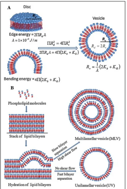
Figure 2.1 Schematic showing the liposome formation process explained by energetic considerations ... 48
Figure 2.2 Energy diagram of lipid aggregates as they move from phospholipid aggregates to SUVs ... 49
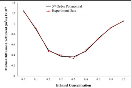
Figure 2.3 Mutual diffusion coefficient for the binary mixture ethanol-water dependent on concentration. In black, the experimental data, in red, the 5th order polynomial ... 58 Figure 2.4 Mixing of ethanol and water in a microfluidic device. Experimental vs.
numerical modeling ... 59 Figure 2.5 PDM. The micromixer consists of two inlets a micromixing channel
and an outlet ... 60 Figure 2.6 Micromixer fabrication process ... 61 Figure 2.7 Micromixing imaging A) Food dyes imaging in two dimensions. In
blue-stained water and in orange ethanol B) Fluorescence dyes imaging in three dimensions ... 62 Figure 2.8 Mixing imaging experimental setup ... 63 Figure 2.9 Lipid preparation process. From left to right, lipid mixture, chloroform
removal, and organic solvent dilution ... 64 Figure 2.10 Liposome production experimental setup A) Schematic
B) Photography of the functional experimental setup ... 65 Figure 2.11 Particle size distribution A) By number B) By volume C) By intensity
... 68 Figure 2.12 Zetasizer S90 workflow. From left to right, inserting the sample,
equipment, and results ... 69 Figure 2.13 NTA Measurements A) Nanosight NS500 B) Sample results are taken
from NS500 C) Image of the scattered light of nanoparticles ... 71 Figure 2.14 Diagram showing the potential as a function of the distance from the
particle’s surface in a colloidal dispersion ... 72 Figure 2.15 Philips Tecnai T12 electron microscope ... 73 Figure 2.16 General methodology for investigating the liposome formation process
in Dean Flow Dynamics based micromixers ... 75 Figure 3.1 Dean Forces-based micromixers A) CEA based on flow lamination
B) PDM, based on alternatively shifting centripetal forces ... 78 Figure 3.2 A) Screening experiments steps B) Lipids used in the screening
XXI
Figure 3.3 Experimental setup V) DMPC:CHOL:D 5:4:1 10 mM X) DMPC:CHOL 1:1 40 mM Y) DMPC: CHOL 1:1 10 mM Z) DMPC: CHOL: DHP 5 :4 :1 40 mM ... 81 Figure 3.4 Fit screening model for size A) Main effects plots for size B) Pareto
chart of effects ... 82 Figure 3.5 Normalized size distribution by intensity of two samples, produced by
a CEA micromixer, in blue a Z-Average of 236.6 nm and in orange 160.7 nm for a FRR of 1 and 5 ... 83 Figure 3.6 Fit screening model for PDI A) Main effects plots for PDI B) Pareto
chart of effects ... 84 Figure 3.7 Fit screening model for zeta potential A) Main effects plots for PDI.
B) Pareto chart of effects ... 85 Figure 3.8 Periodic disturbance micromixer A) Microchannel dimensions
B) Microscope image of the PDM C) Schematic representation of the liposome formation process ... 87 Figure 3.9 CCCR design graphical representation ... 90 Figure 3.10 Surface Response Model derived from the Central Composite
Circumscribed Rotatable Design Data. The model level of significance is p < 0.05 A) 3-dimensional B) Contours representation ... 94 Figure 3.11 PDI Surface Response Model as well as the R2, R2-adjusted, and R2
-predicted. The model has a p < 0.05 A) 3-dimensional B) Contours representation ... 95 Figure 3.12 Zeta potential measurements result for the nine different conditions.
The error bars show 1.96σ, indicating the limits of 95%
confidence (n = 3) ... 97 Figure 3.13 Z-average (nm) vs. FRR (n = 3). Error bars indicate +/− 1 standard
deviation (SD) for samples and SE fit for the model prediction ... 98 Figure 3.14 Mixing efficiency at different FRRs. Each data point corresponds to a
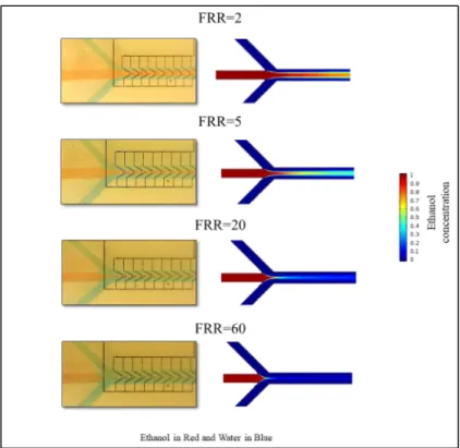
cross-section for a total of 11 data points from 1–11 ... 100 Figure 3.15 Numerical simulations comparing the concentration profiles at FRR =
1 and FRR = 3 at a constant TFR = 18 mL/h A) Top view of the mixing channels and position of the cross-sections B) Cross-sections at different FRRs ... 101
Figure 4.1 Micromixing in PDM at a FRR = 1 and TFR = 18 mL/h (A) Confocal image showing a top view of the PDM (B) Micromixer numerical model with the ethanol flow in red and the water flow in blue. Milli-Q. The bar represents the ethanol concentration (C) Cross-sections of the confocal images and their numerical model counterparts ... 107 Figure 4.2 Mixing index versus time for a constant FRR = 8.56 at various TFR
(5-20 mL/h). Each data point corresponds to the 11 different cross sections in the diagram below ... 108 Figure 4.3 Size distribution by number normalized for the same sample produced
at FRR = 1 and TFR = 18 mL/h. Characterized in the DLS (blue) and NTA (orange) ... 110 Figure 4.4 Size distribution by number normalized for the same sample produced
at FRR = 1 and TFR = 18 mL/h. Characterized in the DLS (blue) and NTA (orange) ... 111 Figure 4.5 Effect of the TFR on various liposome properties ... 112 Figure 4.6 The effect of FRR over liposomes properties ... 114 Figure 4.7 Initial lipid concentration effects over liposome size ... 116 Figure 4.8 Liposome properties produced using DSPC, cholesterol, and DHP in a
molar ratio 5:4:1 ... 118 Figure 4.9 Temperature effects on liposome properties ... 120 Figure 4.10 Liposomes stability after 50 days diameter mean +/- 1 SD A) Liposome
sample synthesized at FRR = 12 and TFR = 18 mL/h, nm B)
FRR = 8.56 TFR = 20 ml/h T = 70 °C ... 121 Figure 4.11 Liposomes produced at FRR = 8.56 TFR=18 mL/h, initial lipid
concentration of 10 mM, T = 40 °C made of DMPC:CHOL:DHP at a molar ratio 5:4:1 three months after production ... 121 Figure 5.1 Liposomes synthesized using an FRR = 8.56 and TFR = 18 mL/h at 40
℃ using different organic solvents with a 10 mM lipid concentration A) Z-Average and PDI. The bars indicate +/- 1 SD n = 3
B) Size Distribution by Intensity Normalized ... 130 Figure 5.2 Liposomes synthetized using an FRR = 8.56 and TFR = 18 mL/h at
40 ℃ using different organic solvents with a 10 mM lipid concentration A) Diameter by number. The bars indicate +/- 1 SD n = 3 B) Size Distribution by Number Normalized ... 131
XXIII
Figure 5.3 Nanoparticle formation process. The increase of the aqueous solvent concentration, and hence the polarity of the mixture, causes the lipids to self-assemble in disk-shaped structures, which finally bend and close into liposomes ... 132 Figure 5.4 Zeta Potential of liposomes synthesized using an FRR = 8.56 and
TFR = 18 mL/h at 40 ℃ ... 133 Figure 5.5 Liposomes synthesized using two different organic solvents and
different lipid concentrations at 40 ºC, with an FRR = 8.56 and TFR = 18mL/h A) Average size and PDI. The bars indicate +/- 1 SD n = 3 B) Size Distribution by Intensity Normalized for ethanol C) Size Distribution by Intensity Normalized for Transcutol® ... 135 Figure 5.6 Zeta Potential of liposomes synthesized using two different organic
solvents and different lipid concentrations at 40 ºC, with an FRR = 8.56 and TFR = 18mL/h. The bars indicate +/- 1 SD n = 3 ... 136 Figure 5.7 Liposomes synthetized using two different organic solvents and
different production temperatures at 10 mM lipid concentration, with an FRR = 8.56 and TFR = 18mL/h A) Average size and PDI. The bars indicate +/- 1 SD n =3 B) Size Distribution by Intensity Normalized for ethanol C) Size Distribution by Intensity Normalized for Transcutol® ... 137 Figure 5.8 Zeta Potential of liposomes synthesized using two different organic
solvents and different production temperatures at 10 mM lipid concentration, with an FRR = 8.56 and TFR = 18mL/h. The bars indicate +/- 1 SD n = 3 ... 138 Figure 5.9 Liposome size evolution synthetized with ethanol at 40 ºC using an
FRR = 8.56 and TFR = 18mL/h. 1-day vs. 50 days after synthesis characterization A) Size Distribution by Intensity of the least changing sample synthesized with a 5 mM lipid concentration B) Size Distribution by Intensity of the most changing sample synthesized with a 40 mM lipid concentration ... 139 Figure 5.10 Liposome size evolution synthetized with Transcutol® using an
FRR = 8.56 and TFR = 18mL/h. 1-day vs. 50 days after synthesis characterization A) Size Distribution by Intensity of the least changing sample produced at 40 ºC and with a 40 mM lipid concentration B) Size Distribution by Intensity of the most changing sample produced at 70 ºC and with a 10 mM lipid concentration ... 140 Figure 5.11 Liposome size evolution synthetized with Transcutol® using an
characterization A) Size Distribution by Intensity of the least changing sample produced at 40 ºC and with a 40 mM lipid concentration B) Size Distribution by Intensity of the most changing sample produced at 70 ºC and with a 10 mM lipid concentration ... 141 Figure 6.1 Microfabrication process of 3D printed-PLA-COC device A) PLA
device and COC “ceiling” B) 3D printing the microfluidic device
C) A completed device with connectors ... 144 Figure 6.2 Factors influencing gene delivery systems production ... 148
LIST OF ABBREVIATIONS ACS American Chemical Society
AFA Adaptative focused acoustics ANOVA Analysis of variance
AO Antioxidants
CAGR Compound annual growth rate
CA-MD Chaotic advection-based microfluidic device CCCR Central composite circumscribed rotatable
CEA Contraction-expansion array
CHOL Cholesterol
CL Cationic liposomes
CM Crisscross micromixer
COC Cyclic olefin copolymer
CONACyT Consejo Nacional de Ciencia y Tecnología
DEPC 1,2-Dierucoyl-sn-glycero-3-phosphocholine
DHP Dicetyl phosphate
DiIC18 1,1'-dioctadecyl-3,3,3',3'-tetramethylindocarbocyanine perchlorate
DLIN-MC3-DMA
(6Z,9Z,28Z,31Z)-heptatriacont-6,9,28,31-tetraene-19-yl 4-(dimethyl amino) butanoate
DLS Dynamic light scattering
DMPC 1,2-dimyristoyl-sn-glycero-3-phosphocholine DMPG Dimyristoylphosphatidylglycerol
DODAC N-N-dioleoyl-N, N-dimethyl ammonium chloride DoE Design of experiments
DOPC 1,2-dioleoyl-sn-glycero-3-phosphocholine DOPE 1,2-dioleoyl-sn-glycero-3-phosphoethanolamine DOPS 1,2-dioleoyl-sn-glycero-3-phospho-L-serine
DOTAP Dioleoyl-3-trimethylammonium propane
DOTMA 1,2-di-O-octadecenyl-3-trimethylammonium propane DPPG 1,2-Dihexadecanoyl-sn-Glycero-3-Phospho-1'-rac-glycerol
DS Delivery systems
DSPC 1,2-distearoyl-sn-glycero-3-phosphocholine DSPG 1,2-distearoyl-sn-glycero-3-phospho-(1'-rac-glycerol) EMA European medicines agency
EPC Egg 1,2-dioleoyl-sn-glycero-3-ethylphosphocholine EPG Egg L-α-phosphatidylglycerol
EPR Enhanced retention and permeability effect
EVs Extracellular vesicles
FDA Food and Drug Administration FDM Fused deposition modeling FRR Flow rate ratio
GMP Good manufacturing practices HSPC L-α-phosphatidylcholine
XXVII
IEEE Institute of Electrical and Electronics Engineers iLiNP Invasive lipid nanoparticle production device
IPA Isopropyl alcohol
Lipid T Lipid mixture type
LNPs Liposome nanoparticles
LUV Large unilamellar vesicles
ME Mixing efficiency
MHF Microfluidic hydrodynamic focusing
MI Mixing index
MLV Multilamellar vesicles
MR Magnetic resonance
Mtype Mixer type
NDS Nanoparticle delivery system NTA Nanoparticle tracking analysis
OLV Oligolamellar vesicles
PA Phosphatidic acid
PBS Phosphate buffer saline solution PC Phosphatidylcholine PCS Photo correlation spectroscopy
PDI Polydispersity index
PDM Periodic disturbance mixer PDMS Polydimethylsiloxane
PE Phosphatidylethanolamine PEG-DSPE 1,2-distearoyl-sn-glycero-3-phosphoethanolamine-N-[amino(polyethylene glycol)-2000] PG Phosphatidyglycerol PI Phosphatidylinositol
PLA Poly-lactic acid
PLGA Poly lactic-co-glycolic acid
POPC 1-palmitoyl-2-oleoyl-sn-glycero-3-phosphocholine PS Phosphatidylserine
RSC Royal Society of Chemistry RSM Response surface methodology SAW Surface acoustic waves
SHM Staggered herringbone micromixer siRNA Small interfering ribonucleic acid
SL Stealth liposomes
SLA Stereolithography SM Sphingomyelin SU-8 Substrate with 8 epoxy SUV Small unilamellar vesicles
TEM Transmission electron microscopy TFF Tangential flow filtration
XXIX
UM Uveal melanoma
VFF Vertical flow focusing
LIST OF SYMBOLS
µ Dynamic viscosity
A Lipid area
Ado Surface area of disk 0
c Concentration D Diffusion coefficient De Dean number DH Hydraulic diameter dh Hydrodynamic diameter Ds0 Diameter of sphere 0 Ds1 Diameter of sphere 1
e Lipid bilayer thickness
F External force
h Half diameter in a pipe.
j Diffusion flux k Boltzmann constant N Molar flux n Number NA Avogadro’s number p Pressure P Production rate
Qas Aqueous solvent flow rate
Qos Organic solvent flow rate
rh Particles Ratio or Liposome Ratio
R Volumetric source for the species RC Ratio of curvature Re Reynolds number T Temperature u Flow velocity x Distance X Increasing proportion η Viscosity ν Kinematic viscosity ρ Fluid density σ Standard deviation
INTRODUCTION
Liposomes are vesicles made of lipids. Lipids are organized in lipid bilayers forming sphere shape structures that can encapsulate multiple substances. This property makes liposomes suitable for drug delivery applications. Along 50+ years in liposome research, various preparation methods have been proposed; however, the vast majority of them are batch-based, and mostly production rates are only suitable for laboratory scale production. Moreover, reproducibility is a significant concern required to meet the standards of regulatory agencies. Batch methods do not have the required consistency. These problems delay or block the research, development, and production of liposomes and prevent them from reaching patients worldwide (Hua, de Matos, Metselaar, & Storm, 2018).
Between 1995 and 2005, significant advances were made in microfabrication, especially in soft lithography allowing researchers to produce microfluidic devices with channels in the order of micrometers in relatively easy few steps. These advancements opened an entirely new world of opportunities to investigate the micromixing phenomenon and its applications.
Micromixers offer a suitable alternative for liposome production. Tunable size monodispersed liposome nanoparticles (LNPs) can be produced in a continuous flow, improving reproducibility. Unfortunately, micromixers yield, in general, is too low for industrial-scale production, while the ones that are suitable for mass production clog and are challenging to fabricate. Other mixing strategies and micromixers designs might help to solve this conundrum. However, only a few micromixers have been studied and explicitly characterized for liposome production; furthermore, important variable interactions have not been addressed. This work objective is to investigate liposome formation in micromixers that are based on Dean’s flow dynamics. This type of micromixers uses chaotic advection and Taylor dispersion to accelerate the mixing process. The conditions under which LNPs are produced, as well as crucial factors that direct and control liposome physicochemical characteristics such as size, size distribution, and zeta potential were investigated.
0.1 Rationale
Pharmacy word originates from the Greek word pharmakon, which has different meanings, including remedy and poison. For years, this dichotomy has been accepted. This acceptance meant for patients that sometimes the cure was worse than the disease. Several therapeutic agents must be administered at high doses, increasing toxic effects. For patients, removing or reducing the poisonous part of the remedy would mean a better quality of life. Having a way of releasing the correct dose at the right location without producing side effects, the magic bullet of Paul Ehrlich was an elusive feat until recently (Ehrlich, 1913).
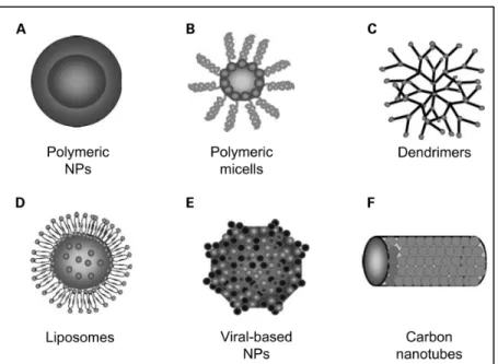
Delivery systems (DS) consists of a shell or structure that functions as a carrier and a cargo that can include genes, drugs, imaging labels, among others. This cargo could be enclosed inside the transport vehicle as well as ligated to its exterior. Most of the DS used in the biomedical field are in the range of 3-200 nm. DS protect their cargo from degradation, offering an extended circulation time inside the human body. Due to their properties such as size and modified surfaces added with specific moieties, DS can target specific organs, tissues, and cells. DS that are in the range of nanometers are called nanoparticle delivery systems (NDS). There are several types of nanoparticles, such as polymeric conjugates, micelles, dendrimers, viral nanoparticles, carbon nanotubes, and liposomes (Cho, Wang, Nie, Chen, & Shin, 2008). Figure 0.1 shows the different types of nanoparticles.
In terms of market potential, nanopharmaceuticals are expected to grow to 168.91 billion by 2026, with a compound annual growth rate (CAGR) of 22.1%, as shown in Figure 0.2. This market growth is supported by liposomes and polymer delivery systems (Business-Wire, 2019).
3
Figure 0.1 Type of nanodrugs particles Taken from Cho et al. (2008, p.1312)
Figure 0.2 Nanomedicines market value Adapted from Business-Wire (2019)
Liposomes have proven to be one of the most versatile nanoparticles, considering the number of approved products in the market. The first approved nanodrug was a liposomal carrier (Doxil®). Nanoparticles applied in the medical field is growing (Havel, 2016). This type of nanoparticles has established a precedent and paved the way for regulations in the area of
nanopharmaceuticals (Food & Administration, 2018). So far, 17 liposome-based drug formulations have been approved (Leung, Amador, Wang, Mody, & Bally, 2019).
Liposomes applications range from drug delivery systems, gene delivery systems, vaccines imaging, and analytical applications. These vesicles can encapsulate both imaging labels and therapeutic substances, also known as theragnostics (Pattni, Chupin, & Torchilin, 2015). Additionally, applications in the field of cosmeceuticals have been explored (Rahimpour & Hamishehkar, 2012; Van Tran, Moon, & Lee, 2019). Figure 0.3 summarizes Liposomes applications.
Figure 0.3 Liposome applications summary
NDS have the potential to add value to existing products, extending their product life as patented formulations as well as reducing toxicity. One example of this is the antibiotic field, where their indiscriminate use caused the resistance of bacteria, requiring increasing the level of dosage just below toxic values for effective treatment. Old formulations that right now are
5
not effective anymore could be reused through NDS. Currently, a few liposomal antibiotic formulations are under experimentation (Gao et al., 2014).
However, there are still challenges in the field of nanopharmaceuticals. Liposome based delivery systems are getting more sophisticated with new added bioactive molecules. At a laboratory-scale, this increased complexity translates into lengthy production processes that prevent innovative formulation production at the industrial scale. Moreover, these production methods must comply with good manufacturing practices (GMP), so they can be included in late clinical stages and commercialization (Hua et al., 2018). The nanopharmaceuticals industry looks for integrative liposome production systems with high reproducibility and scalability
Microfluidics devices, specifically micromixers, offer an approach that can produce liposomes in a continuous-flow production, improving reproducibility (Andreas Jahn et al., 2010). However, these microfluidic devices are not exempt from challenges. Low production yield and toxic solvent remains are still the main problems to be solved.
In this dissertation, a new microfluidic configuration is proposed to increase the production rate while keeping control over final liposome characteristics such as size, size distribution, and zeta potential. The mixing inside the proposed device is based on Dean flow dynamics. This device can produce liposomes of controlled size, and it can be useful for creating other types of NDS.
0.2 Problem statement
Liposomes are used in medical imaging, general delivery, and analysis systems (Pattni et al., 2015). These vesicles encapsulate drug formulations, genes, imaging labels, and proteins. The biodegradable components of liposomes provide them with the capacity to interact safely with biological systems. Moreover, by adding surface moieties and controlling the size, it is possible to target distribution towards specific organs and tissues, increasing drug circulation time, and
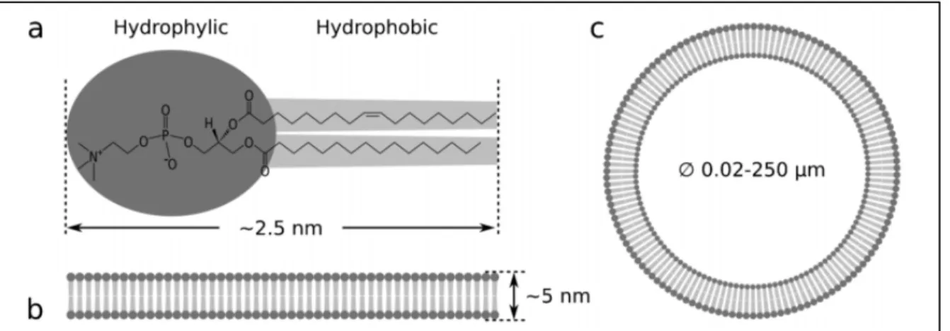
improving drugs therapeutic window (Allen & Cullis, 2013; Gabizon et al., 1994; Matsumura & Maeda, 1986; Safra et al., 2000). Figure 0.4 shows a typical liposome with potential contents and surface ligands (Liu & Boyd, 2013).
Figure 0.4 Structural liposome model, encapsulated, and surface elements
Taken from Liu & Boyd (2013, p.392)
Liposomes physicochemical characteristics such as size, size distribution, and zeta potential are crucial for general delivery applications. Inside the human body, for example, large liposomes >2000 nm are rapidly filtered in liver and spleen, whereas vesicles with size from 100 to 200 nm escape to this filtration and accumulate in tumors. Particles < 5 nm are filtrated by kidneys (Blanco, Shen, & Ferrari, 2015; Hak Soo et al., 2007). The size, shape, and surface properties also play a crucial role in nanoparticle accumulation, as shown in Figure 0.5Moreover, liposomes monodispersity is a determinant quality factor, defining if all the prepared particles will finish at the desired place. LNPs ranging between 150 to 200 nm are necessary because they accumulate in tumors due to the enhanced retention and permeability effect (EPR). This effect establishes that particles of specific sizes accumulate selectively in solid tumors. Particles pass through the large fenestrations in the endothelial cells caused by abnormal angiogenesis (Matsumura & Maeda, 1986). Current commercially available
lipid-7
based nanoparticles are in the size range between 45 and 180 nm (Sedighi et al., 2019). On the other hand, zeta potential determines how particles interact with the human body by controlling circulation time or particle entrapment by cells (P. L. Felgner et al., 1987; Khadke, Roces, Cameron, Devitt, & Perrie, 2019). Thus, controlling liposome physicochemical properties is crucial for delivering the right substance to the right place.
Figure 0.5 Nanoparticle accumulation Taken from Blanco et al. (2015, p.947)
The control of liposome characteristics requires systematic exploration of the factors influencing liposome properties in each new proposed micromixer. The relationship between these variables is complex. Statistical modeling can shine a light on this subject. For micromixers based on molecular diffusion and chaotic advection, this modeling has proved useful in identifying and controlling liposomes size, size distribution as well as transfection efficiency in gene delivery applications (Tiago A. Balbino et al., 2013; Kastner et al., 2014). However, for Dean flow dynamics-based micromixers, there are not reported results of such characterization.
Even though several liposome drug formulations have been approved, such as Doxil®, among others, production methods are still behind regarding industrial-scale purposes. The fragility of the production methods was proved in November 2011, when Doxil® production was
suspended due to manufacturing and quality problems. Patients were not able to access their vital medicine worldwide. In February 2013, the Food and Drug Administration (FDA) needed to approve a similar drug version called Lipodox to solve the shortage problem (Barenholz, 2012) partially. Production problems represent a latent risk that faces liposomal formulations if no action is taken to research better manufacturing approaches. At an industrial level, there are two significant problems: reproducibility and scalability.
First, the lack of reproducibility of commonly used batch methods makes new formulations challenging to manufacture at industrial-scale, while production methods require multiple steps that might further degrade nanoparticles quality (Stavis, Fagan, Stopa, & Liddle, 2018). The reproducibility is crucial for maintaining high-quality standards; it is decisive to meet the requirements of the regulatory agencies, such as the FDA or the European Medicines Agency (EMA) (Hafner, Lovrić, Lakoš, & Pepić, 2014).
Second, the scalability of the production methods is pivotal to ensure a reliable medication supply to patients. Current commercial production methods mostly rely on conventional laboratory-scale techniques such as thin-film hydration or ethanol injection assisted by sonication and membrane extrusion that require additional steps for cargo loading (Hua et al., 2018).
Microfluidics devices, specifically micromixers, are capable of producing size-controlled LNPs in continuous-flow. Liposome cargo can be added in a single step. Moreover, these devices can be parallelized for an increased production rate while keeping the same control over liposome properties resulting in process intensification as well as reducing waste and cost (Shallan & Priest, 2019).
Liposomes produced using micromixers have typically highly monodispersed to low polydispersed populations. However, lipid concentration remains too small for scaling-up. Usually, the initial lipid concentration in a typical micromixer based on molecular diffusion is of the order of 5 to 80 mM and, after passing through the micromixer, is diluted even more
9
from 10 to 50 times to achieve a liposome size below 100 nm. If we compare this value with commercial formulations such as Doxil® that is in the order of 8 mM, it becomes clear why novel micromixer approaches are not yet substituting available methods for industrial-scale productions.
Interesting solutions to overcome the liposome final dilution problem have been proposed. One is the Staggered Herringbone Micromixer (SHM), which reduces the dilution of lipid concentration required for small liposomes (Belliveau et al., 2012). Another is the vertical flow focusing (VFF) (R. R. Hood & DeVoe, 2015), which increments the flow in the device by increasing height in microchannels. These two devices can yield tens to hundreds of mg/h of liposomes. However, these micromixers are challenging to produce because they include 3D features in the case of SHM or high aspect ratio channels, as in the VFF. New devices with simpler geometries that increase liposome production rate are needed. Dean flow dynamics-based micromixers are in general, easy-to-fabricate. There are a few that have been used for liposome production (Kimura et al., 2018; Jisun Lee et al., 2013; Valencia et al., 2010; Y. Wu, Li, Mao, & Lee, 2012).
An essential part of liposome production using the principle of nanoprecipitation is mixing an organic solvent into an aqueous solvent, which causes a polarity change. It has been shown that a fast mixing produces small and uniformly-sized liposomes (Andreas Jahn et al., 2010). Numerical modeling provides the tools to better understand the mixing process. Current used numerical models for studying mixing in liposome production not always include variables that depend on the ethanol concentration (Amrani & Tabrizian, 2018; Kimura et al., 2018). In binary mixtures, density, viscosity, and the mutual diffusion coefficient change as concentration changes. Only a few authors have used models that include viscosity and mutual diffusion coefficient dependent on concentration (Renee R. Hood, DeVoe, Atencia, Vreeland, & Omiatek, 2014; Andreas Jahn et al., 2010). However, none has added density to numerical model calculations as far as the author knows it.
Finally, the solvent remains in liposome production using micromixers are inherent to the process itself. Lipids must be diluted first in an organic solvent, to later be mixed with an aqueous solvent to produce liposomes. Harmful leftovers can interact with the liposome cargo reducing their stability, capabilities, or in the worst-case scenario, they can damage the biological systems that they interact with. An inline filtration or the substitution of organic solvent with some other similar substances could improve the usability of produced liposomes. A better understanding of how organic-aqueous solvent mixtures affect liposome size, size distribution and zeta potential would be useful for controlling such characteristics. Only a few studies have tried to describe this influence in micromixers for liposome production (Webb et al., 2019).
In summary, controlling liposome physicochemical characteristics is crucial for their functionality. Thus, modeling which factors influence these properties is the first step towards liposome functionalization. Additionally, reliable liposome production methods that can be easily scalable from laboratories to commercial manufacture are required to ensure the supply chain to patients worldwide. Micromixers offer a solution; however, current devices are cumbersome to fabricate. Dean flow dynamic-based micromixers have the potential to produce controllable-size liposomes, with easy-to-fabricate devices. Finally, conventional organic solvents could be removed or replaced by substances capable of improving liposome therapeutic properties.
0.3 Research objectives
The general objective of this research work is to investigate liposome production in Dean Flow dynamics-based micromixers. The research objectives include the evaluation of the factors influencing liposome properties to produce liposomes in a controllable way, with a final concentration and size comparable to commercially available formulations.
11
0.3.1 Specific objectives
1. Production of LNPs with controlled physicochemical characteristics such as size, size distribution, and zeta potential.
2. Preparing liposomes in the range of 50 to 200 nm as commercially available formulations with homogenous size.
3. The removal or substitution of the potentially harmful solvent remains.
4. Achieving liposome production yields in the order of tens to hundreds of mg/mL.
0.4 Research approach and main hypothesis
In this work, numerical modeling was used to investigate the mixing of two fluids in microfluidic devices. The central hypothesis of this dissertation is that a fast and uniform mixing or, in other words, changes in polarity, will result in smaller liposomes with homogeneous size populations. This hypothesis was further developed first by identifying which factors control liposome physicochemical characteristics. Later, flow-related factors were statistically modeled. Then, through confocal images and liposome characterization, the relationship between mixing and LNPs properties was further investigated.
Additionally, other molecular factors influencing liposome properties were considered, such as temperature, lipid type, and initial lipid concentration. Finally, by using different organic solvents, we extended the mixing hypothesis to investigate how by modulating the polarity change rate, average liposome size can be controlled too. All previously mentioned studies were focused on Dean flow dynamics-based micromixers.
0.5 Main contributions
The main contributions of this dissertation are the following:
1. A method to fabricate micromixing channels with optical windows using biocompatible materials and 3D printing. This fabrication method enables the production of microfluidic devices at low-cost with reduced steps compared with conventional soft lithography. At the same time, it allows optical access to the mixing channels to evaluate mixing performance. This method can be extended to produce different types of micromixers for applications, including liposome production, where materials need to be biocompatible. This contribution resulted in the following conference paper publication.
Lopez, R., Nerguizian, V., & Stiharu, I. (2018, 24-27 June 2018). Low-Cost 3D-Printed PLA-COC Micro Hydrodynamic Focused Device. Paper presented at the 2018 16th IEEE International New Circuits and Systems Conference (NEWCAS). (Presented and published)
2. The identification of the most important factors controlling liposome characteristics in Dean flow dynamics-based micromixers. A Plackett-Burman experimental design was used to evaluate in a reduced number of experimental runs what factors contributed to each of the liposome properties studied in this work (liposome size, size distribution, and zeta potential). This contribution resulted in the following conference poster presentation.
López Salazar, R. R., Ocampo, I., Bergeron, K.-F., Alazzam, A., Mounier, C., Stiharu, I., & Nerguizian, V. (2019). Assessment of the Factors Influencing Liposome Size in Dean Forces Based µmixers. Paper presented at the µTAS 2019 The 23rd International Conference on Miniaturized Systems for Chemistry and Life Sciences Basel, Switzerland. (Presented as poster).
3. The design and fabrication of a micromixer based on Dean flow dynamics, capable of producing LNPs. The periodic disturbance mixer (PDM). This micromixer uses semicircular structures to guide the flow inside the microchannels to a circular path that
13
produces centripetal forces. These forces change their vector direction and speed periodically. These two changes combined contribute to an improved mixing process. Later, we demonstrated the performance and utility of this new micromixer. The conception and design of this mixer were aided by numerical modeling and previous works in the field.
4. A validated numerical model of binary mixtures mixing with viscosity, density, and mutual diffusion coefficient in function of the organic solvent concentration. This model couples Navier-Stokes equations with convection-diffusion equations. The model was used to investigate the relationship between mixing efficiency with liposome properties. This model is useful in studying and evaluate mixing performances in different types of micromixers.
5. The creation of a statistical model using design of experiments (DoE) and response surface methodology (RSM) for optimizing the factors that control liposome size and size distribution. Once the factors that control liposome properties were identified, the evaluation of what levels resulted in which responses was required. The presented model was used for further contributions to locate an experimental region where nanosized liposomes were produced with a low polydispersity index. The same methodology can be used to create new models if liposome production conditions change (new lipids, concentrations, or microfluidic devices).
6. The evaluation of the mixing efficiency and organic solvent concentration profile influence over liposomes properties in a PDM device. Liposome production in micromixers depends on the polarity change rate caused by the mixing of the organic and aqueous solvent. This change rate controls liposome size and size distribution; it is unique for each micromixer type. We found out through numerical simulations and experiments this relationship for the PDM. This contribution, as well as previously mentioned 3,4,5, resulted in the following journal publication.
López, R. R., Ocampo, I., Sánchez, L.-M., Alazzam, A., Bergeron, K.-F., Camacho-León, S., Mounier, C., Stiharu, I., & Nerguizian, V. (2020). Surface Response Based Modeling of Liposome Characteristics in a Periodic Disturbance Mixer. Micromachines, 11(3), 235. (Published)
7. The parametric evaluation of both flow and molecular related factors influencing liposome production in a PDM device. Through numerical simulations, confocal imaging, and parametric experimentation, we evaluated other factors previously identified in the screening experiments such as concentration, temperature, and primary lipid type and their influence over liposome properties. With this extended set of experiments, the formulation of liposomes under various production conditions can be simplified.
8. The demonstration of the fatty acid chain length role in liposome properties and its independence of the liposome zeta potential. To investigate the effect of the fatty acid chain length in liposome properties, we substituted the primary lipid in our liposome forming mixture with a one with a longer fatty acid chain. This lipid has the same head terminal group. We found out that as a result of this change, liposomes size increased, and the size distribution was modified. Also, we found out that the zeta potential of liposomes was the same as the shorter acid chain length lipid. This result indicates that liposome zeta potential is only influenced by the lipid terminal head groups. This contribution resulted in the following conference paper.
López, R. R., G. Font de Rubinat, P., Sánchez, L.-M., Alazzam, A., Stiharu, I., & Nerguizian, V. (2020). Lipid Fatty Acid Chain Influence over Liposome Physicochemical Characteristics Produced in a Periodically Disturbed Micromixer. Paper presented at the IEEE Nano 2020, Montreal. (Accepted and to be presented as oral presentation).
9. A model that related initial lipid concentration to liposome size. An analytical model explaining the relationship between concentration and liposome size was presented. This model allows evaluating if intermediate-disk shaped lipid structures are disrupted while liposomes are in the process of being formed. This contribution and contributions 7 and 8 derived in the following submitted journal paper.
15
López, R. R., Ocampo, I., G. Font de Rubinat, P., Sánchez, L.-M., Alazzam, A., Tsering, T., Bergeron, K.-F., Camacho-León, S., Burnier, J. V., Mounier, C., Stiharu, I., & Nerguizian, V. (2020). Parametric Study of the Factors Influencing Liposome Physicochemical Characteristics in a Periodic Disturbance Mixer. Manuscript submitted.
10. The replacement of conventional organic solvents with Transcutol® for liposome nanoparticle production in microfluidic devices. The use of organic solvents for liposome production using micromixers requires to dilute the lipids as a part of the nanoprecipitation process. However, most of the conventional organic solvents are toxic at a certain level. Therefore, additional steps are required to filter these residues. By contrast, Transcutol® HP has a history of safe use in applications such as food, care products, and drug solubilizer in topical, transdermal oral and injectable human products. We demonstrated for the first time using micromixers that liposomes can be produced using Transcutol® as organic solvent. This solvent has reduced toxicity and enhanced transdermal drug delivery, saving filtration steps this contribution derived in the following conference poster presentation.
López, R. R., G. Font de Rubinat, P., Sánchez, L.-M., Ocampo, I., Alazzam, A., Bergeron, K.-F., Mounier, C., Stiharu, I., & Nerguizian, V. (2020). Transcutol®: A promising drug solubilizer suitable for in continuous flow liposome production in a Periodic Disturbance μmixer. Paper presented at the 7th FIP Pharmaceutical Sciences World Congress (PSWC2020), Montreal, Canada. (Accepted and to be presented as poster)
11. The demonstration of the polarity change rate influence over liposome properties. In micromixers, the liposome formation process is controlled by the polarity change rate caused by the mix of the order of milliseconds of the organic and the aqueous solvent. Another way of controlling this process is by selecting specific organic solvents. We investigated the influence of the organic solvents over liposome properties. We found out that the polarity gradient plays a crucial role in controlling liposome size. Moreover, we found out that this variable does not influence liposome zeta potential. We further investigate Transcutol® as a replacement for ethanol. It was demonstrated that
Transcutol® produced smaller liposomes over different concentrations and temperature conditions. This contribution and contribution 10 derived in the following journal paper.
López, R. R., G. Font de Rubinat, P., Sánchez, L.-M., Alazzam, A., Bergeron, K.-F., Mounier, C., Stiharu, I., & Nerguizian, V. (2020). The Effect of Different Organic Solvents in Liposome Properties Produced in a Periodic Disturbance Mixer: Transcutol®, a potential organic solvent replacement. Manuscript submitted
12. The characterization of physicochemical properties of extracellular vesicles (EVs) derived from Uveal Melanoma cancer cells. Liposomes and EVs share common characteristics such as lipidic membranes and the capacity to encapsulate biomolecules. As liposomes, EVs physicochemical characteristics play a key role in their interactions inside the human body. Additionally, EVs are studied as biomarkers and their potential link to cancer dissemination. Using the same methods to investigate liposome properties, we characterized EVs derived from healthy and UM cell lines. We found a difference in physicochemical properties between EVs derived from metastatic, primary, and non-cancerous cells. This characterization is the first step towards the use of EVs-like liposomes to investigate cancer dissemination and cell to cell communication. This work resulted in the following conference poster presentation.
López, R. R., Tsering, T., Bustamante, P., G. Font de Rubinat, P., Stiharu, I., Burnier, J. V., & Nerguizian, V. (2020). Unraveling uveal melanoma-derived extracellular vesicles: a physicochemical characterization. Paper presented at the Association for Research in Vision and Ophthalmology (ARVO 2020), Baltimore, USA. (Accepted and to be presented as poster).
0.6 Thesis organization
Chapter 1 reviews literature, including an introduction to liposomes, their classification, and applications. Next, a detailed literature review of the micromixers used to produce these vesicles is presented divided by mixing principles.
17
Chapter 2 deepens the theory of liposomes formation in micromixers using the nanoprecipitation method as well as mixing theory. This chapter also includes a general description of the methods and materials used in this work.
Chapter 3 is divided into two parts. In the first one, it identifies the most important variables controlling liposome physicochemical characteristics in a PDM. In the second one, the most statistically significant variables are used to create an experimental design to produce a statistical model capable of predicting liposome size. This model is compared with experimental results. Finally, numerical simulations are introduced to understand better the role of mixing in liposome properties.
Chapter 4 evaluates the relationship between mixing conditions and liposome physicochemical characteristics. In this chapter, we assess the PDM performance in liposomes production through confocal image microscopy, simulations, and experiments. We also add three additional variables related to molecular factors: temperature, concentration, and lipid fatty acid chain length. Finally, a model relating initial lipid concentration and liposome size is presented.
Chapter 5 explores a way of modulating liposome size by using different organic solvents. We introduce a hypothesis of how polarity change rate control liposome size. Finally, in this chapter, it is proposed the substitution of the organic solvent ethanol, by Transcutol for the first time in liposome production using micromixers. This solvent has potential applications in transdermal drug delivery systems.
Chapter 6 describes future work focused on translating this platform to applications in the medical field. The chapter includes innovative ways of producing devices at low-cost, geometry optimization for enhanced yield, pumpless liposome production approaches, EVs characterization, and gene delivery optimization.
Appendix I summarizes the works published during the Ph.D. studies. These publications include journal papers, conference presentations and popular science.
CHAPTER 1 LITERATURE REVIEW
1.1 Introduction
Since the first description of phospholipid systems (Bangham, Standish et Watkins, 1965), liposomes have been used for a variety of applications, some of the most used are related to drug delivery systems (Allen & Cullis, 2013), medical imaging (Arrieta et al., 2014), and gene delivery (Buck, Grossen, Cullis, Huwyler, & Witzigmann, 2019). After more than 50 years of research, these vesicles have shown several advantages, such as their biodegradable and biocompatible components that allow them to safely interact with the human body as well as their capacity of encapsulating several formulations (Gregoriadis, 1973).
1.2 Liposomes
In general, liposomes can be described as sphere-shaped vesicles structured in one or more lipid bilayers. Liposomes are formed by amphiphilic molecules called lipids, composed of a hydrophilic head and two hydrophobic tails. These amphiphilic molecules are insoluble in water except when they are assembled in lipid bilayers with polar heads of the outer layer pointing outwards and the polar heads of the inner layer pointing inwards. This configuration leaves space inside the inner bilayer for hydrophilic formulations and between the bilayers for hydrophobic formulations (Pattni et al., 2015). Figure 1.1a shows the general chemical structure of phospholipids. Figure 1.1b shows the size of a typical lipid bilayer and Figure 1.1c a liposome representation (van Swaay & deMello, 2013).
Figure 1.1 a Structure of POPC lipid molecule b Lipid bilayer thickness c Lipid vesicle
Taken from Van Swaay & deMello (2013, p.753)
The lipids are commonly formed by two hydrocarbon chains esterified to a glycerol backbone or constituted from a ceramide. The hydrophobic part is connected to a polar head that might contain phosphate (“phospholipids”) or carbohydrate units (“glycolipids”).
There are head groups of the amphiphilic molecules that define the surface properties of liposomes formed from them. For instance, the phosphatidylcholine (PC) produces zwitterionic liposomes (Ulrich, 2002).
On the other hand, positively charged lipids such as Dioleoyl-3-trimethylammonium propane (DOTAP) are used to create lipoplexes (Tiago Albertini Balbino, Azzoni, & de la Torre, 2013). These liposomes are capable of condensing DNA due to electrostatic interactions and interact with biological membranes. They are used to transfect cells.
Another example is the PEGlylated liposomes, also known as stealth liposomes. The Polyethylene Glycol (PEG) in the head groups of the lipids masks them against opsonization and further destruction by the Mononuclear Phagocyte System (MPS). In this way, liposomes have longer circulation times inside the human body (Allen & Cullis, 2013). Table 1.1 shows examples of lipid head-groups and the chemical graphical representation of those examples.
21
Table 1.1 Headgroups classification with examples Adapted from Ulrich (2002, p.131)
Type
of headgroup Examples Graphical Example
Zwitterionic Phosphatidylcholine (PC), phosphatidylethanolamine (PE), sphingomyelin (SM) Negatively Charged (Therapeutics)
Phosphatidic Acid (PA), phosphatidyglycerol (PG), phosphatidylserine (PS), phosphatidylinositol (PI), cardiolipin. Positive charged (Transfection) DOTAP, DOTMA, DODAC PEGylated (Stealth liposomes) PEG-DSPE 1.2.1 Classification of liposomes
Liposomes can contain multiple bilayers; they are called multilamellar vesicles (MLV) or one single bilayer called unilamellar vesicles. Similarly, classification regarding size is accepted, such as large unilamellar vesicles (LUV), small unilamellar vesicles (SUV), and giant unilamellar vesicles (GUV) (van Swaay & deMello, 2013). Additionally, liposomes classification can also follow function or composition. Table 1.2 shows the common liposomes classifications.
Table 1.2 Liposome classification Adapted from Pattni et al. (2015, p.10940)
Based on lamellarity or size Based on composition
Multilamellar vesicles (MLV) >500 nm Conventional liposomes
Oligolamellar vesicles (OLV) 100-1000nm Long-circulating liposomes (e.g. PEGylated) Giant unilamellar vesicles (GUV) >1000nm Cationic Liposomes
(e.g. used for transfection) Large unilamellar vesicles (LUV) >100nm Stimuli-sensitive
Small unilamellar vesicles (SUV) 20-100 nm Immunoliposomes Liposome nanoparticles (LNPs) 20-100 nm
unilamellar
Imaging Liposomes
1.2.2 Liposomes applications
The capability of liposomes to encapsulate multiple cargos makes them versatile delivery systems.
Drug delivery systems are one of the most prolific fields for liposomes applications. Since the beginning of the field, liposomes were viewed as potential carrier candidates for drugs (Gregoriadis & Ryman, 1971). The first-ever approved nanodrug approved by the FDA was a liposomal formulation composed of cholesterol and a lipid called phosphatidylcholine. It encapsulates Doxorubicin®, a chemotherapy that interferes with the DNA function of rapidly dividing cells (Barenholz, 2012). As opposed to its free form, the liposomal formulation Doxil® showed to accumulate in tumors due to the EPR, reducing side effects (Gabizon et al., 1994). Later, PEGylated liposomes helped to improve circulation time (Safra et al., 2000). PEGylation helps to avoid opsonization and further elimination by Mononuclear Phagocyte System (MPS). By contrast, in some cases, where the disease is affecting precisely the MPS such as leishmaniasis, PEGylation is not necessary because MPS uptake is desirable (New, Chance, Thomas, & Peters, 1978). Currently, liposomes drug delivery applications extend beyond cancer, in antibiotics (Campardelli, Trucillo, & Reverchon, 2018), vaccines (Kanra et
23
al., 2004), and antiviral applications (Croci et al., 2016). New applications include particles that encapsulate two different drugs in one carrier, opening the door to custom therapies (Joshi et al., 2016; Ma, Kohli, & Smith, 2013). Several liposome-based medications have developed and approved by regulatory agencies. Table 1.3 shows liposome approved formulations.
Table 1.3 Approved liposome formulations Adapted from Leung et al. (2019, p.12) Approval
Year
Trade Name Active Agent Lipid Composition Indication
1993 Epaxal (Discontinued) Inactivated hepatitis A virus. DOPC:DOPE (75:25 molar ratio) Hepatitis A
1995 Doxil Doxorubicin HSPC:Cholesterol:
PEG 2000-DSPE (56:39:5 molar ratio) Ovarian, breast cancer, Kaposi´s sarcoma 1995 Abelcet Amphotericin B DMPC:DMPG (7:3 molar ratio) Invasive fungal infections.
1996 DaunoXome Daunorubicin DSPC:Cholesterol
(2:1 molar ratio)
AIDS-related Kaposi´s
sarcoma 1996 Amphotec Amphotericin B Cholesteryl sulphate:
Amphotericin B (1:1 molar ratio)
Several fungal infections
1997 Ambisome Amphotericin B HSPC:DSPG:Cholest erol:Amphotericin B (2:0.8:1:0.4) Presumed fungal infections 1997 Inflexal V (recalled) Inactivated hemaglutinine of influenza virus strains A and B DOPC:DOPE (75:25 molar ratio) Influenza 1999 Depocyt (discontinued) Cytarabine/Ara-C Cholesterol:Triolein: DOPC:DPPG (11:1:7:1 molar ratio) Neoplastic meningitis